Toxic action of copper on the membrane system of a marine
diatom measured by flow cytometry
Angeles Cid*, Pablo Fidalgo, Concepción Herrero and Julio Abalde
Laboratorio de Microbiologia, Departamento de Biologia Celular y Molecular,
Universidade da Coruña, Coruña, Spain
Cytometry, Volume 25, Issue 1, pages 32–36, 1 September 1996
Manuscript Accepted: 3 APR 1996, Manuscript Received: 17 JUN 1995
This is the peer reviewed version of the following article:
Cid, A., Fidalgo, P., Herrero, C. and Abalde, J. (1996), Toxic action of copper
on the membrane system of a marine diatom measured by flow cytometry.
Cytometry, 25: 32–36. doi:
10.1002/(SICI)1097-0320(19960901)25:1<32::AID-CYTO4>3.0.CO;2-G
ABSTRACT
Flow cytometric measurements were used to investigate the toxic action of copper
on some Phaeodactylum tricornutum membrane systems. Throughout the time of metal exposure, the percentage of viable cells decreased as copper concentration increased.
The forward scatter signal increased as a result of copper exposure. After 72 h of metal
exposure, cultures with 0.5 and 1 mg l−1 of copper showed an important increase in the
peroxidase activity in comparison with control cells. Cells cultured with copper
presented alterations in the membrane potential, increasing as copper concentration
increased, after 96 h of metal exposure. Results obtained in this work showed that
copper induced a degenerative process in P. tricornutum cells, closely related with alterations or disorders in membrane systems.
Keywords:
Flow cytometry; microalgae; viability; forward scatter; peroxidase activity;
membrane potential
Copper is an essential micronutrient for growth, metabolism, and enzyme
activities of various algae, cyanobacteria, and other organisms; however, it is also a
proven inhibitor of algal growth at high concentrations (5). The increasing occurrence of heavy metals, copper included, has stimulated many studies on the toxicity to aquatic
microorganisms, and the need for convenient methods for assayed pollutants toxicity
has become evident. The response of microalgae to a toxicant is typically measured
using population-based parameters (11, 12), such as specific growth rate, biomass, cell
yield, chlorophyll fluorescence, and primary production. The bulk population based
endpoints used in algal toxicity tests did not supply information on the distribution of
responses among the individual cells within the population. Flow cytometry is an
alternative to the standard algal population-based endpoints, since it allows the rapid
and quantitative measurement of responses of individual algal cells to a toxic stress.
Microorganisms, and microalgae in particular, are the first organisms affected by
heavy metals discharges in aquatic environments (9) because they are directly in contact
with the medium, separated only by the cytoplasmic membrane and the cell wall.
regulating biochemical and physiological events, so any alteration produced in the
environment provokes changes in microorganisms membranes.
The present work studies the effect of different copper concentrations on growth
and different parameters closely related to cellular membranes, in the marine microalgae
Phaeodactylum tricornutum. Flow cytometric measurements were used to investigate the mode of toxic action of copper to some P. tricornutum membrane systems.
MATERIALS ANDMETHODS
Algal Cultures
Phaeodactylum tricornutum Bohlin (Bacillariophyceae) (isolated from Ria de Arousa waters by Dr. J. Fábregas, University of Santiago, Spain) was cultured in batch
conditions in seawater filtered through a 0.45 µm Millipore filter, and autoclaved at
120°C for 60 min. Microbiological studies on heavy metals are generally performed in
synthetic growth media, the constituents and properties of which can greatly influence
the free concentrations, and thus toxicity, of metals. Because of this, the assays were
carried out in raw, unenriched sea water, with no inorganic nutrients added. P. tricornutum grows normally in raw sea water, as has been previously shown (3). Salinity of seawater was 35% and the initial pH of the cultures was 7.6. Cultures were
grown in KIMAX test tubes, containing 40 ml of seawater. The tubes were previously
rinsed with nitric acid and washed several times with redistilled water. Cultures were
maintained at 18 ± 1°C and 140 µmol photon m-2 s-1, with a darkdight cycle of 12:12 h.
Initial cell density was 2.4 x 10⁵ cells ml-1. Copper concentrations assayed were 0.05,
0.10, 0.50, and 1 mg Cu l-1, added as copper chloride; control cultures without copper
were also included. All experiments were carried out in triplicate.
Measurement of Growth
Growth of the microalgal cultures was measured by counting daily culture
aliquots in a Neubauer hemocytometer, during the 96 h of copper exposure. Growth
µ = (ln Nt – ln N0)/ ln2 (t – t0)
analysis, should be used. Probit analysis of growth data was carried out using the
SPSS-PC + software.
Flow Cytometry Determinations
Forward scatter and cell viability, peroxidase activity, and membrane potential
were determined during copper exposure (96 h) by flow cytometry (FCM), using a
FACScan flow cytometer (Becton Dickinson Instruments, San Jose, California),
equipped with an argon-ion excitation laser (488 nm). Fluorescence signals were
collected at 90º to the light beam, split by a dichroic mirror, and detected by
photomultiplier tubes (PMT). Scattered light was removed from fluorescence
measurements using a 515 nm laser blocking pair. Autofluorescence from chlorophyll a
was separated from the green and orange fluorescences of the different fluorochromes,
using short pass filters. The interval of fluorescence collected by the different PMT
were 530-560 nm for the green fluorescence (FL1 channel), 560-590 nm for the orange
fluorescence (FL2), and 660-700 nm for the red fluorescence (FL3). Chlorophyll a red fluorescence histograms were used to set gating levels, excluding particles without red
fluorescence, which are obviously non-algal particles.
Cell viability. The fluorescence of cells stained with propidium iodide (PI; Sigma Chemical Co.) was measured to study cell viability. PI is a fluorescent dye that
intercalates with double-stranded nucleic acids to produce red fluorescence when
excited by blue light. It is unable to pass through intact cell membranes; however, when
the cell dies the integrity of the cell membrane fails, PI is able to enter and stain the
nucleic acids (14). In this way, PI can be used to discriminate betwen live
nonfluorescent cells and non-viable fluorescent cells; the orange fluorescent emission of
cells ml-1 were stained with PI to a final concentration of 60 µM, during an incubation
possible changes in cell volume. Only viable cells were analyzed.
Peroxidase activity. Flow cytometry techniques have important advantages over conventional biochemical assays of enzyme activities, particularly as cells can be
assayed under near physiological conditions (19). Dihydroethidium, also called
hydroethidine (HE; Molecular Probes, Inc.), is a chemically reduced fluorophore.
Cytoplasmic dihydroethidium has blue fluorescence, but when intracellular peroxidases,
in combination with reactive oxygen species (peroxide and superoxide), catalyze the
oxidative reaction, ethidium, a highly red fluorescent product, is obtained (2, 8). The
orange fluorescent emission of this compound was also collected in the FL2 channel.
Aliquots of 2.4 x 10⁵ cells ml-1 were stained with HE to a final concentration of 10.3
mM. The incubation time was 30 min.
Membrane potential. Flow cytometry was first demonstrated to be applicable to analysis of membrane potential by Shapiro et al. (20), and the techniques used
subsequently are fundamentally unaltered. The dyes used for this purpose are lipophilic
to permit passage of lipid bilayers, and are positively charged as the interior of the cell
and of the mitochondria are negative; once the cells are equilibrated with the probe,
depolarization (decrease in potential difference) will cause release of the dye into the
medium, and hyperpolarization (increase in potential difference) will cause uptake of
the dye (15). The dyes used were 3,3'-dihexyloxacarbocyanineine, abbreviated
DiOC6,(3) (Sigma Chemical Co.), and rhodamine 123 (Rh123; Sigma Chemical Co.)
(8). Final concentrations used were 0.35 µM for DiOC6(3) and 26 µM for Rh123. A
centrifugation step is necessary in the staining procedure with Rh123, to eliminate the
excess of dye in the medium. The green fluorescent emission of these compounds were
collected in the FL1 channel (530-560 nm). When these dyes were used as membrane
non-viable cells to be gated out of analyses on the basis of its orange fluorescence (FL2
channel).
Data Analysis
Data were statistically analyzed by an one-way analysis of variance (ANOVA)
and, when differences observed were significant, means were compared by the multiple
range Duncan test, at a level of significance of 0.05.
For each cytometric parameter investigated, 104 events (cells) were analyzed per
condition and fluorescence measurements were in the logarithmic scale. Data collection
was performed using the list mode. The mean of fluorescence for any given population
was provided by the instrument software (LYSIS II program; Becton Dickinson
Instruments).
Since results obtained by flow cytometry are qualitative, they are treated in a
special way, making possible the comparison of data. Except in the study of the
viability, data were expressed as a percentage (%) of the fluorescence (or forward
scatter signal) of the control cells according to the equation of Reader et al. (16):
%F= 100 - [00(Fc - Ft)/Fc]
where %F is the percentage of fluorescence of the P. tricornutum cells; Fc, the mean fluorescence of control cells; and Ft; mean fluorescence of copper-treated cells.
RESULTS
Growth
Copper affected the growth of the marine diatom Phaeodactylum tricornutum
(Fig. 1). There are not significant differences between control cultures, without copper,
and cultures with 0.05 mg l-1 of copper (P < 0.05), with growth rates of 1.16 and 1.10 doublings day-1, respectively. As copper concentration increased in the medium, the
growth decreased; a copper concentration of 1 mg l-1 did not allow the growth of this
diatom, with a growth rate close to 0. (Fig. 1). The EC50, of copper for growth was
Fig. 1. Growth curves of P. tricornutum cultures with different copper concentrations (mg l-1). Results are the means of three replicates.
Cell Viability
The evolution of cell viability did not show important variations in the first 24 h
of copper exposure, but the proportion of viable cells decreased after 48 h in cultures
with 0.5 and 1 mg l-1 (Table 1). After 96 h of copper exposure, the percentage of viable
cells decreased to 76, 14 and 8% in cultures with 0.10, 0.50, and 1 mg l-1, respectively.
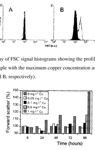
Forward Scatter
Copper provoked an increase in the FSC signal of P. tricornutum cells (Figs. 2 and 3). The highest copper concentration assayed, 1 mg l-1, provoked an important
increase in FSC after 24 h of copper exposure, while the effect of the remaining copper
concentrations were not visible until 48or 72 h of metal exposure (Fig. 3). After 96 h of
copper exposure, differences in forward scatter between control and all cultures with
copper occurred, being maximum for cultures with 1 mg l-1 of copper, which provokes
an increase of 49% in FSC, calculated as described before. This increase in forward
scatter is correlated with an increase in the volume of the cells, observed using an
Fig. 2. Typical overlay of FSC signal histograms showing the profiles of a control sample (solid histogram) and a sample with the maximum copper concentration assayed (1 mg l-1), after 8 and 96 h of culture (A and B, respectively).
Fig. 3. Forward scatter, after copper exposure, of P. tricornutum cells. Data are expressed as the percentage of the FSC signal of control cells, according to the equation cited in the text. Results are the means of three replicates.
Peroxidase Activity
Figure 4 represents data on the changes observed in the peroxidase activity during
the 96 h culture, expressed as percentage of the hydroethidine fluorescence of the
control cells, according with the equation described by Reader et al. (16). After 48 h of
copper exposure, viable cells exposed to copper showed a higher peroxidase activity
(Fig. 4). After 72 h of copper exposure, only cultures with 0.5 and 1 mg l-1 of copper
presented an important increase in the peroxidase activity respect to the control cells,
Fig. 4. Variations in the peroxidase activity after copper exposure of P.tricornutum cells. Hydroethidine was used as the fluorescent probe to evaluate the peroxidase activity. Data are expressed as the percentage of the fluorescence of control cells, according to the equation cited in the text. Results are the means of three replicates
Membrane Potential
Possible variations in membrane potential were measured after 24 and 96 h of
copper exposure, using DiOC6(3). After 24 h of culturing, differences in membrane
potential were not found using this fluorochrome (Figs. 5 and 6); but after 96 h of metal
exposure, all cells cultured with copper presented alterations in the membrane potential,
which increased as copper concentration increased (Fig. 6).
The lipophilic cationic dye rhodamine 123 has been used for investigations of
mitochondrial structure and function; it accumulates in energized mitochondria as a
result of their membrane potential (18). Copper has also provoked an increase in the
membrane potential studied using Rh123, following the same pattern described for the
assays carried out using DiOC6(3) (Fig. 7): after 96 h of metal exposure, all cultures
with copper presented higher membrane potential than control cultures.
FIG. 6. Variations in the membrane potential after copper exposure of P. tricornutum cells, using 3,3'dihexyloxacarbocyanine as the fluorescent probe. Data are expressed as the percentage of the fluorescence of control cells, according to the equation cited in the text. Results are the means of three replicates.
FIG. 7. Variations in the membrane potential after copper exposure of P. tricornutum cells, using rhodamine 123 as the fluorescent probe. Data are expressed as the percentage of the fluorescence of control cells, according to the equation cited in the text. Results are the means of three replicates.
DISCUSSION
Some metals play indispensable roles in cell growth and maintenance of
metabolic functions, but when their concentrations in the environment increase above a
threshold, many cellular changes can be detected as a response to the stress provoked.
Results obtained indicated that growth of Phaeodactylum tricornutum cultures was affected by copper (Fig. l).
Copper concentrations assayed provoked an increase in the forward scatter signal
of P. tricornutum cells detected by flow cytometry, being maximum for cultures with 1 mg l-1 of copper (Figs. 2 and 3). Microscopical analysis of these cells have shown an
increase in size, probably due to the incapacity to finish the cell division because of
observed microscopically and the increase in the forward scatter signal. Other authors,
using microscopic techniques, have observed an increase in the cellular volume in
different microalgal species exposed to high concentrations of different heavy metals (1,
17, 21, 22).
The main characteristic of cell death, whether from senescence, acute stress, or
aging, seems to be the loss of the cell's ability to maintain homeostasis (4,6). Cellular membranes are selective, dynamic barriers that play an essential role in regulating
biochemical and physiological events. The viability of P. tricornutum cells decrease throughout 96 h of copper exposure (Table 1 ), showing a progressive loss of their
membrane integrity, like occurs in the aging or senescence process (23). Whereas
senescence represents endogenously controlled degenerative processes leading to death,
aging encompasses a wide array of passive or nonregulated, degenerative processes
driven primarily by exogenous factors (23).
Membranes could be expected to be highly prone to free radical attack inasmuch
as unsaturated fatty acids are major components of most membrane lipid bilayers. The
consequences of free radical attack on membranes are numerous and include the
induction of lipid peroxidation (lo), lysis (7), and fatty acid deesterification (13).
Senescence is an active process initiated by some combination of internal and
environmental triggers, and membrane deterioration is an early and fundamental feature
of this process. Results obtained in this work show important changes in the
cytoplasmic membrane. Peroxidase activity increased in cells exposed to the higher
copper concentrations assayed (Fig. 4), where cell viability decreased. The peroxidase activity detected seems to be directly correlated to the progressive loss of function and
structural integrity of the cell membrane, leading to the cell death or decrease of cell
viability.
After 96 h of copper exposure, results obtained by flow cytometry showed that
cells cultured with this metal presented an increase in the membrane potentials,
increasing as copper concentration increased (Figs. 6 and 7). These changes in the
membrane potentials can be associated with alterations provoked by the peroxidation of
membrane lipids as consequence of free radical attack (copper in this case), and also
Results obtained in this work showed that copper induced a degenerative process
in Phaeodactylum tricornutum cells, closely related with alterations or disorders in membrane systems.
ACKNOWLEDGMENTS
The authors thank Dr. Gunter Valet for his scientific support in the functional
analysis of the marine phytoplankton by flow cytometry.
LITERATURE CITED
1. Bolaños, L, García-González M, Mateo P, Bonilla I: Differential toxicological response to cadmium in Anabaena strain PCC7119 grown with NO3− and NH4+ as nitrogen source. J
Plant Physiol 140: 345–349, 1992.
2. Bucana C, Saiki I, Nayar R: Uptake and accumulation of the vital dye hydroethidine in neoplastic cells. J Histochem Cytochem 34:1109–1112, 1986.
3. Cid A, Herrero C, Torres E, Abalde J: Copper toxicity on the marine microalga Phaeodactylum tricornutum: effects on photosynthesis and related parameters. Aquat Toxicol 31: 165–174, 1995.
4. Davies I, Sigee DC: Cell ageing and cell death: Perspectives. In: Cell Ageing and Cell Death, Davies, I, Sigee, DC, (eds). Cambridge University Press, London, U. K. 1984, pp 347–350.
5. Erickson SJ: Toxicity of copper to Thalassiosira pseudonana in unenriched inshore seawater. J Phycol 8: 318–323, 1972.
6. Gahan PB: Revestible and irreversible damage in plant cells of different ages. Cell Ageing and Cell Death, Davies, I, Sigee, DC, (eds). Cambridge University Press, London, U. K. 1984, pp 155–169.
7. Goldstein IM, Weissmann G: Effects of generation of superoxide anion on permeability of liposomes. Biochim Biophys Res Commun70: 452–458, 1977.
9. Hughes MN, Poole RK: Metals and micro-organisms. Chapman and Hall Ltd, London, U. K., 1989.
10.Kellogg EW, Fridovich I: Superoxide, hydrogen peroxide and singlet oxygen in lipid peroxidation by the xanthine oxidase system. J Biol Chem 250: 8812–8817, 1975.
11.Lewis MA: Proposed New Standard Guide for Conducting Static 96-h Toxicity Test with Microalgae. Draft 13, ASTM CommitteeE47.01, Philadelphia, 1987.
12.Miller WE, Greene JC, Shiroyama T: The Selenastrum capricornutum Printz Algal Bottle Test. EPA 600/9-78-018, U. S. Environmental Protection Agency, Corvallis, Oregon, 1978. 13.Niehaus WG: A proposed role of the superoxide anion as a biological nucleophile in the
deesterification of phospolipids. Bioorg Chem7: 77–84, 1978.
14.Ormerod MG: Analysis of DNA. General methods. In: Flow Cytometry. A Practical Approach, OrmerodMG (ed). Oxford University Press, Oxford, U. K., 1990, pp 69–87. 15.Rabinovitch PS, June CH: Intracellular ionized calcium, membrane potential, and pH.
In: Flow Cytometry. A Practical Approach,OrmerodMG (ed). Oxford University Press, Oxford, U. K., 1990, pp 161–185.
16.Reader S, Marion M, Denizeau F: Flow cytometric analysis of the effects of tri- n-butylin chloride on cytosolic free calcium and thiol levels in isolated rainbow trout hepatocytes. Toxicology 80: 117–129, 1993.
17.Riisgard HV, Nielsen KN, Sogaard-jensen B: Further studies on volume regulation and effects of copper in relation to pH and EDTA in the naked marine flagellater Dunaliella marina. Mar Biol 56: 267–276, 1980.
18.Shapiro HM: Cell membrane potential analysis. In: Methods in Cell Biology, vol. 33: Flow Cytometry, Darzynkiewicz, Z, Crissman, HA (eds). Academic Press, Inc., San Diego, CA, 1990, pp 25–35.
19.Shapiro HM: Practical Flow Cytometry. Third Edition. Wiley-Liss, Inc., New York, 1995. 20.Shapiro HM, Natale PJ, Kamentsky LA: Estimation of membrane potentials of individual
lymphocytes by flow cytometry. Proc Nat Acad Sci 76: 5728–5731, 1979.
22.Stokes PM, Hutchinson TC, Krauter K: Heavy-metal tolerance in algae isolated from contaminated lakes near Subury, Ontario smeltors. Can J Bot 51: 2155–2168, 1973.
23. Thompson JE: The molecular basis for membrane deterioration during senescence.