Molecular y Biomedicina
Neuropathic pain induced by nerve injury involves
epigenetic changes and chromatolytic damage in the
somatosensory nervous system. Effects of
miR-30c-5p gain and loss of function
Director:
Dr. María Amor Hurlé González
Codirector:
Dr. Mónica Tramullas Fernández
Tesis doctoral presentada por Raquel Francés Romero para
optar al Grado de Doctor por la Universidad de Cantabria
doctor, del departamento de Fisiología y Farmacología, Facultad de Medicina, Universidad de Cantabria,
EXPONEN:
Que han llevado a cabo las funciones de dirección de la Tesis Doctoral de Dña
Raquel Francés Romero, Licenciada en Biotecnología con el título:
“Neuropathic pain induced by nerve injury involves epigenetic changes and chromatolytic damage in the somatosensory nervous system. Effects of miR-30c-5p gain and loss of function”.
Y que dicho trabajo se encuentra terminado y reúne los requisitos necesarios para su presentación como Memoria de Doctorado, con el objeto de poder optar al grado de Doctor por la Universidad de Cantabria.
Y para que conste y surta los efectos oportunos, expedimos la presente certificación en Santander, a 8 de Enero de 2019.
- Beca predoctoral de Neurociencia: Fundación Tatiana Pérez de Guzmán el Bueno.
- Instituto de investigación sanitaria Marqués de Valdecilla
- Ministerio de Economía y Competitividad a través de los Proyectos del Plan Estatal: SAF2013-47434-R y SAF2016-77732-R
Como escribía Severo Ochoa, “Mi consejo a los estudiantes de ciencia es que, si desean ardientemente investigar, deberían hacerlo por todos los medios. Difícilmente alguna otra cosa te dará tanta satisfacción y, sobre todo, tal sentido de logro”. Y es así como me siento al finalizar esta tesis. Es por esto que quiero agradecer a todas las personas que han contribuido directa o indirectamente para que este trabajo pudiera llevarse a cabo.
Quiero empezar estos agradecimientos dando las gracias a mi directora Maruja Hurlé por confiar en mi y darme la oportunidad de realizar la tesis bajo su dirección. Gracias por acogerme como a un miembro más del laboratorio desde el primer día, por animarme siempre y por la buena orientación que siempre he recibido. Sus consejos, apoyo y conocimientos han sido claves en la realización de esta tesis doctoral. Pero sobre todo gracias por hacerme sentir una parte importante del laboratorio.
También quiero darle las gracias a mi co-directora Mónica Tramullas por sus consejos, ayuda y dedicación durante todos estos años. Gracias también por su todo el tiempo y paciencia dedicados en la elaboración de esta tesis. A María Carcelén, que ha sido mi compañera de fatigas durante los últimos años y que continúa con la investigación. Gracias por su entusiasmo, ayuda y paciencia. A Raquel García por sus innumerables y siempre acertados consejos y a AnaB por los buenos ratos que hemos pasado y por ofrecer siempre su ayuda.
Al resto de compañeros de laboratorio, Nieves García, Ana Cayón, María Navarro, Roberto de la Fuente y Rosmarí de la Puerta por ofrecerme su ayuda siempre que la he necesitado y sobre todo por todos los buenos momentos. Al grupo de la Dra.Carmen Martínez-Cué: Noemí Rueda, Eva García, Verónica Vidal, Susana García y Sara Lantigua por su ayuda y su siempre buena disposición.
A los Dres. Miguel Lafarga y Mª Teresa Berciano y a todo su grupo por abrirme las puertas de su laboratorio, por su constante ayuda, transmitiéndome generosamente sus conocimientos y por permitirme usar sus infraestructuras. A Oriol Narcís, que siempre me ha ayudado cuando le he necesitado y me ha hecho reír infinitas veces. A Jorge Mata, que ha
colaborado especialmente en la elaboración de esta tesis. Gracias por todas las técnicas y conocimientos que me ha transmitido y por su paciencia y constante apoyo día a día.
Al grupo del Dr.Juan Hurlé: JuanAn, Sonia, Montse, Susana, Nacho y Cristina por haberme ayudado en tantos experimentos siempre que lo he necesitado. Al grupo de la Dra. Claire Francastel del Centro de Epigenética y destino celular de París por aceptarme en su laboratorio, poniendo a mi disposición todo lo que necesitase y por hacer que disfrutara tanto mi estancia en París. Al Dr. Javier Ayesta por su ayuda durante estos años, especialmente en las correciones finales.
Al grupo del Dr. Ángel Pazos, a Álvaro Díaz, Elena Castro, Fuen y Eva Florensa por ayudarme siempre que lo he necesitado.
ABBREVIATIONS ... 21
JUSTIFICATION AND OBJECTIVES ... 29
INTRODUCTION ... 35
1.Pain ... 35
1.1 The nociceptive system ... 35
1.1.1 Pain modulation circuits ... 43
1.2 Types of pain ... 45
1.2.1 Nociceptive pain ... 45
1.2.2 Neuropathic pain ... 46
2. Epigenetics ... 51
2.1 Chromatin structural organization ... 51
2.1.1 Types of chromatin ... 52
2.2 Epigenetics, an overview ... 54
2.2.1 DNA methylation ... 56
2.2.1.1 Role and location of DNA methylation ... 57
2.2.1.2 DNA methylation in disease ... 58
2.2.1.3 DNA methylation in pain ... 59
2.3 Histone modifications ... 62
2.3.1 Histone methylation ... 62
2.3.2 Histone acetylation ... 63
2.3.3 Histone modifications and disease ... 64
2.3.4 Histone modifications and pain ... 64
2.4 Non-coding RNAs:microRNAs(miRNAs) ... 66
2.4.1 Characteristics and biogenesis of miRNAs ... 66
2.4.2.1 miRNAs in the nervous system ... 73
2.4.2.2 miRNAs and neuropathic pain ... 74
2.4.2.3 microRNA-30c (miR-30c-5p) ... 77
2.5 Epigenetic modifiers as therapeutic targets for chronic pain ... 78
2.5.1 Analgesics as epigenetic modulators for chronic pain ... 79
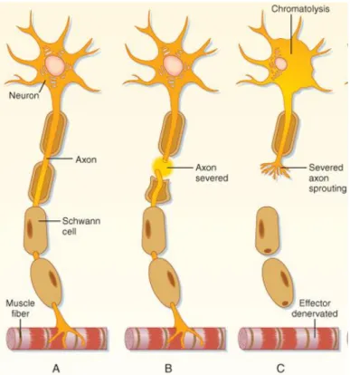
3. Neuronal stress reaction after the injury to the axon ... 80
3.1 The cytoplasm ... 80
3.1.1 Neuronal chromatolysis ... 80
3.2 The nucleus ... 82
3.2.1 The nucleolus ... 83
3.2.2 The Cajal body ... 86
SPECIFIC OBJETIVES ... 91
MATERIAL AND METHODS ... 95
1. Animals ... 95
1.1 Experimental model of neuropathic pain ... 95
1.1.1 Traumatic peripheral neuropathy model: Spared Nerve Injury ... 95
1.1.2 Assessment of the response to mechanical stimuli: Von Frey test ... 96
1.2 Treatments and experimental groups... 97
1.3 Processing of biological material ... 99
1.3.1 Spinal cord and dorsal root ganglion extraction ... 99
1.3.2 Perfusion technique ... 100
1.3.3 Obtaining neuronal dissociates ... 100
2. Microscopy techniques ... 101
2.1.1 5’-Methylcitosine detection ... 103
2.2 Conventional transmission electron microscopy ... 103
3. Molecular biology techniques ... 104
3.1 Protein analysis by western blot ... 104
3.2 mRNA and miRNAs expression by quantitative PCR ... 106
3.2.1 mRNA and miRNA extraction in SDH and DRG ... 106
3.2.2 mRNA and miRNA reverse transcription ... 106
3.2.3 Oligonucleotide primer design for quantitative PCR ... 107
3.2.4 Detection of gene expression by quantitative real time PCR 109 3.3 DNA isolation ... 110
3.3.1 Methylation sensitive qPCR (ms-qPCR) ... 110
4. Cell culture studies ... 111
4.1 Dual luciferase reporter assay... 112
5. Quantification and statistical analysis of relative gene expression ... 112
RESULTS ... 117
1. Epigenetic changes in the somatosensory nervous system associated with neuropathic pain and its modulation by mirna-30c-5p ... 117
1.1 The development of mechanical allodynia in rats is modulated in opposite ways by an miR-30c-5p mimic and an miR-30c-5p inhibitor ... 117
1.2 Dorsal root ganglion and spinal dorsal horn neurons present increased global levels of DNA methylation after sciatic nerve injury in rats ... 119
1.2.1 Dorsal root ganglion and spinal dorsal horn neurons present increased mRNA expression levels of DNMTs after sciatic nerve injury in rats ... 123
1.3 miR-30c-5p mimic and miR-30c-5p inhibitor modulate global DNA methylation in the rat dorsal root ganglion and spinal dorsal horn
neurons after sciatic nerve injury in rats ... 124
1.3.1 miR-30c-5p mimic and miR-30c-5p inhibitor modulate the expression of DNMT3a, DNMT3b and DNMT1 in dorsal root ganglion and spinal dorsal horn neurons after sciatic nerve injury in rats .. 129
1.4 Luciferase reporter assays revealed post-transcriptional regulation of DNMT3a and DNMT3b by miR-30c-5p in Hela and SH-SY5Y cells . 134 1.5 Modulation of miR-30c-5p in rats subjected to sciatic nerve injury results in methylation changes of Nfyc and TGFβ-1 in the SDH ... 135
1.6 Trimethylation of lysine 9 on histone H3 (H3K9me3) in dorsal root ganglion and spinal dorsal horn neurons after sciatic nerve injury in rats and its modulation by miR-30c-5p mimic and miR-30c-5p inhibitor . 138 2. Morphologic, ultrastructural, and functional changes induced by miR-30c-5p modulation in primary sensory neurons after sciatic nerve injury ... 146
2.1 Effects of miR-30c-5p modulation on the protein synthesis machinery in dorsal root ganglion neurons after sciatic nerve injury 146 2.2 Effects of miR-30c-5p modulation on the nucleolar cytoarchitecture of dorsal root ganglion neurons after sciatic nerve injury ... 151
2.3 Effects of miR-30c-5p modulation on the Cajal bodies of dorsal root ganglion neurons after sciatic nerve injury ... 156
DISCUSSION ... 161
CONCLUSIONS ... 181
RESUMEN EN ESPAÑOL ... 185
21
AGO: Argonaute
APS: Amonium persulafte ATP: Adenosine triphosphate
BDNF: Brain-derived neurotrophic factor bp: Base pairs
BSA: Bovine serum albumin ºC: Celsius degrees
C.elegans: Caernohabditis elegans CaCl2: Calcium dichloride
CB: Cajal body
CCI: Chronic constriction injury cDNA: Complementary DNA CNS: Central nervous system cm: Centimeters
CO2: Carbon dioxide
COX: Cyclooxigenase CSF: Cerebrospinal Fluid Ct: Cycle threshold
DFC: Dense fibrillar component
DGCR8: Digeorge syndrome chromosomal or critical region 8
(microprocessor complex)
DICER: Double strand RNA binding domain DMEM: Dulbecco’s modified eagle medium DMSO: Dimethylsulphoxide
DNA: Deoxyribonucleic acid DNMTs: DNA methyltransferases DNMT1: DNA methyltransferase 1 DNMT3a: DNA methyltransferase 3a DNMT3b: DNA methyltransferase 3b DNMT3L: DNMT3-Like protein
22
DRG: Dorsal root ganglia
DROSHA: Ribonuclease type III
EDTA: Ethylenediaminetetraacetic acid EGTA: Ethylene glycol tetraacetic adic FBS: Fetal bovine serum
FC: Fibrillar center
FITC: Fluorescein isothiocyanate g: Grams
GC: Granular component h: Hour
H+: Hydrogen molecule
HAT: Histone acetyltransferase HCl: Hydrochloric acid
HDAC: Histone deacetylase HDMs: Histone demethylases
HEPES: 4-(2-hydroxyethyl)-1-piperazineethanesulfonic acid HMTs: Histone methyltransferases
H2O: Water
H2O DEPC: Diethylpyrocarbonate water
H3K9me3: Histone 3 trimethylated in lysine 3
IASP: International Association for the Study of Pain IF: Immunofluorescence K: Lysine K+: Potassium molecule KCl: Potassium Chloride kDa: Kilodalton KDM: Lysine demethylase kg: kilograms
KH2PO4: Potassium phosphate monobasic
KMT: Lysine methyl-transferase Lamb1: Lamina b1
23 M: Molar m: Meter MBPs: Methyl-binding proteins mg: milligrams Mg2+: Magnesium molecule MgCl2: Magnesium chloride microRT-PCR: Micro-retrotranscriptase PCR miRISC: MiRNA-induced silencing complex miRNA: MicroRNA
ml: Mililiters mM: Millimolar mm: Millimeters
mRNA: Messenger RNA ms: Miliseconds
ms-qPCR: Methylation sensitive qPCR mT: Melting temperature
MOR: Mu opioid receptor Na+: Sodium molecule
Na2HPO4: Sodium phosphate dibasic
NaCl: Sodium chloride
NaH2PO4: Monosodium phosphate
NB: Nissl bodies
nBLAST: nucleotide basic local alignment search tool ncRNA: Non-coding RNA
Nfyc: Nuclear transcription factor Y subunit gamma ng: Nanograms
nm: Nanometer nM: Nanomolar
NMDA: N-methyl-D-aspartate recepot No: Nucleolus
24
NS: Nervous system
NSAIDs: non-steroidal anti-inflammatory drugs O2: Oxygen molecule
PAG: Periaqueductal gray PBS: Phosphate buffered saline PCR: Polimerase chain reaction PFA: Paraformaldehyde
PI: Propidium Iodide
PNS: Peripheral nervous system Pre-miRNA: MiRNA precursor Pre-rRNA: Pre-ribosomal RNA Pri-miRNA: Primary miRNA PSL: Partial sciatic ligation PVDF: Polyvinylidene difluoride
qPCR: Quantitative Polimerase chain reaction R: Arginine
RAN-GTP: Ras-related nuclear protein Guanosine-5-triphosphate rpm: Revolutions per minute
RER: Rough endoplasmic reticulum RNA: Ribonucleic acid
rRNA: Ribosomal RNA RT: Room temperature
RT-PCR: Retrotranscriptase PCR RVM: Rostral ventromedial medulla s: Seconds
SAM: S-adenosyl-L-methionine SDH: Spinal dorsal horn
SDS: Sodium dodecyl sulfate SEM: Standard error mean SI: Signal intensity
25
SNL: Spared nerve ligation
snoRNPs: Small nucleolar ribonucleoproteins snRNPs: Small nuclear ribonucleoproteins
Suv39h1: Suppressor of variegation 3-9 homolog 1 T: Temperature
TBS-T: Tris Buffer Saline- Tween 20 TGB-β: Transforming growth factor β TNF: Tumor necrosis factor
TRBP: TAR RNA binding protein
TRPA1: Transient receptor potential cation channel subfamily A member 1 TRPM8: Transient receptor potential cation channel subfamily M member 8 U: Units
UBF: Upstream binding factor UTR: Untranslated region V: Volts WB: Western blot 5’-hmC: 5’-hydroxymethyl cytosine 5-HT: Serotonin 5’-MeC: 5’-methylcytosine µl: Microliters µm: Micrometers
29
Pain is an unpleasant experience that alerts the body of actual or potential tissue damages or diseases. Physiological pain is proportional to the intensity of the stimulus that caused it and triggers protective responses directed to the defense of the organism. Pain usually disappears after the healing of the triggering lesion. However, in certain situations of neural damage or inflammation, pain may persist long after the healing of the lesion.
Chronic pain is a highly prevalent condition among the general population (~20 %), which constitutes a serious burden on life quality of patients and an important public health problem, with socio-economic and health related consequences. Chronic pain is considered a pathological process in itself, which requires specific treatment.
A particularly devastating form of pathological pain is neuropathic pain which arises as a consequence of a lesion or disease affecting the somatosensory system, peripheral or central. According to data from The Spanish Pain Society, the prevalence of neuropathic pain is estimated in 8–10 % of the general population in Spain. Common causes of neuropathic pain include, among others, metabolic (diabetes) and infectious (herpes zoster, VIH) diseases, traumatisms (surgery, spinal cord injury), tumors, neuralgias, stroke, peripheral ischemia, etc.
The therapeutic approach to neuropathic pain is basically focused on four pharmacological groups: antidepressants, antiepileptics, local anesthetics, and several opioids. However, management neuropathic pain is still a challenge because the response to pharmacological treatments is generally limited. Thus, the number-needed-to-treat, for 50% pain relief in one patient, ranges from 3 to 5 of patients. Moreover, the drugs currently available do not have the capacity to modify the evolutionary course of neuropathic pain, and lack preventive/curative properties.
Our incomplete understanding of the cellular and molecular mechanisms responsible for neuropathic pain establishment and chronification has limited the identification of novel therapeutic targets for its prevention and/or release. Therefore, their study is a priority field
In the recent years, a rapidly increasing body of evidence implicates epigenetic modifications, such as DNA methylation, histone modifications and
30
microRNAs (RNAs), in the long-lasting aberrant expression of crucial pain-related genes in the somatosensory nervous system, underlying pain chronification after neural injuries.
In this regard, our group has been studying for several years the role of miRNA-related epigenetic mechanisms involved in the persisting neural adaptations triggered by peripheral nerve damage, which contribute to the development of neuropathic pain. We have previously demonstrated a key role for miR-30c-5p in the development and persistence of neuropathic pain, as well as its potential value as biomarker and therapeutic target, both in an experimental animal model and in patients. Our preclinical study shows that the severity of neuropathic pain following spared nerve injury (SNI) in rats is directly related with miR-30c-5p overexpression in relevant regions of the nociceptive pathway. Consistently, the administration in the cisterna magna of miR-30c-mimic accelerates the development of allodynia. In contrast, the administration of a miR-30c-5p specific inhibitor, at the time of nerve injury, prevents the development of allodynia. But most importantly, the rats treated with miR-30c-5p inhibitor several weeks after allodynia is fully established recover the normal nociceptive sensitivity. In both cases, the animals remained free of neuropathic pain until their sacrifice, two months after nerve injury. Overall, our results in experimental animals point to a relevant contribution of miR-30c-5p to mechanical allodynia development, and make it a promising target for treating neuropathic pain states.
The relevance of these findings was transferred to the clinic, in a group of patients with ischemia of the lower extremities. The logistic regression analysis allowed us to develop a model in which the circulating levels of miR-30c-5p in CSF or in plasma discriminate with great sensitivity and specificity those patients who suffer from neuropathic pain of those who are free of pain. Online software has revealed that the de novo DNA methyltransferase DNMT3b enzyme is found among the predicted transcript targets for miR-30c-5p.
Increasing evidence points to the involvement of epigenetic processes in the aberrant expression of genes encoding ion channels, receptors, neurotransmitters, modulators, cytokines, etc., which underlie the pathological chronification of pain. These studies allow us to propose that the
31
persistence of the neuronal excitability of the chronic pain syndromes could be related to dynamic epigenetic changes that modify the phenotype of neuron and glial cells.
DNA methylation, which associates heterochromatin formation and transcription silencing, constitutes a major epigenetic mark in mammals. Recent studies suggest that DNA methylation changes play a crucial role in the process of pain chronification. However, we are still far from understanding how DNA methylation contributes to pain chronification and, specifically, the role played by the crosstalk between miRNAs and this epigenetic mark in the neuropathic pain setting. In this regard, it has been reported that deregulation of miR-30c-5p, under several pathological conditions, brings about changes in the expression and function of DNMTs with pathophysiological implications. In addition, the “de novo” DNMT3b is consistently predicted as a target for miR-30c-5p in the online miRNA bioinformatics tools (TargetScan4.0, miRanda and miRbase).
DNA methylation represses gene transcription through several mechanism including physically blocking the binding of transcription factors or functioning as docking sites for transcriptional repressors. Therefore, it is possible that some epigenetic mechanisms are playing a key role in the pathologic neural plasticity processes that lead to the chronic neuropathic pain state. How DNMTs contribute to neuropathic pain genesis and establishment is still elusive and the possibility to interfere in the painful process through modulation of epigenetic mechanism mediated by miR-30c-5p in vivo modulation could suppose the opening of new therapeutic perspectives for the treatment of pathological pain situations highly resistant to conventional pharmacological treatment.
On the other hand, little is known about the changes that occur at a cellular level in the DRG neurons following the neuropathic lesion. DRGs are the first step in the nociceptive pathway and probably the first neurons to be affected. Although structural and functional alterations in some cellular organelles have been described in other neurodegenerative diseases such as lateral amyotrophic sclerosis or spinal muscular atrophy, we have not found any published data that investigate the effect of sciatic nerve injury, which causes neuropathic pain in animal models, at a more cellular level. Understanding
32
the cellular alterations caused by a sciatic nerve lesion and how miR-30c-5p modulation could induce changes in the organization and functions of the cellular components are essential to understand the pathophysiology and aberrant plasticity mechanism involved in the neuropathic pain disease caused after a nerve damage.
Considering all this data, we hypothesized that epigenetic modifications and cellular alterations are involved in the pathological plasticity of the nervous system that underlies establishment and development of neuropathic pain after a neuronal lesion. Furthermore, miR-30c-5p could be involved in the aberrant epigenetic mechanisms underlying neuropathic pain by modulating the activity of DNA methyltransferases. Based on our hypothesis, we proposed the following general objectives:
I. To stablish the global levels of DNA and histone methylation and its distribution pattern in two pain-related areas, the dorsal root ganglia and the dorsal horn of the spinal cord, in rats subjected to sciatic nerve injury and evaluate the effects of miR-30c-5p-gain- and –loss- of function in vivo.
II. Asses the cellular alterations (protein synthesis machinery and nucleolar organization and structure) in neurons of the DRG that occur in rats exposed to sciatic nerve injury and the consequences of miR-30c-5p modulation.
35
1. Pain
The International Association for the Study of Pain (IASP) defines pain as “an unpleasant sensory and emotional experience associated with actual or potential tissue damage”. Pain, unlike other conditions, is the most important element of interference in the life of the affected person at a biological, psycho-emotional and social level. For this reason, it is considered a key indicator of health, welfare and quality of life. Pain is an indispensable sensation that under physiological conditions warns the body to protect itself from a tissue damage or disease. The painful physiological feeling is proportional to the intensity of the stimulus and triggers protective responses directed to the defense of the organism. The pain usually disappears after the healing of the lesion that caused it. However, in specific conditions of neural damage or inflammation, pathologic plasticity events occur in the nervous system (Woolf and Ma, 2007; Basbaum et al., 2009; Li et al., 2016; Gwak et al., 2017). This leads to abnormal painful sensations in the nociceptive system such as prolonged and persistent pain when the lesion that caused it disappeared or a disproportionate painful response to the causal stimulus. In these situations, pain loses its protective function and it stands as a pathological process that requires specific treatment (Cerveró, 2009). Prolonged suffering caused by chronic pain is one of the main reasons of medical consultation as chronic pain can produce a negative impact on the physical, psycho-emotional, personal, family, work, social and economic level of the affected person and their environment. Chronic pain is considered a pathological process itself, refractory to conventional analgesic drug therapy (Cerveró, 2009).
1.1 The nociceptive system
The nociceptive system is the one responsible for the detection and processing of the noxious stimuli, transforming it into electrical signals, which are then conducted to the central nervous system (CNS). The neurophysiological processes that participate in pain are:
36
a) Activation and sensitization of the peripheral nociceptors
Pain receptors are called nociceptors. A nociceptor is a primary nociceptive-sensory neuron that responds to potential damage stimuli by sending nerve signals to the dorsal horn of the spinal cord. The cellular bodies of the nociceptors are located inthe dorsal root ganglion or the trigeminal ganglion (Flórez, 2007; Woolf and Ma, 2007). Nociceptors are able to distinguish between innocuous and noxious stimuli as they are able to encode the intensity of a stimulus between a range of noxious intensities, while they are not able to respond or respond irregularly to low intensity stimuli (Woolf and Ma, 2007). Nociceptors are distributed through the body and are present in many body tissues including the skin, viscera, muscles, joint, meninges and connective tissue. The rest of body tissues have lower levels of nociceptive endings. These receptors transmit the information through nerve fibers that are classified according to the speed of transmission, which is directly correlated with the diameter of axons of the neurons and whether or not they are myelinated. Nociceptive fibers have been classified on the basis of their conduction speed and sensitivity (Meyer et al., 2006; Flórez, 2007; Dubin and Patapoutian, 2010) (Figure 1):
- Aα/β fibers are highly myelinated and of large diameter axons (6-20 µm), therefore allowing rapid signal conduction (30-120 m/s). They have a low activation threshold and usually respond to light touch and transmit non-noxious stimuli.
- Aδ fibers are lightly myelinated and have medium diameter (1-5 µm), and hence have intermedium conduction speed (12-30 m/s). They respond to mechanical and thermal stimuli, although they can also respond to innocuous stimuli. They carry the first feeling of pain, what is also called “rapid pain” (about 300 ms) and are responsible for the initial adaptive response to acute pain (the withdrawal response). It is a well delimited, localized (epicritic) and sharp pain.
- C fibers are unmyelinated and have small diameter axons (0.3-1.5 µm). Hence, they demonstrate the slowest conduction velocity (0.4-2 m/s). C fibers are polymodal, responding to mechanical, chemical and thermal harmful stimuli although they can also respond to innocuous stimuli. They are
37
responsible of the transmission of the “second pain”or “slow pain” (approximately 0.7-1. S). It is a bad localized (protopathic) and burning pain.
- -
Figure 1: Anatomy of the nociceptors. Types of sensitive primary neurons and
degree of myelination; primary afferents include those with large-diameter myelinated (Aβ), small-diameter myelinated (Aδ), and unmyelinated (C) axons (Taken from Rathmell and Fields, 2015).
Nociceptors can be classified according to their localization and the type of the stimulus that they respond to. There are four types of nociceptors:
- Cutaneous nociceptors: They present a high stimulation threshold and
only become activated in response to intense stimuli. They are inactivated when there are no noxious stimuli. There are two main categories of cutaneous nociceptors:
i) Aδ mechanical nociceptors: They are located in the superficial layers of the dermis, with branches that extend to the epidermis and respond to noxious stimuli of mechanical type (sharp pain). ii) C-polymodal nociceptors: They are free endings in the skin and
respond well to noxious mechanical, thermal and chemical stimuli. They are very sensitive, leading to the phenomenon called primary hyperalgesia. They are called “polymodal nociceptors” as they can respond to a wild range of noxious stimulus. Nociceptors can also be classified according to the differential expression of channels
38
that cause heat sensations (TRPV1), (TRPM8) and the ones that are activated by chemicals (TRPA1) (Basbaum et al., 2009).
- Visceral nociceptors: They innervate internal organs such as the heart,
lungs, vascular system, testis, uterus and ureter. Most of the visceral nociceptors are free nerve endings of C, and in some cases, Aδ fibers. - Joint nociceptors: The joint capsule, ligaments, periosteum and joint fat have C and Aδ fiber endings that respond to low threshold joint movements or noxious ones and to factors released by tissue damage such as inflammation.
- Muscle nociceptors: Muscles are innervated by Aδ fibers and respond
to allogenic molecules (potassium ions, bradykinin or serotonin) or sustained muscle contractions and C fiber terminations that respond to noxious muscle stimulus such as pressure, heat or muscle ischemia. It is a diffuse pain, difficult to locate, dull and continuous.
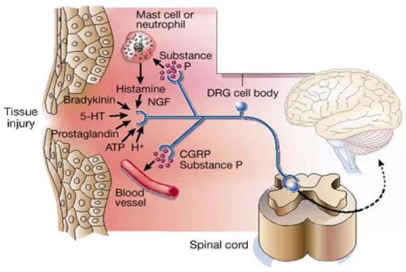
- b) Nociceptive stimuli transmission through the primary afferents Tissue pain triggers the production and release of chemical mediators with allogenic properties from the primary sensory terminal and from non-neural cells (for example, fibroblast, mast cells, neutrophils and platelets) in the immediate environment of the peripheral sensory ends or nociceptors (Figure 2). Some of these molecules are: ions (H+ and K+), bradykinin, prostaglandins, serotonin, noradrenalin, histamine, substance P, thromboxanes, protons, cytokines, tumor necrosis factor (TNF) and neurotrophins (specially the nervous growth factor), are also produced during inflammation. Some of these factors directly activate the nociceptor while other factors act in a synergic way, increasing the response or sensitizing (decrease the threshold) of the nociceptor, which play a crucial role in the primary hyperalgesia processes. Activation of the nociceptor not only transmits afferent messages to the spinal cord dorsal horn (and from there to the brain), but also initiates the process of neurogenic inflammation. These factors also activate many non-neuronal cells, including mast cells and neutrophils which in turn contribute as additional elements to the inflammatory process that are able to stimulate and sensitize peripheral nociceptors and spinal cord pathways of painful transmission (Julius and Basbaum, 2001; Woolf and Ma,2007).
39
Figure 2: Inflammatory mediators released at the site of tissue injury. Some
of the main components that facilitate the inflammatory processes and stimulate pain transmission include peptides (bradykinin), lipids (prostaglandins), neurotransmitters (serotonin (5-HT) and ATP) and neurotrophins (NGF). (Taken from Julius and Basbaum, 2001).
c) Modulation and integration of the nociceptive response at the dorsal
horn of the spinal cord level
The axons of primary afferent nociceptors enter the posterolateral sulcus of the spinal cord, ending in the dorsal horn of the spinal cord gray matter. The primary neuron of the pain transmission pathway has an ending in the periphery, the body in the dorsal root ganglia (DRG) and the central ending in the dorsal horn of the spinal cord (SDH). Several excitatory neurotransmitters are released in the transmission of the nociceptive impulse from the periphery to the second neuron in the SDH. Nevertheless, the spinal cord is not only a point in the transmission of nociception, but represents a place of important interactions where a nociceptive impulse is allowed to follow the way towards higher structures or is totally or partially blocked (Meyer et al.,2006; Todd and Koerber,2006).
The gray matter of the spinal cord is organized into a series of layers called “Rexed laminae”. The central terminals of the primary afferent fibers end in
40
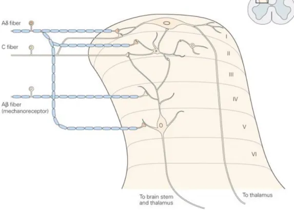
the gray matter of the dorsal horn of the spinal cord. Most of the primary nociceptive neurons terminate superficially in laminae I, II, IV and V of the dorsal horn (Figure 3), where they synapse with the secondary afferent neurons to carry pain information to the brain (Flórez, 2007; D’Mello et al., 2008).
Figure 3: Schematic representation of the Rexed laminae organization in the
dorsal horn of the spinal cord and afferent Aβ, Aδ y C fibers. (Taken from Kandel, 2000).
The distribution of the cells and fibers within the gray matter of the spinal cord exhibits a pattern of lamination. Laminae I, also called the marginal zone, contains mainly supraspinal projection neurons that respond exclusively to noxious stimuli, sending direct connections to the thalamus and to the different spinal segments. Lamina II is also called the substantia gelatinosa of Rolando. This layer is composed of tightly packed excitatory (glutamatergic) e inhibitory (glicinergic and GABAergic) interneurons that are essential for nociception processing and respond to noxious stimuli. The majority of the neuron axons in Rexed lamina II receive information from sensory dorsal root ganglion cells as well as from descending dorsolateral fasciculus fibers. They send axons to Rexed laminae III and IV. Lamina IV receives predominantly non-noxious information. Lamina V is composed by
41
wide dynamic range neurons that are able to identify different pain intensities. Aδ fibers end in the I and V lamina, C fibers in the I, II and V and Aβ in the II, IV and V. Generally, nociceptive afferents end in lamina I and II and V, and the non-nociceptive afferents of low threshold end in the deep II, IV and V layers. Lamina X is also related to the visceral pain nociceptive transmission.
d) Transmission through the ascending projection pathways
The ascending spinal cord projections anatomically connect second-order neuron of the spinal cord and the upper nerve centers. They are located in the ventral, lateral and dorsal funiculi on each side of the spinal cord (Figure
4) (Bonica, 2001; Dostrovsky and Craig, 2006; Flórez, 2007; García-Porrero
and Hurlé, 2014).
Figure 4: Ascending and descendending pain pathways. A)
Anterolateral-spinotalamic somatostetic system. Spinothalamic tract fibers are represented in green; spinoreticular tracts in black; and spinomesencefalic tracts in violet.
42
The spinothalamic tract
, conveys nociception, temperature, non-discriminative touch and pressure information to the somatosensory region of the thalamus. It is composed of a ventral (anterior, paleospinothalamic) and a lateral (neospinothalamic) pathway. 50% of the neurons of this tract are located in the lamina I, but also in the IV-V and V-VIII ones. 90% of the neurons decussate to the contralateral side of the spinal cord, the ones that decusse in the Lamina I form the spinothalamic lateral tract (Aδ y C afferents; thermoanalgesic sensibility) and the ones from the V-VII form the spinothalamic anterior tract (afferents Aβ, Aδ y C; crude touch and pressure sensations). Main projections connect with the third-order neuron in the medial and intralaminar nucleus of the thalamus.e) Pain processing in upper centers (encephalic structures)
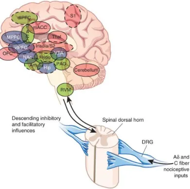
The classic pain pathway consists of a three-neuron chain that transmits information from the periphery to the spinal cord and relays the signal to the thalamus before terminating in the cerebral cortex. Ascending projections allow the anatomic connections between the second-order neuron of the spinal cord and the upper nervous centers, so that the perceived intensity of the painful impulses is correlated with an increase in the activity of a great number of brain structures (Apkarian et al., 2011; Hayati and Badariah,2014) (Figure 5). The main areas of the brain involved in pain perception are:
- Primary somatosensory cortex plays an important role in pain perception.
It is the main area on which pain is perceived, location and intensity is assessed. Its activation is modulated by cognitive factors such as attention or previous experiences, which alter pain perception.
- The thalamus is one of the structures that receives projections from multiple ascending pain pathways. The thalamic nuclei are involved in the sensory discriminative and affective motivational components of pain. The structure is not merely a relay center but is involved in processing nociceptive information before transmitting the information to various parts of the cortex (Melzack and Casey, 1968). Spinal lamina I neurons project extensively to the ventrobasal complex (ventral posterolateral + ventral posteromedial) and to the posterior thalamic nuclei (Dado et al., 1993). The nociceptive neurons from the ventrobasal complex mainly
43
project to the primary somatosensory cortex and this pathway constitutes the lateral pain system that plays an important role in the discrimination of stimuli.
- The amygdala integrates the aversive component of the painful experience, such as anxiety, fear avoidance, dangerous or painful situations.
- The nucleus Raphe magnus is involved in pain mediation; it sends projections to the dorsal horn of the spinal cord to directly inhibit pain. When stimulated, it releases serotonin, participating in the endogenous analgesia system.
- The locus coeruleus is the noradrenergic center and is involved in the descendent control of pain and the emotional component of pain.
- The periaqueductal gray (PAG) is the primary control center for descending pain modulation and the defensive behavior. Activation of this area releases encephalin by neurons that project to the nucleus Raphe magnus, producing a release of serotonin that is carried through the descendent pathways producing an inhibitory effect over the entrance of noxious stimuli.
- The hypothalamus is responsible for regulating hunger, thirst, response to pain, levels of pleasure, anger and aggressive behavior. Paraventricular and ventrolateral nuclei have neuroendocrine functions, such as pain arousal, thermoregulation, changes in blood pressure and other homeostatic function.
1.1.1 Pain modulation circuits
Contrary to the nociceptive ascending centripetal transmission, endogenous inhibitory system is descending and centrifugal. There are mechanisms that act to inhibit pain transmission at the spinal cord level and via descending inhibition from upper centers. Pain descending modulating systems exert an inhibitory and activator action over the nociceptive afferent, using some molecules such as noradrenaline, serotonin, opioids and cannabinoids (Ossipov et al., 2010). Specifically, descending systems are originated in the periaqueductal grey (PAG), the rostral ventromedial medulla (RVM), including
44
the nucleus Raphe magnus, amygdala, the paragigantocellular reticular nucleus and neurons of the adjacent reticular formation.
Figure 5: Ascending pathways, subcortical and cortical structures related with pain processing. PAG, periaqueductal gray; PB, prabraquial nuclei; VMpo,
núcleo ventromedial del tálamo; MDvc: núcleo dorso medial del tálamo; VPL, núcleo lateral ventro-posterior del tálamo; ACC, corteza cingular anterior; PCC, corteza cingular posterior; HT, hipotálamo; S-1 y S-2, áreas corticales somatosensoriales primera y segunda; PPC, complejo parietal posterior; SMA, área motora suplementaria; AMYG, amígdala; PF, corteza prefrontal. (Taken from Price, 2000).
The PAG in the midbrain and the rostral ventromedial medulla are two important areas of the brain involved in descending inhibitory modulation. PAG receives afferents from the prefrontal limbic cortex, amygdala, hypothalamus and spinal neurons. Thus, it receives information about the emotional and motivational state of the person and information regarding the somatic afferents. The balance of this activity over the PAG will allow to regulate pain sensibility. When stimulate, PAG activates encephalin-releasing neurons that projects to the Raphe nuclei. This last structure produces serotonin (5-HT) that descends to the dorsal horn of the spinal cord where it forms excitatory connections with the inhibitory interneurons of the laminae II. When activated, these interneurons release encephalin or dynorphin
45
(endogenous opioid neurotransmitters) which bind to mu opioid receptors on the axons of incoming fibers carrying pain signals. Two types of neuronal system have been described in the Raphe magnus nucleus: the “on” cell system that increases the activity after the nociceptive stimulus, remaining the whole time that the motor response lasts and favors pain transmission; and the “off” cell system that exerts an inhibitory influence over that transmission. Both systems influence each other and their activity is alternating, so when some are activated, the others are inhibited.
“Off” cells are characterized by interrupting their activity before the reflex response occurs and their activity is promoted by endogenous and exogenous opioids (Bonica, 2001; Flórez, 2007; Ossipov et al., 2010).
One of the mechanisms consists in the activation of inhibitory interneurons present in the spinal cord when they contact with dendrites or bodies of the ascending projecting neurons carrying cutaneous sensory. Activation of inhibitory interneurons inhibits and modulates pain transmission information carried by the pain fibers. This presynaptic inhibition system affects all primary afferent fibers and acts as an auto-control mechanism of afferent impulses of complementary sensory fibers (Aliaga et al., 2009).
1.2 Types of pain
1.2.1 Nociceptive pain
Also known as physiologic pain, arises from actual or threatened damage to non-neural tissue and is due to the activation of nociceptors. It is divided into somatic or visceral pain. Somatic pain affects the skin, muscles, joints, articulations or bones (well localized and limited to the injured area). Visceral pain affects organs (bad localized, referred and accompanied with vegetative responses) (Bonica, 2001). Inflammatory nociceptive pain is triggered by tissue breakdown, high pressures, burns, intense or prolonged cold, chemical lesions that arise an inflammatory response that releases a huge amount of molecules that stimulate nociceptors.
It is an acute, sharp, aching, throbbing, dull, of a medium-high intensity, short duration pain that usually can be controlled if the cause of the irritation ends and its function is to alert the body from intense thermal, mechanical and chemical noxious stimuli (endogenous and exogenous). Nociceptive pain
46
can be temporal, but sometimes, depending on the pathology can generate recurrent stimuli producing chronic nociceptive pain. This type of pain activates C and Aδ fibers and is mostly mediated by TRPV channels (Basbaum et al., 2009).
1.2.2 Neuropathic pain
According to the IASP, neuropathic pain is a type of pain that arises as a direct consequence of a lesion or disease of the somatosensory nervous system (IASP, 2011). Neuropathic pain can be classified according to the location of the neuronal damage in:
- Peripheral neuropathic pain affects the peripheral nervous system (PNS). It originates from a damage to the peripheral nerve, plexus, dorsal root ganglion or root. Diabetes is the most common cause of this type of neuropathy, although it can also be caused by traumatic or herpetic injuries, autoimmunity disorders, chronic renal disease, metabolopaties, prolonged exposure to extreme cold, direct pressure in a nerve, etc. - Central neuropathic pain affects the central somatosensory system,
originating from a damage of the brain or spinal cord. The most frequent causes of central neuropathic pain are medullar lesions, brain ischemia, multiple sclerosis, etc.
- Mixed neuropathic pain which affects the CNS and PNS.
Neuropathic pain is a type of chronic pain caused by the injury or illness of the somatosensory nervous system. Some individuals develop a persistent pain after the healing of a nerve injury. This pain is characterized by exaggerated responses to painful stimulus (hyperalgesia), pain in response to harmless stimulus (allodynia) and spontaneous pain which can remain for months or years after the healing time (Cerveró, 2009). Often, patients with neuropathic pain do not response to currently available treatments (Finerup et al., 2015; Colloca et al., 2017). Neuropathic pain is a very prevalent pathological process, which affects 3-10% of the general population and constitutes the most frequent cause of demand for medical care. Secondary neuropathy after chronic ischemia of the lower extremities, the phantom limb syndrome after amputation, tumor processes, diabetic neuropathy, postherpetic neuralgia, fibromyalgia, etc are frequent causes of neuropathic pain (Hsu and Cohen, 2013; van Hecke et al.,2014).
47
Pain chronification after a neural lesion is a consequence of pathological plasticity process that is established in structures of the CNS and PNS responsible for reception, processing and modulation of nociceptive sensitivity (Basbaum et al., 2009; Li et al., 2016; Gwak et al., 2017). The final result of these pathological adaptations is a neuronal hyperexcitability of very long duration in front of noxious or even harmless stimulus. The hyperreactivity of the PNS and CNS causes sub-threshold stimulus to be able to evoke electrical response in the specific pathways. In these situations, peripheral stimulation causes hyperalgesia and hyperesthesia (exaggerated reactions to physiological responses), allodynia (painful perception to non-harmful thermal or tactile stimulus), hyperpathy (exaggerated response to repetitive stimulus) and dysesthesia (abnormal perception of daily stimulus). Spontaneous pain can also appear without apparent stimulus, occupying a peripheral nervous territory.
Although the molecular and cellular mechanism that contribute to the phenomenon of sensitization have been exhaustively studied, we still do not know what are the elements that determine the persistence of pain after the healing of neural damage and what factors condition the individual susceptibility to suffer this type of pathology.
Peripheral sensitization (primary hyperalgesia):
The first element in the course of neuronal sensitization is the inflammatory process in the area of the injury. Tissue damage causes the release of numerous mediators from the local primary nociceptive neurons, cells resident in the damaged area and from inflammatory infiltrated cells from the bloodstream (Basbaum et al., 2009; Echeverry et al., 2013). Ectopic (outside the habitual place where they should be generated) potential actions are generated within the signaling nociceptive channels. This ectopic environment propitiates that axons transmit continuously and without the need of a stimuli an action potential that is interpreted as a pain feeling in the upper brain areas. The different elements of the so-called "inflammatory soup" (H+, proteases, adenosine, bradykinin, interleukin, adenosine triphosphate (ATP), histamine, prostaglandins, neurotrophins, factor tumor necrosis (TNF), substance P, peptide related to the calcitonin gene (CGRP), neuroregulin, chemokines, etc) (i) directly excite the membrane of the nociceptor, activating it; (ii) cause48
sustained changes in gene expression (ionic channels, receptors coupled to G proteins, receptors with tyrosine kinase activity, etc) in the primary nociceptive neurons of the DRG and in the satellite glia, which lead to their hypersensitivity; and (iii) they recruit new inflammatory elements to the injured area (Basbaum et al.,2009). There is also a decrease in the activation threshold of many ionic channels of the nociceptors, specially TRPV1 and vaniloid receptor that plays a crucial role in the peripheral sensitization, allowing the entrance of Na+ and Ca+2, in the damaged area an in the proximal area of the injury (Davis et al., 2000: O'Neill et al., 2012). All this leads to a reduction of the nociceptive threshold and consequent hypersensitivity to thermal stimuli and mechanical (peripheral sensitization). All of this results in pain hypersensitivity symptoms confined to the site of the inflamed tissue. This is referred to as the zone of primary hyperalgesia.
Central sensitization (secondary hyperalgesia): Chronic neuropathic pain
often extends spatially beyond the area of the initially involved root or nerve to create a zone of secondary hyperalgesia, which often becomes independent of the initial noxious event. These symptoms cannot be explained by changes in the PNS, but rather reflect changes in spinal and supraspinal networks that culminate in a functional shift of the sensory system from physiological high-threshold nociception to pathological low-high-threshold pain hypersensitivity (Ikoma et al.,2003). The sustained activity of peripheral terminations of the nociceptor promotes a profuse release of amino acids and excitatory neuropeptides (P substance, glutamate and nitric oxide) to the second-order neurons of the dorsal horn of the spinal cord, leading to postsynaptic changes, as well as the phosphorylation of NMDA and AMPA receptors and to an overexpression of Na+ voltage dependent channels. At this level, activated immunocompetent glial cells and infiltrated lymphocytes release soluble mediators that diffuse to neighboring zones modulating presynaptic and postsynaptically neuronal excitability (Salter and Beggs, 2014). The interactions between primary afferents, second-order neurons, activated spinal glia and infiltrates inflammatory (Grace et al., 2014) lead to central sensitization, characterized by: (i) threshold of response to reduced afferent stimuli; (ii) hyperexcitability and activity neuronal spontaneous; (ii)49
expansion of connectivity and synaptic area of influence; (iii) transformation of non-nociceptive neurons into nociceptives.
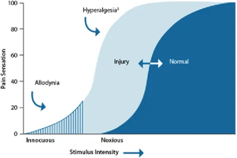
Mechanisms similar to these operate not only in the spinal cord, but also in supraspinal levels such as the somatosensory, anterior cingulate, prefrontal and insular cortex, amygdala or gray matter periaqueductal (Baron et al., 2010). They have been described even in brain areas not directly associated with the processing of sensory information. This aberrant plasticity results in the apparition of pain in response to innocuous not-painful stimulus (allodynia) and exaggerated pain after a stimulus that in normal conditions is painful, which becomes much more painful (hyperalgesia) in the area of the lesion as well as in remote spraspinal areas, which may persist for very long periods of time (Figure 6) (Campbell et al., 2006; Latremoliere and Woolf, 2009).
Figure 6: Representation of the difference between allodynia and hyperalgesia. In allodynia, an innocuous stimulus is perceived as painful. With
hyperalgesia, there is an increased sensitivity to injury (Taken from Medscape).
Loss inhibitory systems:
Inhibitory interneurons of the spinal cord are responsible for restringing the development of hyperalgesia and allodynia after neural damage. In neuropathic pain, there is a loss in the number of these inhibitory interneurons. In the remaining neurons there is a decrease in the expression of inhibitory receptors (in the primary afferents and in postsynaptic neurons) after the nerve injury that contributes to the50
sensitization and exacerbation of pain due to a reduction in the transmission of the supraspinal inhibitory signals (Kohno et al., 2005). The dorsal horn of the spinal cord receives afferent fibers from the supraspinal centers that have a modulator function in the descendent control of pain: the loss of descendent inhibitory signals (opioid, serotonergic and noradrenergic) that are originated mainly in the PAG and in the locus coeruleus, contributes to hyperalgesia and central sensitization as well as pain chronification (Fields et al., 2006). With the loss of neuronal input (deafferentation) the spinothalamic tract neurons begin to fire spontaneously, a phenomenon designated "deafferentation hypersensitivity” (Hanakawa, 2012).
Not only neurons are involved in neuropathic pain but also Schwann cells, satellite cells of the dorsal root ganglia, microglia, astrocytes and components of the immune system (P substance, bradykinin, PRGC, NO, macrophages, lymphocytes T, cytokines, etc.) (Scholz et al., 2007; Austin and Mohalem-Taylor, 2010). Microglia constitutes one important source of inflammatory mediators, thus inducing the release of allogenic molecules and respond to pro-inflammatory signals released by other non-neuronal cells, mainly immune cells (Grace et al.,2011; Mika et al.,2013).
Following peripheral injury, microglia proliferate, become hypertrophic and activated and secrete molecules which sensitize sensory neurons in the spinal cord. The best-characterized mechanism involves the activation of microglial purinergic receptors P2X4 by ATP secreted from damaged neurons, astrocytes or both. This stimulates microglia to release brain-derived neurotrophic factor (BDNF), which activates its neuronal receptors (Tsuda, 2016; Malcangio, 2016). Glia have emerged as key contributors to pathological and chronic pain mechanisms. Upon activation, these mediators cause an inflammatory reaction, hyperemia and chemotaxis, contributing to the nociceptive sensibility and inducing the appearance of hyperalgesia and allodynia (Costigan et al., 2009; Chiu et al., 2012).
The intercommunication neuron-astrocyte-microglia after the injury of the CNS seems to be based on an exchange of molecules such as cytokines (Milligan and Watkins, 2009, Baron et al., 2010; Calvo et al., 2012; Skaper et al., 2012; Taves et al., 2013). Transforming growth factor-β (TGF- β) constitutes a family of pleiotropic, contextually acting cytokines (Massagué,
51
2012). Emergent evidence supports a protective role for TFG-β signalings againts the pathological neural plasticity underlying neuropathic pain in animal models (Echeverry et al., 2009; Tramullas et al., 2010; Lantero et al., 2012; Echeverry et al., 2013). Recently is has been discovered the existence of crossed interactions between TGF-β signaling and some microRNAs, through feedback circuits (Butz et al., 2012). Most of the elements of the TGF-β signaling pathway are regulated by miRNAs and, at the same time, TGF-β signaling increases the biogenesis of a subgroup of miRNAs, leading to a bidirectional interaction. Some of the miRNAs interrelated to TGF-β have been related to the aberrant neuronal plasticity that underlies the development of tolerance to opioid analgesia (Rodríguez, 2012). Previous results from our group also evidence that the interaction between miR-30c-5p and its target TGF-b modulated the endogenous opioid system (Tramullas et al., 2018). Therefore, we postulate that therapies focused on modulating the crosstalk between TGF-β signaling and miRNAs could constitute an alternative strategy for chronic pain treatment.
2. Epigenetics
2.1 Chromatin structural organization
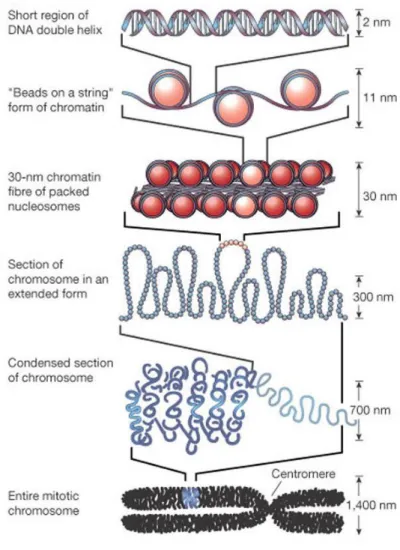
Genome of eukaryotic organisms face the enormous challenge of packing an incredibly long linear molecule of DNA into a restricted nuclear volume. To solve this problem, DNA is tightly packaged and associated with basic proteins called histones, forming the chromatin. The basic repeat element of the chromatin is the nucleosome, which consist of a histone octamer and 146 base pairs of DNA wrapped 1.7 turn tightly around it. This histone octamer consists of a central (H3/H4)2 tetramer flanked on either side by two H2A/H2B dimers. Each nucleosome is separated from the next by linker DNA (10-80 base pair long), associated with histone H1 (Felsenfeld and Groudine, 2003). This DNA-nucleosome complex forms a fiber of 11 nm in diameter known as “beads on a string” (Olins and Olins, 1974; Woodcock, 1973). These fibers are then coiled to a helical structure known as the 30 nm fiber, which in turn is condensed to form chromosomes which are visible through light microscope in dividing cells (Figure 7)(van Holde and Zlatanova, 1995).
52
The structure of the chromatin is highly dynamic and it can switch between the heterochromatin (condensed) and the euchromatin (relaxed) form. This flexible structure allows the chromatin to function properly in the cell to package DNA into the nucleus, to strengthen the DNA during mitosis and meiosis and to control gene expression, DNA replication and DNA repair (Felsenfeld and Groudine, 2003). To achieve this high level of coordination in the nuclear processes, cells have developed several mechanisms to spatially and temporally modulate chromatin structure and function on specific loci in the genome. These mechanisms involve chromatin remodeling, incorporation of histone variants and covalent modifications of histones. The “histone tails” (the amino terminal ends of histones) are extended outside the nucleosome core. Thus, they are accessible to enzymes for chemical modifications which in turn affect the histone-DNA interaction and modulate chromatin structure. Several different types of histone modifications are known, including acetylation, methylation, phosphorylation, ubiquitination, sumoylation and decimation. The combination of these modifications would produce over a million different possibilities for each nucleosome (Bhaumik et al., 2007; Turner, 2007). Unsurprisingly, this astounding condensation of the genome represents a sizeable obstacle to DNA-templated processes such as transcription, replication, and DNA repair. Eukaryotic genomes have dealt with this problem by dynamically manipulating chromatin structure in order to expose underlying DNA sequences. Since histones are intimately associated with DNA, they play an important role in this process.
2.1.1 Types of chromatin
Euchromatin:
For transcription to be possible, chromatin must have an open conformation to facilitate the access to the transcription machinery. This state of “accessible” chromatin is called euchromatin and it is visualized as pale regions of the nucleus, in the optical microscope images, or as scattered chromatin domains with electron microscopy. In neurons, euchromatin represents a large proportion of the genome. This is because the neurons are cells that express a great number of genes that are “ready” to be quickly activated, as is the case of proto-oncogenes (Nagel et al., 2016).53
Figure 7: The organization of DNA within the chromatin structure. The lowest
level of organization is the nucleosome, in which two superhelical turns of DNA (a total of 165 base pairs) are wound around the outside of a histone octamer. Nucleosomes are connected to one another by short stretches of linker DNA. At the next level of organization, the string of nucleosomes is folded into a fiber of about 30 nm in diameter, and these fibers are then folded into higher-order structures (Taken from Alberts et al., 2002).
In the microenvironment of the euchromatin, DNA is very dynamic and can be “unrolled” so that it can be accessed by the transcription, replication or repair machinery of transcription. A determining factor in the configuration of the euchromatin is the histones post-transcriptional modification pattern, “the histone code” characterized by high levels of acetylation of nucleosomal histones as well as by the trymethylation in H3K4, H3K36 and K3K79. This open conformation of chromatin not only facilitates the access of the molecular machinery that carries out the different cellular processes, but also
54
increases the vulnerability to DNA damage, given its greater exposure to all types of genotoxic agents (Kouzarides, 2007; Wagner et al.,2012; Mahrez et al.,2016; Hocher et al.,2018).
Heterochromatin:
The chromatin organization, in two main functional states, was firstly described by Heitz in 1928. While euchromatin constitutes an open and accessible conformation, heterochromatin is highly condensed and transcriptionally inactive. One important function of heterochromatin is to protect the DNA from the transcriptional machinery. Furthermore, heterochromatin is divided into facultative and constitutive. Facultative heterochromatin contains the genes that encodes proteins but should remain silenced during cell differentiation. On the other hand, constitutive heterochromatin corresponds to areas of the genome that are permanently silenced and generally lacks genes that encode proteins. In most of the organisms, constitutive heterochromatin occupies a delimited volume that is grouped in the pericentromeric and telomeric regions. These areas, poor in genes are formed by tandem repeat sequences, also called satellite, that can range from 5 bp to hundreds (Eymery et al.,2009; Sullivan et al.,2017; Tosolini et al.,2018).The heterochromatin is characterized by some post-translational modifications in the histones that conform the nucleosomes. The most frequent mark in heterochromatin histones is the global hypoacetylation, which contributes to the chromatin packaging. Additionally, the heterochromatin is enriched in specific methylation marks. One of the main mark of constitutive heterochromatin is the trymethylation of the histone H3 in the lysine 9 (H3K9me3) while facultative heterochromatic is marked by the trymethylation of the same histone but in the lysine 27 instead on the lysine 9 (H3K27me) and by the trymethylation of histone H4 on lysine 20 (H4K20me3) (Eymery et al., 2009; Shirai et al., 2017; Tosolini et al., 2018; Zhang et al., 2018). Despite having different trymethylation marks, the global result is the same, the chromatin fiber packaging.
2.2 Epigenetics, an overview
Complex organism need epigenetics. While the genetic information in all the cells of an organism is identical, cellular identity and function are highly
55
diverse. This means that some specialized genes that determine the phenotype of differentiated cells are permanently turned on, and other genes, active in some other cell types, are permanently turned off. Moreover, this cellular identity is mitotically stable, and even when differentiated cells such as fibroblasts or lymphocytes are extracted from their endogenous environment and maintained in the artificial medium of cell culture, maintain their phenotypes through cell division. These terminally differentiated cells are derived from undifferentiated precursors called stem cells. Here an undifferentiated cell divides to produce a differentiated cell, and another undifferentiated stem cell. In the case of bone marrow stem cells, a variety of blood cell types are produced. Thus in addition to its relative stability in differentiated cells, genomic output must be highly malleable during the process of cellular differentiation. This astounding regulatory feat is achieved by a wide range of phenomena that can be collectively grouped under the definition of ‘epigenetic’, a term initially coined by Conrad Waddington in 1942 to entail, “processes by which genotype gives rise to phenotype” (Waddington, 2012).
The word “epigenetics” means “in addition to changes in the genetic sequence” and refers to those heritable changes in gene expression that occur without changes in the DNA sequence (Waddington, 1942). This term has evolved to include all those processes that alter gene activity without changing the DNA sequence and lead to modifications that can be transmitted to the daughter cells (Wu et al., 2001). All cells of multicellular complex organisms contain the same genetic information but during development, each single cell differentiates into a specific phenotype without any changes in the DNA sequence. This feature implies that the accuracy of epigenetics modifications is crucial for maintaining the genome integrity and the cell phenotype. Aberrant epigenetic modifications are associated with different heritable and non-heritable diseases. Indeed, epigenetics contributes to the understanding of mechanisms underlying different diseases for which genetic mutations are not the only cause. Epigenetic marks include a variety of gene regulatory events, such as chromatin structure remodeling, histone modifications, DNA methylation and small noncoding RNA, that do not entail changes in the DNA sequence. Therefore, epigenetic events regulate gene
56
expression at both transcription (histone modification and DNA methylation) and translation (small noncoding RNA) levels.
2.2.1 DNA methylation
DNA methylation is an epigenetic modification essential for maintaining genomic stability, specifying cell fate, genomic imprinting (Li et al., 1993), X-chromosome inactivation and stabilization (Heard et al., 1997; Sado et al., 2000), protection against retroviruses and transposons (Walsh et al., 1998) and gene expression regulation (Bird, 2002).
Cytosine nucleotides can be methylated in the 5’ position on their pyrimidine ring. Cytosine nucleotides which directly precede guanine nucleotides are known as CpG dinucleotides (Bird, 1986) (where ‘p’ represents the phosphate bond between cytosine and guanine). DNA methylation occurs almost exclusively in the symmetrical CG context and affects approximately 80% of all CpGs (Ehrlich et al., 1982). DNA methylation is also found at sites other than CpGs sequences. This type of methylation is referred to as non-CpG methylation and includes methylation at cytosines followed by adenine, thymine or another cytosine. Non-CpG methylation is nearly absent in adult somatic cells and comprises only 0.02% of total methyl-cytosine in differentiated somatic cells (Lister et al.,2009; Laurent et al.,2010). Non-CpG methylation seems to have different functions in mouse and human brain tissue. Specifically, it is likely to be correlated with gene activity in human brain tissue, but is negatively correlated with gene activation in the mouse frontal cortex (Lister et al., 2009). Recent studies have, however, shown that non-CpG methylation is high in pluripotent stem cells and several other cell types (such as oocytes) and is important for gene regulation (Sharma et al., 2015; Patil et al., 2016). More recently, it has been discovered that non-CpG methylation plays a role in cardiac gene programing during development (Zhang et al., 2016).
The enzymes responsible for DNA methylation are the DNA methyl transferases (DNMTs). These enzymes catalyze the transfer of the methyl group from S-adenosyl-L-methionine (SAM) to the 5’ position of the cytosine ring (Figure 8). DNMT1, 2, 3A, 3B and 3L are the main DNMTs that belong