1
Self-Assembling Elastin-Like Hydrogels for Timolol Delivery:
2
Development of an Ophthalmic Formulation Against Glaucoma
3
Alicia Ferna
ndez-Colino,
́
†,§,∥Daniela A. Quinteros,
‡,∥Daniel A. Allemandi,
‡Alessandra Girotti,
† 4Santiago D. Palma,
*
,‡and F. Javier Arias
*
,†5†Bioforge Lab, University of Valladolid, CIBER-BBN, Paseo de Belen 19, 47011 Valladolid, Spaiń
6‡Unidad de Investigacion y Desarrollo en Tecnología Farmacé ́utica (UNITEFA), CONICET and Departamento de Ciencias 7 Farmaceuticas, Facultad de Ciencias Químicas, Universidad Nacional de Có ́rdoba, Ciudad Universitaria, 5000-Córdoba, Córdoba, 8 Argentina
9
*
S Supporting Information10 ABSTRACT: This work focuses on improving the eff ective-11 ness of current therapies against glaucoma by incorporating 12 self-assembled polymers into the ophthalmic formulation. To 13 that end, we first studied the influence of the dispersing 14 medium on the mechanical performance of self-assembling 15 elastin-like (EL) and silk-elastin-like (SEL) hydrogels by 16 conducting rheological tests. These polymers were subse-17 quently incorporated into the antiglaucoma formulation, which 18 contains timolol maleate (TM) as active ingredient, andin vivo
19 tests, namely adhesion tests and intraocular pressure measurements (IOP), were performed in New Zealand rabbits. An 20 enhanced reduction in IOP due to the presence of the polymers was observed. Moreover, differences in the effectiveness between 21 both EL- and SEL-hydrogels, which can be explained on the basis of the different rheological properties displayed by these two 22 systems, were also encountered. The results point to the potential of this system as a basis for the development of an ophthalmic 23 formulation against glaucoma.
24 KEYWORDS: glaucoma, silk-elastin-like recombinamers, elastin-like recombinamers, thermo-gelling, ophthalmic formulation
1. INTRODUCTION
25Glaucoma is the second leading cause of blindness worldwide1 26and is a multifactorial, progressive and neurodegenerative 27disease characterized by atrophy of the optic nerve and loss of 28retinal ganglion cells that can eventually lead to loss of visual 29acuity and visual field. High intraocular pressure (IOP) is 30considered to be the greatest risk factor for the development of 31glaucoma, therefore most treatments involve the chronic 32application of eye drops containing hypotensive agents. 33Timolol maleate (TM) is a small, hydrophilic molecule (432 34Da) which is the US Food and Drug Administration’s (FDA) 35“gold standard” drug for the treatment of high IOP.2 Indeed, 36the IOP-lowering potential of this β-receptor antagonist has 37been reported to be between 20% and 25% from the initial 38values.3
39 Topical instillation of this and other hypotensive drugs is 40preferred in order to minimize systemic side effects.4 41Ophthalmic drug delivery is one of the most interesting and 42challenging endeavors facing the pharmaceutical sector as the 43anatomy, physiology, and biochemistry of the eye render this 44organ exquisitely impervious to foreign substances.5 As such, 45most drugs are hardly absorbed, with bioavailabilities ranging 46from 1% to 10%. Among other factors, such low bioavailabilities 47are a consequence of a rapid and extensive loss of the 48formulation from the precorneal area due to the turnover of
49
lacrimal drainage, which decreases the residence time of the
50
formulation on the eye surface and hampers the efficiency of
51
this route.5 Consequently, repeated and frequent applications
52
of topical ophthalmic formulations are usually required to
53
achieve the desired therapeutic effect. Glaucoma treatments are
54
usually associated with adverse reactions generated by frequent
55
exposure of the eye to drugs and excipients. With regard to
56
excipients, preservatives can induce ocular surface alterations
57
that contribute to the development of secondary ophthalmic
58
diseases, such as dry eye syndrome, which, in turn, can
59
compromise patient compliance. However, the elimination of
60
preservatives from ophthalmic formulations is not always
61
sufficient to avoid side effects on the ocular surface. The
62
development of topical ophthalmic formulations for the
63
treatment of this disease therefore presents a challenge.6 As
64
such, the incorporation of new components with beneficial
65
properties into ophthalmic formulations that are also able to
66
increase the bioavailability of the drug is of great interest in this
67
field.7
Received: July 17, 2017
Revised: October 9, 2017
Accepted: November 10, 2017
Published: November 10, 2017
Article
pubs.acs.org/molecularpharmaceutics
68 The incorporation of viscosifying agents that are able to 69increase the residence time of the formulation in the eye is 70gaining increasing attention. Among these compounds, in situ 71gel-forming formulations, which undergo a phase transition 72from a liquid to a semisolid gel upon exposure to physiological 73environments, are a promising approach. These formulations 74should be free-flowing liquids at room temperature to allow 75easily reproducible administration into the eye as a drop. They 76should also undergo an in situ phase transition to form a gel 77that is able to withstand shear forces in the cul de sac and of 78sustaining drug release under physiological conditions.8 As 79such, biomaterials science is fast becoming a cornerstone to 80help to meet the therapeutic challenges faced by ophthalmol-81ogists when treating glaucoma.6,9
82 An excellent approach to in situ hydrogel formation takes 83advantage of the self-assembling nature displayed by some 84protein-based polymers.10 Many self-assembling motifs have 85been explored11,12in materials science, with those inspired by 86the sequence of elastin13(elastin-like polymers, ELPs), silk14 87(silk-like polymers, SLPs) or by a combination of the two (silk 88elastin like polymers, SELPs) being especially relevant. 89Furthermore, in order to ensure strict control over the 90sequence, chain complexity and monodispersity, recombinant 91DNA technology has been implemented to bioproduce this 92class of materials, therefore a new term, namely recombinamer, 93has been established to refer to the polymers produced using 94recombinant technology.15,16
95 The ELP or their recombinant counterparts, known as
96elastin-like recombinamers (ELR),17are artificial polypeptides 97that are bioinspired in the natural elastin. The native elastin is 98an insoluble protein formed by the interaction of various 99molecules of tropoelastin (its soluble precursor). Tropoelastin 100is composed of two different types of domains, namely cross-101linking domains and elastomeric structural domains, which are 102formed by repeating sequences as poly(VPG), poly(VPGG),
103poly(GVGVP), poly(IPGVG), poly(VAPGVG), where V
104stands for valine, P for proline, G for glycine, A for alanine, 105and I for isoleucine. The sequences of the elastin-based artificial 106polymers mimic these patters, and the vast majority have the 107formula (VPGXG)n, wherenis the number of repetitions and 108“X”represents any amino acid except proline. An amphiphilic 109ELR tetrablock copolymer from this family, which contains two 110different kinds of blocks, and was developed to generate 111micropatterned biocompatible hydrogels, has been reported 112previously.18 The hydrophobic blocks, which follow the 113patterned poly(IPGVG), are responsible for physical cross-114linking of the hydrogel (by means of hydrophobic contacts)
115when the system reaches body temperature due to the
116characteristic inverse temperature transition (ITT)19 experi-117enced by these materials. Briefly, this transition involves the 118conformational change of the hydrophobic elastin-like domain
119
from a soluble, random state at low temperature to an
120
aggregated state characterized by a succession ofβ-turns above
121
a specific temperature20,21known as the transition temperature
122
(Tt). The remaining two blocks of the molecule are
123
characterized by the presence of VPGEG pentapeptides,
124
where E stands for glutamic amino acid. The amino acid
125
glutamic displays a carboxylate group which is ionized at neutral
126
pH and therefore it provides a hydrophilic character. Therefore,
127
VPGEG-containing blocks are a random, soluble state under
128
physiological conditions (37 °C, neutral pH),22 and exert a
129
water-retention function required for an hydrogel state.
130
Silk-like recombinamers are bioinspired by the sequence of
131
silk.23 One of the most popular motifs is the hexapeptide
132
GAGAGS (where A and S stand for the amino acids alanine
133
and serine, respectively), which is naturally present in the heavy
134
chain of silkfibroin produced by the wormBombyx mori.24The
135
interest in this domain is due to its ability to mediate
136
irreversible and stable physical interactions by adopting a β
-137
sheet conformation. In this respect, the combination of both
138
kinds of domains has given rise to the so-called silk-elastin like
139
recombinamers. (EIS)x2, which belongs to this family, is
140
constituted by a combination of EL blocks and SL blocks, with
141
the former being found in a tetrablock, thermally triggered,
142 f1
amphiphilic molecule, equivalent to that of (EI)x2 (Figure 1).
143
This material self-organizes from a sol state to a stablefibrous
144
gel state.22 In (EIS)x2, EL blocks clearly dominate the final
145
structure compared to other SELRs found in the literature. The
146
small proportion of SL blocks to EL blocks used in this
147
construct allows to maintain the self-assembling properties of
148
the EL-blocks.
149
Self-assembly processes of protein-based polymers (e.g., ELR
150
and SELR) have been reported to be influenced by
environ-151
mental conditions, such as pH, temperature, and sonication,25
152
and this feature has been exploited to create devices that are
153
able to sense surrounding stimuli.26 Similarly, this fact opens
154
the door to further tuning the properties of a given material by
155
controlling the external inputs to which it is exposed. With
156
regard to ELR self-assembly, it has been reported that the
157
composition of the dispersing medium plays a pivotal role in
158
determining the nanometer-size features of the resulting
159
micelle-like ELR nanoparticles.27,28 However, many of these
160
studies have focused on the change of properties on a
161
nanoscale, and little attention has been paid to the possible
162
effects on macroscale performance.
163
In light of the above, this work focuses on determining the
164
influence of such variables on the physical properties of the
165
hydrogel, as well as the translation of such fundamental studies
166
to the practical aim of developing an ophthalmic antiglaucoma
167
formulation. Thus, four different types of aqueous solutions,
168
namely deionized water, glucose 5%, NaCl 0.9%, and PBS
169
(phosphate buffered saline), were selected according to their Figure 1.Schematic diagram showing the different domains of the recombinamers (EI)x2 and (EIS)x2.
DOI:10.1021/acs.molpharmaceut.7b00615 Mol. PharmaceuticsXXXX, XXX, XXX−XXX
170relevance for their envisaged biomedical application, PBS and 171NaCl 0.9% and glucose 5% are highly used solutions in 172biomedical research and clinics, and the three of them provide a 173osmolarity equivalent to that found in physiological conditions 174(270−330 mOsmol/L). Deionized water was included as the 175control dispersing medium, characterized by the absence of 176solutes.In situgelation behavior in these solutions was studied. 177 Furthermore, the use of both recombinamers as components 178of an ophthalmic formulation against glaucoma was also 179evaluated by performing in vivo irritation tests, adhesion tests 180and IOP measurements in New Zealand rabbits.
2. EXPERIMENTAL SECTION
181 2.1. Materials. TM was supplied by Parafarm (Buenos
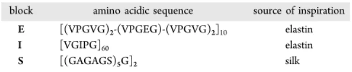
182Aires, Argentina) and sodium chloride by Cicarelli Reagents 183(Rosario, Argentina). Glucose 5% was supplied by Roux Ocefa 184(Buenos Aires, Argentina), and (EI)x2 and (EIS)x2 were 185produced by recombinant technology and purified by Bioforge 186laboratory as reported elsewhere.15,18,22The nomenclature used 187for referring to each block contained in each recombinamer is
t1 188provided in Table 1 and the amino acid sequences of the
t2 189different recombinamers are shown inTable 2.
190 Deionized water was used in all experiments. PBS
191(phosphate buffered saline: KH2PO4 0.0144% (w/v); NaCl 1920.9% (w/v); and Na2HPO40.0795% (w/v), pH 7.4) and NaCl 193were of analytical grade and were used without further 194purification.
195 2.2. Production of (EI)x2 and (EIS)x2. The gene
196sequences encoding for (EI)x2 and (EIS)x2 were available in 197the laboratory from previous studies18,22 and had been 198constructed through standard molecular biologic techniques. 199Specifically, we used the iterative- recursive directional ligation 200(RDL) method,29 which allows controlled and sequential 201concatenation of the gene segments, resulting in a multiblock-202coding gene. The multiblock-coding genes sequences encoding 203for (EI)x2 and (EIS)x2 were subcloned into a modified version 204of pET-25(+) expression vector. Finally, they were transformed 205into the E. coli strain BL21 Star (DE3) (Invitrogen) for 206subsequent expression and production of the recombinamers. 207The purification protocol consisted of sequential rounds of 208inverse transition cycling (ITC). The purity and molecular 209weight of the recombinamers were routinely determined by 210sodium dodecyl sulfate polyacrylamide gel electrophoresis 211(SDS-PAGE), NMR (nuclear magnetic resonance) analysis 212amino acid analysis, and mass spectrometry (MALDI-TOF/ 213MS).
214
2.3. Rheology Tests.The mechanical properties of (EI)x2
215
and (EIS)x2 hydrogels were measured in a controlled stress
216
rheometer (AR2000ex, TA Instruments) equipped with a
217
Peltier plate temperature control. Thus, 350 μL of each
218
recombinamer solution at 15 wt% in the corresponding
219
dispersing medium (deionized water, glucose 5%, NaCl 0.9%,
220
and PBS) were placed on the Peltier plate of the rheometer
221
precooled to 5°C. A parallel plate geometry with a diameter of
222
20 mm was used. Temperature ramp experiments were
223
performed by heating the sample from 5 to 37 °C. The
224
heating rate was 2.5°C/min, and the reverse process (cooling)
225
was performed under the same conditions. A constant strain of
226
0.5% (within the linear viscoelastic region) and a frequency of
227
10 Hz were used.
228
2.4. Differential Scanning Calorimetry (DSC). DSC
229
experimentswere performed using a Mettler Toledo 822e with
230
liquid-nitrogencooler. Both temperature and enthalpy were
231
calibrated using a standard indiumsample. (EI)x2 and (EIS)x2
232
samples for the DSC measurements wereprepared at 15 wt % in
233
PBS, NaCl 0.9%, deionized water and glucose 5%.A volume of
234
20μL of the corresponding sample was placed inside astandard
235
40 μL aluminum pan and sealed hermetically. The heating
236
program for DSC measurements included an initial isothermal
237
step (5 min at 0°C to stabilize the temperature and the state of
238
the tetrablock), followed by heating from 0 to 60°C at 5°C/
239
min.
240
2.5. In Vitro Erosion Testing of the Recombinamers.
241
(EI)x2 and (EIS)x2 solutions at 15 wt% (1 mL) were prepared
242
in dextrose 5% and kept at 35°C to achieve a gel state. Then,
243
3.5 mL of buffer solution pH 6.8 was added with gentle shaking
244
at 50 rpm. Atfixed times, 250 μL samples were removed and
245
replaced by fresh buffer. The erosion of the hydrogels was
246
determined using Biuret reagent (CuSO4 at 2% + NaOH at 247
40%). The concentration of (EI)x2 and (EIS)x2 in the released
248
medium was determined by UV spectrophotometry at a
249
maximum absorbance wavelength of 540 nm (UV VIS
250
TERMO Evolution 300). All experiments were performed in
251
triplicate.
252
2.6. In Vitro Drug-Release Studies. Solutions of (EI)x2
253
and (EIS)x2 (15 wt %) containing TM (0.5 w/v.%) (1 mL)
254
were prepared and subsequently kept at 35 °C for 10 min to
255
ensure hydrogel formation. Thereafter, 3.5 mL of buffer
256
solution pH 6.8 was added with gentle shaking at 50 rpm. At
257
appropriate intervals, 250 μL samples were removed and
258
replaced with fresh buffer. The quantity of TM in the release
259
medium was determined by high-performance liquid
chroma-260
tography (HPLC, seeSection 2.6). Each sample was assayed in
261
triplicate (n = 3). Mathematical analysis was performed by
262
adjusting the experimental values to the Korsmeyer−Peppas
263
model.
= ·
∞
M
M k t
n
t
264
In the above,Mtis the cumulative amount of drug released at 265
timet,M∞is the cumulative amount of drug released at infinite
266
time, k is a rate constant incorporating characteristics of the
Table 1. Correspondence of the Abbreviations Used To Name Each Block that Composes Each Recombinamer
block amino acidic sequence source of inspiration
E [(VPGVG)2-(VPGEG)-(VPGVG)2]10 elastin
I [VGIPG]60 elastin
S [(GAGAGS)5G]2 silk
Table 2. Amino Acid Sequence of Each Recombinamer
abbreviated name amino acid sequence
(EI)x2 MESLLP-{[(VPGVG)2-(VPGEG)-(VPGVG)2]10[VGIPG]60}2-V
(EIS)x2 MESLLP-{[(VPGVG)2- (VPGEG)-(VPGVG)2]10[VGIPG]60)-[V(GAGAGS)5G]2}2-V
DOI:10.1021/acs.molpharmaceut.7b00615 Mol. PharmaceuticsXXXX, XXX, XXX−XXX
267macromolecular network of the system and the drug, and nis 268the release exponent, which is related to the mechanism of drug 269release. Ifn= 0.5, the release is governed by Fickian diffusion, 270whereas n= 1 indicates that molecules are released by surface 271erosion; both mechanisms play a role in release for n values 272between 0.5 and 1.30
273 2.7. HPLC Determinations of TM. The HPLC system
274consisted of a Waters HPLC pump and a Waters HPLC
275detector set at 295 nm. Samples were chromatographed on a
276reversed-phase Luna C18 column (250 × 4.6 mm, 5 mm,
277Phenomenex) and a 2×8 mm precolumn of the same material, 278with the mobile phase having a flow rate of 1 mL/min and 279consisting of trifluoroacetic acid 0.05% (v/v) in acetonitrile 280(40:60, v/v), which was filtered and degassed before use. The 281column was thermostated at 25 °C.
282 2.8. Cytocompatibility. The HFF-1 (human foreskin
283fibroblast) cell line was used as cell model to test the 284cytocompatibility of the recombinamers. Thus, 7500 HFF-1 285cells were seeded onto 96-well culture plates, then the culture 286medium was removed after 5 h and replaced with 100μL of the 287corresponding recombinamer solution at 25 μM in culture
288medium (DMEN). A 100 μL aliquot of DMEN (with no
289recombinamer) was added in the case of the negative controls. 290Live and dead staining (LIVE/DEAD Viability/Cytoxicity 291Assay Kit, Invitrogen) was used according to the manufacturer’s 292instructions, andfluorescence intensity emission was measured
293at 425 and 620 nm after excitation at 485 and 525 nm
294(SpectraMax M5e microplate reader, Molecular Devices), 295respectively, after culture for 24 h. The number of live and 296dead HFF-1 cells was calculated from thefluorescence intensity 297using a calibration curve obtained with known numbers of 298HFF-1 cells seeded on 96-well plates (from 1000 to 20 000 cells 299per mL, using 100 μL of DMEN medium). Statistical analysis 300was performed by one way analysis of variance (ANOVA). 301Images of the cells after Live and dead staining were taken with 302a Nikon Eclipse Ti-SR (Japan)fluorescence microscope. Three 303independent experiments, each in triplicate, were performed for 304each recombinamer.
305 2.9. In Vivo Mucoadhesion Tests. Solutions of each 306recombinamer at 15 wt % with sodiumfluorescein at 0.25% in 307dextrose solution at 5% were prepared, and 50 μL of the 308corresponding solution was placed in the inferior conjunctival 309fornix of the right eyes of three rabbits. The left eyes were used 310as controls and were treated with 50 μL of commercial 311fluorescein at 0.25%.
312 The behavior of each solution in terms of residence and 313adherence on the eye surface was evaluated using a binocular 314indirect ophthalmoscope (Neitz IO- small pupils, Tokyo, 315Japan) and 20 diopter lens (Nikon, Tokyo, Japan) and a score 316was calculated for each time point according to the parameters t3 317presented in Table 3. A one-way ANOVA statistical analysis
318was performed and the Holm−Sidak method was applied.
319 2.10. In Vivo Study of the Hypotensive Efficacy: IOP
320Determinations.Experiments were performed in both eyes of
321nonsedated normotensive male New Zealand white rabbits (2− 3222.5 kg). The animals were kept in individual cages with free 323access to food and water and maintained in a controlled 12/12 324h light/dark cycle. Animal management procedures conformed
325to the ARVO (Association for Research in Vision and
326Ophthalmology) resolution on the use of animals in research, 327the European Communities Council Directive (86/609/EEC),
328and the Institutional Care and Use Committee of the
329
Chemistry Faculty of Córdoba University (Córdoba,
Argenti-330
na) reviewed and approved the protocols.
331
The corresponding recombinamer, namely (EI)x2 or
(EIS)-332
x2, was dissolved at 15 wt % in glucose 5% (w/v) solution with
333
0.5% TM (Parafarm), then 50 μL of each formulation was
334
placed into the conjunctival fornix of the eye of the rabbits (n=
335
15 for each formulation). As controls for the recombinamer
336
effect, additional rabbits (n= 20) were treated with glucose 5%
337
(w/v) solution with TM 0.5% (with no recombinamer). In
338
order to establish the basal IOP of the animals, additional eyes
339
(n = 18) were treated with glucose 5% (w/v) solution alone
340
(with no recombinamer or TM). IOP was measured using a
341
Tonovet rebound tonometer (Tiolat, Helsinki, Finland), which
342
allows IOP to be assessed without the need for topical
343
anesthesia. For each eye, IOP was set at 100% with two basal
344
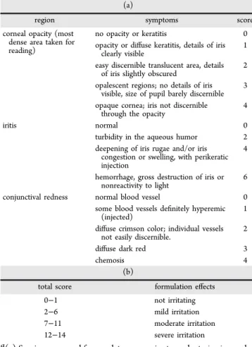
readings taken 30 min before and immediately before the
345
instillation. The evolution of the IOP was measured each hour,
346
for a period of up to 8 h.
347
2.11.In VivoOcular Irritation Test.The potential ocular
348
irritancy and/or damaging effects of the samples tested were
349
evaluated using a slightly modified version of the Draize test.31
350
Thus, for each recombinamer sample, a test was carried out on
351
three New Zealand rabbits using a volume of 50 μL of
352
recombinamer solution at 15 wt % in dextrose 5% with TM at
353
0.5%. This solution was placed in the conjunctival fornix of the
354
right eyes, with the left eyes being used as negative control. The
355
commercial solution Zopirol 0.5% TM (Elea Visual) was used
356
as the negative irritation control. Postexposure evaluations of
357
the conjunctiva, cornea and iris were performed by external
358
observation under adequate illumination, and additional
359
information was obtained using a binocular indirect
ophthalmo-360
scope (Neitz IO- small pupils Tokyo, Japan) and 20 diopter
361
lens (Nikon, Tokyo, Japan). For each observation, one drop of
362
fluorescein salt (0.25%) was instilled to contrast the potential
363
corneal injury. The ocular irritation or damage was scored by
364 t4
successive inspections at 0, 30, 60, 90, and 120 min according
Table 3. Proposed Score Rating for Evaluating Bioadhesion
Intensity in Vivoa
tissue/region appearance score
cornea complete 4
3/4 cornea 3
1/2 cornea 2
1/4 cornea 1
nothing 0
conjuntival sac abundant 3
medium 2
Scarce 1
nothing 0
lachrymal meniscus 2 mm 3
1 mm 2
thin line 1
nothing 0
eyelid wet 0
not wet 1
nose wet 0
not wet 1
aThe presence of the formulation is assessed by inspecting several
regions and tissues in the eye, and a score is assigned according to the observed appearance. A total score encompassing the global behavior of the formulation is obtained by summing the scores obtained for each region.
DOI:10.1021/acs.molpharmaceut.7b00615 Mol. PharmaceuticsXXXX, XXX, XXX−XXX
t4 365to the outcomes listed inTable 4a), and the total score for each 366formulation at each time point was calculated by summing the
367scores obtained for each region. The effects of the formulation 368in terms of irritation, namely no irritation or mild, moderate, or 369severe irritation, were established by comparing the total score 370obtained for each condition withTable 4b).
3. RESULTS AND DISCUSSION
371 3.1. Rheology and DSC. Rheological measurements of
372(EI)x2 and (EIS)x2 at 15 wt % in the four different types of 373dispersing media under study, namely PBS, NaCl 0.9%, glucose 3745%, and deionized water, all of them at physiological pH, were 375performed in order to check the possible influence of the 376composition of the different media on the mechanical 377performance.
f2 378 Figure 2shows the recorded Ǵ (elastic or storage modulus) 379and Ǵ́ (viscous or loss modulus). The gel point is usually 380considered as the point from which the storage modulus 381surpasses the loss modulus, which indicates that the sample has 382transitioned from fluid-like behavior to viscoelastic solid
383behavior. Therefore, the gelation temperature can be
384determined as the crossover temperature between Ǵ-Ǵ́.33 As 385shown in Figure 2, at low temperatures, all the samples 386displayed Ǵ and Ǵ́values of just a few pascals, and Ǵ́ > Ǵ. 387However, this situation changes for all eight samples (both 388recombinamers, each in four different types of dispersing
389
media) in the temperature range 10−22 °C, in which Ǵ
390 t5
surpasses Ǵ́, indicating gel formation (Table 5), as.34
391
Additionally, DSC experiments were performed in order to
392
check the Tt of the recombinamers (Figure S1). It is worth
393
noting that the resulting values are close to the temperature
394
gelation values measured by rheology (Table 5). This similarity
395
is due to the gelation mechanism that governs these materials,
396
in which EL moieties undergo conformational changes from an
397
extended state (below theTt) to a folded one (above theTt).
398
This folded state involves the establishment of hydrophobic
399
contacts, which result in gel formation. For the four conditions
400
tested, (EIS)x2 displayed a slightly higher gelling temperature
401
(between 1 and 2 °C higher) than (EI)x2 under the same
402
conditions (Table 5). The presence of the hydrophilic amino
403
acid serine in the sequence of the SL motif could be responsible
404
for the observed increase in the gelation temperature for
405
(EIS)x2 with respect to (EI)x2.
406
As regards (EI)x2, the gel state was not stable over the whole
407
range of temperatures measured when the dispersing media
408
were NaCl 0.9% or PBS (Figure 2a,c). Although a gel state was
409
clearly present at 20 °C, the mechanical performance of the
410
material was markedly decreased at physiological temperature
411
(37°C). In other words, the gel state was close to disappear for
412
the sample with PBS (Figure 2a) and was almost negligible in
413
the sample with NaCl 0.9% (Figure 2c). It is remarkable that
414
this behavior contrasts with the behavior experienced by the
415
system when the dispersing media were deionized water or
416
glucose 5%, in which there were no signs of instability and the
417
gel states were maintained at 37°C (the value of Ǵwas clearly
418
higher than Ǵ́;Figure 2e,g, andTable 5). This behavior is also
419
patently clear by looking at the tanδvalues. Tanδrepresents
420
the ratio of viscous modulus (Ǵ́) to elastic modulus (Ǵ), and it
421
is therefore a quantifier of the dominance of the elastic or the
422
viscous behavior. As shown inTable 3, tanδvalues were higher
423
when the dispersing medium was PBS or NaCl than when
424
glucose 5% or deionized water were used, indicating a more
425
viscous and less elastic behavior of the former.
426
A similar tendency to that observed for (EI)x2 was found for
427
(EIS)x2, namely the presence of NaCl 0.9% and PBS provoked
428
certain instability in the gel state at 37 °C. However, in this
429
case, this instability did not result in the disappearance of the
430
gel state, as Ǵ was clearly higher than Ǵ́ under these conditions
431
(Figure 2). The slight reduction in the mechanical properties of
432
the gel state observed upon warming from 20 to 37 °C when
433
the dispersing medium was NaCl 0.9% or PBS was not
434
encountered when the medium was deionized water or glucose
435
5%.
436
In summary, the presence of NaCl 0.9% or PBS exerts a
437
destabilizing effect on the maintenance of the gel state at 37°C
438
of both (EI)x2 and (EIS)x2 samples. This destabilization is
439
more prominent for (EI)x2 than for (EIS)x2 due to the
440
presence of the SL block in the latter, which is able to improve
441
the mechanical performance.22
442
Furthermore, no signs of instability or a reduction in the
443
mechanical performance of (EI)x2 and (EIS)x2 when changing
444
the temperature of the samples from 20 to 37°C are observed
445
when the dispersing medium is deionized water or glucose 5%.
446
Taken together, these findings point to an influence of the
447
composition of the dispersing medium on the hydrophobic
448
contacts mediated by the EL-blocks. Such blocks are present in
449
both recombinamers and are responsible for triggering the
450
thermo-gelling process of both systems. When the composition
451
of the dispersing media is analyzed, it can be deduced that the
Table 4. Evaluation Parameters for Ocular Irritationa
(a)
region symptoms score
corneal opacity (most dense area taken for reading)
no opacity or keratitis 0 opacity or diffuse keratitis, details of iris
clearly visible
1
easy discernible translucent area, details of iris slightly obscured
2
opalescent regions; no details of iris visible, size of pupil barely discernible
3
opaque cornea; iris not discernible through the opacity
4
iritis normal 0
turbidity in the aqueous humor 2 deepening of iris rugae and/or iris
congestion or swelling, with perikeratic injection
4
hemorrhage, gross destruction of iris or nonreactivity to light
6
conjunctival redness normal blood vessel 0 some blood vessels definitely hyperemic
(injected)
1
diffuse crimson color; individual vessels not easily discernible.
2
diffuse dark red 3
chemosis 4
(b)
total score formulation effects
0−1 not irritating
2−6 mild irritation
7−11 moderate irritation 12−14 severe irritation
a(a) Scoring proposed for regulatory agencies to evaluatein vivoocular
irritation. The total score is calculated from the sum of all scores obtained for the cornea, iris, and conjunctivae. Adapted from refs
31,32. (b) Formulation effects corresponding to each score value.
DOI:10.1021/acs.molpharmaceut.7b00615 Mol. PharmaceuticsXXXX, XXX, XXX−XXX
452
presence of NaCl plays an important role in this destabilization
453
as it is the only component present in both PBS and NaCl 0.9%
454
solutions, and at the same concentration. The destabilization of
455
the gel state exerted by both media is similar, and the tan δ
456
values (Table 5, II) clearly point to a decrease in the elastic
457
behavior in both dispersing media (PBS and NaCl 0.9%).
458
However, the remaining components of PBS (i.e., KH2PO4and 459
Na2HPO4) seem to accentuate the destabilization of the EL 460
block-mediated gelation to a small extent as the complex
461
modulus (i.e., the overall resistance to deformation, that
462
encompasses both the elastic and the viscous moduli) for
463
(EI)x2 in PBS is slightly lower, and tan δ is higher, than in
464
NaCl 0.9% (Table 5). Moreover, the concentration of KH2PO4 465
and Na2HPO4in PBS is about 10-times lower than the NaCl 466
concentration, which could explain the almost negligible effect
467
of these compounds on the stability when compared to the
468
marked effect of NaCl.
469
It is also noticeable that the modulus displayed by (EI)x2 in a
470
dispersing medium lacking salts, namely deionized water or
471
glucose 5% (Figure 2e,g), was higher than that displayed by
472
(EIS)x2 (Figure 2f,h). This effect could be explained by the
473
lower amount of EL moieties per molecule in relative terms
474
when comparing (EIS)x2 to (EI)x2. Thus, EL moieties
475
constitute 100% of (EI)x2 molecules but only 90% in the Figure 2.Storage (Ǵ) and loss moduli (Ǵ́) for (EI)x2 (left) and (EIS)x2 (right) recombinamer solutions (15 wt%) as a function of temperature in different dispersing media. Measurements were performed at 10 Hz. (a) and (b) PBS. (c) and (d) NaCl 0.9%. (e) and (f) Deionized water. (g) and (h) Glucose 5%.
Table 5. (I) Compilation of the Temperatures at which the
Crossover between Ǵ and Ǵ́ Takes Place for (EI)x2 and
(EIS)x2 in the Different Dispersing Media, and the
Concomitant Complex Modulus (G*) Obtained at 37°C for
Each Recombinamer and (II) Further Detailed Mechanical
Properties (Ǵ, Ǵ́, and tan δ) Displayed by (EI)x2 and
(EIS)x2 in Different Dispersing Media at 37 °C
(I)
Ǵ-Ǵ́crossover (°C) G*(37°C) (Pa)
(EI)x2 (EIS)x2 (EI)x2 (EIS)x2
PBS 18.9 20.8 105 1340
NaCl 0.9% 15.7 16.5 570 1200
deionized water 15.7 17.0 5630 3200
glucose 5% 14.9 15.7 5760 3180
(II)
(EI)x2 (EIS)x2
Ǵ(Pa) Ǵ́(Pa) tanδ Ǵ(Pa) Ǵ́(Pa) tanδ
PBS 84 65 0.77 1320 237 0.18
NaCl 0.9% 520 340 0.65 1150 202 0.18
deionized water 5600 623 0.11 3190 331 0.10 glucose 5% 5740 515 0.09 3160 300 0.10
DOI:10.1021/acs.molpharmaceut.7b00615 Mol. PharmaceuticsXXXX, XXX, XXX−XXX
476case of (EIS)x2. Importantly, neither of these hydrogels was 477destabilized upon changing the temperature from 20 to 37°C 478in these dispersing media.
479 In light of the above, it is clear that dispersing media exert an 480influence on the mechanical properties of both systems, 481although the underlying mechanism responsible for the 482different responses remains unclear. Numerous studies have 483reported the influence of salts on the ITT behavior of ELRs, 484and it is well-known that salts cause a concentration-dependent 485decrease in Ttand an increase in the transition enthalpy.27,35 486With regard to the nanostructured properties of ELR, NaCl has 487been reported to have an influence on the size and shape of the 488resulting nano-objects.27 However, to the best of our 489knowledge, this is the first time that the influence of salts on 490the macroscale behavior of an elastin-based material, namely 491the mechanical performance, has been reported. Diblockcopo-492lypeptide amphiphilic polymers containing charged and hydro-493phobic segments have been reported to be weakened by the 494presence of NaCl, and this effect has been attributed to a 495screening of polyelectrolyte charges, with the authors of this 496contribution stating that highly charged segments contribute to 497gel formation.36Mehta et al. also showed that NaCl can shield 498electrostatic interactions and impact the modulus of the gels.37 499In the case in hand, in other words a weakening of the gel state 500in these elastin-based systems, further research is required in 501order to understand the underlying molecular phenomena that 502result in such changes in the rheological properties. However, 503the aforementioned studies induce us to consider that the 504interaction between the NaCl and the negative charges of the 505glutamic residues of the recombinamers (Table 1andTable 2) 506may be responsible of the decrease in gel stability.
507 From a practical point of view, glucose 5% was selected as 508the dispersing medium for (EI)x2 and (EIS)x2 since the 509stability of both systems (Table 5) is enhanced in this medium. 510 3.2.In Vitro Drug Release Studies.Clear difference can f3 511readily be seen in the drug-release profiles shown inFigure 3.
512Specifically, the (EIS)x2 system shows a more sustained release 513when compared to its (EI)x2 counterpart, with the percentage 514release after 8 h being 80.39% and 40.04% TM for (EI)x2 and 515(EIS)x2, respectively. These data point to a relationship 516between the mechanical properties of the hydrogels and the 517rate of drug release. Thus, in concordance with its enhanced 518mechanical performance when compared to (EI)x2, (EIS)x2 519shows the most sustained release.
520 Drug delivery data were fitted to the Korsmeyer−Peppas 521model in order to further analyze the experimental results and
522
to obtain quantitative information that could facilitate the
523 t6
comparison of both release profiles (Table 6).
524
The rate constantk was lower for the (EIS)x2 formulation
525
than for its (EI)x2 counterpart, thus confirming a slower drug
526
release by the former. Thenvalue obtained for (EIS)x2 (n=
527
0.58) and for (EI)xs (n= 0.62) points to Fickian diffusion as
528
the main mechanism governing the release. However, erosion
529
processes are also involved in drug delivery as the values
530
obtained are higher than 0.5. Specifically, thenvalue for (EI)x2
531
is higher than that for (EIS)x2, which points to a higher
532
influence of erosion processes for (EI)x2. Consequently,
533
erosion tests were performed to corroborate this (see next
534
section).
535
3.3. In Vitro Erosion Testing of the Recombinamers.
536
Erosion tests were performed in order to determine possible
537
differences in the stability between the two recombinamers. As
538 f4
shown in Figure 4, (EIS)x2 displays a significantly lower
539
erosion than (EI)x2. This is in agreement with the presence of
540
the SL motif in (EIS)x2 as said motif has been reported to
541
provide enhanced stability against erosion in an excess of
542
aqueous media inin vitrotests. This performance is likely to be
543
translated into an increase in the residence of ophthalmic
544
formulations containing (EIS)X2. Consequently, the next set of
545
experiments was performedin vivoto test this hypothesis.
546
3.4. Cytocompatibility. Cytocompatibility assays were
547
performed in order to check the suitability of these
548
recombinamers for biomedical applications. A fibroblast cell
549
line (HFF-1) was used as cell model to test the cytocompat-Figure 3.Release profiles of TM from (EI)x2 and (EIS)x2 hydrogels.
Table 6. Values Obtained after Fitting the Drug Delivery
Profiles to the Korsmeyer−Peppas, Higuchi, and Zero-Order
Kinetics Models
formulations TM(EI)x2 TM(EIS)x2
Korsmeyer−Peppas model
k(hs−n) 22.685±0.973 14.930±0.887
n 0.619±0.026 0.588±0.036
R2 0.988 0.973
Higuchi model
k(hs−1/2) 26.988±0.724 16.965±0.469
R2 0.962 0.958
zero-order model
k(hs) 11.831±0.694 7.390±0.496
R2 0.824 0.764
Figure 4.Erosion profiles of (EI)x2 and (EIS)x2 at 15 wt %.
DOI:10.1021/acs.molpharmaceut.7b00615 Mol. PharmaceuticsXXXX, XXX, XXX−XXX
550ibility asfibroblasts are the predominant cell type in the ECM 551and, thereof, represent one of the main portals of exposure to 552biomaterials. A quantitative analysis was performed by 553measuring the corresponding fluorescence emitted by both 554calcein and EtDH-1 under three test conditions, namely HFF-1 555culture treated with (i) (EI)x2, (ii) (EIS)x2, or (iii) without any
556recombinamer (control) for 24 h, as detailed in the
557experimental section.
558 No statistically significant differences in cell viability were
f5 559found between the treatment groups (Figure 5a) and
560microscopic observation of the cells corroborated thesefindings 561(Figure 5b−d). Furthermore, no morphological differences 562were observed between the fibroblasts treated with the 563recombinamers and the control fibroblasts. As such, these 564results show the cytocompatible nature of these recombinamers 565and further support their potential application in the biomedical 566field.
567 3.5. In Vivo Mucoadhesion Tests. Adhesion tests were 568performed in order to determine any differences in retention of 569the formulation on the eye surface that could potentially affect 570topical absorption of the drug.
f6 571 Figure 6 shows that, immediately after instillation of the 572formulations (t= 0), adhesion seems to be higher for (EIS)x2 573than for (EI)x2, with both formulations presenting a higher 574score than the control, although the apparent differences are 575not statistically significant. This trend was maintained
576throughout the study, and between 5 and 15 min, the
577differences between the three groups were statistically 578significant, withp< 0.05 in all cases. No statistically differences 579were detected between the control and the formulation
580containing (EI)x2 between 30 to 45 min, whereas the
581(EIS)x2 formulation still presented statistically significantly 582higher adhesion properties, and this formulation could still be 583detected at 75 min.
584
Thus, formulations containing either of the two
recombi-585
namers displayed better adhesion than the control. This is
586
clearly important as regards the development of ophthalmic
587
formulations since rapid washing-out and shear-thinning of
588
mucoadhesive systems is a considerable obstacle that must be
589
overcome when developing drug carriers to be administered in
590
anatomical sites such as the ocular surface, where the clearance
591
time for the tear film is 5−10 s.38,39Herein we show that the
592
incorporation of either of these two recombinamers results in
593
an increase in the residence time of the formulation on the eye
594
surface. Moreover, differences were also detected between
595
(EI)x2 and (EIS)x2, with the latter being more effective at
596
increasing adhesion. This increase in adhesion can be related to
597
the enhanced rheological properties and lower erosion
598
displayed by the (EIS)x2 system when compared to (EI)x2
599
when NaCl is present, as is the case for the eye surface.
600
3.6. In Vivo Study of the Hypotensive Efficacy: IOP
601 f7
Determinations. As shown in Figure 7, formulations
Figure 5. LIVE and DEAD differential staining of HFF-1 cells following 24 h of TC-PS (tissue culture−polystyrene surface) culture in the presence of DMEN medium supplemented with the corresponding recombinamer. (a) Representation of the percentage of viability (with respect to the control) after 24h of culture of HFF-1 cells in the presence of (EI)x2 and (EIS)x2. Three experiments were performed, each in triplicate. Data are expressed as mean±standard deviation. (b)−(d) Representative fluorescence microscopy images. Live cells appear in green whereas dead cells appear in red.
Figure 6.Adhesion score versus time for the recombinamer solutions and control (solution without recombinamer),n= 3 rabbits. Data are expressed as mean± standard deviation. Statistical significance (p< 0.05) is marked with an asterisk.
Figure 7.IOP evolution in normotensive New Zealand rabbits after administration of different TM formulations. Gray circles: IOP at different times after administration of TM 0.5% solution in glucose 5% (n= 20 eyes). White circles: basal IOP (glucose 5% solution with no hypotensive agent) (n= 10 eyes). Green circles: IOP at different times after administration of the formulation containing TM 0.5% in (EIS)x2 at 15 wt % in glucose 5% (n= 15 eyes). Blue circles: IOP at different times after administration of the formulation containing TM 0.5% in (EI)x2 at 15 wt % in glucose 5% (n= 15 eyes). Data are expressed as mean±standard error.
DOI:10.1021/acs.molpharmaceut.7b00615 Mol. PharmaceuticsXXXX, XXX, XXX−XXX
602containing TM produced a decrease in the IOP, with no 603statistical differences being detected between them in thefirst 3 604h. However, this situation changed at 4 h postadministration, 605when the IOP for eyes treated with the formulation lacking 606recombinamer was statistically significantly higher than that 607displayed by the eyes treated with (EIS)x2 (p< 0.05). From 4 h 608onward, the TM solution presented no hypotensive effects, 609while those formulations containing TM and the respective
610recombinamer, namely (EI)x2 or (EIS)x2, maintained a
611reduced IOP, with this effect being more pronounced for the 612formulation containing (EIS)x2. In this case, the hypotensive 613effect lasted for more than 8 h.
614 Thesefindings are in concordance with our initial hypothesis, 615in which we speculated that elimination of the drug as a result 616of lacrimal turnover may be minimized by the use ofin situ gel-617forming systems, thus leading to an enhanced hypotensive 618effect of the formulation. Furthermore, the differences 619encountered between (EI)x2- and (EIS)x2-containing systems 620agree with an enhanced stability of the latter under saline 621conditions. As NaCl constitutes one of the main components of 622lacrimal fluid,40,41it is expected to diffused over time into the 623gel-formulation and decrease its mechanical properties, 624especially in the case of (EI)x2 (as shown in the rheological 625tests). Therefore, it is rational to suppose that the formulation 626containing (EIS)x2 displays a longer residence time than its 627counterpart containing (EI)x2, as was also demonstrated 628experimentally in the in vitro erosion tests and the in vivo 629adhesion tests, thus leading to an increased hypotensive effect. 630The results show that the formulation containing (EIS)x2 had a 631more sustained effect than a formulation containing just TM, in 632which the hypothesive effect lasted only 4 h. Although 633hypotensive effects of up to 8 h are also displayed by 634commercially available preservative-containing formulations, 635such as Timoftol (FrosstLaboraties, Madrid, Spain) or Timolol 636Sandoz (Frosst Laboratories),42it is important to note that the 637hypotensive effect achieved by the formulation containing 638(EIS)x2 was achieved without the use of preservatives. The 639inclusion of preservatives, such as benzalkonium chloride,43is 640believed to favor TM penetration, and therefore its therapeutic 641efficacy, due to disruption of the hydrophobic barrier of the 642corneal epithelium. However, this adjuvant effect is produced at 643the expense of an increased toxicity and serious side-effects on 644the eye surface.44,45 As such, preservative-free antiglaucoma 645eyedrops are believed to improve patient compliance and 646adherence in the medical treatment of this disease, and the 647introduction of preservative-free formulations that maintain 648efficacy is an important step toward the development of 649ophthalmic solutions.46Moreover, some studies have pointed 650to a possible role of the inclusion of polymers in the 651antiglaucoma formulations in the reduction of ocular toxicity, 652thereby protecting the ocular surface in long-term therapies.42 653 The results reported herein support the feasibility of using 654(EIS)x2 as a component in a preservative-free antiglaucoma 655formulation while maintaining the efficacy of the commercial 656benzalkonium chloride-containing versions. Moreover, the 657thermogelling behavior of this system allows easy self-658administration, thus providing an advantage with respect to 659other polymeric systems in which preformed scaffolds are 660incorporated into the conjunctival sac, which can lead to patient 661discomfort.47,48
662 3.7.In VivoOcular Irritation Test.In order to evaluate the 663safety of the formulation containing (EI)x2 or (EIS)x2 15 wt % 664with TM at 0.5% in dextrose solution when applied topically to
665
rabbit eyes, irritation tests were performed as described in the
666 t7
experimental section. As shown in Table 7, no irritation was
667
observed for either of the recombinamers as the score was less
668
than 1 for all the times tested. Moreover, no significant
669
differences were detected between the recombinamer
for-670
mulations and the control group. The absence of irritation is in
671
agreement with the reported biocompatible nature of this class
672
of materials,49 together with the aqueous base of the
673
formulation and the lack of any preservatives, which maximizes
674
the potential utility of these devices as drug-delivery systems.
4. CONCLUSIONS
675
Although numerous studies have been conducted in the
676
development of new antiglaucoma formulations in order to
677
reduce IOP to a greater extent, further research is still required.
678
In this sense, the combination of biomaterials science and
679
pharmacology is a must in order to find new solutions and
680
approaches to overcome the current problems of ophthalmic
681
drug delivery. Herein we have evaluated thermosensitive elastin
682
and silk-elastin-like recombinamers as innovative
pharmaceut-683
ical dosage forms for the topical administration of TM.
684
Aqueous dispersions of recombinamers remained veryfluid at
685
low temperatures, which facilitated drug incorporation and
686
administration. However, they were able to change into a gel
687
form at physiological temperature so that TM could come
688
entrapped inside the gel and experienced a sustained release.In
689
vivostudies conducted in New Zealand rabbits showed that the
690
incorporation of these recombinamers into a pharmaceutical
691
formulation containing TM prolonged its retention on the
692
preocular surface, leading to a greater decrease in the IOP. This
693
effect was more evident in the case of the silk-elastin-like
694
recombinamer (EIS)x2, which is in agreement with the
695
enhanced stability of this material in the presence of a saline
696
aqueous environment, as it is the scenario of the eye surface.
697
Furthermore, these recombinamers can be placed in the eye
698
without causing irritating effects or tear turnover, as
699
demonstrated by the irritation tests, thereby maximizing the
700
potential utility of these devices as drug-delivery systems.
701
Therefore, (EIS)x2 constitute a novel and versatile type of
702
hydrogel to address the critical issues that ophthalmic drug
703
delivery entails.
704
In view of the above, and considering the potential offered by
705
recombinant technology to develop further designs, the next
706
step will focus on the development of a battery of
707
recombinamers based on these initial designs, in order to
Table 7. Irritation Scores Obtained for the Two
Formulations Tested, Namely (EI)x2 and (EIS)x2 Hydrogel at 15 wt % Containing TM 0.5% in the Dispersing Medium Glucose 5% and the Commercial Solution Zopirol 0.5% TM
as Controla
time (min)
sample 30 60 90 120
(EI)x2 0.33±0.58 0.33±0.58 1±0 1±0 (EIS)x2 0.67±0.58 0±0 0±0 0±0 control 0.50±0.55 0.17±0.41 0.33±0.51 0.67±0.51
aThree rabbits were used for each recombinamer formulation, with the
right eyes being treated with the recombinamer solution and the left eyes being treated with the negative control. Data are expressed as mean ± standard deviation. Standard deviation values of zero are possible due to the sensory nature of this class of test.
DOI:10.1021/acs.molpharmaceut.7b00615 Mol. PharmaceuticsXXXX, XXX, XXX−XXX
708further improve these outcomes. Specifically, different guest 709residues will be engineered in the amino acid sequence in order 710to further increase the retention in the preocular surface, 711besides facilitating the handling of their aqueous solutions at 712room temperature.
713
■
ASSOCIATED CONTENT714
*
S Supporting Information715The Supporting Information is available free of charge on the 716ACS Publications website at DOI: 10.1021/acs.molpharma-717ceut.7b00615.
718 DSC scans for 15 wt% (EIS)x2 and (EI)x2 solutions
719 (PDF)
720
■
AUTHOR INFORMATION721Corresponding Authors
722*E-mail:[email protected]; Tel. +54-351-5353865. 723*E-mail:[email protected]; Tel. +34-983-185855.
724ORCID
725F. Javier Arias:0000-0001-8584-3768 726Present Address
727§Department of Biohybrid & Medical Textiles (BioTex) at
728AME-Institute of Applied Medical Engineering, Helmholtz 729Institute & ITA-InstitutfürTextiltechnik, RWTH Aachen 730University, Pauwelsstr. 20, 5207. Aachen, Germany.
731Author Contributions ∥
732 A.F.-C. and D.A.Q. contributed equally to this work.
733Notes
734The authors declare no competingfinancial interest.
735
■
ACKNOWLEDGMENTS736The authors are grateful for the European Social Fund (ESF)
737and the European Regional Development Fund (ERDF)
738funding from the EU (NMP-2014-646075,
HEALTH-F4-7392011-278557, PITN-GA-2012-317306, and
MSCA-ITN-2014-740642687), the MINECO (MAT2015-68901-R,
MAT2016-74179435-R, and MAT2016-78903-R), the JCyL (projects
742VA244U13 and VA313U14), the CIBER-BBN,, the Instituto 743de Salud Carlos III under the“Network Center of Regenerative 744Medicine and Cellular Therapy of Castilla and Leon”, the 745assistance of the “Consejo Nacional de InvestigacionesCientif-́ 746icas y Tecnicas (CONICET)́ ”, and Universidad Nacional de 747Córdoba.
748
■
ABBREVIATIONS749ELR, elastin-like recombinamer; SELR, silk-elastin like 750recombinamer; TM, timolol maleate; ITT, inverse temperature 751transition; ITC, inverse transition cycling; Tt, transition 752temperature; IOP, intraocular pressure
753
■
REFERENCES (1)754 Quigley, H. A.; Broman, A. T. The number of people with 755glaucoma worldwide in 2010 and 2020.Br. J. Ophthalmol.2006,90
756(3), 262−267. (2)
757 Marquis, R. E.; Whitson, J. T. Management of glaucoma: focus on 758pharmacological therapy.Drugs Aging2005,22(1), 1−21.
(3)
759 Schmidl, D.; Schmetterer, L.; Garhofer, G.; Popa-Cherecheanu, 760A. Pharmacotherapy of glaucoma.J. Ocul. Pharmacol. Ther. 2015,31
761(2), 63−77. (4)
762 Nieminen, T.; Lehtimäki, T.; Mäenpaä̈, J.; Ropo, A.; Uusitalo, H.; 763Kahö ̈nen, M. Ophthalmic timolol: Plasma concentration and systemic
764 cardiopulmonary effects.Scand. J. Clin. Lab. Invest.2007,67(2), 237−
765 245.
(5)Urtti, A. Challenges and obstacles of ocular pharmacokinetics and766 767 drug delivery.Adv. Drug Delivery Rev.2006,58(11), 1131−5.
(6) Knight, O. J.; Lawrence, S. D. Sustained drug delivery in 768 769 glaucoma.Curr. Opin. Ophthalmol.2014,25(2), 112−7.
(7) Quinteros, D.; Vicario-de-la-Torre, M.; Andres-Guerrero, V.; 770 771 Palma, S.; Allemandi, D.; Herrero-Vanrell, R.; Molina-Martinez, I. T.
772 Hybrid formulations of liposomes and bioadhesive polymers improve
773 the hypotensive effect of the melatonin analogue 5-MCA-NAT in
774 rabbit eyes.PLoS One2014,9(10), e110344.
(8)Gratieri, T.; Gelfuso, G. M.; Rocha, E. M.; Sarmento, V. H.; de 775 776 Freitas, O.; Lopez, R. F. A poloxamer/chitosan in situ forming gel with
777 prolonged retention time for ocular delivery.Eur. J. Pharm. Biopharm.
778
2010,75(2), 186−93.
(9)Fernandez-Colino, A.; Bermudez, J. M.; Arias, F. J.; Quinteros, 779 780 D.; Gonzo, E. Development of a mechanism and an accurate and
781 simple mathematical model for the description of drug release:
782 Application to a relevant example of acetazolamide-controlled release
783 from a bio-inspired elastin-based hydrogel.Mater. Sci. Eng., C2016,61,
784 286−92.
(10)Altunbas, A.; Pochan, D. Peptide-Based and Polypeptide-Based 785 786 Hydrogels for Drug Delivery and Tissue Engineering. InPeptide-Based
787
Materials; Deming, T., Ed.; Springer Berlin Heidelberg: 2012; Vol. 788
310, pp 135−167.
(11)Zhao, X.; Zhang, S. Molecular designer self-assembling peptides. 789 790
Chem. Soc. Rev.2006,35(11), 1105−10.
(12) Fernandez-Colino, A.; Arias, F. J.; Alonso, M.; Rodriguez- 791 792 Cabello, J. C. Amphiphilic Elastin-Like Block Co-Recombinamers
793 Containing Leucine Zippers: Cooperative Interplay between Both
794 Domains Results in Injectable and Stable Hydrogels.
Biomacromole-795
cules2015,16(10), 3389−98.
(13)Rodríguez-Cabello, J. C.; Arias, F. J.; Rodrigo, M. A.; Girotti, A. 796 797 Elastin-like polypeptides in drug delivery.Adv. Drug Delivery Rev.2016,
798
97, 85−100.
(14)Altman, G. H.; Diaz, F.; Jakuba, C.; Calabro, T.; Horan, R. L.; 799 800 Chen, J.; Lu, H.; Richmond, J.; Kaplan, D. L. Silk-based biomaterials.
801
Biomaterials2003,24(3), 401−416.
(15) Rodriguez-Cabello, J. C.; Girotti, A.; Ribeiro, A.; Arias, F. J.802 803 Synthesis of genetically engineered protein polymers (recombinamers)
804 as an example of advanced self-assembled smart materials. In
805
Nanotechnology in Regenerative Medicine; Navarro, M., Planell, J. A., 806 Eds.; Humana Press, 2012; Vol.811, p 319.
(16) Rodriguez-Cabello, J. C.; Pierna, M.; Fernandez-Colino, A.;807 808 Garcia-Arevalo, C.; Arias, F. J. Recombinamers: combining molecular
809 complexity with diverse bioactivities for advanced biomedical and
810 biotechnological applications. Adv. Biochem. Eng./Biotechnol. 2010,
811
125, 145−79.
(17) Girotti, A.; Fernandez-Colino, A.; Ló ́pez, I. M.; Rodríguez- 812 813 Cabello, J. C.; Arias, F. J. Elastin-like recombinamers: Biosynthetic
814 strategies and biotechnological applications.Biotechnol. J.2011,6(10),
815 1174−1186.
(18) Martin, L.; Javier Arias, F.; Alonso, M.; Garcia-Arevalo, C.; 816 817 Rodriguez-Cabello, J. C. Rapid micropatterning by
temperature-818 triggered reversible gelation of a recombinant smart elastin-like
819 tetrablock-copolymer.Soft Matter2010,6(6), 1121−1124.
(19)Li, B.; Alonso, D. O. V.; Daggett, V. The molecular basis for the 820 821 inverse temperature transition of elastin.J. Mol. Biol.2001,305(3),
822 581−592.
(20) Urry, D. W. Molecular Machines: How Motion and Other 823 824 Functions of Living Organisms Can Result from Reversible Chemical
825 Changes.Angew. Chem., Int. Ed. Engl.1993,32(6), 819−841.
(21) Tamburro, A. M.; Guantieri, V.; Pandolfo, L.; Scopa, A. 826 827 Synthetic fragments and analogues of elastin. II. Conformational
828 studies.Biopolymers1990,29(4−5), 855−870.
(22) Fernandez-Colino, A.; Arias, F. J.; Alonso, M.; Rodriguez- 829 830 Cabello, J. C. Self-organized ECM-mimetic model based on an
831 amphiphilic multiblock silk-elastin-like corecombinamer with a
DOI:10.1021/acs.molpharmaceut.7b00615 Mol. PharmaceuticsXXXX, XXX, XXX−XXX
832concomitant dual physical gelation process.Biomacromolecules 2014, 83315(10), 3781−93.
(23)
834 Numata, K.; Kaplan, D. L. Silk-based delivery systems of 835bioactive molecules.Adv. Drug Delivery Rev.2010,62(15), 1497−508.
(24)
836 Hardy, J. G.; Römer, L. M.; Scheibel, T. R. Polymeric materials 837based on silk proteins.Polymer2008,49(20), 4309−4327.
(25)
838 Luo, Q.; Dong, Z.; Hou, C.; Liu, J. Protein-based supra-839molecular polymers: progress and prospect. Chem. Commun.
(Cam-840bridge, U. K.)2014,50(70), 9997−10007. (26)
841 Klaikherd, A.; Nagamani, C.; Thayumanavan, S. Multi-stimuli 842sensitive amphiphilic block copolymer assemblies. J. Am. Chem. Soc.
8432009,131(13), 4830−8. (27)
844 Pinedo-Martín, G.; Santos, M.; Testera, A. M.; Alonso, M.; 845Rodríguez-Cabello, J. C. The effect of NaCl on the self-assembly of 846elastin-like block co-recombinamers: Tuning the size of micelles and 847vesicles.Polymer2014,55(21), 5314−5321.
(28)
848 Pinedo-Martín, G.; Castro, E.; Martín, L.; Alonso, M.; 849Rodríguez-Cabello, J. C. Effect of Surfactants on the Self-Assembly 850of a Model Elastin-like Block Corecombinamer: From Micelles to an 851Aqueous Two-Phase System.Langmuir2014,30(12), 3432−3440.
(29)
852 Rodriguez-Cabello, J. C.; Girotti, A.; Ribeiro, A.; Arias, F. J. 853Synthesis of genetically engineered protein polymers (recombinamers) 854as an example of advanced self-assembled smart materials. Methods
855Mol. Biol. (N. Y., NY, U. S.)2012,811, 17−38. (30)
856 Ritger, P. L.; Peppas, N. A. A simple equation for description of 857solute release I. Fickian and non-fickian release from non-swellable 858devices in the form of slabs, spheres, cylinders or discs.J. Controlled
859Release1987,5(1), 23−36. (31)
860 Wilhelmus, K. R. The Draize eye test.Surv. Ophthalmol.2001, 86145(6), 493−515.
(32)
862 Chambers, W. A.; Green, S.; Gupta, K. C.; Hill, R. N.; Huntley, 863K.; Hurley, P. M.; Lambert, L. A.; Lee, C. C.; Lee, J. K.; Liu, P. T.; et al. 864Scoring for eye irritation tests.Food Chem. Toxicol.1993,31(2), 111− 8655.
(33)
866 Mehta, S. B.; Lewus, R.; Bee, J. S.; Randolph, T. W.; Carpenter, 867J. F. Gelation of a monoclonal antibody at the silicone oil-water 868interface and subsequent rupture of the interfacial gel results in 869aggregation and particle formation.J. Pharm. Sci.2015,104(4), 1282− 87090.
(34)
871 Tung, C.-Y. M.; Dynes, P. J. Relationship between viscoelastic 872properties and gelation in thermosetting systems.J. Appl. Polym. Sci.
8731982,27(2), 569−574. (35)
874 Reguera, J.; Urry, D. W.; Parker, T. M.; McPherson, D. T.; 875Rodriguez-Cabello, J. C. Effect of NaCl on the Exothermic and 876Endothermic Components of the Inverse Temperature Transition of a 877Model Elastin-like Polymer.Biomacromolecules2007,8(2), 354−358.
(36)
878 Nowak, A. P.; Breedveld, V.; Pakstis, L.; Ozbas, B.; Pine, D. J.; 879Pochan, D.; Deming, T. J. Rapidly recovering hydrogel scaffolds from 880self-assembling diblock copolypeptide amphiphiles.Nature2002,417
881(6887), 424−8. (37)
882 Mehta, S. B.; Carpenter, J. F.; Randolph, T. W. Colloidal 883Instability Fosters Agglomeration of Subvisible Particles Created by 884Rupture of Gels of a Monoclonal Antibody Formed at Silicone Oil-885Water Interfaces.J. Pharm. Sci.2016,105(8), 2338−48.
(38)
886 Sosnik, A.; das Neves, J.; Sarmento, B. Mucoadhesive polymers 887in the design of nano-drug delivery systems for administration by non-888parenteral routes: A review. Prog. Polym. Sci. 2014,39(12), 2030− 8892075.
(39)
890 Greaves, J. L.; Wilson, C. G. Treatment of diseases of the eye 891with mucoadhesive delivery systems.Adv. Drug Delivery Rev.1993,11 892(3), 349−383.
(40)
893 Murube, J.; Paterson, A.; Murube, E., Classification of Artificial 894Tears. In Lacrimal Gland, Tear Film, and Dry Eye Syndromes 2; 895Sullivan, D., Dartt, D., Meneray, M., Eds.; Springer US, 1998; Vol.438, 896pp 693−704.
(41)
897 Tiffany, J. M. Tears in health and disease.Eye 2003, 17(8), 898923−926.
(42)
899 Andres-Guerrero, V.; Vicario-de-la-Torre, M.; Molina-Martinez, 900I. T.; Benitez-del-Castillo, J. M.; Garcia-Feijoo, J.; Herrero-Vanrell, R.
901 Comparison of the in vitro tolerance and in vivo efficacy of traditional
902 timolol maleate eye drops versus new formulations with bioadhesive
903 polymers.Invest. Ophthalmol. Visual Sci.2011,52(6), 3548−56.
(43)Noecker, R.; Miller, K. V. Benzalkonium chloride in glaucoma 904 905 medications.Ocul. Surf.2011,9(3), 159−62.
(44)Pisella, P. J.; Pouliquen, P.; Baudouin, C. Prevalence of ocular 906 907 symptoms and signs with preserved and preservative free glaucoma
908 medication.Br. J. Ophthalmol.2002,86(4), 418−423.
(45)Jaenen, N.; Baudouin, C.; Pouliquen, P.; Manni, G.; Figueiredo, 909 910 A.; Zeyen, T. Ocular symptoms and signs with preserved and
911 preservative-free glaucoma medications. Eur. J. Ophthalmol.2007,17
912 (3), 341−349.
(46)Ishibashi, T.; Yokoi, N.; Kinoshita, S. Comparison of the short-913 914 term effects on the human corneal surface of topical timolol maleate
915 with and without benzalkonium chloride. J. Glaucoma2003,12(6),
916 486−90.
(47)Gonzalez, A.; Tartara, L. I.; Palma, S. D.; Alvarez Igarzabal, C. I. 917 918 Crosslinked soy protein films and their application as ophthalmic drug
919 delivery system.Mater. Sci. Eng., C 2015,51, 73−9.
(48)Calles, J. A.; Tartara, L. I.; Lopez-Garcia, A.; Diebold, Y.; Palma, 920 921 S. D.; Valles, E. M. Novel bioadhesive hyaluronan-itaconic acid
922 crosslinked films for ocular therapy.Int. J. Pharm.2013,455(1−2),
923 48−56.
(49)Rincon, A. C.; Molina-Martinez, I. T.; de Las Heras, B.; Alonso, 924 925 M.; Bailez, C.; Rodriguez-Cabello, J. C.; Herrero-Vanrell, R.
926 Biocompatibility of elastin-like polymer poly(VPAVG) microparticles:
927 in vitro and in vivo studies. J. Biomed. Mater. Res., Part A2006,78A
928 (2), 343−351.
DOI:10.1021/acs.molpharmaceut.7b00615 Mol. PharmaceuticsXXXX, XXX, XXX−XXX