Universidad de Valladolid
Facultad de Ciencias
Departamento de Física de la Materia Condensada, Cristalografía y Mineralogía
SYNTHESIS AND CHARACTERIZATION OF
AMPHIPHILIC ELASTIN-LIKE RECOMBINAMERS:
DEVELOPMENT OF SELF-ASSEMBLING
NANOPARTICLES AND HYDROGELS
A Master Thesis for the Degree of M.Sc. Molecular Nanoscience and
Nanotechnology presented by
Sergio Acosta Rodríguez
Supervised by
AGRADECIMIENTOS
En primer lugar quiero dar las gracias a mi director de TFM, muchas gracias
Carlos por dirigirme en este trabajo y por haberme dado la oportunidad de poder formar
parte de este grupo.
También quiero agradecer a Javi y a todos los seniors; Matilde, Alessandra,
Merche y Luis, toda la ayuda recibida desde el primer momento, y gracias a los cuales he
podido aprender tantísimo.
A todos mis compañeros, muchas gracias por la atmósfera de trabajo de la que
disfrutamos en el grupo, y por vuestra actitud, siempre dispuestos a ayudar en las
dificultades que surgen en el día a día.
Por último quiero agradecer enormemente el apoyo de las personas que
comparten mi día a día. A mis amigos, por estar ahí, por compartir conmigo tantos buenos
momentos en este tiempo. A Cris muchas gracias por aguantarme, escucharme y apoyarme
en todo momento. Y a mis padres y a mi hermana, mil gracias por creer en mí y porque sin
vuestra confianza y apoyo nunca podría haber llegado hasta aquí.
Dans les champs de l'observation, le hasard ne favorise que les esprits préparés.
CONTENTS
ABSTRACT ... 4
1 INTRODUCTION ... 7
1.1 Introduction to nanoscience and nanotechnology ... 7
1.2 Protein based materials: ELASTIN-LIKE RECOMBINAMERS (ELRs) ... 7
1.2.1 Elastin-like recombinamers (ELRs) ... 8
1.2.2 Block Copolymers ... 11
2 MATERIALS AND METHODS ... 16
2.1 MATERIALS... 16
2.1.1 Chemical Reagents ... 16
2.1.2 Other materials ... 17
2.1.3 Molecular biology materials ... 17
2.1.4 Bacterial strain ... 19
2.1.5 Culture media ... 20
2.1.6 Buffers ... 20
2.1.7 Elastin-like recombinamers (ELRs) ... 21
2.2 METHODS ... 23
2.2.1 DNA agarose gel electrophoresis ... 23
2.2.2 Plasmid purification ... 24
2
2.2.4 DNA dephosphorylation ... 25
2.2.5 DNA fragments purification from an agarose gel ... 25
2.2.6 Ligation reaction ... 25
2.2.7 Cloning on the pDrive/ p7 vector ... 25
2.2.8 Transformation of competent cells ... 25
2.2.9 Glycerol stock preparation ... 26
2.2.10 DNA Sequencing ... 27
2.2.11 Production and purification of recombinant polymers ... 27
2.2.12 Denaturing polyacrylamide gel electrophoresis ... 28
2.2.13 Experimental techniques ... 30
2.2.14 Experimental techniques performed by external services ... 32
3 RESULTS AND DISCUSSION ... 34
3.1 DESIGN, CONSTRUCTION AND RECOMBINANT PRODUCTION ... 34
3.2 MOLECULAR CHARACTERIZATION ... 39
3.2.1 SDS-PAGE ... 39
3.2.2 MALDI-ToF and HPLC analysis ... 39
3.3 Determination of the Inverse Temperature Transition (ITT) by Differential Scanning Calorimetry (DSC) as a function of pH and solvent ... 44
3.4 ANALYSIS OF THE ABILITY TO ASSEMBLE NANOPARTICLES AND HYDROGELS ... 46
3
3.4.2 Cryo-Transmission Electron Microscopy (Cryo-TEM) ... 52
3.4.3 Rheological characterization ... 54
4 CONCLUSION ... 61
4
ABSTRACT
Nanotechnology is one of the science fields with a great development in the last
years, with an imperative necessity to produce systems with specific functionalities at
nanometric scale. Nanotechnology has provided sophisticated tools that have
revolutionized many areas of knowledge, such as biomedical science, where enables
improving efficiency and accuracy of current diagnostic techniques, and developing safer
and more effective therapeutics1.
In many cases, these systems are inspired by nature, trying to mimic nanometric
structures formed by proteins and or other macromolecules in the living tissues and cells.
Elastin-Like-Recombinamers (ELRs) are excellent candidates to develop systems that
mimic the extracellular matrix structure due to their smart behavior and their recombinant
nature. And they could be applied for instance, to develop treatments for connective tissue
diseases as arthrosis.
The recombinant nature is one of the most important features of these polymers,
that allows to produce them by recombinant expression in Escherichia coli from
pre-designed genes. So we are able to design new materials with enormous potential that
integrate new properties incorporated by genetic engineering for specific applications in
nanomedicine.
The study shown here is focused on the development of new systems intended
for injectable hydrogels in regenerative medicine, and for spherical nanocarriers in drug
delivery. Based on the ability of self-organization into nanostructures demonstrated by
previous amphiphilic tetrablock ELR2, three new copolymers have been developed varying the individual blocks size, of the original tetrablock ELR. Thus, the aim of this
investigation is to shed light about the influence of the block sizes on the physicochemical
properties and on the structuration of the nanoparticles and of the hydrogels, with the final
5
RESUMEN
La nanotecnología es uno de los campos de la ciencia que mayor desarrollo ha
experimentado en los últimos años, gracias a la necesidad imperiosa de producir nuevos
sistemas con funciones específicas a escala nanométrica. La nanotecnología ha
proporcionado sofisticadas herramienta, que han revolucionado múltiples áreas del
conocimiento, como es el caso de la ciencia biomédica, donde ha permitido mejorar la
eficiencia y la precisión de las técnicas diagnósticas actuales, y el desarrollo de terapias
más efectivas y seguras1.
Estos sistemas, en muchos casos, están inspirados en la naturaleza, intentan
imitar las nanoestructuras formadas por proteínas u otras macomoléculas en tejidos y
células vivas. Los Recombinámeros del tipo Elastina (ELRs) son unos excelentes
candidatos para el desarrollo de sistemas que mimetizen la matriz extracelular debido a su
comportamiento inteligente y a su naturaleza recombinante. Pudiendo ser empleados por
ejemplo, para el desarrollo de tratamientos contra enfermedades del tejido conectivo, como
la artrosis.
Su naturaleza recombinante es una de las principales características de estos
polímeros, ya que permite su producción mediante expresión recombinante en Escherichia
coli a partir de los genes pre-diseñados. Así, vamos a ser capaces de diseñar nuevos
materiales con un potencial enorme que integre nuevas propiedades incorporadas a través
de ingeniería genética para aplicaciones específicas en nanomedicina.
El estudio expuesto se centra en el desarrollo de nuevos sistemas pensados para
su aplicación como hidrogeles inyectables en medicina regenerativa, y para su aplicación
como nanotransportadores esféricos para liberación de fármaco. Basándonos en la
capacidad de auto-organización en nanoestructuras demostrada por ELR tetrabloques
anfifílicos previos, tres nuevos copolímeros han sido desarrollados variando el tamaño de
los bloques individuales del ELR tetrabloque original. De este modo, el objetivo de esta
investigación es arrojar luz sobre la influencia del tamaño de los bloques en las
propiedades físico-químicas y en la estructuración de las nanopartículas y de los
hidrogeles, con la finalidad de sentar las bases que permitan llevar a cabo un diseño
7
1
INTRODUCTION
1.1
Introduction to nanoscience and nanotechnology
Nanoscience is the study of the properties of matter at the nanoscale. Many
research fields study these phenomena, like physics (e.g. quantum effects), chemistry (e.g.
supramolecular chemistry; colloids, micelles, or polymer molecules) or biology and
biochemistry (e.g. interactions in cell signaling, nanomachines)3. Nanotechnology arises from the knowledge derivate
from Nanoscience, and is commonly defined as the
understanding, control, and restructuring of matter on the order
of nanometers to develop new materials with new or superior
properties or functions4.
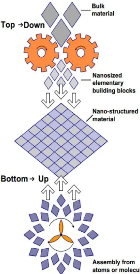
Nanotechnology encompasses two main approaches:
The “top-down” approach (physics) in which larger structures are reduced in size to
the nanoscale while maintaining their original
properties without atomic-level control (e.g.,
miniaturization in the domain of electronics) or
deconstructed from larger structures into their
smaller parts.
The “bottom-up” approach (chemistry), in which materials are engineered from atoms
or molecular components through a process of
assembly or self-assembly. In this work, this approach has been used to
get hydrogels based on cross-linked nanoparticles from individual
protein chains.
1.2
Protein based materials: ELASTIN-LIKE RECOMBINAMERS (ELRs)
Protein based-materials have drawn attention of numerous researchers in recent
8
years as promising advanced biomaterials for use in the field of biomedicine, especially asa result of recent improvements in recombinant DNA technology, which allow us to design
and manufacture materials by exploiting the abilities of natural proteins5. Some of the most widely studied protein-derived materials are the so-called elastin like recombinamers
(ELRs), taking into account its recombinant nature this new nomenclature was proposed6 in replacement of the more conventional terminology elastin- like polymers (ELPs)7-9, which include those first chemically synthesized materials.
1.2.1
Elastin-like recombinamers (ELRs)
Elastin is an elastic, insoluble protein that is widely distributed in vertebrate
tissues, such as lung, skin, major vascular vessels, or tendon10, where elasticity and resilience are a key requirement11. It is well-known for its extreme durability and ability to deform reversibly12,13. The origin of these properties reside in the structure of the recurrent sequences (VPGVG, VPGG, VGVAPG) found in the soluble elastin precursor
tropoelastin14.
ELRs are a family of repetitive artificial biopolymers all exhibiting a smart
behavior. These polymers are a genetically engineered biomaterials inspired by natural
elastin and the majority of its members are based on repeats of the pentapeptide sequence
Val-Pro-Gly-Xaa-Gly, where Xaa is any natural amino acid except proline15.
ELRs are a highly interesting biomaterials because their relevant characteristics.
The natural elastin‟s mechanical properties are combined with other properties such as
biocompatibility, stimuli-responsive behavior and the ability to self-assemble16,17. This class of smart polymers exhibits an inverse temperature transition (ITT) which allows them
to undergo a reversible phase transition from a soluble to an insoluble state upon
increasing or lowering the temperature above a specific threshold, the transition
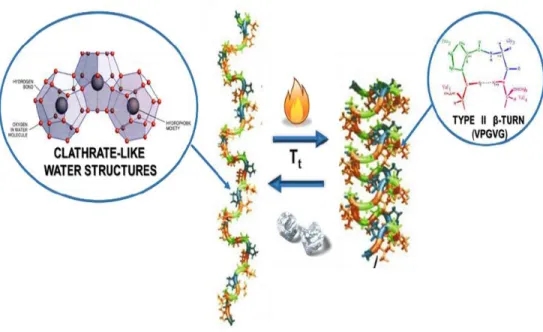
temperature (Tt)15,18,19. Below the Tt the polymer chains remain disordered in a relatively
extended state with a random coil conformation, and fully hydrated10. This hydrophobic hydration is characterized by an ordered clathrate-like water structure surrounding the
apolar moieties of the polymer (Figure 2). This structure is somewhat similar to that
described for crystalline gas hydrates, although more heterogeneous and of varying
perfection and stability11,20. When temperature surpasses the Tt, and according to Urry‟s
9
that leads to phase separation. That “coacervate” is composed of about of 63% water and37% polymer21. In the folded state, the polymer chain adopts a dynamic, regular, non-random structure called β spiral, which involves one type II β turn per pentamer stabilized
by intra-spiral inter-turn and inter-spiral hydrophobic contacts6 (Figure 2).
Figure 2: Schematic representation of the thermal transition of ELRs from an extended state (low temperatures, hydrophobic moieties surrounded by clathrate water structures) to a folded state (type II β-turn in VPGVG pentapeptides)
The process begins with the formation of filaments composed of three-stranded
dynamic polypeptide β-spirals, which grow up to various hundred nanometers before
settling into a visible separated state6,14.(Figure 3)
Figure 3 Mechanism of the ELRs’ ITT. From left to right: spiral formation, formation of twisted filaments or β-spiral supercoil and their aggregation into microaggregates. Reproduced from reference22.
10
or extrinsic (adding a substance), will alter the clathrate structure and consequently willmodify the Tt 23
. The ELP concentration and intrinsic factors like the modification of the
guest residue, Xaa, and to a certain extent, the length of the polypeptide chain are factors
that affect the transition temperature. The transition temperature can also be modulated by
other physiological changes, such as changes in the pH, addition of extrinsic factors as salt,
organic solutes or changing pressure24.
Generally, Tt increases as the mean polarity increases and vice-versa.
Additionally, if a chemical group that can be present in two different states of polarity
exists in the polymer chain, and these states are reversibly interconvertible by appliance of
an external stimulus, the polymer will exhibit two different Tt values 25,26
. This change in
the Tt, opens a working temperature window in which the polymer isothermally and
reversibly switches between the folded and unfolded states in response to environmental
changes. This effect of changing the Tt is the basis of the Δ Tt mechanism and has been
exploited to obtain pH, electric potential, light, chemical and other responsive
interconvertible energy processes25. The hydrophobic paradigm, involving the ITT and the Δ Tt mechanism, for protein folding and function and the intrinsic capability of performing
several energy interconversions allows new strategies for the development of ELP
derivatives and working temperatures. Experimental studies on the ITT exhibited by ELPs
and based on the factors that control hydrophobic folding and assembly of model proteins
resulted in a set of five phenomenological axioms for the protein engineering of PBPs
capable of inverse temperature transitions27,28:
Axiom 1: The temperature intervals for the hydrophobic folding and assembly transition of a host protein or protein-based polymer with
different guest substituents becomes a functional measure of their
relative hydrophobicity.
Axiom 2: Heating to raise the temperature from below, to above, the temperature interval for hydrophobic folding and assembly of
macromolecules can drive contraction with the performance of
mechanical work.
11
macromolecule can itself, drive hydrophobic folding and self-assemblyat constant temperature.
Axiom 4: Two or more different functional groups of a macromolecule, each of which can be acted upon by a different energy input that changes
the temperature interval for hydrophobic folding and assembly, become
coupled one to another by being part of the same hydrophobic folding
and self-assembling domain, that is, the energy input acting on one
functional constituent alters the property of another functional
constituent as an energy output.
Axiom 5: More hydrophobic domains make more efficient the energy conversions involving constituents undergoing conversion between more
and less hydrophobic states.
1.2.2
Block Copolymers
Block copolymers have been the subject of numerous studies due to their ability
to undergo self-assembled phase separation resulting in different complex morphologies.
Self-assembly procedures have drawn attention over a number of years, with the
self-assembly of nanoparticles being a particularly active field. Self-assembled polymer
nanoparticles and hydrogels tend to be obtained from amphiphilic macromolecules.
Generally speaking, a solution of these amphiphilic molecules in a solvent that only
specifically solvates one part of the molecule will result in aggregation due to interaction
of the solvent with the solvophobic blocks of the molecule. The hydrophobic parts tend to
form aggregates as this collapse is more entropically favorable than the ordination of water
molecules around each hydrophobic segment. On the other hand, hydrophilic parts are
dissolve in water as the formation of hydrogen bonds with water molecules is higher
enthalpic compensated than the interaction between hydrophilic parts.
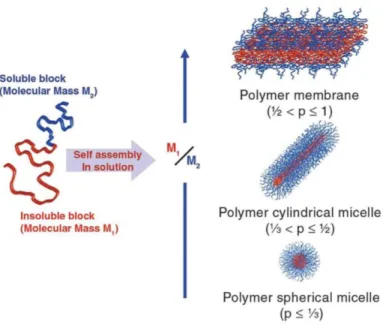
1.2.2.1 Physical properties of Block Copolymers
Block copolymers are polymers composed of two or more covalently linked
chemically distinct sequences (blocks).Thus, block copolymers can be designed as a
12
structures29.The degree of order and morphology of those aggregates are dependent on
concentration and volume ratio between the hydrophobic blocks and the hydrophilic
blocks, known as insoluble soluble ratio (ISR). Below a certain concentration, critical
aggregation concentration (CAC), the hydrophobic blocks are capable of maintaining the
molecules dissolved. On the contrary, once above the CAC block copolymers begins to
self- assembly resulting from the separation from the solvent of the hydrophobic block.
The CAC decreases as the ISR and the molecular mass increases30.
The dimensionless packing parameter (p) is the ratio between the molecular
volumes of the solvent-phobic chain and the volume occupied by the copolymer in the
assembly, defined as29:
Where a0 is the optimal surface area of the solvent-phobic segment at the
interface of between both blocks resulting from the balance between solvophilic
solvophobic interactions, v is the volume and d is the length of the solvophobic block.
Generally speaking, if p ≤ 1/3 the self-assembled structure result in spherical micelles,
while 1/3 < p ≤ ½ correspond to cylindrical micelles and a p values between ½ and 1 are
related to polymer membranes (Figure 4).
Theoretically speaking, the most stable assembly would be an infinity large
membrane and an infinity long cylinder. Nevertheless, it goes without saying that the
system is force to finite dimensions, thus a certain level of curvature (molecular
frustration) is required in order to avoid the contact between the solvent and the insoluble
parts31.
Wormlike structures will come as a result of the stabilization by end-caps of
cylindrical micelles if the molecular frustration is restricted to a specific part of the
assembly. In the same way, under these local confinement of the molecular frustration,
stabilization of membranes will consist on the curvature of the edges giving rise to
13
Figure 4 Different geometries formed by block copolymers in selective solvent conditions.On the other hand, if all the molecules share the molecular frustration then
stabilization of cylinders and membranes will result in the formation of toroidal micelles
(as cylinders bend) and vesicles (membranes closure)
Energetically speaking, the formation of vesicles and worm-like micelles is more
favorable than disk-like and toroidal micelles. But, it has been demonstrated that those less
likely structures can be stabilized by the increase of the molecular mass of the
copolymer33,34, which will make the local frustration unfavorable, or by introducing extra interaction between block copolymers35.
1.2.2.2 Protein-based block copolymers
Protein-based block copolymers consist of a type block copolymer in which
some or all of the building blocks are composed by protein inspired materials, peptide
sequences. Throughout the careful and specific selection and positioning of amino acid
residues it is possible the production of polymers with an absolute control over the
hydrophobicity patterns or secondary structures, which give rise to a wide selection of
tailor-made materials36.
As it has been previously described, see 1.2.2.1, the microstructure formation of
synthetic block copolymer is highly influence by the ISR, but the phase separation
14
protein-based block copolymers. When dealing with protein inspired materials, intra andinter-molecular bonding, together with chain conformation, are to be taken also into
account in the process of structures formation. Thus, a different phase behavior from that
of synthetic block copolymers should be expected, mainly due to normally occurring
interactions in protein, such as electrostatic interactions, peptide backbone rotational
restrictions, high hydrogen bonding or hydrophobic interactions, are usually absent from
the synthetic block copolymer systems. The supramolecular organization in proteins is
mostly directed by two structural elements, α-helix and β-sheet. All in all, the conditions
under which structures (microphase separation) self-assemble and subsequence phase
diagram relationships is yet to be determine for most of protein-based copolymers.
Protein-based block copolymer can be divided into two main groups; synthetic polymer-peptide
block copolymer (hybrids) and protein/peptide block copolymers.
1.2.2.3 Elastin-Like block co-Recombinamers (ELbcRs)
Recent advances in recombinant DNA techniques have provided the tools needed
to produce block corecombinamers with the desired sequence, depending on the
application, from simple amino acids with an absolute degree of control and complexity
superior to those of synthetic polymers.
Amphiphilic elastin-like block co-recombinamers (ELbcRs) can form nano- or
micro-sized structures37,38To this end, ELR-based amphiphilic tetrablock copolymers have been synthesized in which the amphiphilicity of the component blocks is achieved by
substituting the amino acid (X) in the guest position of the pentamer VPGXG by a
hydrophilic (glutamic acid), or hydrophobic (isoleucine) amino acids. Nanoparticle
formation occurs when the elastin-like block co-recombinamer (ELbcRs) solution is heated
above the characteristic Tt of the hydrophobic block, at which point the co-recombinamer
chains organize themselves by hiding the hydrophobic blocks from the aqueous
environment, thus reaching a minimum free energy situation.
Mean polarity, molecular weight, amino-acid sequence and molecular
architecture are parameters influencing the final morphology and size of the final
15
Figure 5 Schematic representation of the formation of a physical hydrogel through the micellation of atetra-block ELR. Below Tt the monomers are extended, once above Tt the hydrophobic tetra-block (black) aggregates forming nanoparticles. If the concentration is increased that aggregation leads to the formation of a water
swollen network resulting in the hydrogel formation.
ELbcRs have also been reported as materials capable of forming physical
hydrogels at a sufficiently high concentration. In this sense, tetrablocks
(polar-apolar-polar-apolar) has been reported to self-assemble into a physical hydrogel once above the
Tt. These materials are designed to be injectable self-gelation systems with high
applicability in biomedical applications, such as tissue repair or as drug delivery systems
for local therapies. In order to fulfill the requirement of injectability the material is
required to be a low viscosity liquid below the physiological temperature. In this sense, the
monomers are completely unassociated in aqueous solution below the Tt and once above
the Tt the hydrophobic elastin domains undergo a phase separation associating into
16
2
MATERIALS AND METHODS
2.1
MATERIALS
2.1.1
Chemical Reagents
All the reagents employed on this work are listed on Table 1
Reactive
Brand
Acrylamide/Bis-acrylamide Amresco
Agarose Seakem. Cambrex
Ammonium persulphate (APS). Sigma-Aldrich
Ampicillin Apollo Scientific
Bromophenol blue Sigma-Aldrich
Chloridric acid Merck
Copper chloride Sigma-Aldrich
Dimethyl sulfoxide (DMSO) Carlo Erba
Ethanol Merck
Glycerol Sigma-Aldrich
Glycine Sigma-Aldrich
Mineral oil Sigma Aldrich
Phenylmethylsulfonyl fluoride (PMSF). Apollo Scientific
Phosphate buffered saline (PBS) Gibco
Sodium chloride (NaCl)- Sigma-Aldrich
Sodium dodecyl sulfate (SDS) Sigma-Aldrich
17
Tetramethylethylenediamine (TEMED). Sigma-Aldrich
Ultrapure water Millipore
β-Mercaptoethanol. Sigma-Aldrich
Table 1 Reagents employed and suppliers.
2.1.2
Other materials
During the realization of this work light scattering techniques have been
employed, where glass test tubes were used. The contamination of such tubes with organic
residues, dust or grease may interfere with the results obtained, being necessary to pay
special attention to the cleaning of the tubes employed. An optimum cleaning is achieved
by rinsing the tubes out with distilled water followed by a wash with a mixture soap-water
(Hellmanex ®II special for optical cleaning). To remove soap traces the tubes are
extensively washed with water. Finally, the tubes are rinsed with water type I and allowed
to dry in an oven at 60°C for at least 4 hours.
Other glass materials after washing and rinsing several times with distilled water
were sterilized on an autoclave (Autotester E-75). Other laboratory materials like tips,
conical tubes, Eppendorf tubes, falcon tubes, etc. are bought sterile or are sterilized when
needed on an autoclave (Autotester E-75, 20 minutes 120°C one atmosphere).
2.1.3
Molecular biology materials
2.1.3.1 Restriction enzymes
The restriction enzymes used in this work are listed below:
DpnI, EarI, EcoRI, SapI, XbaI, XhoI (Thermo Fisher).
All enzymes are used according to the manufacturer instructions.
2.1.3.2 Other enzymes
The following enzymes have been employed, being all of them purchased to
18
T4 DNA Ligase, Shrimp Alkaline Phosphatase (SAP), FastAP phosphatase.
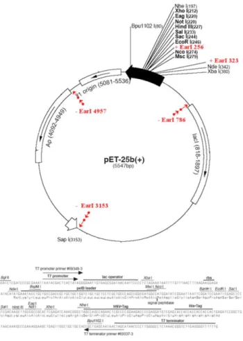
2.1.3.3 Cloning and expression vectors
The DNA fragments employed were cloned in pDrive cloning vector (Qiagen),
Figure 6.
Figure 6 pDrive cloning vector (Qiagen).
For the expression of the different recombinant polimers a p7 expression vector
has been employed Figure 7. The p7 expression vector was constructed in our laboratory
19
Figure 7 Scheme of p7expression vector based on Navagen’s pET-25b(+) vector2.1.3.4 Other reagents
Two kits were used for the plasmid and DNA purification either from an
Escherichia coli (E.coli) culture: NucleoSpin Plasmid (Macherey-Nagel) and Quantum
Prep Plasmid Midiprep Kit (Bio-Rad); or from an agarose gel: PureLink Quick Gel
Extraction Kit (Life Technologies).
2.1.4
Bacterial strain
The E. coli strains used on this work have the following genotypes:
a) XL1-Blue Competent Grade/Subcloning Grade (Stratagene): endA1 supE44
hsdR17 thi1 recA1 gyrA96 relA1 lac [F‟ proAB lacIq ZΔM15 Tn (Tetr)].
20
c)
BLR (DE3) (Novagen): F- ompT hsdSB (rB- mB-) gal dcm Δ (srl-recA) 306::Tn10(Tetr) (DE3).
2.1.5
Culture media
The culture media used for bacteria growth and transformation are listed below:
a) Luria-Broth (LB)(Pronadisa): Concentration 25g/L.
b) Terrific Broth (TB) (Formedium): 55.85 g/L + 8mL/L Glycerol.
c) LB-Agar: LB 25 g/l + 1.5 % (p/v). Agar (Fluka).
d) SOC Broth. (Sigma Aldrich).
2.1.6
Buffers
During the performance of the work presented on this thesis different buffers
were employed:
a) PBS (pH=7,4): 5mM, NaCl 137 mM, KCl 2.7 mM, 10mM Na2 HPO4 , KH2PO4
1.8 mM
b) TAE: 40 mM Tris-acetate, 1mM pH=8 EDTA.
c) TE (sonication buffer): 10 mM pH 8 Tris-base, 1 mM pH=8 EDTA, 1mM PMSF.
d) TBS (washing buffer):20 mM pH 8 Tris-base, 140 mM NaCl.
e) Running buffer: Tris-base 25 mM pH=8,3, glicina 192 mM y SDS 0,1% (w/v).
f) DNA loading buffer: 30% (v/v) glycerol, 0.1% (w/v) SDS, 0.05% (w/v)
bromophenol blue (BPB), 50mM Tris pH 8, 0.05mM EDTA.
g) Protein loading buffer: Tris 1MpH 6.5 312.5 mM, SDS 10%(w/v), Glycerol ( v/v), β-Mercaptoethanol 25%(v/v), bromophenol blue (BPB) 2% (v/v).
21
2.1.7
Elastin-like recombinamers (ELRs)
All the elastin-like recombinamers (ELRs) employed in the development of this
work have been designed and produced in our laboratory (G.I.R. Bioforge) by recombinant
DNA techniques. Those recombinamers have been specifically designed for the realization
of this work, and these have been produced by E. Coli fermentation and purified taking
advantage of both the smart nature and the reversible thermo-dependent segregation
showed by this kind of materials, by inverse transition cycling (ITC)8.
DNA corresponding the individual blocks E50, I40 and I60 were cloned by Dr.
García-Arévalo, while the gene of the amphiphilic tetrablock (E50I60)2 was constructed
by the Dr. Martín Maroto. Despite that, I reconstructed this tetrablock to verify the
sequence. Also three new amphiphilic tetrablocks were constructed. Table 2 shows all the
tetrablocks employed, the abbreviation, molecular weight (Mw), and amino acid sequence.
Elastin-like
recombinamer (ELR)
abbreviation
Amino acid sequence
Molecular
weight (Mw)
(E50I60)
2MESLLP
[[(VPGVG)2-VPGEG-(VPGVG)2]10
(VGIPG)60]
V
22
E100I60E50I60
MESLLP
[(VPGVG)2-VPGEG-(VPGVG)2]20
(VGIPG)60
[(VPGVG)2-VPGEG-(VPGVG)2]10
(VGIPG)60
V
113931.8 Da
E50I60E50I100
MESLLP
[(VPGVG)2-VPGEG-(VPGVG)2]10
(VGIPG)60
[(VPGVG)2-VPGEG-(VPGVG)2]10
(VGIPG)100
V
110098.20
Da
(E50I100)
2MESLLP
[[(VPGVG)2-VPGEG-(VPGVG)2]20
(VGIPG)100]2
V
127038.7 Da
23
2.2
METHODS
2.2.1
DNA agarose gel electrophoresis
DNA agarose gel electrophoresis are used to separate and check the appearance
and size of DNA fragments form either a plasmid or from an enzymatic digestion with
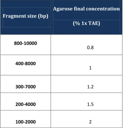
endonucleases. Different concentrations (in 1x TAE), are applied according the sizes of the
DNA fragments and the kind of gel, analytical or preparative, being the first one used to
assess the rightness of a purified plasmid and the second one to obtain DNA for further
use. The different agarose concentrations and their resolution capability are listed in the
Table 3.
Fragment size (bp)
Agarose final concentration
(% 1x TAE)
800-10000
0.8
400-8000
1
300-7000
1.2
200-4000
1.5
100-2000
2
Table 3 Resolution for linear DNA in electrophoresis of different agarose gel concentrations.
The gels are prepared adding in a glass-made Erlenmeyer flask the quantity of
agarose and a volume of buffer according to the gel concentration and size. The agarose is
melted on the microwave, after weight and hydration, until the formation of a gel. Once
melted, the flask with the gel is weighted again and ultrapure deionized water is added
24
After cooling down to 60ºC the gel is casted in a horizontal tray with the desired comb.The samples are applied adding 0.2 volumes of 5x loading buffer. A fixed
voltage, between 2 and 7 V/cm – according to each sample, is then applied. The
electrophoresis is run having as references the color markers (Table 4). Last, the gel is
stained for 10 to 30 minutes in a 1x GelRed™ solution, and the DNA bands are visualized
by exposition to UV light in a Viber Lourmat, TFX-20M transilluminator.
TAE 1x –BPB
% Agarose
2900 0.30
1650 0.50
1000 0.75
500 1
370 1.25
200 1.75
150 2
Table 4 Relation between linear DNA migration and bromophenil blue (BPB).
2.2.2
Plasmid purification
The plasmids employed in this work were purified, using the commercial systems
listed above, following the manufacturer‟s instructions. DNA was eluted with ultrapure
water or Elution buffer from the kits. For applications where higher DNA concentration is
required only half of the recommended elution volume is used, the elution water is used at
65°C and the time incubation was increased up to 10 minutes to enhance the purification
yield. The eluted DNA is stored under -20ºC.
2.2.3
DNA digestion with restriction enzymes
Reaction conditions (temperature, concentration, time of reaction, buffer) for the
digestion are supplied by the enzyme manufacturer. The rate of digestion is controlled by
25
2.2.4
DNA dephosphorylation
Dephosphorylation reaction conditions (temperature, time of reaction, buffer) are
supplied by the phosphatase manufacturer. For the p7 expression vector two different
consecutive phosphatases were used and the incubation time was enlarged until one hour.
2.2.5
DNA fragments purification from an agarose gel
The target DNA band is first separated and visualized in an analytic agarose gel
of an appropriated concentration and stained with GelRed™Nucleic Acid (as indicated in
2.2.1), secondly, the band is extracted from the gel with the help of a scalpel. Minimum
quantity of agarose should be cut during band extraction.
The purification of the fragment is carried out using the commercial system
“PureLink Quick Gel Extraction Kit” (Life Technologies), following the protocol indicated
by the manufacturer.
2.2.6
Ligation reaction
The reaction ligation is carried out in a final volume of 15μL by mixing the insert
with the vector, in a molar relation from 1:1 to 5:1, and T4 DNA ligase as enzyme with its
corresponding buffer following the specifications indicated by the supplier. The reaction is
conducted during 1 hour at room temperature or during 24 hours at 4°C.
2.2.7
Cloning on the pDrive/ p7 vector
The ligation reaction is interrupted by the inactivation of the T4 DNA ligase
by incubation during 10 minutes at 70°C. Once the ligation reaction is concluded, a
certain quantity of it is used to transform competent cell as specified below.
2.2.8
Transformation of competent cells
2.2.8.1 Transformation of XL1 blue subcloning grade competent cells
This bacterial strain has an efficiency ≥ 106
transformants per μg of DNA.
Plasmid DNA to be amplified by cloning is transformed in this bacterial strain following
26
2.2.8.2 Transformation of XL1 blue competent cellsThis bacterial strain has an efficiency ≥ 108
transformants per μg of DNA.
Ligation products were transformed into this bacterial strain following the protocol
specified by the supplier.
2.2.8.3 Transformation of BLR (DE3) competent cells
This bacterial strain is transformed with the expression plasmid p7 following the
method TSS reagent (“Transformation and Storage Solution”)40. This method is a combination of two steps from the transformation procedure, first we obtained competent
cells and second the cells are stored at -80ºC or transformed resulting in transformation
efficiency that goes up to the 107 cfus (colony forming units) per microgram of DNA. A single colony, isolated and grown in a LB-agar plate, is used to inoculate 100
mL of LB medium (plus antibiotic) and is grown at 37ºC with shaking (250rpm), until
reach a OD600=0.3-0.4. At this point the metabolism and cell growth is stopped by
incubation on ice for 5 minutes. The cell suspension is centrifuged at 3000rpm (1100Gx)
for 10minutes at 4ºC. The supernatant is discarded and the pellet is re-suspended in 10mL
of cold 1xTSS solution, and is mixed gently (TSS1x is LB broth containing 10% (wt/vol,
Mw 8000) polyethylene glycol, 5% (vol/vol) dimethyl sulfoxide, and 50 mM Mg2+ at pH 6.5) Now the competent cells are ready to be transformed. 150 μL competent cells are
aliquoted to 1.5 mL Eppendorf tubes and are storage at -80ºC (pre-treated with liquid
nitrogen).
At the moment of the transformation, an aliquot is defrosted in ice, and about
1-10 ng of plasmid in final volume of 1-1-10μL are added to the mix. The cellular suspension
plus the plasmidic DNA are kept on ice thirty minutes. A 0.85 ml of pre-warmed LB is
added and the suspension is incubated one hour at 37ºC with shaking (250rpm). And
finally, 50-200 μL of the transformation mix is plated in LB-agar plus the antibiotic plates
that are incubated for 16-20 hours at 37ºC.
2.2.9
Glycerol stock preparation
To maintain and store the clones with interest, glycerol stocks were made. The
27
(250rpm.) on LB or LB with 0.5% of glucose (for the expression strains) plus antibiotic,until reaching an OD600= 0.6-0.8. At this point 0.1 volumes of 80% sterile glycerol are
added and the cells are added to a cryovial and are stored at -80°C.
2.2.10
DNA Sequencing
The automatic DNA sequencing was made at Cenit Support Systems S.L.L.-
Scientific park of Salamanca (Villamayor, Salamanca).
2.2.11
Production and purification of recombinant polymers
2.2.11.1 Recombinant polymer’s expression
During the biosynthesis of the four different tetrablocks employed in this work
the expression vector p7 has been employed. p7 has been obtained in our laboratory by
mutagenesis of pET25b (+) by Dr. Alessandra Girotti. The final constructions were
transformed on the bacterial strain BLR (DE3) following the above mentioned protocol
(see 2.2.8.3).
ELRs expression starts inoculating the desired colony in liquid LB medium plus
antibiotic and 1% of glucose at 37 °C with orbital shaking (250 rpm.) during
approximately 6 hours. This culture is used as inoculum for a fresh TB medium (plus
antibiotic), in a volume ratio of 1:500, not exceeding the 25% of the capacity of the
Erlenmeyer used. This culture is grown for 14-16 hours at 37 ° C with orbital shaking
(250rpm.).
For large batches production, as this case, a 15L Bioreactor is used (Applikon
Biotechnology), allowing the full control of variables like temperature, pH, OD600 and
oxygen concentration, regulating all of them if needed, improving the yield of the
bioproduction process. It is inoculated with 1L of the pre-incubated cell suspension to a
final volume of 15L of TB medium and fermentation time varies from 14 to 16h, setting
temperature at 37ºC, pH at 7, oxygen control at 50% of the initial oxygen concentration
and stirring at 499rpm.
2.2.11.2 Bacteria disruption
28
by cooling it down to 4ºC and cells are centrifugated and washes with Washing buffer (see2.1.6)until having a clear supernatant. Then the pellet is re-suspended in a volume VTE of
TE per liter of culture (see 2.1.6):
VTE=5*Vculture*OD600
Cell suspension is kept at 4ºC and 10μg/mL of PMSF protease inhibitor is added.
Bacteria are disrupted (lysated) by changing pressure disruption employing a
Constant Cell Disruption System (Model TS 0.75KW, Constant System). Finally, the
lysate is centrifuged at 4ºC for 60 minutes at 15000xg. The supernatant contains the
recombinant polymer biosynthesized.
2.2.11.3 Purification of the recombinant protein-based polymer
The purification of ELRs starts from the supernatant obtained at the end of the
lysis process (see 2.2.11.2), taking advantage of the ELRs‟ smart nature and inverse
temperature transition (ITT). Therefore, the purification process is based on successive
cycles of precipitation (heating) and resuspension (cooling) of the supernatant, named
Inverse Transition Cycling (ITC).
The ELRs biosynthesized in this work had glutamic acid residues in their
sequence (see Table 2), which at a pH above its pKa are depronated and therefore
negatively charged what increase significantly their Tt 41
. In order to reduce the Tt and in
this sense, facilitate the precipitation of the ELRs, NaCl is added during the purification
process until a 2M concentration is achieved.
Finally, after the last purification cycle, the re-suspended polymer is dialyzed
against cold ultrapure type I water. The suspension is then adjusted to pH 7, passed
through a 0.2 μm PES (Polyethersulphone) filter, and lyophilized and stored at -20°C.
Purification steps, as well as the final product, are checked by polyacrylamide gel
electrophoresis.
2.2.12
Denaturing polyacrylamide gel electrophoresis
The protein polyacrylamide gel electrophoresis in the presence of sodium
29
described by Laemmli42 to accomplish the separation of a proteins mixture by their molecular weight, being the gel composed of two different sub-gels: stacking andresolving.
There are almost no differences between the proteins‟ effective charge because
the SDS strongly interacts with proteins providing them with approximately one negative
charge per each amino acid residue. The denaturing conditions are obtained because the
SDS denatures the quaternary and tertiary structure of the proteins by breaking the
non-covalent interactions. Besides, β-mercaptoethanol (a reducing agent) is added to the
samples in order to break the disulfide bonds that might exist. This process is facilitated
with the heating of the samples during 5 minutes at 95ºC.
A “MiniVE vertical electrophoresis system” from Hoefer (Amersham Pharmacia
Biotech, Pittsburg, USA) electrophoretic system was employed to perform the
polyacrylamide electrophoresis. The total percentage of acrylamide (%T) in the resolving
gel varies according to the molecular weight of the polypeptide we want to separate. The
optimal %T for a determined size range is presented in Table 5.
Target size range
(kDa)
%T in resolving gel
24-205 7.5%
14-205 7.5%
14-66 12.5%
14-45 15%
Table 5 Optimal %T according to the polypeptide target size range.
The composition of a resolving and stacking for a gel X%T is detail in Table 6.
Reactive
Resolving gel
Stacking gel
Acrylamide 40% 10% (w/v) 4% (w/v)Tris-HCl pH 8.8 375 mM ---
Tris-HCl pH 6.8 --- 125 mM
SDS 10% 0.1% (w/v) 0.1% (w/v)
APS 10 % 0.05% (w/v) 0.05% (w/v)
TEMED 0.05% (w/v) 0.08% (w/v) *both gels are prepared in ultrapure type I water
Table 6 Composition of the resolving and stacking gel in a gel 10%T.
30
Thermo Fisher) is loaded together with the samples in order to know the molecular weightof each band43. Staining is performed according to Lee‟s method44: Ten minutes incubation at room temperature of the gel in a 0.3M Copper chloride solution followed by a washing
step in distilled water for 5 minutes. Pictures are taken by „Gel Logic 100 Imaging System‟
camera system and „Kodak 1D Image Analysis (Kodak)‟ software.
2.2.13
Experimental techniques
2.2.13.1 Dynamic light scattering
Dynamic light scattering (DLS) is a technique for measuring the size of particles
normally in the sub-micron region. Typically, DLS is concerned with measurements of
dispersed particles or suspended macromolecules in a liquid medium, measuring the
particles Brownian motion and relating it to the particles‟ size.
Brownian motion is the random movement of particles suspended in a fluid due
to their collision with the solvent molecules that surround them. Thus, the random motion
will be affected by different factors, mainly the size of the molecules (the bigger the
molecules, the slower they move), the viscosity of the solvent (the more viscous the
solvent, the slower the molecules move) and temperature. Temperature is a crucial
parameter, both temperature stability and temperature accurate knowledge are required,
due to its influence on the solvent viscosity and owing to, temperature instability will lead
to convection currents in the sample, thus non-random motion, resulting in incorrect size
interpretation.
The velocity of the Brownian motion is defined by the translational diffusion
coefficient (D), which is used to calculate the size of the particles by using the
Stokes-Einstein equation:
Where kB is the Boltzmann constant, T is the temperature and η is the viscosity.
It is worth noting, that DLS refers to how a particle diffuse within a fluid so the calculated
diameter is a hydrodynamic diameter (Dh), (effective molecule diameter + hydration layer,
31
Figure 8 Representation of particle with its hydration layer, hydrodynamic radius.Light scattering measurements were performed using a Zetasizer nano ZSP
(Malvern instruments) equipped with a 10 mW He-Ne laser at a wavelength of 633 nm.
2.2.13.2 Zeta potential
Z-potential analysis is a technique for determining the surface charge of
nanoparticles in solution. The Z-potential of the ELR was monitored at 37°C using a
Zetasizer nano ZSP apparatus (Malvern instruments). The Z-potential values, which were
determined using the Smolukowski equation relating ionic mobility to surface charge, were
plotted as the average of 10 repeated measurements.
2.2.13.3 Differential scanning calorimetry (DSC)
Differential scanning calorimetry (DSC) is a technique in which the difference in
the amount of heat required to increase the temperature of a sample and reference is
measured as a function of temperature.
DSC experiments were performed on a Mettler Toledo 822e with liquid-nitrogen
cooler. Both temperature and enthalpy are calibrated with a standard sample of indium.
The solutions for the DSC experiments were prepared at 50 mg/mL in water or an aqueous
buffered solution (PBS). 20 μL of the solution were placed inside a standard 40-μL
aluminum pan and sealed hermetically. The same volume of the employed solvent was
placed in the reference pan. Both, sample and reference are heated at a constant velocity.
The heating program included an initial isothermal stage (5 min at 0º C for
stabilization of the temperature and the state of the polymers), followed by heating at 5º
32
2.2.13.4 RheologyRheology is the study of flow and deformation of materials under applied forces.
In this sense, it was used to analyze the thermogelification process of the different
tetrablock-ELRs, by studying their mechanical properties in order to obtain the storage and
loss moduli of the hydrogel.
Trhe viscoelastic properties of 250, 275 and 300 mg/mL solutions (final volume
of 300μL) of each tetrablock-ELRs in ultrapure water and in PBS buffer were evaluated
using a controlled stress rheometer (AR-2000ex, TA Instruments). A 12 mm Standard steel
parallel plate was used to characterize the rheological properties at a constant strain of
0.3% and a frequency of 1 Hz, mineral oil was used at the edge of the samples to prevent
solvent evaporation in the hydrogels.
To characterize the gelation kinetics of the tetrablock-ELRs hydrogels,
time-sweep experiments were performed at 25ºC and 37ºC Strain time-sweeps were carried out
across a strain range of 0.01-15%. Temperature ramps were performed from 5ºC to 40ºC
(heating rate: 5ºC/min).
2.2.14
Experimental techniques performed by external services
2.2.14.1 Amino-acid analysis
The amino acid composition of the ELRs employed during this work was
determined by Laboratorio de Técnicas Instrumentales (University of Valladolid).
After addition of a known quantity of α-aminobutyric acid as internal pattern the
samples were hydrolyzed (6M HCl, 1% Phenol and 2.5 hours at 155°C) and evaporated.
The powder was resuspended in 1mL of 20mM HCl and a 1/10 dissolution was prepared.
The quantification of the less represented amino acids was made from the most
concentrated sample and the quantification of the most represented amino acids from the
1/10 dissolution. One aliquot of each dissolution was derivatizated according to the
AccQ-Tag Waters method and analyzed by HPLC with UV detection, using a WATERS600
HPLC gradient system with a WATERS2487 detector.
2.2.14.2 MALDI-TOF Mass Spectrometry Analysis
33
Laboratorio de Técnicas Instrumentales (University of Valladolid).2.2.14.3 Cryo-Transmission electron microscope (Cryo-TEM)
(Cryo-TEM imaging acquisition of buffered solutions was performed at CIC
bioGUNE Structural Biology Platform, Bilbao).
Samples were prepared through rapid vitrification of the liquid samples in the
automated vitrification robot Vitrobot™ Mark IV (FEI).The specimens were observed with
a JEM-2200FS/CR transmission electron microscope (JEOL, Japan), equipped with an
ULTRASCAN 4000 SP (4008×4008 pixels) cooled slow-scan CCD camera (GATAN,
34
3
RESULTS AND DISCUSSION
3.1
DESIGN, CONSTRUCTION AND RECOMBINANT PRODUCTION
The use of block copolymers in nanotechnology has acquired a great interest in
the last years. They enable the possibility of creating different structures in the nanometer
scale thanks to their self-assembly properties in an easy and non-expensive way using the
bottom up approach45.
In this study, to facilitate the rational engineering of the physico-chemico
properties of hydrogels based on amphiphilic tetrablock ELRs, three new tetrablock
recombinamers with different molecular weight and composition were designed, taking as
point of departure, the amphiphilic tetrablock E50I60E50I60 (A in Figure 9). A
recombinamer that is comprised by two hydrophilic blocks, L-Glutamic acid containing
block (E-block) and two hydrophobic blocks, L-Isoleucine containing block (I-block).
The identity and sequence of the individual block units within the polymer
dictates the nature of the supramolecular assembly, for this reason two of the new
tetrablocks incorporated an extra-block at the N-terminal end (B in Figure 9) and at the
C-terminal end (C in Figure 9). Thus, the influence of an „asymmetric‟ block could be
evaluated.
35
And finally, a tetrablock ELR that incorporated an extra hydrophobic contentpreserving the proportionality between its two diblocks was also designed (D in Figure 9).
The construction of the different genes was performed following the guidelines
described in material and methods. The different tetrablock genes were constructed on
pDriveAll vector starting from the individual blocks E50, I40 and I60. All the
constructions were assessed by agarose gel electrophoresis in every step of the process and
by DNA sequencing. Figure 10 and 11 show the results of the different steps of the genetic
engineering process. Figure 10 presents the starting building blocks in the cloning plasmid
pDriveAll (Lanes 1-3) and the diblocks constructed as a middle point before obtaining the
final tetrablocks (Figure 11 shows the desired tetrablock ELRs in the expression plasmid
p7).
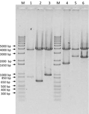
Figure 10 Enzymatic analysis with the EcoRI endonuclease of the colonies containing the plasmid pDrive All and the inserts: Lane 1: E50 (750 bp); Lane 2: I40 (600 bp); Lane 3: I60 (900 bp); Lane 4: E50I60 (1650 bp); Lane 5:
E100I60 (2400 bp); Lane 6: E50I100 (2250 bp); Lanes M: DNA marker 1Kb Plus.DNA agarose.
The digestion with EcoRI produced two bands, an upper band corresponding to the pDrive plasmid and a
36
Figure 11 Enzymatic analysis with the XhoI and XbaI endonucleases of the colonies containing the expressionplasmid p7 and the inserts: Lane 1: (E50I60)2 (3321 bp); Lane 2: E100I60E50I60 (4071 bp); Lane 3:
E50I60E50I100 (3921 bp); Lane 4: (E50I100)2 (4521 bp); Lanes M: DNA marker 1Kb Plus.DNA agarose.
The digestion with XhoI combined with XbaI produced two bands, an upper band corresponding to the p7
plasmid and a lower one corresponding to the insert plus 168bp.
Once the constructs were obtained, the following step was to introduce into the
expression vector p7, and subsequent transforming of the E. coli strain BLR (DE3).
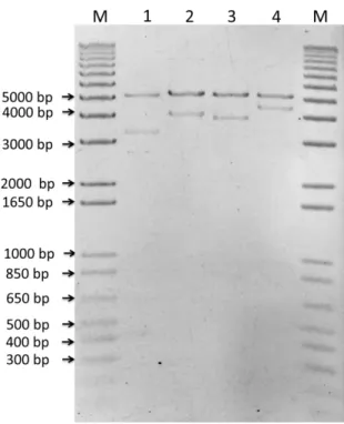
Expression was qualitatively assessed by SDS-PAGE. The overexpressing colonies were
selected taking into consideration the protein bands pattern. In the following figure
(Fig.12) we can observe a SDS-PAGE assay for screening the capacity to produce one of
37
Figure 12 SDS-PAGE of E. coli BLR (DE3) colonies expressing E100I60E50I60 stained with Cooper.M: Protein Marker (Unstained marker); Lanes 1 to 8: total protein fraction of eight BLR (DE3) ELR
E100I60E50I60 producing transformants after overnight induction in TB medium; Lane 0: untransformed BLR
as negative control of recombinamer production.
The screening assay showed a slightly variation in recombinamer production
between the colonies, nevertheless it is very important to perform first, a small scale
experiment of several transformants in order to choose the most suitable for large scale
recombinamer expression (Figure 12, Lane 4 in this case).
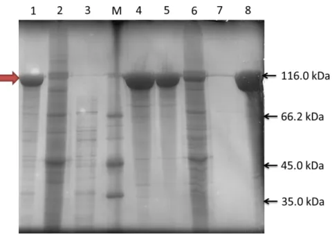
Then, the polymers were produced in large scale and appropriately purified by
Inverse Transition Cycling (ITC). The recombinant proteins were purified from the soluble
protein fraction as described on 2.2.11.3. As can be seen in the Figure 13, a high level of
purity is achieved after two IT cycles, despite that three IT cycles were carried out for
ensuring that a level of purity greater than 95% is obtained. Following the three IT cycles,
38
Figure 13 E50I60E50I100 purification analysis by 10% SDS-PAGE stained with Cooper.Electrophoretic SDS-PAGE image of different stages of the purification procedure of the tetrablock ELR
E50I60E50I100. Lane 1: Overnight cold supernatant from the disrupted supernatant; Lane 2: Overnight cold
precipitate from the disrupted supernatant; Lane 3: First heating supernatant; Lane 4: First heating
precipitate; Lane 5: Overnight cold-dissolved precipitate from first heating; Lane 6: Overnight cold-dissolved
supernatant from first heating; Lane 7: Second heating supernatant; Lane 8: Second heating precipitate. M:
39
3.2
MOLECULAR CHARACTERIZATION
3.2.1
SDS-PAGE
Figure 14 Analysis of the different purified tetrablock-ELRs. 10% SDS-PAGE stained with Cooper.
Lane 1– running pattern of 5µg of the purified recombinamer (E50I60)2. Lane 2– Of the purified
E100I60E50I60. Lane 3– Of the purified E50I60E50I100. Lane 4– Of the purified (E50I100)2.
The band although is slightly higher than the theoretical size is within the 20% range, typical of this
polymer’s, where the ELR run in a polyacrylamide gel. The numbers on the left side of the image correspond
to the size in kDa of protein marker reference bands. M: Protein Marker Unstained.
SDS-PAGE analysis lets us verify the purity of the polymer and the correct
molecular weight. As we can see, the purity level is over 95%, which demonstrates that the
ITC purification method is highly effective and easy to perform.
3.2.2
MALDI-ToF and HPLC analysis
Both, MALDI-ToF and Amino Acid Composition analyses are techniques that
confirm the correctness of the expressed recombinant biopolymers. The first method give
the information about the protein molecular weight and the theoretical value should be
40
second, although affected by higher experimental error, gives information about the proteinsequence and if there are present any other amino acid not predicted by the DNA sequence.
3.2.2.1 MALDI-ToF Mass Spectrometry Analysis
The MALDI-ToF confirmed the monodisperse character of the purified
tetrablock-ELRs. The differences between the theoretical molecular weight and the value
experimentally determined are within the experimental error associated with the technique.
There are three peaks in each spectrum. The peaks correspond to the whole
tetrablock-ELR, and to the double and triple charged species.
The spectrum for the tetrablock (E50I100)2 is missing because it was impossible
to perform. The molecular weight of this polymer is 127kDa, and it is on the limit
resolution of this technique. Nor has it been possible to find the double charged specie.
Nevertheless, as we could see in the electrophoretic SDS-PAGE analysis, the molecular
weight of the polymer and its purity correspond to the expected value.
41
Figure 16 MALDI-ToF mass spectrometry spectrum of the tetrablock E100I60E50I60.Figure 17 MALDI-ToF mass spectrometry spectrum of the tetrablock E50I60E50I100.
3.2.2.2 Amino acid Composition Analysis (HPLC)
(E50I60)2
Amino Acid
42
Alanine 0 0.37
Aspartic Acid 0 0.47
Glycine 440 432.83
Glutamic Acid 21 22.01
Isoleucine 120 119.34
Leucine 2 1.91
Lysine 0 0.22
Methionine 1 0.44
Proline 221 221.02
Serine 1 1.09
Valine 301 304.8
TOTAL 1107 1104.5
Table 7 Amino acid composition of the tetrablock ELR (E50I60)2, calculated by HPLC.
E100I60E50I60
Amino Acid Residues Theoretical Value Experimental Value
Glycine 640 647.23
Glutamic Acid 41 37.82
Isoleucine 120 145.94
Leucine 2 1.96
Methionine 1 1.68
Proline 321 322.14
Serine 1 1.68
43
TOTAL 1607 1607.21
Table 8 Amino acid composition of the tetrablock ELR E100I60E50I60, calculated by HPLC.
E50I60E50I100
Amino Acid Residues Theoretical Value Experimental Value
Glycine 520 524.24
Glutamic Acid 21 24.74
Isoleucine 160 156.54
Leucine 2 2.54
Methionine 1 334.83
Proline 261 261.32
Serine 1 1.33
Valine 341 334.83
TOTAL 1307 1307.65
Table 9 Amino acid composition of the tetrablock ELR E50I60E50I100, calculated by HPLC.
(E50I100)2
Amino Acid
Residues
Theoretical
Value
Experimental
Value
Glycine 600 623.52
Glutamic Acid 21 30.6
Isoleucine 200 196.77
Leucine 2 4.31
44
Proline 301 310.05
Serine 1 2.82
Valine 381 343.38
TOTAL 1507 1513.23
Table 10 Aminoacid composition of the tetrablock ELR (E50I100)2, calculated by HPLC.
3.3
Determination of the Inverse Temperature Transition (ITT) by
Differential Scanning Calorimetry (DSC) as a function of pH and
solvent
Inverse Temperature Transition (ITT) can be determined by Differential
Scanning Calorimetry (DSC) a technique described elsewhere41. In order to determine the ITT of the tetrablock-ELRs and the solvent and composition dependence three solutions
were prepared for each tetrablock (two of those solutions in ultrapure water, at
pH<pKGlutamic acid and at pH≈7), and the other in PBS buffer (1X).
The solutions for the DSC experiments were prepared at 50 mg/mL in both water
and in PBS. The heating program of a typical DSC experiment included an initial
isothermal stage (5 min at 0º C for stabilization of the temperature and the state of the
polymers), followed by heating at 5º C/min from 0º C to 60º C. The results obtained are
45
Figure 18 DSC thermograph for a heating cycle (5°C min-1) for the four tetrablock ELRs at 50 mg.mL-1.Mw (Da)
Water
(pH<pK
Glu)
Water
(pH
≈7)
PBS (1X)
(E50I60)
2 93157.7 15.2 21 16.5E100I60E50I60
113931.8 15 19.8 15.6E50I60E50I100
110098.20 12.2 15.5 12.8(E50I100)
2 127038.7 13.2 15.6 12.4Table 11 Transition temperatures of the polymers under study in water and PBS.
The ITT is a parameter strongly influenced by the composition of the ELRs but
also depends on the molecular mass, concentration, the degree of ionization of any