Brain insulin signaling in Alzheimer's disease
48
0
0
Texto completo
(2) TRABAJO DE FIN DE GRADO (TFG) - MEDICINA. EL/LA PROFESOR/A TUTOR/A hace constar su AUTORIZACIÓN para la Defensa Pública del Trabajo de Fin de Grado y CERTIFICA que el/la estudiante lo ha desarrollado a lo largo de 6 créditos ECTS (150 horas). TÍTULO del TFG: Brain insulin signaling in Alzheimer’s disease. ALUMNO/A:. Miguel Navarro Burgal. DNI: 29212947A PROFESOR/A TUTOR/A: Ana María Sánchez-Pérez. Fdo (Tutor/a): .......................................................... COTUTOR/A INTERNO/A (Sólo en casos en que el/la Tutor/a no sea profesor/a de la Titulación de Medicina):. Fdo (CoTutor/a interno): ...................................................... 2.
(3) Introduction ……………………………………………………………………………………. 4. The role of insulin in the brain …………………………………………………………….. 6. The Brain Insulin Signaling Pathway ………………………………………………………. 7. Insulin-mediated AD pathology …………………………………………………………….. 9. Aβ oligomers neurotoxicity and brain insulin signaling in AD ……………………….. 9. Tau phosphorylation and neurofibrillary tangles (NFTs) …………………………….... 12. Neuronal metabolic stress, neuroinflammation and impaired insulin signaling in AD …………...………………………………………………………………………………... 14. Dysfunctional insulin signaling in AD as a target for treatment ……………………... 16. GLP-1 signaling as a promising therapeutic target …………………………………….. 18. Agmatine ……………………………………………………………………………………….. 19. Concluding remarks ………………………………………………………………………….. 20. 3.
(4) Brain insulin signaling in Alzheimer’s disease. Abstract Alzheimer’s disease (AD) is a devastating neurodegenerative disorder and the most common cause of dementia affecting elderly people. Its pathological hallmarks are the presence of extracellular aggregates of amyloid-β peptide (Aβ), also known as senile plaques, and intracellular clusters of hyperphosphorylated tau protein, commonly called neurofibrillary tangles (NFTs). Classically, the brain was considered to be insulin-insensitive, but evidence has proved the contrary and now it is widely accepted that insulin and IGF-1 have neuromodulatory actions. Insulin signaling is involved in various neuronal functions such as synapse transmission, cognition, learning and memory. Insulin resistance, together with AβOs neurotoxicity, neuronal metabolic stress and neuroinflammation, has been shown to play a role in AD pathogenesis. In this review we will describe the connection between neuronal insulin signaling and the pathogenesis and progression of AD, as well as the new promising therapeutic approaches focused on the insulin signaling pathway that are being researched, such as intranasal insulin or insulin sensitizing agents. Keywords: Alzheimer’s disease, brain insulin signaling, amyloid-β oligomers, tau hyperphosporylation, neuroinflammation, TNF-α, stress kinases.. Introduction Alzheimer’s disease (AD) is a devastating neurodegenerative disorder and the most common cause of dementia affecting elderly people. Around thirty-five million people are now estimated to be affected by AD worldwide. It is expected that the prevalence of AD and dementia will double in the next decades mainly due to extension of life expectancy in developed countries and to metabolic risk factors exposure (1). For example, common metabolic diseases like obesity and diabetes mellitus have been described as AD risk factors (2). Indeed, neuropathological studies have revealed that AD brains present several markers of inflammation, insulin resistance and endoplasmic. 4.
(5) reticulum (ER) stress (3-8). Early onset forms of the disease have familial aggregation and are generally caused by rare autosomal dominant mutations in genes codifying for proteins involved in Aβ production like the amyloid precursor protein (APP), presenilin1 and presenilin-2. Nonetheless, the big majority of AD cases has a late onset presentation and is sporadic (9). AD still has no proven preventative intervention, the pharmacological treatments are modestly useful only for some symptoms, and, to this date, there are no effective disease-modifying therapies. From a cellular point of view, AD is associated with aberrant protein processing. Its pathological hallmarks are the presence of extracellular aggregates of amyloid-β peptide (Aβ), also known as senile plaques, and intracellular clusters of hyperphosphorylated tau protein, commonly called neurofibrillary tangles (NFTs) (10,11). Clinically, AD is characterized by a progressive loss of cognitive abilities, of which the most affected are memory and learning (12-14). Historically, classical AD pathophysiological theories were principally focused on the amyloid/tau model. Nevertheless, data from last decades has shown that AD is a heterogeneuos disorder, in which, of course, amyloid and tau pathology play an important role, but within a more intricated and complex mechanism. Hence, recent evidence supports the idea of AD as a form of dementia caused by degenerative metabolic dyshomeostasis, that typically manifests in the elderly as a result of a cumulative, lifelong impact on peripheral tissues and the brain (2,15). One of the widely studied metabolic impairments associated with AD is insulin resistance. Thereupon, in this report we will focus on how insulin signaling impairment affects AD. Classically, since the discovery of insulin in 1922, the brain was considered to be insulininsensitive, but evidence has proved the contrary and now it is widely accepted that insulin has neuromodulatory actions in the brain. It has been described that the risk of AD is at least two-fold higher in patients with type 2 diabetes mellitus (16) and several studies have shown that brain insulin resistance has a key role in AD-like alterations (17-20). Insulin/IGF-1 signaling is involved in various neuronal functions such as synapse transmission and cognition (21) learning and memory (22) as well as physiological processes like dendritic sprouting, neuronal stem cell activation, cell growth, and both repair and neuroprotection from stressors (23-26). Moreover, insulin affects, direct or indirectly, different pathways, enzymes and factors like PI3K/AKT, GSK-3β or Aβ oligomers, among others, as we will discuss later on. It is then proposed that defective insulin signaling in AD contributes to hyperphosporylation of tau, deposition of Aβ, neuroinflammation, synaptic plasticity disruption and consequently,. 5.
(6) memory and cognition impairment (27). All the growing evidence indicating that impaired insulin signaling is a putative factor governing AD pathology has led researchers to consider AD as a brain-specific form of diabetes, often termed as “type 3 diabetes” (28). In this review we will describe the connection between neuronal insulin signaling and the pathogenesis and progression of AD, as well as the new therapeutic approaches focused on the insulin signaling pathway that are being researched, such as intranasal insulin or insulin sensitizing agents. Our aim is to expose in a very clear way the current knowledge regarding the topic without forgetting that intense research must follow in the next years in order to answer several open questions, and more importantly, in the hope to find an effective treatment for this terrible disease.. The role of insulin in the brain As we have mentioned, for a long time it was thought that the brain was insensitive to insulin, and not only insulin effects on the brain have been discovered since increasing evidence also suggests that insulin is produced locally in the brain and its levels are variable coupling with neural demands across the different brain regions (29,30). Additionally, pancreatic insulin can actively cross the blood-brain barrier (BBB) and circulate in the cerebrospinal fluid (CSF) (31). Brain insulin concentrations can be 10to 100-fold greater than plasma levels, particularly in the hypothalamus, hippocampus, cortex, olfactory bulb, substantia nigra and pituitary gland (32,33). Similar to insulin, IGF-1 is present in the human brain and is able to cross the BBB (34). The role of insulin/IGF-1 signaling in the neuroendocrine brain, regulating the peripheral metabolism via insulin receptors (IRs) and insulin-like growth factor receptors (IGFRs) is well known (35). However, accumulating evidence indicates that IRs and IGFRs are not only present in the neuroendocrine brain but are also widely distributed throughout the encephalon. IRs and IGFRs are especially abundant in brain regions intrinsically related to memory such as the forebrain, including the hippocampus, a fundamental region in the generation of new memories (36-38). The discoveries about the IR distribution led researchers to question its role in neuronal function. Consequently, several studies were carried out analyzing this fact and now it is quite well established that insulin signaling is crucial for neural development,. 6.
(7) synaptic plasticity, synaptogenesis, learning and memory in animal models (39-42). However, some paradoxical evidence exists contrary to this notion, as is the case of one study using NIRKO mice (mice with neuron-specific disruption of the IR gene) which showed no spatial memory alterations or even some benefits related to the disruption of the insulin signaling pathway in the subjects studied (43). One possible explanation for this fact could be the existence of compensatory mechanisms that stimulate the insulin-signaling pathway via other receptors such as the GLP-1 or IGF1R. Nevertheless, despite of this counterpoint, the majority of reports have concluded that insulin/IGF resistance impairs optimal brain function. To exemplify, deficient neuronal insulin receptor signaling causes brain developmental abnormalities (44,45), monoamine metabolism impairments and depressive-like behavior (46), reduces synaptic density and plasticity (47,48) and increases AD-related tau phosphorylation (43). Supporting this trend, in 2012 Nisticò and colleagues showed that haploinsufficiency of the B-subunit of the insulin receptor impairs long-term potentiation (LTP) and memory acquisition in mice, remarking the key role of insulin receptors in memory formation (41). Additionally, intracerebroventricular administration of streptozotocin, a toxic compound for the insulin-producing beta cells of the pancreas, has been demonstrated to impair brain insulin signaling, leading to both reduced short-term object-recognition memory and long-term spatial memory (49). Moreover, as we will further develop, exogenous insulin can improve neural transmission, brain metabolism and memory in animal models and in humans (26). In animal models, insulin increases synaptic density (50), facilitates LTP (51), stimulates neurogenesis (52), prolongs cultured neurons survival under physiological (53) or stress conditions (54,55) and improves spatial learning and memory (56,57). In humans, clinical trials using intranasal insulin report some benefits in individuals with mild cognitive impairment (MCI) and AD patients. We will analyze this issue in the sections below.. The Brain Insulin Signaling Pathway IR and IGF-1R are tetrameric membrane glycoproteins of the receptor tyrosine (Tyr) kinase superfamily composed of two extracellular α subunits (binding region) and two transmembrane β subunits (commands the tyrosine kinase activity) (58). Insulin and IGF-1 peptides use identical intracellular signaling machinery and can activate both IR. 7.
(8) and IGF-1R (see figure 1A) but they have higher affinity for their own respective receptors (59). Additionally, insulin receptors are able to modulate neurotransmission by altering GABAergic and glutamatergic receptor activity. NMDA glutamatergic receptors can increase the opening of its associated Ca2+ channels (60) and IR activation affects GABA transmission by recruiting functional GABA receptors to the postsynaptic site (61). In order to simplify and clarify the pathway we will focus on the main molecules involved in the AD insulin signaling pathophysiology. Thus, the insulin signaling profile has two major branches: 1) the phosphatidylinositol 3 kinase (PI3K)/protein kinase B (AKT) pathway. 2) the mitogen-activated protein kinase pathway (MAPK)/extracellular signal-regulated kinase cascade (62). Ligand binding leads to the autophosphorylation of the IR followed by tyrosine phosphorylation of the insulin receptor substrate (IRS) protein family (mainly IRS-1 and IRS-2 in the CNS), triggering the activation of the downstream pathways mentioned (PI3K and MAPK) (63). PI3K/AKT, for example, which seems to be the major insulin-dependent pathway involved in AD cognitive decline, targets diverse downstream pathways including mTORC1, GSK3β and the FoxO transcription factors family (42). mTORC1-mediated protein synthesis is important for synaptic plasticity (64). mTORC1 is also involved in the regulation of neuronal autophagy. Dysregulation of mTORC1-dependent neuronal autophagy ends in neuronal cell death and neurodegeneration (65). GSKβ regulates important neuronal aspects such as polarity, progenitor cell proliferation and neuroplasticity (66). Furthermore, of special relevance for this report is the fact that GSKβ can phosphorylate the tau protein, which is well-known for its implications in the pathogenesis of AD. Insulin stimulates GSKβ phosphorylation reducing its enzymatic activity. Hence, any situation potentially able to decrease GSKβ activity, such in brainspecific insulin resistance, increases tau hyperphosphorylation, a hallmark of AD (67,43). Moreover, the MAPK cascade plays a main role in cell proliferation, differentiation, cytoskeletal reorganization and gene expression contributing to the survival of neuronal cells (68). As the reader will notice, IRS is a critical switch in the insulin-signaling pathway. Interestingly, the phosphorylation of IRS-1 on multiple serine (Ser) residues can inhibit the IRS-1 activity, leading to insulin resistance (69).. 8.
(9) Insulin-mediated AD pathology Based on experimental and post-mortem brain studies the brain insulin signal transduction system has been proposed to be involved in the etiopathogenesis of AD. Early studies showed a clear reduction of IRs and receptor-kinase activity markers in AD brain tissue while posterior investigations have reported increased concentrations of both cytosolic IRs and IGF-1Rs compared to plasma membrane levels, suggesting that neurons could become resistant to insulin/IGF-1 signaling in the course of AD (5,70,71). Furthermore, reduced levels of IRS-1 and IRS-2, key components of the signaling, are disease-stage related and strongly correlate with neurofibrillary tangle pathology (5). Since insulin and IGF-1 share the same downstream pathway and adaptors it is hard to determine the relative contribution of IRs and IGF-1Rs to signaling disruption in AD. Various studies have been carried exploring this issue but several questions still remain open (43,72). Brain insulin resistance resulting from intracerebroventricular injection of streptozotocin has been shown to be mediated by a reduced IR gene expression in the hippocampus and the frontoparietal cortex (73), a reduced IGF-1R gene expression in the cortex and striatum (74) and an increased activation of GSK-3 in the hippocampus (75). Moreover, insulin resistance induced by intracerebroventricular injection of streptozotocin in AD transgenic mice exacerbates mice neuropathology (76). In addition, an increase in the levels of insulin resistance biomarkers have been found in the hippocampi of non-diabetic AD patients, reinforcing the connection between insulin signaling alterations and dementia (7,77,78). Summing up, evidence demonstrates that AD can be considered a brain-specific form of diabetes. AD brains display insulin resistance (see figure 1B).. Aβ oligomers neurotoxicity and brain insulin signaling in AD Even though Aβ oligomers (AβOs) have been known for decades to be a pathological hallmark of AD, just recent discoveries have clinically linked these neurotoxins with AD insulin resistance. Initially, it was thought that large insoluble Aβ fibrils, deposited as amyloid plaques and easily recognized in AD brains, were the cause of neuronal death in AD. Subsequently, this hypothesis proved to be not very accurate, due to. 9.
(10) postmortem analysis of patients with abundant brain amyloid deposits which had shown neither cognitive nor intellectual deterioration (79). Evidence has established that the best correlate to the extent of dementia is not amyloid burden, but synapse loss. Finally, small oligomers have been more recently shown to be implicated in AD neuronal disruption. The so called Aβ oligomers (AβOs) are not easily found in anatomopathological studies, and are now considered to be responsible of synapse failure (79,80). Townsend and colleagues described in 2007 that soluble Aβ peptides could bind to neuronal insulin receptors, inhibiting its autophosphorylation (81) (see figure 1B). One year later, another research group showed that the exposure to AβOs caused impairment in the neuronal insulin receptor activity and triggered the removal of IRs from the dendritic plasma membrane (82). Curiously, these IRs are not degraded but accumulated inside the cell body in a non-functional state (83). This redistribution mechanism has been proposed to be one of the leading causes of neuronal insulin resistance in AD brains and seems to be mediated by NMDA receptors and calcium/calmodulin-activated kinase IIα (CamKIIα) (5,7,83). According to these evidences, AβOs disrupt IRs homeostasis, impairing the normal insulin signaling functioning and thus contributing to AD and dementia. On the other hand, insulin signal contributes to the clearance of AβOs, as shown by Zhao and colleagues. Their study concluded that insulin receptor dysfunction impairs cellular clearance of neurotoxic AβOs (84). As a result, a question comes up: can we neutralize AβOs toxicity by stimulating the insulin receptor with its natural ligand, i.e. insulin? Evidence showed that insulin pretreatment counteracted oligomer-induced neurotoxicity (81) and absence of synapse loss (83). In addition, insulin pretreatment prevented the pathogenic binding of AβOs to IR, suggesting that they may bind to the same site, or close by (83). Therefore, one of the mechanisms by what insulin improves insulin signaling and benefits dementia patients is by preventing AβOs neurotoxicity. Theoretically, increased levels of insulin would raise the percentage of insulin-bound receptors, hence increasing the potential availability for signaling, obviously in a scenario where insulin resistance is not yet established or at least not fully established. This is one of the reasons why insulin is thought to be more effective in AD early phases than in the late stages of the disease, as we will discuss further on. As we see, different actors are involved in the scenario adding complexity to the equation, such is the case of the insulin degrading enzyme (IDE). IDE can degrade Aβ in neuronal and microglial cell cultures, eliminating its neurotoxic effects (85-87). IDE expression is in turn controlled by insulin levels via the PI3K pathway (88,89), so if the insulin levels. 10.
(11) drop, there will be less IDE and consequently more Aβ. Moreover, one study showed that IDE knockout mice exhibited elevated levels of cerebral Aβ (90). On the contrary, overexpression of IDE in the AβPP double transgenic mice reduced brain Aβ levels, preventing Aβ plaque formation (88). Another study showed that AD brains had lower Aβ degrading activity by IDE compared to controls (91). There are still a lot of open questions to answer, for instance, one study found that extreme hyperglycemia per se can independently raise Aβ (92). Another example is that chronic hyperinsulinemia can down-regulate insulin BBB transport (93) and this obviously would have an impact on Aβ. Thus, it is important to have a wide approach and have all these ideas on mind while unraveling the process. Aβ oligomers neurotoxicity is not the only mechanism involved in AD insulin signaling impairment. Some reports have shown that abnormal serine phosphorylation of IRS-1 occurs in the hippocampus and other cortical regions in AD (94). Interestingly, it has been described that AβOs increase neuronal IRS-1 phosphorylation at different serine residues while decreasing tyrosine residues phosphorylation (6). This is very important because IRS phosphorylation at serine or tyrosine residues determines the extension of insulin actions. IRS-1 inhibition by P-Ser phosphorylation is mediated by PKR and JNK kinases in response to oxidative stress and inflammatory processes (6,95-97). Abnormal PKR and JNK activity promotes neuronal degeneration and they are overactivated in AD (6,96,97) This IRS-1 phosphorylation pattern was found in postmortem AD brains (7). Neuroinflammation is, thus, very likely to play an important role in the development of brain insulin resistance in AD (98). In fact, tumor necrosis factor α (TNF-α), a main cytokine synthesized and secreted by microglia, the brain resident macrophages, activates JNK and PKR stress kinases (99). The circulating levels of TNF-α are increased in AD patients and anti-TNF-α therapies are able to improve cognition and neuropathology in AD mouse models (96,100-102). Other studies have shown that AβOs promote the hyperreactivity of both astrocytes and microglia, as well as the stimulation of TNF-α expression and release (103,104). AβOs not only promote the expression and release of pro-inflammatory cytokines, but it might also be impeding microglial cells from scavenging amyloid deposits through phagocytosis (104,105). Is it possible to counteract the neuronal impact of AβOs by stimulating antiinflammatory actions or by enhancing amyloid clearance? Several teams are working on these questions (106-108). Meanwhile, other approaches target the TNF-α receptor 1 (TNFR1) by genetic ablation (96) or by using infliximab, a monoclonal antibody, to. 11.
(12) neutralize the soluble TNF-α. Both strategies have been proven to protect neurons from AβO-induced IRS-1 inhibition (6) and to block cognitive impairment in mice injected with oligomers i.c.v. (96). As we have denoted, evidence shows that the development of insulin resistance in AD probably has some mechanisms in common with diabetes but it is also important to consider that presumably this is not the only mechanism involved, based on the findings of how an increase in cytokine levels may impact neuronal function and contribute to AD pathogenesis (98,109). Cytokine receptors proximity to synapses may be contributing to insulin signaling defects (110,111). On the whole, it seems that pro-inflammatory signaling plays a key role in AβO-induced synapse loss and memory impairment in mice (96). Future research will disclose more details about the specific mechanisms involved. Another actor involved seems to be the protein mammalian target of rapamycin (mTOR), a well-known downstream signaling effector in neurons and other cell types. Although the impact of mTOR in AD is not yet fully understood, we know that mTOR kinase activity is crucial for insulin-stimulated protein synthesis and that it also mediates synaptic plasticity and memory formation (112,113,114). Regarding AβOs, evidence shows that impaired mTOR-dependent autophagy triggered by insulin resistance enhances brain Aβ levels (115). There is controversy about how insulin signaling affects mTOR and the consequences of mTOR disregulation, because both high and low levels might impair neuronal function so the hypothesis is that an intermediate optimal range is needed for proper brain homeostasis. Additionally, mTOR function is affected by multiple regulators so it is not surprising to find controversial results. Tau phosphorylation and neurofibrillary tangles (NFTs) As mentioned above, under normal conditions, insulin and IGF-1 activate the PI3K/AKT pathway inactivating GSK-3β and resulting in a controlled tau phosphorylation. Thus, any reduction in brain insulin signaling will decrease GSK-3β phosphorylation, which in turn will increase abnormal tau phosphorylation (84,116). Tau hyperphosphorylation promotes the accumulation of neurofibillary tangles (NFTs) in neurons and induces cell death (117,118). IGF-1 and IRS-2 gene deletion in knockout mice models leads to increased levels of tau phosphorylation (65,119). Furthermore, streptozotocin administration also induces tau hyperphosphorylation by inhibiting insulin signaling. 12.
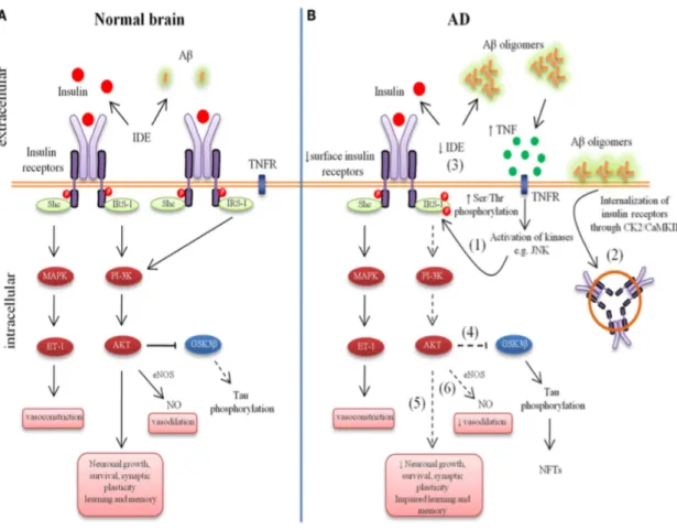
(13) (49,120). GSK-3β has been most extensively studied as a tau kinase, but it is also involved in Aβ production (121). Interestingly, peripheral hiperinsulinemia may result in active GSK-3β and high tau protein deposits (122,123). This is likely due to the serine phosphorylation of IRS as a result of the negative feedback induced by hypersinsulinemia (124).. Figure 1. Aberrant brain insulin signaling in Alzheimer’s disease (AD). Schematic outline of neuronal insulin signaling in the normal brain (A) and AD brain (B). Under physiological conditions, insulin binding to its receptor triggers phosphorylation of insulin receptors substrate-1 (IRS-1). This results in phophoinositide 3-kinase (PI3K) activation and downstream cellular responses that facilitate neuronal growth, neuronal survival, synaptic plasticity, learning and memory, etc. Activation of the IR can result in both vasodilatation and vasoconstriction and under physiological conditions there is a balance of both processes to regulate the immediate metabolic requirements of various tissues. In AD, accumulation of amyloid-β oligomers (AβOs) leads to increased tumor necrosis factor-alpha (TNF-α) levels and activation of stress kinases such as c-Jun N-terminal kinase (JNK) resulting in inhibitory serine phosphorylation of IRS-1 (1). Aβ oligomers cause removal of IRs from the cell surface mediated by Casein Kinase 2 (CK2) and Ca2+/Calmodulin-Dependent Kinase II (CaMKII) and redistribute them to cell bodies (2). Insulin resistance lowers the expression of Aβ-degrading insulin degrading enzyme (IDE) (3). Lowered IDE. 13.
(14) expression further decreases the availability of IDE for Aβ degradation. The reduction in brain insulin signaling increases GSK-3β activity (4), which increases abnormal tau phosphorylation. Deficient insulin signaling leads to impairment in nerve growth, synaptic plasticity, learning and memory, etc. (5). Aberrant phosphorylation of IRS causes an imbalance in homeostatic regulation of vascular function (6). This decreased production of NO may result in decreased cerebral blood flow and increased proinflammatory cytokines and reactive oxygen species production. Extracted and modified from: Bedse G, Di Domenico F, Serviddio G and Cassano T (2015) Aberrant insulin signaling in Alzheimer’s disease: current knowledge. Front. Neurosci. 9:204. doi: 10.3389/fnins.2015.00204.. Neuronal metabolic stress, neuroinflammation and impaired insulin signaling in AD We have described the importance of AβOs neurotoxicity in AD neuropathology. Recapitulating, AβOs promote neuronal stress by inducing neuroinflammation, with consequent abnormal rising in TNF-α levels and ROS which in turn leads to overactivation of JNK/PKR stress kinases signaling (6,96,125,126). The activation per se is not problematic, in fact, stress kinases activation regulates homeostasis. However, their exceeding or lengthened actions provoke cell damage and later apoptosis (126-129). Not only JNK and PKR are involved, AD brains have also been reported to present abnormal phosphorylation levels of PERK (3,130), p38 MAPK (131) and IKK (7). All these stress kinases are thought to be core factors of neuronal dysfunction. JNK, PKR and IKK mediate AβO-induced IRS-1 inhibition in hippocampal neurons (see figure 2) (6,96). Studies have shown that PKR and JNK blockage protects, at least partially, from cognitive impairments in mouse models of AD (96,132). Thus, neuroinflammation plays a leading role in AD. Human and mice models of AD have shown elevated markers of inflammation (98,133,134). There is evidence of gliosis and central infiltration of peripheral immune cells in AD mouse models (98,107,133,134,136,137). Amyloid aggregates, predominantly oligomers as we have mentioned, promote a neuroinflammatory profile that causes synaptic and neuronal damage through TNF-α, cytokines and stress signaling (96,134,138-141). Furthermore, it is possible that AD could be more susceptible to peripheral inflammation as the BBB of AD transgenic mouse models has been described to be more permeable (142). Most interestingly, reducing neuroinflammation can counteract memory deficits in AD mouse models (101,102,143,144), so that probably future research will reveal new therapeutic targets aiming to stop or slow down AD cognitive impairment. Likewise, the. 14.
(15) insulin signaling pathway is clearly affected by neuroinflammatory processes. AD metabolic stress theory is relatively recent compared to the amyloid and hyperphosphorylated tau classical theories. Although the specific mechanisms responsible for the synaptic defects have not been fully elucidated, little by little our understanding of the factors involved as well as their interrelation is growing. In line with the metabolic approach, we also know that excessive ROS levels are part of AD etiology and AβOs may be the causal agents, via Ca2+-related mitochondrial dysfunction that could trigger an aberrant excitatory stimulation of NMDA receptors (124,145). This is relevant because oxidative stress and neuronal insulin signaling seem to be connected, as studies disclose that insulin blocks AβO-induced oxidative stress in the brain (83,146).. Figure 2. AβOs trigger neuronal metabolic stress in Alzheimer’s disease (AD). AβOs instigate an inflammatory response which involves increased TNF-α levels. TNF-α promotes the activity of stress kinases like JNK, PKR and IKKα. Stress kinases provoke the inhibitory phosphorylation (pSer) of IRS-1, causing defective insulin signaling and leading to neuronal dysfunction, synapse deregulation, memory and cognitive impairment.. 15.
(16) Dysfunctional insulin signaling in AD as a target for treatment As discussed above, defective insulin signaling is intimately associated with AD neuropathology and memory impairment. Therapies targeting brain insulin signaling are expected to improve AD treatment. Agents like insulin, metformin, GLP-1 agonist, PPAR-γ agonists and agmatine are being researched. Evidence shows that insulin treatment, both intravenously and intranasally, can modestly improve performance on memory tasks in healthy individuals and in patients with AD or mild cognitive impairment (MCI) (147-153). Both acute and chronic intranasal insulin treatment (160 IU/day) enhance memory and mood in healthy adults (154-157) and in patients with MCI or in the early stages of AD at various doses (10, 20 and 40 IU) (see figure 3) (153). Systemic administration of insulin is not a good option for treating AD due to the risk of developing hypoglycemia and because insulin affects other hormones like cortisol, among others. Intranasal delivery is a non-invasive way of bypassing the BBB via the nasal epithelia with a small effect on peripheral glucose levels (153). On the other hand, gender and the presence of the APOE4 allele modulate the effects of insulin (149,153). For example, only APOE4 negative individuals showed improved declarative memory after insulin treatment (158). However, we do not know if it correlates with an increment in insulin signaling or if it offers protection against neuronal damage, in any case, neither theory excludes the other (159). The rise of hippocampal insulin. and. insulin. signaling. in. mice. also. increases. memory.. Nevertheless, one study showed that mice with type 2 diabetes induced by a high fat diet did not present any improvement with insulin therapy (57). This fact supports the theory that once insulin resistance has been established, it is probable that insulin treatment will not be very benefitial, so it is likely that the biggest benefits from insulin treatment come from its application at the initial phases of the disease, as we have mentioned before. Recent investigations have pointed out the possibility of insulin working through indirect pathways to influence cognition. For instance, Novak and colleagues reported that intranasal insulin increases regional cerebral blood flow and cognition in type 2 diabetes patients (160). Determining the exact mechanisms through which intranasal insulin enhances cognition and whether neuronal insulin signaling is required or not, will unlock new specific approaches for treatment without potential harmful side effects like raising Aβ or tau. Having on mind that insulin is probably not very effective in the late stages of AD, new research has focused in the search of antidiabetic drugs that replicate insulin cellular actions independently from insulin. 16.
(17) receptors activation. Such is the case of peroxisome proliferator-activated receptor γ (PPAR-γ) agonists, metformin and GLP-1 agonists. PPAR-γ agonists are insulin sensitizers with neuroprotective effects. In AD research, rosiglitazone and pioglitazone have been shown to improve the microglia ability of clearing amyloid deposits. Moreover these molecules can modify gene expression and restore both memory and cognition impairment in AD mouse models (107,108,161163). Additionally, PPAR-γ stimulation also improves synapse density in cell cultures (84) and reduces Aβ levels in AD transgenic mice (164). Another option tested for AD has been the insulin-sensitizer agent metformin, a widely used antidiabetic drug. Metformin was able to reduce AD-related neuropathology in a diabetes mice model (165) and to ameliorate insulin resistance in neuronal cell cultures (166). Metformin was also found to reduce tau phosphorylation in mice (167). Furthermore, metmorfin, as well as physical exercise or thiazolidinediones (TZD) improve insulin action via the inhibition of iNOS and mTOR signaling (168,169). Nonetheless, as could be guessed, further research about metformin is needed, first, because it has not been clinically studied in AD patients, and secondly, because some studies have observed that it may increase Aβ production (170,171). At the same time, considering the metabolic hypothesis of AD, adopting a healthy lifestyle and strategies aimed at lifelong control of blood glucose levels may produce benefits to preserve cognition in the elderly and to prevent AD development. Lately, the National Institute of Health (NIH) has selected intranasal insulin administration as one of the two therapeutic strategies receiving substantial funding as part of the National Alzheimer’s Plan in the US. This plan is part of an initiative aimed at finding a therapeutic treatment to cure AD by 2025. In a double-blind and placebocontrolled phase II/III clinical trial, subjects will be treated with intranasal insulin or placebo for one year; subsequently all subjects will receive the active drug for a 6 months period. Primary outcome measures include effects on cognitive status, brain structure on MRI, changes in cerebrospinal fluid (CSF), AD markers and both significance of gender and APOE E4 status for insulin response (172).. More. extensive, detailed and multi-center clinical trials are still needed to establish the beneficial effects of intranasal insulin on cognition and to evaluate its potential sideeffects.. 17.
(18) Figure 3. Intranasal insulin improves memory in patients with MCI or in the early stages of AD (153). Mean memory saving scores (±SEM) in patients with early stage Alzheimer’s disease and mild cognitive impairment (MCI) at baseline (day 0) and after 21 days of treatment with placebo (n=12) and insulin (2 × 20 IU/day; n=13), respectively. Insulin-treated patients showed increased memory savings over 21-day period relative to placebo. *P value smaller than 0.05 Extracted from: Brain Insulin Signaling and Alzheimer's Disease: Current Evidence and Future Directions Helgi B. Schiöth & Suzanne Craft & Samantha J. Brooks & William H. Frey II & Christian Benedict Mol Neurobiol (2012) 46:4–10 DOI 10.1007/s12035-011-8229-6.. GLP-1 signaling as a promising therapeutic target Glucagon-like peptide 1 (GLP-1) is an incretin and neuropeptide first known to stimulate pancreatic insulin secretion after feeding. GLP-1 receptors are present in several brain regions, mostly on large neurons such as pyramidal neurons at the hippocampus or cortex and in the Purkinje neurons of the cerebellum (173-175). The use of insulin in non-diabetic patients, even intranasal via, is not exempt of risk, not only because it can cause hypoglycemia but because it may accelerate insulin desensitisation in the brain. Thereupon, new alternatives focused on insulin signaling have been proposed, mainly drugs first developed to treat type 2 diabetes, as GLP-1 analogs (176-178). Up to date, three drugs of this class have been approved for type 2 diabetes treatment: Exendin-4, lixisenatide and liraglutide (179,180), which in turn are under investigation for the treatment of neurodegenerative diseases such as AD. The. 18.
(19) interesting aspect about these drugs is that they have small side effects, they do not directly affect blood sugar levels and they can be administered to non-diabetic patients (181,182). Moreover, these drugs can cross the BBB (183), so they are potentially effective in treating neurodegenerative disorders of the CNS. GLP-1 analogs can shield cultured neurons from stressors, reduce apoptosis and increase cell division (184,185). They also protect synapses from injurious effects caused by amyloid deposits in the hippocampus (186,187). GLP-1, just like insulin and IGF-1, interferes with PI3K and MAPK second messenger signaling pathways (188). Neuroprotective effects have been observed in AD animal models after GLP-1 mimetics treatment (189). Exendin-4 proved itself capable of reducing AD associated biomarkers, as well as endogenous levels of beta-amyloid in the mouse brain (190). Likewise, lixisenatide has been shown to boost stem cell proliferation and to protect the hippocampus from beta amyloid effects regarding memory, learning and synaptic plasticiy in rats (191,192). In addition, liraglutide displayed similar effects as lixisenatide, rescued memory formation and reduced chronic inflammation response related to AD (193-195). GLP-1 analogs also have been stated to induce neurite outgrowth and to protect against excitotoxic cell death in cell cultures (184,185,196). Based on these very promising preclinical studies, clinical trials with GLP-1 mimetics in AD patients are being carried. Such is the case of liraglutide, which is being tested for efficacy in AD (http://clinicaltrials.gov/ct2/ show/NCT01843075).. Agmatine Agmatine is an endogenous aminoguanidine compound derived from L-Arginine which has been used in models of several diseases such as stroke, diabetes, spinal cord injury and cognitive decline (197). Kang and colleagues showed that agmatine administration improved and rescued the reduced expressions of p-IRS1, p-Akt, and pGSK-3β in the brain of high-fat diet-fed mice (198,199). It also reduced Aβ burden and p-tau accumulation. In addition, agmatine improved impairments in learning and memory functions in high-fat diet-fed mice (200). It has been reported as well that agmatine can improve insulin sensitivity by activating I2-imidazoline receptors in the adrenal glands of diabetic animal models (200). It may also reduce the phosphorylation of iNOS and JNK in the brain of type 2 diabetes mice with AD-like alterations (201,202). Agmatine improves hippocampus-dependent spatial learning by rescuing. 19.
(20) blunted insulin signaling in the region (198). However, agmatine could also improve hippocampus-dependent spatial learning and memory, not only by rescuing insulin signaling but due to its neurotransmitter function (203,204). Further research is needed to clarify the direct connection between brain insulin resistance and agmatine. The drug has the potential to rescue some AD-like alterations so maybe it could help us manage the disease in the future combined with other treatments.. Concluding remarks AD is a dreadful disorder both for the patient and for his family. A family that has to witness, impotent, how the disease slowly takes away their beloved one. Until date, no therapy has proven to be effective in stopping or reversing the disease. The 21st century has turned out to be a scenario where metabolic disorders represent an important public health problem, with an impact potentially comparable to that of infectious diseases on the 20th century. We can easily see this trend by taking a glance at the leading causes of mortality and morbidity worldwide. Cardiovascular diseases, cancer, diabetes and obesity, all have a metabolic component. The same goes with dementia and AD. In this review, we have centered our attention on analyzing the relation between insulin resistance and AD, on how insulin signaling disruption promotes AD-like pathology and the mechanisms involved. Insulin resistance, AβOs neurotoxicity, neuronal metabolic stress and neuroinflammation all have been proven to play a role in AD pathogenesis and to be interconnected. Due to its complexity, a lot of questions still remain open, but we hope that sooner than later, the answers will come so we can develop better therapies to manage the disease. Intranasal insulin, GLP-1 analogs, anti-TNF-α antibodies, gene therapy and agmatine have been the most promising treatments up to date, with some of them in the clinical trial phase. Other treatments like metformin, PPAR-γ agonists, lipoic acid and antioxidant compounds are also being explored but with controversial or doubtful results. The future will reveal whether we were able to cure the disease or not, now it is our time to make the effort and tilt the balance towards the yes, or maybe we will not have to, if the anti-aging progress does the trick first, but that is another topic.. 20.
(21) Acknowledgments I would like to thank all my family for the support and strength they have given me during all these 6 years. Thank you as well to my tutor and professor Ana María Sánchez Pérez for her help and advice during this work and during my Erasmus mobility last year. Thank you to all the professors and clinicians who have inspired me during this journey. And last but not least, thank you to professor Conrado Martínez Cadenas for his help during my last faculty year. “Above all, do not fear difficult moments, the best comes from them.” Rita Levi-Montalcini - Italian Neurologist and Nobel laureate.. References 1. Alzheimer’s. Association.. 2013. Alzheimer’s. disease. facts. and. figures.. Alzheimers Dement 2013;9:208–245. 2. De Felice FG. Alzheimer’s disease and insulin resistance: translating basic science into clinical applications. J Clin Invest 2013;123:531–539. 3. Hoozemans, J. J. M., Veerhuis, R., Haastert, E. S., Rozemuller, J. M., Baas, F., Eikelenboom, P., et al. (2005). The unfolded protein response is activated in Alzheimer’s disease. Acta Neuropathol. 110, 165–172. doi: 10.1007/s00401005- 1038-0. 4. Steen, E., Terry, B. M., Rivera, E. J., Cannon, J. L., Neely, T. R., Tavares, R., et al. (2005). Impaired insulin and insulin-like growth factor expression and signaling mechanisms in Alzheimer’s disease – is this type 3 diabetes? J. Alzheimers Dis. 7, 63–80. 5. Moloney, A. M., Griffin, R. J., Timmons, S., O’connor, R., Ravid, R., and O’neill, C. (2010). Defects in IGF-1 receptor, insulin receptor and IRS- 1/2 in. 21.
(22) Alzheimer’s disease indicate possible resistance to IGF-1 and insulin signalling. Neurobiol. Aging 31, 224–243. doi: 10.1016/j.neurobiolaging.2008. 04.002.. 6. Bomfim, T. R., Forny-Germano, L., Sathler, L. B., Brito-Moreira, J., Houzel, J. C., Decker, H., et al. (2012). An anti-diabetes agent protects the mouse brain from defective insulin signaling caused by Alzheimer’s disease-associated Aβ oligomers. J. Clin. Invest. 122, 1339–1353. doi: 10.1172/jci57256. 7. Talbot, K., Wang, H., Kazi, H., Han, L., Bakshi, K. P., Stucky, A., et al. (2012). Demonstrated brain insulin resistance in Alzheimer’s disease patients is associated with IGF-1 resistance, IRS-1 dysregulation and cognitive decline. J. Clin. Invest. 122, 1316–1338. doi: 10.1172/JCI59903. 8. O’Neill, C. (2013). PI3-kinase/Akt/mTOR signaling: impaired on/off switches in aging, cognitive decline and Alzheimer’s disease. Exp. Gerontol. 48, 647–653. doi: 10.1016/j.exger.2013.02.025. 9. Karran E, Mercken M, De Strooper B (2011) The amyloid cascade hypothesis for Alzheimer's disease: an appraisal for the development of therapeutics. Nat Rev Drug Discov 10:698–712. 10. Selkoe, D., Mandelkow, E., Holtzman, D., 2012. Deciphering Alzheimer disease. Cold Spring Harb. Perspect. Med. 2, a011460. 11. Serrano-Pozo,. A.,. Frosch,. M.P.,. Masliah,. E.,. Hyman,. B.T.,. 2011.. Neuropathological alterations in Alzheimer disease. Cold Spring Harb. Perspect. Med. 1, a006189. 12. Ballard, C., Gauthier, S., Corbett, A., Brayne, C., Aarsland, D., Jones, E., 2011. Alzheimer’s disease. Lancet 377, 1019–1031. 13. Castellani, R.J., Rolston, R.K., Smith, M.A., 2010. Alzheimer disease. Dis. Mon. 56, 484–546.. 22.
(23) 14. LaFerla, F.M., Green, K.N., Oddo, S., 2007. Intracellular amyloid-beta in Alzheimer’s disease. Nat. Rev. Neurosci. 8, 499–509. 15. Craft S. Alzheimer disease: insulin resistance and AD—extending the translational path. Nat Rev Neurol 2012;8:360–362. 16. Craft, S., Watson, G.S., 2004. Insulin and neurodegenerative disease: shared and specific mechanisms. Lancet Neurol. 3, 169e178. 17. Haan, M.N., 2006. Therapy Insight: type 2 diabetes mellitus and the risk of lateonset Alzheimer's disease. Nat. Clin. Pract. Neurol. 2, 159e166. 18. Jayaraman, A., Pike, C.J., 2014. Alzheimer's disease and type 2 diabetes: multiple mechanisms contribute to interactions. Curr. Diab Rep. 14, 476. 19. Kim, B., Feldman, E.L., 2012. Insulin resistance in the nervous system. Trends Endocrinol. Metab. 23, 133e141. 20. Ma, L., Wang, J., Li, Y., 2015. Insulin resistance and cognitive dysfunction. Clin. Chim. Acta 444, 18e23. 21. Zhao, W.Q., Alkon, D.L., 2001. Role of insulin and insulin receptor in learning and memory. Mol. Cell Endocrinol. 177, 125e134. 22. Kim, B., Feldman, E.L., 2015. Insulin resistance as a key link for the increased risk of cognitive impairment in the metabolic syndrome. Exp. Mol. Med. 47, e149. 23. Stockhorst U, de Fries D, Steingrueber HJ, Scherbaum WA. Insulin and the CNS: effects on food intake, memory, and endocrine parameters and the role of intranasal insulin administration in humans. Physiol Behav 2004; 83(1): 47– 54. 24. Hoyer S. Glucose metabolism and insulin receptor signal transduction in Alzheimer disease. Eur J Pharmacol 2004; 490(1–3): 115–125.. 23.
(24) 25. van Dam PS, Aleman A. Insulin-like growth factor-I, cognition and brain aging. Eur J Pharmacol 2004; 490(1–3): 87–95. 26. Freiherr J, Hallschmid M, Frey WH, 2nd, Brunner YF, Chapman CD, Hölscher C, Craft S, De Felice FG, Benedict C. Intranasal insulin as a treatment for Alzheimer’s disease: a review of basic research and clinical evidence. CNS Drugs 2013; 27(7): 505–514. 27. Craft S, Cholerton B, Baker LD. Insulin and Alzheimer's disease: untangling the web. J Alzheimers Dis 2013, 33 Suppl 1: S263–275. 28. de la Monte SM, Wands JR (2005) Review of insulin and insulin-like growth factor expression, signaling, and malfunction in the central nervous system: relevance to Alzheimer’s disease. J Alzheimers Dis 7:45–61. 29. Devaskar, S.U., Giddings, S.J., Rajakumar, P.A., Carnaghi, L.R., Menon, R.K., Zahm, D.S., 1994. Insulin gene expression and insulin synthesis in mammalian neuronal cells. J. Biol. Chem. 269, 8445–8454. 30. Kuwabara, T., Kagalwala, M.N., Onuma, Y., Ito, Y., Warashina, M., Terashima, K., Sanosaka, T., Nakashima, K., Gage, F.H., Asashima, M., 2011. Insulin biosynthesis in neuronal progenitors derived from adult hippocampus and the olfactory bulb. EMBO Mol. Med. 3, 742–754. 31. Banks, W.A., 2012. Role of the blood–brain barrier in the evolution of feeding and cognition. Ann. N. Y. Acad. Sci. 1264, 13–19. 32. Frolich, L., Blum-Degen, D., Bernstein, H. G., Engelsberger, S., Humrich, J., Laufer, S., et al. (1998). Brain insulin and insulin receptors in aging and sporadic Alzheimer’s disease. J. Neural Transm. 105, 423–438. doi: 10.1007/s007020050068.. 24.
(25) 33. Van Der Heide, L. P., Ramakers, G. M., and Smidt, M. P. (2006). Insulin signaling in the central nervous system: learning to survive. Prog. Neurobiol. 79, 205–221. doi: 10.1016/j.pneurobio.2006.06.003. 34. Duarte, A. I., Moreira, P. I., and Oliveira, C. R. (2012). Insulin in central nervous system: more than just a peripheral hormone. J. Aging Res. 2012:384017. doi: 10.1155/2012/384017. 35. Schwartz, M.W., 2005. Diabetes, obesity, and the brain. Science 307, 375–379. 36. Bondy, C.A., Cheng, C.M., 2004. Signaling by insulin-like growth factor 1 in brain. Eur. J. Pharmacol. 490, 25–31. 37. Broughton, S., Partridge, L., 2009. Insulin/IGF-like signalling, the central nervous system and aging. Biochem. J. 418, 1. 38. Ghasemi, R., Haeri, A., Dargahi, L., Mohamed, Z., Ahmadiani, A., 2012. Insulin in the brain: sources, localization and functions. Mol. Neurobiol. 47, 145–171. 39. Zhao, W.Q., Chen, H., Quon, M.J., Alkon, D.L., 2004. Insulin and the insulin receptor in experimental models of learning and memory. Eur. J. Pharmacol. 490, 71–81. 40. Ohno, H., Kato, S., Naito, Y., Kunitomo, H., Tomioka, M., Iino, Y., 2014. Role of synaptic phosphatidylinositol 3-kinase in a behavioral learning response in C. elegans. Science 345, 313–317. 41. Nisticò , R., Cavallucci, V., Piccinin, S., Macrı`, S., Pignatelli, M., Mehdawy, B., Blandini, F., Laviola, G., Lauro, D., Mercuri, N.B., D’Amelio, M., 2012. Insulin receptor b-subunit haploinsufficiency impairs hippocampal late-phase LTP and recognition memory. Neuromol. Med. 14, 262–269. 42. Fernandez, A.M., Torres-Alema ́n, I., 2012. The many faces of insulin-like peptide signalling in the brain. Nat. Rev. Neurosci. 13, 225–239.. 25.
(26) 43. Schubert, M., Gautam, D., Surjo, D., Ueki, K., Baudler, S., Schubert, D., Kondo, T., Alber, J., Galldiks, N., Ku ̈stermann, E., Arndt, S., Jacobs, A.H., Krone, W., Kahn, C.R., Bru ̈ning, J.C., 2004. Role for neuronal insulin resistance in neurodegenerative diseases. Proc. Natl. Acad. Sci. U. S. A. 101, 3100–3105. 44. de la Monte, S.M., Tong, M., Bowling, N., Moskal, P., 2011. si-RNA inhibition of brain insulin or insulin-like growth factor receptors causes developmental cerebellar abnormalities: relevance to fetal alcohol spectrum disorder. Mol. Brain 4, 13. 45. Chiu, S.-L., Cline, H.T., 2010. Insulin receptor signaling in the development of neuronal structure and function. Neural Dev. 5, 7. 46. Kleinridders, A., Cai, W., Cappellucci, L., Ghazarian, A., Collins, W.R., Vienberg, S.G., Pothos, E.N., Kahn, C.R., 2015. Insulin resistance in brain alters dopamine turnover and causes behavioral disorders. Proc. Natl. Acad. Sci. U. S. A. 112, 3463–3468. 47. Chiu, S.-L., Chen, C.-M., Cline, H.T., 2008. Insulin receptor signaling regulates synapse number, dendritic plasticity, and circuit function in vivo. Neuron 58, 708–719. 48. Dixon-Salazar, T.J., Fourgeaud, L., Tyler, C.M., Poole, J.R., Park, J.J., Boulanger, L.M., 2014. MHC class I limits hippocampal synapse density by inhibiting neuronal insulin receptor signaling. J. Neurosci. 34, 11844–11856. 49. Chen Y, Liang Z, Blanchard J, Dai CL, Sun S, Lee MH, et al. A non-transgenic mouse model (icv-STZ mouse) of Alzheimer's disease: similarities to and differences from the transgenic model (3xTg-AD mouse). Mol Neurobiol 2013, 47: 711–725. 50. Lee, C.-C., Huang, C.-C., Hsu, K.-S., 2011. Insulin promotes dendritic spine and synapse formation by the PI3K/Akt/mTOR and Rac1 signaling pathways. Neuropharmacology 61, 867–879.. 26.
(27) 51. Zhao, W., Wu, X., Xie, H., Ke, Y., Yung, W.-H., 2010. Permissive role of insulin in the expression of long-term potentiation in the hippocampus of immature rats. Neurosignals 18, 236–245. 52. Sousa-Nunes, R., Yee, L.L., Gould, A.P., 2011. Fat cells reactivate quiescent neuroblasts via TOR and glial insulin relays in Drosophila. Nature 471, 508– 512. 53. Apostolatos, A., Song, S., Acosta, S., Peart, M., Watson, J.E., Bickford, P., Cooper, D.R., Patel, N.A., 2012. Insulin promotes neuronal survival via the alternatively spliced protein kinase C II isoform. J. Biol. Chem. 287, 9299–9310.. 54. Di Carlo, M., Picone, P., Carrotta, R., Giacomazza, D., San Biagio, P.L., 2010. Insulin promotes survival of amyloid-beta oligomers neuroblastoma damaged cells via caspase 9 inhibition and Hsp70 upregulation. J. Biomed. Biotechnol. 2010, 147835. 55. Duarte, A.I., Proenc ̧a, T., Oliveira, C.R., Santos, M.S., Rego, A.C., 2006. Insulin restores metabolic function in cultured cortical neurons subjected to oxidative stress. Diabetes 55, 2863–2870. 56. Haj-ali, V., Mohaddes, G., Babri, S.H., 2009. Intracerebroventricular insulin improves spatial learning and memory in male Wistar rats. Behav. Neurosci. 123, 1309–1314.. 57. McNay, E.C., Ong, C.T., McCrimmon, R.J., Cresswell, J., Bogan, J.S., Sherwin, R.S., 2010. Hippocampal memory processes are modulated by insulin and high-fat-induced insulin resistance. Neurobiol. Learn. Mem. 93, 546–553. 58. Vogt MC, Bruning JC. CNS insulin signaling in the control of energy homeostasis and glucose metabolism: from embryo to old age. Trends Endocrinol Metab 2013;24:76–84.. 27.
(28) 59. Conejo, R., and Lorenzo, M. (2001). Insulin signaling leading to proliferation, survival, and membrane ruffling in C2C12 myoblasts. J. Cell Physiol. 187, 96– 108. doi: 10.1002/1097-4652(2001)9999:9999<::aid-jcp1058>3.0.co;2-v. 60. Lin JW, Ju W, Foster K, Lee SH, Ahmadian G, Wyszynski M, Wang YT, Sheng M. Distinct molecular mechanisms and divergent endocytotic pathways of AMPA receptor internal- ization. Nat Neurosci 2000; 3(12): 1282–1290.. 61. Wan Q, Xiong ZG, Man HY, Ackerley CA, Braunton J, Lu WY, Becker LE, MacDonald JF, Wang YT. Recruitment of functional GABA (A) receptors to postsynaptic domains by insulin. Nature 1997; 388(6643): 686–690.. 62. KahnCR,SuzukiR.Insulinactioninthebrainandthepathogenesisof.. Alzheimer’s. Diabetes, Insulin and Alzheimer’s Disease, 1 Research and Perspectives in Alzheimer’s Disease. Berlin: Springer-Verlag; 2010. 63. Taha C, Klip A (1999) The insulin signaling pathway. J Membr Biol 169:1–12.. 64. Stoica L, Zhu PJ, Huang W, Zhou H, Kozma SC, Costa-Mattioli M. Selective pharmacogenetic inhibition of mammalian target of rapamycin complex I (mTORC1) blocks long-term synaptic plasticity and memory storage. Proc Natl Acad Sci U S A 2011;108:3791–3796. 65. Son JH, Shim JH, Kim KH, Ha JY, Han JY. Neuronal autophagy and neurodegenerative diseases. Exp Mol Med 2012;44:89–98.. 66. Salcedo-Tello P, Ortiz-Matamoros A, Arias C. GSK3 function in the brain during development, neuronal plasticity, and neurodegeneration. Int J Alzheimers Dis 2011;2011:189728.. 28.
(29) 67. Schubert M, Brazil DP, Burks DJ, et al. Insulin receptor substrate-2 deficiency impairs. brain. growth. and. promotes. tau. phosphorylation.. J. Neurosci. 2003;23:7084–7092. 68. Adams JP, Sweatt JD. Molecular psychology: roles for the ERK MAP kinase cascade in memory. Annu Rev Pharmacol Toxicol 2002;42:135–163. 69. Yarchoan, M., Toledo, J. B., Lee, E. B., Arvanitakis, Z., Kazi, H., Han, L. Y., et al. (2014). Abnormal serine phosphorylation of insulin receptor substrate 1 is associated with tau pathology in Alzheimer’s disease and tauopathies. Acta Neuropathol. 128, 679–689. doi: 10.1007/s00401-014-1328-5.. 70. Hoyer S (2002) The brain insulin signal transduction system and sporadic (type II) Alzheimer disease: an update. J Neural Transm 109:341–360.. 71. Hoyer S, Fro ̈lich L (2007) Chapter XI: Brain insulin function and insulin signal transduction in sporadic Alzheimer disease. In: Sun M-K (ed) Research progress in Alzheimer’s disease and dementias. Nova Science, New York, pp 205–255. ISBN 1-59454-949-4. 72. Kappeler L, De Magalhaes Filho CM, Dupont J, Leneuve P, Cervera P, Perin L, Loudes C, Blaise A, Klein R, Epelbaum J, Le BY, Holzenberger M (2008) Brain IGF-1. receptors. control. mammalian. growth. and. lifespan. through. a. neuroendocrine mech- anism. PLoS Biol 6:e254. 73. Grünblatt E, Salkovic-Petrisic M, Osmanovic J, Riederer P, Hoyer S (2007) Brain insulin system dysfunction in streptozotocin intra- cerebroventricularly treated rats generates hyperphosphorylated tau protein. J Neurochem 101:757–770.. 29.
(30) 74. Grünblatt E, Hoyer S, Riederer P (2004) Gene expression profile in streptozotocin rat model for sporadic Alzheimer's disease. J Neural Transm 111:367–386. 75. Salkovic-Petrisic M, Tribl F, Schmidt M, Hoyer S, Riederer P (2006) Alzheimerlike changes in protein kinase B and glycogen synthase kinase-3 in rat frontal cortex and hippocampus after damage to the insulin signalling pathway. J Neurochem 96:1005–1015.. 76. Plaschke K, Kopitz J, Siegelin M, Schliebs R, Salkovic-Petrisic M, Riederer P, Hoyer S (2010) Insulin-resistant brain state after intracerebroventricular streptozotocin injection exacerbates Alzheimer-like changes in Tg2576 abetaPP-overexpressing mice. J Alz- heimers Dis 19:691–704.. 77. Akter K, Lanza EA, Martin SA, Myronyuk N, Rua M, Raffa RB (2011) Diabetes mellitus and Alzheimer’s disease: shared pathology and treatment? Br J Clin Pharmacol 71:365–376. 78. Rivera EJ, Goldin A, Fulmer N, Tavares R, Wands JR, de la Monte SM (2005) Insulin and insulin-like growth factor expression and function deteriorate with progression of Alzheimer’s disease: link to brain reductions in acetylcholine. J Alzheimers Dis 8:247–268. 79. Negash S, Bennett DA, Wilson RS, Schneider JA, Arnold SE. Cognition and neuropathology in aging: multidimensional perspectives from the Rush Religious Orders Study and Rush Memory and Aging Project. Curr Alzheimer Res. 2011;8(4):336–340. 80. Terry RD, et al. Physical basis of cognitive altera- tions in Alzheimer disease: synapse loss is the major correlate of cognitive impairment. Ann Neu- rol. 1991;30(4):572–580.. 30.
(31) 81. Townsend, M., Mehta, T., Selkoe, D.J., 2007. Soluble Abeta inhibits specific signal transduction cascades common to the insulin receptor pathway. J. Biol. Chem. 282, 33305–33312.. 82. Zhao, W.Q., De Felice, F.G., Fernandez, S., Chen, H., Lambert, M.P., Quon, M.J., Krafft, G.A., Klein, W.L., 2008. Amyloid beta oligomers induce impairment of neuronal insulin receptors. FASEB J. 22, 246–260. 83. De Felice, F.G., Vieira, M.N.N., Bomfim, T.R., Decker, H., Velasco, P.T., Lambert, M.P., Viola, K.L., Zhao, W.Q., Ferreira, S.T., Klein, W.L., 2009. Protection of synapses against Alzheimer’s-linked toxins: insulin signaling prevents the pathogenic binding of Ab oligomers. Proc. Natl. Acad. Sci. U. S. A. 106, 1971–1976. 84. Zhao, W.-Q., Lacor, P.N., Chen, H., Lambert, M.P., Quon, M.J., Krafft, G.a., Klein, W.L., 2009. Insulin receptor dysfunction impairs cellular clearance of neurotoxic oligomeric Ab. J. Biol. Chem. 284, 18742–18753.. 85. Qiu, W. Q., Walsh, D. M., Ye, Z., Vekrellis, K., Zhang, J., Podlisny, M. B., et al. (1998). Insulin-degrading enzyme regulates extracellular levels of amyloid beta-protein by degradation. J. Biol. Chem. 273, 32730–32738. doi: 10.1074/jbc.273.49.32730.. 86. Vekrellis, K., Ye, Z., Qiu, W. Q., Walsh, D., Hartley, D., Chesneau, V., et al. (2000). Neurons regulate extracellular levels of amyloid beta-protein via proteolysis by insulin-degrading enzyme. J. Neurosci. 20, 1657–1665.. 87. Sudoh, S., Frosch, M. P., and Wolf, B. A. (2002). Differential effects of proteases involved in intracellular degradation of amyloid beta-protein between detergent-soluble and -insoluble pools in CHO-695 cells. Biochemistry 41, 1091–1099. doi: 10.1021/bi011193l.. 31.
(32) 88. Zhao, L., Teter, B., Morihara, T., Lim, G. P., Ambegaokar, S. S., Ubeda, O. J., et al. (2004a). Insulin-degrading enzyme as a downstream target of insulin receptor signaling cascade: implications for Alzheimer’s disease intervention. J. Neurosci. 24, 11120–11126. doi: 10.1523/JNEUROSCI.2860-04.2004. 89. Farris, W., Mansourian, S., Chang, Y., Lindsley, L., Eckman, E. A., Frosch, M. P., et al. (2003). Insulin-degrading enzyme regulates the levels of insulin, amyloid beta-protein, and the beta-amyloid precursor protein intracellular domain in vivo. Proc. Natl. Acad. Sci. U.S.A. 100, 4162–4167. doi: 10.1073/pnas.0230450100. 90. Leissring, M. A., Farris, W., Chang, A. Y., Walsh, D. M., Wu, X., Sun, X., et al. (2003). Enhanced proteolysis of beta-amyloid in APP transgenic mice prevents plaque formation, secondary pathology, and premature death. Neuron 40, 1087–1093. doi: 10.1016/S0896-6273(03)00787-6. 91. Perez, A., Morelli, L., Cresto, J. C., and Castano, E. M. (2000). Degradation of soluble amyloid beta-peptides 1-40, 1-42, and the Dutch variant 1-40Q by insulin degrading enzyme from Alzheimer disease and control brains. Neurochem. Res. 25, 247–255. doi: 10.1023/A:1007527721160. 92. Macauley, S.L., M. Stanley, E.E. Caesar, S.A.Yamada, M.E. Raichle, R. Perez, T.E. Mahan, C.L. Sutphen, and D.M. Holtzman. 2015. Hyperglycemia modulates extracellular amyloid-β concentrations and neuronal activity in vivo. J. Clin. Invest. 125:2463–2467. http://dx.doi.org/10.1172/ JCI79742.. 93. Banks, W.A., J.B. Jaspan, and A.J. Kastin. 1997. Selective, physiological transport of insulin across the blood-brain barrier: novel demonstration by species-speci. c. radioimmunoassays.. Peptides.. 18:1257–1262.. http://. dx.doi.org/10.1016/S0196-9781(97)00198-8 94. Yarchoan, M., Toledo, J.B., Lee, E.B. et al. Acta Neuropathol (2014) 128: 679. doi:10.1007/s00401-014-1328-5. 32.
(33) 95. Bose, A., Mouton-Liger, F., Paquet, C., Mazot, P., Vigny, M., Gray, F., Hugon, J., 2011. Modulation of tau phosphorylation by the kinase PKR: implications in Alzheimer’s disease. Brain Pathol. 21, 189–200.. 96. Lourenco, M.V., Clarke, J.R., Frozza, R.L., Bomfim, T.R., Forny-Germano, L., Batista, A.F., Sathler, L.B., Brito-Moreira, J., Amaral, O.B., Silva, C.A., FreitasCorrea, L., Espı ́rito- Santo, S., Campello-Costa, P., Houzel, J.-C., Klein, W.L., Holscher, C., Carvalheira, J.B., Silva, A.M., Velloso, L.A., Munoz, D.P., Ferreira, S.T., De Felice, F.G., 2013. TNF-a mediates PKR-dependent memory impairment and brain IRS-1 inhibition induced by Alzheimer’s b-amyloid oligomers in mice and monkeys. Cell Metab. 18, 831–843. 97. Paquet, C., Mouton-Liger, F., Meurs, E.F., Mazot, P., Bouras, C., Pradier, L., Gray, F., Hugon, J., 2011. The PKR activator PACT is induced by Ab: involvement in Alzheimer’s disease. Brain Pathol. 22, 219–229. 98. Ferreira, S.T., Clarke, J.R., Bomfim, T.R., De Felice, F.G., 2014. Inflammation, defective insulin signaling and neuronal dysfunction in Alzheimer’s disease. Alzheimer’s Dement. 10, S76–S83. 99. Gregor, M.F., Hotamisligil, G.S., 2011. Inflammatory mechanisms in obesity. Annu. Rev. Immunol. 29, 415–445. 100.. Swardfager, W., Lanctoˆ t, K., Rothenburg, L., Wong, A., Cappell, J.,. Herrmann, N., 2010. A meta-analysis of cytokines in Alzheimer’s disease. Biol. Psychiatry 68, 930–941. 101.. McAlpine, F.E., Lee, J., Harms, A.S., Ruhn, K.A., Blurton-Jones, M.,. Hong, J., Das, P., Golde, T.E., LaFerla, F.M., Oddo, S., Blesch, A., Tansey, M.G., 2009. Inhibition of soluble TNF signaling in a mouse model of Alzheimer’s disease prevents pre-plaque amyloid-associated neuropathology. Neurobiol. Dis. 34, 163–177.. 33.
(34) 102.. Medeiros, R., Prediger, R.D.S., Passos, G.F., Pandolfo, P., Duarte, F.S.,. Franco, J.L., Dafre, A.L., Di Giunta, G., FiYOGESHgueiredo, C.P., Takahashi, R.N., Campos, M.M., Calixto, J.B., 2007. Connecting TNF-a signaling pathways to iNOS expressionin a mouse model of Alzheimer’s disease: relevance for the behavioral and synaptic deficits induced by amyloid-b protein. J. Neurosci. 27, 5394–5404. 103.. Carrero, I., Gonzalo, M.R., Martin, B., Sanz-Anquela, J.M., Are ́valo-. Serrano, J., Gonzalo-Ruiz, A., 2012. Oligomers of beta-amyloid protein (Ab1– 42) induce the activation of cyclooxygenase-2 in astrocytes via an interaction with inter- leukin-1beta, tumour necrosis factor-alpha, and a nuclear factor kappa-B mechanism in the rat brain. Exp. Neurol. 236, 215–227.. 104.. Ledo,. J.H.,. Azevedo,. E.P.,. Clarke,. J.R.,. Ribeiro,. F.C.,. FiYOGESHgueiredo, C.P., Foguel, D., De Felice, F.G., Ferreira, S.T., 2013. Amyloid-b oligomers link depressive-like behavior and cognitive deficits in mice. Mol. Psychiatry 18, 1053–1054. 105.. Heneka, M.T., Kummer, M.P., Stutz, A., Delekate, A., Schwartz, S.,. Vieira-Saecker, A., Griep, A., Axt, D., Remus, A., Tzeng, C., Gelpi, E., Halle, A., korte, M., Latz, E., Golenbock, D.T., 2013. NLRP3 is activated in Alzheimer’s disease and contrib- utes to pathology in APP/PS1 mice. Nature 493, 674–678. 106.. Lee, C.Y.D., Landreth, G.E., 2010. The role of microglia in amyloid. clearance from the AD brain. J. Neural Transm. 117, 949–960.. 34.
(35) 107.. Lourenco, M.V., Ledo, J.H., 2013. Targeting Alzheimer’s pathology. through PPARg signaling: modulation of microglial function. J. Neurosci. 33, 5083–5084. 108.. Mandrekar-Colucci, S., Karlo, J.C., Landreth, G.E., 2012. Mechanisms. underlying the rapid peroxisome proliferator-activated receptor-g-mediated amyloid clear- ance and reversal of cognitive deficits in a murine model of Alzheimer’s disease. J. Neurosci. 32, 10117–10128. 109.. De Felice, F.G., 2013. Alzheimer’s disease and insulin resistance:. translating basic science into clinical applications. J. Clin. Invest. 123, 531–539. 110.. Gardoni, F., Boraso, M., Zianni, E., Corsini, E., Galli, C.L., Cattabeni, F.,. Marinovich, M., Di Luca, M., Viviani, B., 2011. Distribution of interleukin-1 receptor complex at the synaptic membrane driven by interleukin-1b and NMDA stimulation. J. Neuroinflamm. 8, 14. 111.. Steinmetz, C.C., Turrigiano, G.G., 2010. Tumor necrosis factor-a. signaling maintains the ability of cortical synapses to express synaptic scaling. J. Neurosci. 30, 14685–14690. 112.. Bekinschtein, P., Katche, C., Slipczuk, L.N., Igaz, L.M., Cammarota, M.,. Izquierdo, I., Medina, J.H., 2007. mTOR signaling in the hippocampus is necessary for memory formation. Neurobiol. Learn. Mem. 87, 303–307.. 113.. Hoeffer, C., Klann, E., 2010. mTOR signaling: at the crossroads of. plasticity, memory and disease. Trends Neurosci. 33, 67–75. 114.. Manzoni, O.J., Slipczuk, L., Bekinschtein, P., Katche, C., Cammarota,. M., Izquierdo, I., Medina, J.H., 2009. BDNF activates mTOR to regulate GluR1 expression required for memory formation. PLoS ONE 4, e6007.. 115.. Son, S.M., Song, H., Byun, J., Park, K.S., Jang, H.C., Park, Y.J., Mook-. Jung, I., 2012.. Altered APP processing in insulin-resistant conditions is. 35.
(36) mediated by auto- phagosome accumulation via the inhibition of mammalian target of rapamycin pathway. Diabetes 61, 3126–3138. 116.. Perluigi, M., Pupo, G., Tramutola, A., Cini, C., Coccia, R., Barone, E., et. al. (2014). Neuropathological role of PI3K/Akt/mTOR axis in Down syndrome brain.. Biochim.. Biophys.. Acta.. 1842,. 1144–1153.. doi:. 10.1016/j.bbadis.2014.04.007.. 117.. Querfurth, H. W., and Laferla, F. M. (2010). Alzheimer’s disease. N.. Engl. J. Med. 362, 329–344. doi: 10.1056/NEJMra0909142. 118.. Bedse, G., Romano, A., Lavecchia, A. M., Cassano, T., and Gaetani, S.. (2015). The role of endocannabinoid signaling in the molecular mechanisms of neurodegeneration in Alzheimer’s disease. J. Alzheimers Dis. 43, 1115–1136. doi: 10.3233/jad-141635.. 119.. Cheng, C. M., Tseng, V., Wang, J., Wang, D., Matyakhina, L., and. Bondy, C. A. (2005). Tau is hyperphosphorylated in the insulin-like growth factor-I null brain. Endocrinology 146, 5086–5091. doi: 10.1210/en.2005-0063. 120. (2009).. Deng, Y., Li, B., Liu, Y., Iqbal, K., Grundke-Iqbal, I., and Gong, C. X. Dysregulation. of. insulin. signaling,. glucose. transporters,. O-. GlcNAcylation, and phosphorylation of tau and neurofilaments in the brain: implication for Alzheimer’s disease. Am. J. Pathol. 175, 2089–2098. doi: 10.2353/ajpath.2009.090157.. 121.. Jeon S, Park JE, Lee J, Liu QF, Jeong HJ, Pak SC, et al. Illite improves. memory impairment and reduces Aβ level in the Tg-AP- Pswe/PS1dE9 mouse model of Alzheimer’s disease through Akt/ CREB and GSK-3β phosphorylation in the brain. J Ethnopharmacol 2015;160:69-77.. 36.
(37) 122.. Freude, S., L. Plum, J. Schnitker, U. Leeser, M. Udelhoven, W. Krone,. J.C. Bruning, and M. Schubert. 2005. Peripheral hyperinsulinemia promotes tau phosphorylation. in. vivo.. Diabetes.. 54:3343–3348.. http://dx.doi.org. /10.2337/diabetes.54.12.3343. 123.. Becker, K., S. Freude, J. Zemva, O. Stöhr,W. Krone, and M. Schubert.. 2012. Chronic peripheral hyperinsulinemia has no substantial influence on tau phosphorylation. in. vivo.. Neurosci.. Lett.. 516:306–310.. http://dx.doi.org. /10.1016/j.neulet.2012.04.022. 124.. Rui L, Aguirre V, Kim JK, et al. Insulin/IGF-1 and TNF-α stimulate. phosphorylation of IRS-1 at inhibitory Ser307 via distinct pathways. Journal of Clinical Investigation. 2001;107(2):181-189. 125.. De Felice, F. G., Velasco, P. T., Lambert, M. P., Viola, K., Fernandez, S.. J., Ferreira, S. T., et al. (2007). Aβ oligomers induce neuronal oxidative stress through an N-methyl-D-aspartate receptor-dependent mechanism that is blocked by the Alzheimer drug memantine. J. Biol. Chem. 282, 11590–11601. doi: 10.1074/jbc. m607483200.. 126.. De Felice, F. G., and Ferreira, S. T. (2014). Inflammation, defective. insulin signaling and mitochondrial dysfunction as common molecular denominators connecting type 2 diabetes to Alzheimer disease. Diabetes 63, 2262–2272. doi: 10.2337/db13-1954. 127.. Mattson, M. P. (2008). Hormesis defined. Ageing Res. Rev. 7, 1–7. doi:. 10.1016/j. arr.2007.08.007.. 128.. Vallerie, S. N., and Hotamisligil, G. S. (2010). The role of JNK proteins in. metabolism. Sci. Transl. Med. 2:60rv65. doi: 10.1126/scitranslmed.3001007.. 37.
(38) 129.. Hetz, C. (2012). The unfolded protein response: controlling cell fate. decisions under ER stress and beyond. Nat. Rev. Mol. Cell Biol. 13, 89–102. doi: 10. 1038/nrm3270.. 130.. Hoozemans, J. J. M., Van Haastert, E. S., Nijholt, D. A. T., Rozemuller,. A. J. M., Eikelenboom, P., and Scheper, W. (2009). The unfolded protein response. is. activated. hippocampus.. in. Am.. pretangle J.. neurons. Pathol.. in. 174,. Alzheimer’s. disease. 1241–1251.. doi:. 10.2353/ajpath.2009.080814. 131.. Hensley, K., Floyd, R. A., Zheng, N.-Y., Nael, R., Robinson, K. A.,. Nguyen, X., et al. (1999). p38 kinase is activated in the Alzheimer’s disease brain.. J.. Neurochem.. 72,. 2053–2058.. doi:. 10.1046/j.1471-. 4159.1999.0722053.x. 132.. Yoon, S. O., Park, D. J., Ryu, J. C., Ozer, H. G., Tep, C., Shin, Y. J., et. al. (2012). JNK3 perpetuates metabolic stress induced by Aβ peptides. Neuron 75, 824–837. doi: 10.3410/f.717965605.793466623. 133.. Monson, N. L., Ireland, S. J., Ligocki, A. J., Chen, D., Rounds, W. H., Li,. M., et al. (2014). Elevated CNS inflammation in patients with preclinical Alzheimer’s. disease.. J.. Cereb.. Blood. Flow. Metab.. 34,. 30–33.. doi:. 10.1038/jcbfm. 2013.183. 134.. Heneka, M. T., Golenbock, D. T., and Latz, E. (2015). Innate immunity in. Alzheimer’s disease. Nat. Immunol. 16, 229–236. doi: 10.1038/ni.3102. 135.. Yamanaka, M., Ishikawa, T., Griep, A., Axt, D., Kummer, M. P., and. Heneka, M. T. (2012). PPARγ /RXRα-induced and CD36-mediated microglial amyloid-β phagocytosis results in cognitive improvement in amyloid precursor protein/presenilin. 1. mice.. J.. Neurosci.. 1523/JNEUROSCI.1569-12.2012.. 38. 32,. 17321–17331.. doi:. 10..
(39) 136.. Yang, Y. M., Shang, D. S., Zhao, W. D., Fang, W. G., and Chen, Y. H.. (2013). Microglial TNF-alpha-dependent elevation of MHC class I expression on brain endothelium induced by amyloid-beta promotes T cell transendothelial migration. Neurochem. Res. 38, 2295–2304. doi: 10.1007/s11064-013-1138-5.. 137.. Baik, S. H., Cha, M. Y., Hyun, Y. M., Cho, H., Hamza, B., Kim, D. K., et. al. (2014). Migration of neutrophils targeting amyloid plaques in Alzheimer’s disease. mouse. model.. Neurobiol.. Aging. 35,. 1286–1292.. doi:. 10.1016/j.neurobiolaging. 2014.01.003. 138.. Combs, C. K. (2009). Inflammation and microglia actions in Alzheimer’s. disease. J. Neuroimmune Pharmacol. 4, 380–388. doi: 10.1007/s11481-0099165-3.. 139.. Pan, X. D., Zhu, Y. G., Lin, N., Zhang, J., Ye, Q. Y., Huang, H. P., et al.. (2011). Microglial phagocytosis induced by fibrillar beta-amyloid is attenuated by oligomeric beta-amyloid: implications for Alzheimer’s disease. Mol. Neurodegener. 6:45. doi: 10.1186/1750-1326-6-45. 140.. Medinas, D. B., and Hetz, C. (2013). Proteostasis impairment: at the. intersection between Alzheimer’s disease and diabetes. Cell Metab. 18, 771– 772. doi: 10. 1016/j.cmet.2013.11.009.. 141.. Parajuli, B., Sonobe, Y., Horiuchi, H., Takeuchi, H., Mizuno, T., and. Suzumura, A. (2013). Oligomeric amyloid beta induces IL-1beta processing via production of ROS: implication in Alzheimer’s disease. Cell Death Dis. 4:e975. doi: 10. 1038/cddis.2013.503. 142.. Takeda S, Sato N, Ikimura K, Nishino H, Rakugi H, Morishita R.. Increased blood-brain barrier vulnerability to systemic inflammation in an Alzheimer disease mouse model. Neurobiol Aging 2013;34:2064–2070.. 39.
Figure


Documento similar
Protein tyrosine phosphatase 1B (PTP1B) is a negative regulator of insulin signaling and a therapeutic target for type 2 diabetes (T2DM). In this study, we have evaluated the role
Type 2 diabetes, type 2 diabetes adjusted for BMI, fasting glucose, fasting glucose adjusted for BMI, fasting insulin, fasting insulin adjusted for BMI, diastolic blood
On the other hand, we addressed the role of GRK2 in the heart of adult (9 month) mice or of mice fed with high-fat diet, two conditions known to promote insulin resistance. In
The results indicated that Irs2 –/– Ptpn1 +/+ mice present a profound congenital sensorineural deafness before the onset of diabetes and altered cochlear morphology with
Adipocytes of patients with obesity have a lower insulin receptor density and a higher density of beta-3 adrenergic receptors, thus increasing the lipolysis rate
Besides, restrained eaters had lower triglyceride, homeostasis model assessment-insulin resistance (HOMA-IR), fasting insulin, blood glucose, and higher high-density
Abstract: Background Background: Insulin-like growth factor 1 (IGF-1) seems to be involved in the neural circuits associated with social cognition and brain
Incidence of dementia and probable Alzheimer´s disease in a general population: The Framingham study.. Hendiré H., Ogunniyi A.,
