Author’s Accepted Manuscript
Predictors of positive
18F-FDG PET/CT-Scan for large
vessel vasculitis in patients with persistent polymyalgia
rheumaticaPredictors of positive PET for LVV in PMR
Diana Prieto-Peña, Isabel Martínez-Rodríguez, Javier
Loricera, Ignacio Banzo, Mónica Calderón-Goercke,
Vanesa Calvo-Río, Carmen González-Vela, Alfonso
Corrales, Santos Castañeda, Ricardo Blanco, José L.
Hernández, Miguel Á. González-Gay
PII:
S0049-0172(18)30215-4
DOI:
https://doi.org/10.1016/j.semarthrit.2018.05.00710.1007/s00259-018-3973-8
Reference:
YSARH51352
To
appear
in:
Seminars in Arthritis and Rheumatism
Cite this article as: Diana Prieto-Peña, Isabel Martínez-Rodríguez, Javier
Loricera, Ignacio Banzo, Mónica Calderón-Goercke, Vanesa Calvo-Río,
Carmen González-Vela, Alfonso Corrales, Santos Castañeda, Ricardo Blanco,
José L. Hernández and Miguel Á. González-Gay, Predictors of positive
18F-FDG PET/CT-Scan for large vessel vasculitis in patients with persistent
polymyalgia rheumaticaPredictors of positive PET for LVV in PMR,
Seminars
in Arthritis and Rheumatism,
doi:10.1016/j.semarthrit.2018.05.007
This is a PDF file of an unedited manuscript that has been accepted for
publication. As a service to our customers we are providing this early version
of the manuscript. The manuscript will undergo copyediting, typesetting, and
review of the resulting galley proof before it is published in its final citable
form. Please note that during the production process errors may be discovered
which could affect the content, and all legal disclaimers that apply to the
journal pertain.
image
www.elsevier.com/locate/bios © 2019. This manuscript version is made available under the CC-BY-NC-ND 4.0 license http:// creativecommons.org/licenses/by-nc-nd/4.0/
1
PREDICTORS OF POSITIVE
18F-FDG PET/CT-SCAN FOR LARGE VESSEL
VASCULITIS IN PATIENTS WITH PERSISTENT POLYMYALGIA
RHEUMATICA
Authors
Diana Prieto-Peña, MD1*, Isabel Martínez-Rodríguez, MD,PhD2*, Javier Loricera, MD, PhD1*, Ignacio Banzo, MD,PhD2, Mónica Calderón-Goercke, MD1, Vanesa Calvo-Río, MD,PhD1, Carmen González-Vela, MD,PhD3, Alfonso Corrales, MD,PhD1, Santos Castañeda, MD,PhD4, Ricardo Blanco, MD,PhD1¶, José L. Hernández, MD,PhD5,6¶, Miguel Á. González-Gay, MD,PhD1,6,7¶+
Affiliations
1
Epidemiology, Genetics and Atherosclerosis Research Group on Systemic Inflammatory Diseases, Rheumatology Division, Hospital Universitario Marqués de Valdecilla, IDIVAL, Santander, Spain.
2 Nuclear Medicine Division, Hospital Universitario Marqués de Valdecilla, Molecular
Imaging Group IDIVAL, Santander, Spain.
3 Pathology Division, Hospital Universitario Marqués de Valdecilla, IDIVAL, Santander,
Spain. 4
Rheumatology Division, Hospital de La Princesa, IIS-Princesa, Universidad Autónoma
de Madrid (UAM), Madrid, Spain.
5 Internal Medicine Division, Hospital Universitario Marqués de Valdecilla, IDIVAL,
Santander, Spain.
6 University of Cantabria, School of Medicine, Santander, Spain.
7 Cardiovascular Pathophysiology and Genomics Research Unit, School of Physiology,
Faculty of Health Sciences, University of the Witwatersrand, Johannesburg, South Africa.
2
*Dr. Prieto-Peña, Dr I. Martínez-RodríguezandDr.J Loricera shared first authorship. 
Prof. González-Gay and Dr. Hernandez and Dr. Blanco shared senior authorship.
+
Corresponding author:
Prof. Miguel A. González-Gay,
Professor of Medicine, University of Cantabria,
Rheumatology, Division and Epidemiology, Genetics and Atherosclerosis Research Group on Systemic Inflammatory Diseases, Hospital Universitario Marqués de Valdecilla, IDIVAL,
Avenida Cardenal Herrera Oria s/n 39011 - Santander, Spain.
Diana Prieto-Pena, MD. E-mail: [email protected]
Isabel Martínez-Rodríguez, MD, PhD. E-mail: [email protected] Javier Loricera, MD, PhD. E-mail: [email protected]
Ignacio Banzo, MD, PhD. E-mail: [email protected] Mónica Calderón-Goercke, MD. E-mail: [email protected] Vanesa Calvo-Río, MD, PhD. E-mail: [email protected]
Carmen González-Vela, MD, PhD. E-mail: [email protected] Alfonso Corrales, MD, PhD. E-mail: [email protected]
Santos Castañeda, MD, PhD. E-mail: [email protected] Ricardo Blanco, MD, PhD. E-mail: [email protected] José L. Hernández, MD, PhD. E-mail: [email protected] Miguel A. González-Gay, M.D, PhD. E-mail: [email protected]
3
Abbreviations
18
F-FDG: 18F-Fluorodeoxyglucose CI: confidence interval
CRP: C-reactive protein CT: computed tomography
ESR: erythrocyte sedimentation rate GCA: giant cell arteritis
IQR: interquartile range LVV: large-vessel vasculitis OR: odds ratio
PMR: polymyalgia rheumatica
PET/CT: positron emission tomography/computed tomography SD: standard deviation
4
ABSTRACT
Objective: Polymyalgia rheumatica (PMR) is often the presenting manifestation of
giant cell arteritis (GCA). Fluorine-18-fluorodeoxyglucosepositron emission
tomography/computed tomography (PET/CT) scan often discloses the presence of large vessel vasculitis (LVV) in PMR patients. We aimed to identify predictive factors of a positive PET/CT scan for LVV in patients classified as having isolated PMR according to well-established criteria.
Methods: A set of consecutive patients with PMR from a single hospital were
assessed. All of them underwent PET/CT scan between January 2010 and February 2018 based on clinical considerations. Patients with PMR associated to other diseases, including those with cranial features of GCA, were excluded. The remaining patients were categorized in classic PMR (if fulfilled the 2012 EULAR/ACR classification criteria at disease diagnosis; n=84) or atypical PMR (who did not fulfill these criteria; n=16). Only information on patients with classic PMR was assessed.
Results: The mean age of the 84 patients (51 women) with classic PMR was 71.4±9.2
years. A PET/CT scan was positive in 51(60.7%). Persistence of classic PMR symptoms was the most common reason to perform a PET/CT scan. Nevertheless, patients with positive PET/CT scan often had unusual symptoms. The best set of predictors of a positive PET/CT scan were bilateral diffuse lower limb pain (OR=8.8, 95% CI 1.7-46.3; p=0.01), pelvic girdle pain (OR=4.9, 95% CI 1.50-16.53; p=0.01) and inflammatory low back pain (OR=4.7, 95% CI 1.03-21.5; p=0.04).
Conclusion: Inflammatory low back pain, pelvic girdle and diffuse lower limb pain are
predictors of positive PET/CT scan for LVV in PMR.
Key words: polymyalgia rheumatica, giant cell arteritis, large vessel vasculitis, PET/CT
scan, predictors.
5 1. INTRODUCTION
Polymyalgia rheumatica (PMR) is a relatively common disease among individuals of European background [1,2]. It occurs mainly in people older than 50 years [1,2]. Pain and stiffness involving the shoulder girdle and the proximal aspects of the arms are typical features of this condition [3]. Other common manifestations are pain and stiffness in the neck, pelvic girdle and thighs [3]. In most cases, PMR is associated with elevation of acute phase proteins, erythrocyte sedimentation rate (ESR) and C-reactive protein (CRP) [3].
Although PMR symptoms may be observed in a large spectrum of conditions that sometimes mimic a “pure” PMR [4], the most remarkable association of PMR is with giant cell arteritis (GCA) [5]. As occurs with PMR, GCA is also more common in people older than 50 years of European descent, in particular those with Scandinavian background [1,2].
PMR and GCA are often overlapping conditions, and PMR may be the presenting manifestation of GCA [5]. Although in a population based-study almost 50% of patients with classic biopsy-proven GCA had PMR manifestations [6], most studies indicate that the frequency of patients with PMR who have concomitant GCA is approximately 20% [7,8].
The classic pattern of GCA is characterized by the presence of cranial ischemic manifestations. Nevertheless, some patients with GCA present large-vessel vasculitis (LVV) features without headache, abnormal temporal arteries on physical examination or other typical manifestations of this entity [9]. In these cases, the temporal artery yield is lower than in the classic cranial form of GCA [9]. With respect to this, the advent of new imaging techniques has allowed us to identify a large proportion of GCA patients who have LVV involvement without cranial ischemic manifestations. This is especially true when we use fluorine-18-fluorodeoxyglucose (18F-FDG) positron emission tomography/computed tomography (PET/CT) [10].
6
Despite a major advance in the diagnosis of GCA, an issue that remains a challenge for the clinicians who treat PMR is to identify those individuals who have a “silent” underlying LVV. Interestingly, a positive 18
F-FDG PET/CT scan showing the presence of LVV is observed in at least a third of the patients presenting with PMR [3]. Although PMR and GCA exhibit a rapid response to corticosteroids in most cases, the initial dose required for the management of these conditions is certainly different. Thus, whereas 12.5-25 mg/day of prednisone/prednisolone is the initial dose of glucocorticoid recommended by the EULAR expert Committee for the management of PMR [11], this dose is in most cases insufficient to prevent severe ischemic manifestations in patients with GCA [12,13]. Moreover, in some cases, patients initially diagnosed as having isolated PMR experience a relapse that include features of a previously silent GCA [13]. In this regard, Narvaez et al. retrospectively reviewed a series of 167 patients with GCA. Seventy-nine percent of them were diagnosed with GCA by a positive temporal artery biopsy and the remaining by well-established classification criteria. Eighteen (11%) of these 167 patients were initially diagnosed with isolated PMR. At that time, they did not have clinical manifestations of GCA and all of them showed a rapid response to 10-20 mg/day of prednisone, with normalization of the acute-phase proteins. However, during the follow-up, 17 patients had relapses with cranial ischemic manifestations of GCA and 1 patient suffered an upper extremity vascular insufficiency due to stenotic involvement of the left subclavian and axillary arteries. Moreover, 9 of these 18 patients initially diagnosed as having isolated PMR suffered severe ischemic complications of GCA, including visual ischemic complications in 7, with permanent visual loss in 2 of them [13]. These observations highlight the need for a close-follow-up of patients diagnosed as having “pure” isolated PMR. In this regard, an issue of major relevance is to identify those patients with PMR who have LVV involvement in the setting of GCA. Although 18F-FDG PET/CT scan is useful to demonstrate the presence of LVV in patients presenting with PMR, this technique is expensive and associated with radiation exposure.
7
Taking all these considerations into account, in the present study, we aimed to identify predictive factors of a positive 18F-FDG PET/CT scan for LVV in patients presenting with well-defined PMR without cranial manifestations of GCA.
2. PATIENTS AND METHODS
Patients
A set of consecutive patients were prospectively included in the study. They were diagnosed with PMR at a single tertiary care center. All of them underwent 18 F-FDG PET/CT scan between January 2010 and February 2018, based on clinical considerations, to identify the presence of LVV involvement.
Inclusion and exclusion criteria and clinical definitions
During the recruitment period, patients with polymyalgia symptoms associated with cranial ischemic manifestations suggestive of GCA, such as headache, abnormal temporal artery on physical examination, jaw claudication, scalp tenderness or visual ischemic manifestations were excluded. In this regard, none of the patients included in the present study fulfilled the 1990 American College of Rheumatology classification criteria for GCA [14]. Patients with PMR symptoms associated with another underlying inflammatory or neoplastic disease that could mimic PMR were also excluded [3,4].
The remaining patients were categorized into classic PMR (if fulfilled the 2012 EULAR/ACR classification criteria at disease diagnosis) or atypical PMR (patients with PMR symptoms who did not fulfill these criteria) [15]. Most patients (n= 84) were classified as having classic (typical) PMR. At the time of disease diagnosis, these patients were older than 50 years old, had predominant inflammatory shoulder pain and elevation of acute phase proteins (ESR and/or CRP). All of them tested negative for rheumatoid factor and anti-cycle citrullinated peptide antibodies and did not exhibit peripheral arthritis. Based on the attending physician’s decision, a temporal artery biopsy of at least 1 cm in length was performed in 36 of them. In all cases the histology
8
was informed as normal (negative for GCA). A few patients (n= 16) had polymyalgia manifestations but they did not fulfill the 2012 EULAR/ACR classification criteria. Six of them were under 50 years of age at the time of PMR diagnosis (range of age in these 6 patients: 43-48 years). Another 10 patients did not complain of relevant pain and stiffness in the arms and shoulder girdle at any time. However, they had typical inflammatory pain involving the pelvic girdle and the proximal aspects of the limbs. In addition, these 16 patients had elevation of acute phase proteins (ESR and/or CRP). No other conditions were found to be responsible for the polymyalgia syndrome in these 16 patients after at least one year of follow-up since the onset of polymyalgia symptoms. Moreover, none of them developed cranial ischemic manifestations over the extended follow-up period.
The reasons for requesting a PET/CT scan in patients with classic PMR were the following: 1) Persistence of typical classic PMR features despite receiving a treatment with at least 15 mg/day of prednisone. 2) Occurrence of unusual
manifestations, such as inflammatory low back pain or bilateral diffuse lower limb pain (including in some cases intermittent claudication pain on movement in the lower extremities). 3) Development of marked constitutional symptoms (with or without fever) in the follow-up period despite receiving glucocorticoid therapy. 4) Unexplained
increase of acute phase proteins (ESR and/or CRP) despite therapy.
In PMR patients who did not fulfill the 2012 EULAR/ACR classification criteria, a PET/CT scan was requested because of the unusual presentation of symptoms or if PMR patients were under 50 years of age.
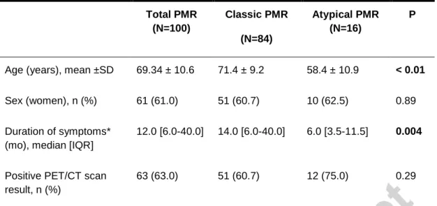
Epidemiological differences between patients with classic and atypical PMR are shown in Table 1. Patients with atypical PMR were younger and had a shorter duration of symptoms when a 18F-FDG PET/CT scan was requested than those with classic PMR. Although the percentage of positive PET/CT scan was greater in the atypical forms of PMR, the difference did not reach statistical significance, possibly due to the small number of patients with atypical PMR (n=16). In addition, in an attempt to identify
9
the presence of LVV in patients with well-defined PMR, only patients with classic presentation at the time of disease diagnosis, who at that time were included in the category of classic PMR were included in the analysis.
Definitions of atypical manifestations
Inflammatory low back pain was considered to be present if the patient
presented low back pain that improved with movement but not with rest, and it was usually predominant at night. Bilateral diffuse lower limb pain was defined when the patient complained of pain in both legs, in the thighs as well as anywhere between the knees and the ankles. Patients with these symptoms often complained of muscle pain on mild exertion such as walking, predominantly in the calves, which improved by a short period of rest [16]. Constitutional symptoms were considered to be present if the patient had asthenia, anorexia and/or weight loss greater than 5% of the normal body weight during the last six months. Fever was defined if the temperature was ≥ 38ºC without any evidence of infectious or neoplastic underlying disease.
Laboratory data
All patients with classic PMR had initially been treated with prednisone at an initial dose of least 15 mg/day. They were still taking prednisone (mean± standard deviation [SD]: 12.1±6.3 mg/day) when the 18F-FDG PET/CT scan was performed. CRP was considered to be abnormal at the time of performing 18F-FDG PET/CT scan if the value was higher than 0.5 mg/dL. At that time, an ESR level above 20 mm/1st hour was also considered elevated.
PET/CT scan equipment and protocol
Patients had to be in a low carbohydrate diet 48 hours before the scan, with reduced physical exercise for 24 hours and fasting state for at least 6 hours before 18 F-FDG administration. Serum glucose level was lower than 160 mg/dL in all the patients.
10
Whole-body PET/CT scan was acquired 180 minutes after intravenous injection of 7 MBq/kg of 18F-FDG using a Biograph LSO Pico 3D (Siemens Healthcare Molecular Imaging, Hoffman Estates, Illinois, USA). A low dose CT scan for attenuation correction and anatomic localization was first obtained, followed by a PET scan (acquiring 250 s/bed position). Images were reconstructed using the ordered subsets-expectation maximization (OSEM) algorithm (2 iterations, 8 subsets). Images were visually
evaluated by two experienced nuclear medicine specialists according to the intensity of the 18F-FDG uptake by the vessel wall at the supraaortic trunks, thoracic aorta,
abdominal aorta, iliac arteries and femoral/tibioperoneal arteries. PET/CT images were visually evaluated grading the vascular FDG uptake in comparison to liver uptake. PET/CT scans were considered positive for active LVV when a pattern of linear uptake was found in the aorta wall and its branches (when involved) with an intensity similar or higher than the liver, according to previous reports [17-19] and the recently published recommendations of the European Association of Nuclear Medicine, The Society of Nuclear Medicine and Molecular Imaging, and the PET Interest Group, and endorsed by the American Society of Nuclear Cardiology [20]. Figures 1A and 1B shows two
representative cases of PMR with negative and positive PET/CT scan for LVV, respectively.
The diagnosis of LVV was established by the combination of clinical data, treatment response, and initial and follow-up PET/CT scan.
Ethical approval was given by the Local Institutional Ethics Committee of Cantabria (Spain).
Statistical analysis
All continuous variables were tested for normality, and results were expressed as mean ±SD or as median and interquartile range (IQR) as appropriate. Student's t test or Mann-Whitney U-test were used to compare continuous variables, and Chi-square-test for categorical variables. Multivariable stepwise logistic regression
11
analyses were conducted to identify the independent set of predictors for a positive 18 F-FDG PET/CT scan. The predicted probability for a positive imaging result was
calculated from the regression model for each patient. The reliability of the model was evaluated using the Hosmer-Lemeshow goodness-of-fit test. The area under the ROC curve and its 95% confidence interval (CI) tested the discriminative ability of the regression model. A p value <0.05 was considered as statistically significant in all the calculations. Data management and analysis were performed using SPSS Statistics for Windows, version 18.0 (SPSS Inc., Chicago, Ill., USA) [21].
3. RESULTS
The mean age of the 84 patients (51 women/33 men) with classic PMR was 71.4±9.2 years. A PET/CT scan was positive in 51(60.7%) of them.
Persistence of classic PMR symptoms, alone or associated to the presence of unusual manifestations and/or constitutional symptoms, was the most common reason for requesting a PET/CT scan (Table 2).
Differences between patients with classic PMR according to PET/CT scan results The main clinical and laboratory differences between patients with classic PMR who had a positive 18F-FDG PET/CT scan for LVV and those in whom this procedure was negative are shown in Table 2. No differences in the age and sex between those with positive and negative 18F-FDG PET/CT scan were observed. It was also the case for the presence of classic cardiovascular risk factors. With regard to the typical PMR features, patients with positive 18F-FDG PET/CT scans had more commonly pelvic girdle involvement than those with negative 18F-FDG PET/CT scans (86.3% versus 36.4%; p<0.01). More importantly, patients with classic PMR who had a positive 18 F-FDG PET/CT scan showed more commonly atypical PMR features, such as
inflammatory low back pain (29.4% versus 9.1%; p= 0.027) or diffuse lower limb pain (52.9% versus 6.1%; p<0.01) at the time of PET/CT scan performance (Figures 2A
12
and 2B). However, no differences between patients with positive and negative 18F-FDG PET/CT scan were observed according to the presence of constitutional symptoms (including fever in this category). Also, the values of laboratory markers of inflammation and the dose of prednisone at the time of 18F-FDG PET/CT scan were similar in both groups (Table 2).
Multivariate logistic regression model showing the best set of predictors for the presence of LVV in the PET/CT scan
Table 3 shows the best set of predictors of a positive 18F-FDG PET/CT-scan for LVV. They were bilateral diffuse lower limb pain (odds ratio-OR=8.8, 95% CI 1.7-46.3; p=0.01), pelvic girdle pain (OR=4.9, 95% CI 1.50-16.53; p=0.01) and inflammatory low back pain (OR=4.7, 95% CI 1.03-21.5; p=0.04) once adjusted for age and sex. Further adjustments, including for diabetes status, did not virtually change these results.
Figure 3 shows the ROC analysis of the full predictive model for a positive 18 F-FDG PET/CT scan result showing LVV in patients with classic PMR (area under the curve 0.85 [95% CI 0.76- 0.93]; p<0.0001). A cut-off point of 0.55 yielded a sensitivity of 86% and a specificity of 64%. The correspondent figures for a cut-off point of 0.70 were 65% and 91%.
4. DISCUSSION
The present study confirms that patients who fulfill well-established
classification criteria for PMR often have LVV. Interestingly, besides the classic pelvic girdle involvement, the presence of atypical symptoms, such as inflammatory low back pain or bilateral diffuse pain in the lower extremities, was a predictor of underlying LVV in these patients at the time of PET/CT scan evaluation.
Experts in the field consider that PET/CT scans may show LVV involvement in at least a third of patients with PMR [22-24]. The results of our study suggest that the
13
prevalence of LVV in patients with well-defined PMR may be higher, reaching in our series up to 60%. Our results were in keeping with a former prospective study of our group that included 40 consecutive patients (27 women/13 men, 68.10±10.27 years) with PMR assessed by 18F-FDG PET/CT scan. In that study, this imaging technique disclosed LVV in 26 of the 40 patients [25]. The high percentage of LVV in our PMR series can be explained in part by the criteria used for the interpretation of PET/CT scan images. As Lavado-Pérez et al. did, we used a more delayed acquisition protocol in comparison to that applied in oncological patients, because it has demonstrated a better visualization of the vessel wall uptake, due to the decrease of the blood pool activity and the increase in the lesion/background ratio [18,19].
Early detection of LVV in patients with PMR is of potential relevance to determine the actual spectrum of the disease [26-28]. 18F-FDG PET/CT has
demonstrated to be very sensitive to make an early diagnosis of LVV [29-36]. However, information aimed to identify clinical and laboratory predictors of a positive 18F-FDG PET/CT for LVV in patients with PMR is scarce [37]. In this sense, in a multicenter retrospective study that included patients with GCA, PMR and other inflammatory disorders assessed by 18F-FDG PET/CT scan, Hooisma et al. reported that an elevated ESR was a positive predictor whereas arthralgia was a negative predictor for LVV [37]. Nevertheless, they concluded that a reliable prediction of the result of the 18F-FDG PET/CT based only on these two parameters was not possible [37]. In the present study we assessed patients with well-defined PMR, including for this purpose only those who fulfilled 2012 EULAR/ACR classification criteria [15]. According to our results, besides pelvic girdle pain, atypical manifestations, such as inflammatory low back pain and lower diffuse limb pain, were predictors of a positive 18F-FDG PET/CT scan result for LVV in patients with PMR. Noteworthy, 18F-FDG PET/CT was negative for LVV in all our patients when it was performed because of a marked unexplained elevation of serum CRP and ESR levels not associated to typical or atypical
14
result for LVV in 18F-FDG PET/CT, based only on an elevation of acute phase proteins is not possible.
A potential limitation of our study may be that our patients were taking prednisone at the time of PET/CT scan assessment. In this regard, glucocorticoids decrease the intensity of vessel wall 18F-FDG uptake [19,38,39]. However, Cimmino et al. [40], demonstrated that 18F-FDG PET/CT scan is useful for the evaluation of LVV in patients with PMR despite a previous treatment with low-dose glucocorticoids. This is especially true if PET/CT scan is performed within the first 3 days after the onset of glucocorticoid therapy [20].
As occurs in the majority of studies of this type, another limitation was the absence of histological confirmation of LVV. Moreover, the size of the study group could also be considered as a potential limitation. However, we think that our series of patients with classic PMR was large enough to disclose predictors of LVV in PMR patients
undergoing 18F-FDG PET/CT scan. Moreover, to the best of our knowledge, the present study constitutes the first attempt to identify the best set of predictors for LVV in a series of patients classified as having PMR according to the 2012 EULAR/ACR classification criteria. In addition, the presence of a positive PET/CT scan in our
patients with persistent PMR manifestations was clinically useful and led us to increase the prednisone dose or to add either methotrexate or anti-Interleukin-6 tocilizumab therapy.
5. CONCLUSIONS
In conclusion, our findings indicate that in patients with classic PMR, besides pelvic girdle pain, the presence of inflammatory low back pain and diffuse lower limb pain may have clinical relevance to identify a LVV by 18F-FDG PET/CT scan. In agreement with experts in the field [28], we feel that higher physician awareness and broader use of vascular imaging techniques to disclose LVV involvement is needed in patients with PMR.
15
REFERENCES
[1]. Salvarani C, Cantini F, Boiardi L, Hunder GG. Polymyalgia rheumatica and giant-cell arteritis. N Engl J Med 2002;34:261-71.
[2]. Gonzalez-Gay MA, Vazquez-Rodriguez TR, Lopez-Diaz MJ, Miranda-Filloy JA, Gonzalez-Juanatey C, Martin J, et al. Epidemiology of giant cell arteritis and polymyalgia rheumatica. Arthritis Rheum 2009;61:1454-61.
[3]. González-Gay MA, Matteson EL, Castañeda S. Polymyalgia rheumatica. Lancet 2017;390:1700-1712.
[4]. Gonzalez-Gay MA, Garcia-Porrua C, Salvarani C, Olivieri I, Hunder GG. The spectrum of conditions mimicking polymyalgia rheumatica in Northwestern Spain. J Rheumatol 2000;27:2179-84.
[5]. Gonzalez-Gay MA. Giant cell arteritis and polymyalgia rheumatica: two different but often overlapping conditions. Semin Arthritis Rheum 2004;33:289-93.
[6]. Gonzalez-Gay MA, Barros S, Lopez-Diaz MJ, Garcia-Porrua C, Sanchez-Andrade A, Llorca J. Giant cell arteritis: disease patterns of clinical presentation in a series of 240 patients. Medicine(Baltimore) 2005;84: 269-76.
[7]. Blockmans D, Stroobants S, Maes A, Mortelmans L. Positron emission tomography in giant cell arteritis and polymyalgia rheumatica: evidence for inflammation of the aortic arch. Am J Med. 2000;108:246–9.
[8]. Ernst D, Baerlecken NT, Schmidt RE, Witte T. Large vessel vasculitis and spondyloarthritis: coincidence or associated diseases? Scand J Rheumatol. 2014;43:246–8.
[9]. Brack A, Martinez-Taboada V, Stanson A, Goronzy JJ, Weyand CM. Disease pattern in cranial and large-vessel giant cell arteritis. Arthritis Rheum 1999;42: 311-7. [10]. Salvarani C, Soriano A, Muratore F, Shoenfeld Y, Blockmans D. Is PET/CT essential in the diagnosis and follow-up of temporal arteritis? Autoimmun Rev 2017;16:1125-1130.
16
[11]. Dejaco C, Singh YP, Perel P, Hutchings A, Camellino D, Mackie S et al. European League Against Rheumatism; American College of Rheumatology. 2015
Recommendations for the management of polymyalgia rheumatica: a European League Against Rheumatism/American College of Rheumatology collaborative initiative. Ann Rheum Dis 2015;74:1799-807.
[12]. Gonzalez-Gay MA, Castañeda S, Llorca J. Giant Cell Arteritis: Visual Loss Is Our Major Concern. J Rheumatol 2016; 43:1458-61.
[13]. Narváez J, Estrada P, López-Vives L, Ricse M, Zacarías A, Heredia S et al. Prevalence of ischemic complications in patients with giant cell arteritis presenting with apparently isolated polymyalgia rheumatica. Semin Arthritis Rheum 2015;45:328-33. [14]. Hunder GG, Bloch DA, Miguel BA, Stevens MB, Arend WP, Calabrese LH et al. The American College of Rheumatology 1990 criteria for the classification of giant cell arteritis. Arthritis Rheum 1990;33:1122-8.
[15]. Dasgupta B, Cimmino MA, Maradit-Kremers H, Schmidt WA, Schirmer M, Salvarani C, et al. 2012 provisional classification criteria for polymyalgia rheumatica: a European League Against Rheumatism/American College of Rheumatology
collaborative initiative. Ann Rheum Dis 2012;71:484–92.
[16]. Leng GC, Fowkes FG. The Edinburgh Claudication Questionnaire: an improved version of the WHO/Rose Questionnaire for use in epidemiological surveys. J Clin Epidemiol 1992;45:1101-9.
[17]. Jamar F, Buscombe J, Chiti A, Christian PE, Delbeke D, Donohoe KJ, et al. EANM/SNMMI guideline for 18F-FDG use in inflammation and infection. J Nucl Med Off Publ Soc Nucl Med 2013;54:647–58.
[18]. Loricera J, Blanco R, Hernández JL, Martínez-Rodríguez I, Carril JM, Lavado C, et al. Use of positron emission tomography (PET) for the diagnosis of large-vessel vasculitis. Rev Esp Med Nucl Imagen Mol 2015;34:372–7.
[19]. Martínez-Rodríguez I, Del Castillo-Matos R, Quirce R, Banzo I, Jiménez-Bonilla J, Martínez-Amador N, et al. Aortic 18F-FDG PET/CT uptake pattern at 60 min (early)
17
and 180 min (delayed) acquisition in a control population: a visual and semiquantitative comparative analysis. Nucl Med Commun 2013;34:926–30.
[20] Slart RHJA; Writing group; Reviewer group; Members of EANM cardiovascular; Members of EANM Infection & Inflammation; Members of Committees, SNMMI Cardiovascular; Members of Council, PET Interest Group; Members of ASNC; EANM Committee Coordinator. FDG-PET/CT(A) imaging in large vessel vasculitis and polymyalgia rheumatica: joint procedural recommendation of the EANM, SNMMI, and the PET Interest Group (PIG), and endorsed by the ASNC. Eur J Nucl Med Mol Imaging. 2018 Apr 11. doi: 10.1007/s00259-018-3973-8.
[21]. SPSS Inc. Released 2009. PASW Statistics for Windows, Version 18.0. Chicago: SPSS Inc.
[22]. Blockmans D, De Ceuninck L, Vanderschueren S, Knockaert D, Mortelmans L, Bobbaers H. Repetitive 18-fluorodeoxyglucose positron emission tomography in isolated polymyalgia rheumatica: a prospective study in 35 patients. Rheumatology (Oxford) 2007;46:672-7.
[23]. Rehak Z, Vasina J, Nemec P, Fojtik Z, Koukalova R, Bortlicek Z, et al. Various forms of (18)F-FDG PET and PET/CT findings in patients with polymyalgia rheumatica. Biomed Pap Med Fac Univ Palacky Olomouc Czechoslov. 2015;159:629–36.
[24]. Rehak Z, Sprlakova-Pukova A, Kazda T, Fojtik Z, Vargova L, Nemec P. (18)F-FDG PET/CT in polymyalgia rheumatica-a pictorial review. Br J Radiol.
2017;90:20170198.
[25]. Lavado-Pérez C, Martínez-Rodríguez I, Martínez-Amador N, Banzo I, Quirce R, Jiménez-Bonilla J, et al. (18)F-FDG PET/CT for the detection of large vessel vasculitis in patients with polymyalgia rheumatica. Rev Esp Med Nucl Imagen Mol 2015;34:275– 81.
[26]. Loricera J, Blanco R, Hernández JL, Carril JM, Martínez-Rodríguez I, Canga A, et al. Non-infectious aortitis: a report of 32 cases from a single tertiary centre in a 4-year period and literature review. Clin Exp Rheumatol 2015;33:19-31.
18
[27]. Possemato N, Salvarani C, Pipitone N. Imaging in polymyalgia rheumatica. Reumatismo. 2018;70:51-58.
[28]. Cimmino MA, Camellino D. Large vessel vasculitis: is it more common than usually assumed? Reumatismo 2017; 69:143-146.
[29]. Prieto-González S, Espígol-Frigolé G, García-Martínez A, Alba MA, Tavera-Bahillo I, Hernández-Rodríguez J, et al. The Expanding Role of Imaging in Systemic Vasculitis. Rheum Dis Clin North Am 2016;42:733-51.
[30]. Meller J, Strutz F, Siefker U, Scheel A, Sahlmann CO, Lehmann K, et al. Early diagnosis and follow-up of aortitis with [(18)F]FDG PET and MRI. Eur J Nucl Med Mol Imaging 2003;30:730-6.
[31]. Löffler C, Hoffend J, Benck U, Krämer BK, Bergner R. The value of ultrasound in diagnosing extracranial large-vessel vasculitis compared to FDG-PET/CT: A
retrospective study. Clin Rheumatol 2017;36:2079-86.
[32]. Bruls S, Courtois A, Nusgens B, Defraigne J-O, Delvenne P, Hustinx R, et al. 18F-FDG PET/CT in the Management of Aortitis. Clin Nucl Med 2016;41:28-33.
[33]. Chrapko BE, Chrapko M, Nocuń A, Stefaniak B, Zubilewicz T, Drop A. Role of 18F-FDG PET/CT in the diagnosis of inflammatory and infectious vascular disease. Nucl Med Rev Cent East Eur 2016;19:28-36.
[34]. Lee YH, Choi SJ, Ji JD, Song GG. Diagnostic accuracy of 18F-FDG PET or PET/CT for large vessel vasculitis : A meta-analysis. Z Rheumatol 2016; 75:924-31. [35]. Kubota K, Yamashita H, Mimori A. Clinical Value of FDG-PET/CT for the
Evaluation of Rheumatic Diseases: Rheumatoid Arthritis, Polymyalgia Rheumatica, and Relapsing Polychondritis. Semin Nucl Med 2017;47:408–24.
[36]. Versari A, Pipitone N, Casali M, Jamar F, Pazzola G. Use of imaging techniques in Large Vessel Vasculitis and related conditions. Q J Nucl Med Mol Imaging
19
[37]. Hooisma GA, Balink H, Houtman PM, Slart RHJA, Lensen KDF. Parameters related to a positive test result for FDG PET(/CT) for large vessel vasculitis: a multicenter retrospective study. Clin Rheumatol 2012;31:861-71.
[38]. Lund-Petersen A, Voss A, Laustrup H. PET-CT findings in patients with polymyalgia rheumatica without symptoms of cranial ischaemia. Dan Med J. 2017;64:5410
[39]. Sondag M, Guillot X, Verhoeven F, Blagosklonov O, Prati C, Boulahdour H, et al. Utility of 18F-fluoro-dexoxyglucose positron emission tomography for the diagnosis of polymyalgia rheumatica: a controlled study. Rheumatol Oxf Engl 2016; 55:1452-7. [40]. Cimmino MA, Zampogna G, Parodi M. Is FDG-PET useful in the evaluation of steroid-resistant PMR patients? Rheumatol Oxf Engl 2008; 47:926-7
FIGURE LEGENDS
Figure 1A. An 88-year-old man with PMR. Although the patient experienced a rapid response to prednisone (15 mg/day), he suffered an unexplained increase of ESR/CRP not associated with polymyalgia symptoms. Because of that, a PET/CT-scan was performed to exclude LVV. Coronal (A), sagittal (B) and axial (C) 18F-FDG PET images ruled out inflammation of large vessels.
Figure 1B. A 69-year-old woman with PMR and persistence of classic PMR symptoms despite prednisone therapy along with unusual symptoms
(inflammatory low back pain) at the time of assessment. Sagittal PET (A) and fused PET/CT (B), axial PET (C) and fused PET/CT (D), and coronal PET (E) images (E) showed inflammation along the thoracic aorta wall (head arrows) and supra-aortic trunks (arrows).
Figure 2. A 63-year-old woman, who initially had with typical PMR features, started to complain of diffuse lower limb pain and intermittent vascular
20
was tapered. Besides typical bursitis in the setting of PMR demonstrated by 18 F-FDG PET/CT in the shoulders and cervical and lumbar interspinous spaces, trochanteric and ischiatic regions of both hips (arrows) (Figure 2A), the images also disclosed the presence of LVV with increased 18F-FDG uptake involving the femoral arteries (Figure 2B). A mild 18F-FDG uptake was also observed at the thoracic aortic wall (Figure 2A).
Figure 3. ROC analyzing the performance of the full predictive model of a positive 18F-FDG PET/CT scan result for LVV in patients with classic PMR. ROC: Receiver operating characteristics.
21
Table 1. Epidemiological differences between classic and atypical PMR.
Table 2. Main clinical features and laboratory findings of 84 patients with classic PMR
when the PET/CT scan was performed.
Total PMR (N=100) Classic PMR (N=84) Atypical PMR (N=16) P
Age (years), mean ±SD 69.34 ± 10.6 71.4 ± 9.2 58.4 ± 10.9 < 0.01
Sex (women), n (%) 61 (61.0) 51 (60.7) 10 (62.5) 0.89
Duration of symptoms* (mo), median [IQR]
12.0 [6.0-40.0] 14.0 [6.0-40.0] 6.0 [3.5-11.5] 0.004
Positive PET/CT scan result, n (%)
63 (63.0) 51 (60.7) 12 (75.0) 0.29
PET/CT: positron emission tomography complemented by computed tomography; PMR: polymyalgia rheumatica; IQR: interquartile range; SD: standard deviation. * At the time of PET/CT scan performance.
22 Classic PMR (N=84) Positive PET/CT (N= 51) Negative PET/CT (N=33) P Sex (women), n (%) 31 (62.7) 19 (57.6) 0.64
Age (years), mean ±SD 70.0 ± 9.2 73.7 ± 9.0 0.09
Cardiovascular risk factors, n (%)
Hypertension 29 (56.9) 24 (72.7) 0.14 Dyslipidemia 18 (35.3) 16 (48.5) 0.23 Diabetes mellitus 10 (19.6) 12 (36.4) 0.09 Current smokers 5 (9.8) 1 (3.0) 0.40 Polymyalgia symptoms, n (%) Neck pain 9 (17.6) 9 (27.3) 0.29
Shoulder girdle pain 33 (64.7) 26 (78.8) 0.17
Pelvic girdle pain 44 (86.3) 12 (36.4) < 0.01
Morning stiffness 11 (21.6) 10 (30.3) 0.44
Unusual symptoms, n (%)
Inflammatory low back pain 15 (29.4) 3 (9.1) 0.027
Diffuse lower limb pain 27 (52.9) 2 (6.1) < 0.01
Constitutional symptoms, n (%) Fever 2 (3.9) 2 (6,1) 0.64 Asthenia 15 (29.4) 8 (24.2) 0.60 Hyporexia 4 (7.8) 7 (21.2) 0.08 Weight loss 9 (17.6) 8 (24.2) 0.46 Laboratory markers, Hb (g/dL), mean ± SD 12.7 ± 1.3 12.2 ± 1.6 0.09
Platelet count (×109/l), mean ± SD 281.4 ± 84.9 263.7 ± 100.3 0.19
CRP (mg/dL), median [IQR] 2.0 [0.6-4.4] 2.0 [1.1-5.3] 0.28
23
Treatment,
Glucocorticoids, n (%) 51 (100.0) 33 (100.0) 0.99
Dose of Prednisone (mg), mean ± SD 12.0 ± 5.7 12.3 ± 7.3 0.88
Months of treatment with Prednisone, median [IQR] 4 [1.0-15.0] 7 [2.5-15.5] 0.31
Methotrexate, n (%) 11 (21.6) 4 (12.1) 0.27
Reasons for PET/CT scan request, n (%)
Persistence of classic PMR symptoms* 8 (15.7) 10 (30.3) 0.19
Presence of unusual symptoms without classic PMR
symptoms* 1 (2.0) 1 (3.0) 0.69
Presence of constitutional symptoms without classic PMR or
unusual symptoms* 1 (2.0) 2 (6.1) 0.69
Unexplained increase of ESR/CRP not associated with
classic PMR symptoms or unusual symptoms 0 (0.0) 4 (12.1) 0.04
Persistence of classic PMR symptoms + unusual
symptoms* 25 (49.0) 5 (15.2) 0.003
Persistence of classic PMR symptoms + constitutional
symptoms* 4 (7.8) 9 (27.3) 0.04
Persistence of classic PMR symptoms + constitutional
symptoms + unusual symptoms* 12 (23.5) 2 (6.1)
0.07
CRP: serum C reactive protein; ESR: erythrocyte sedimentation rate; Hb: hemoglobin; PET/CT: positron emission tomography complemented by computed tomography; PMR: polymyalgia rheumatica; IQR: interquartile range; SD: standard deviation. *In most cases associated with mild or moderate elevation of ESR and/or CRP.
24
Table 3. Multivariate logistic regression model showing the best set of predictors of a
positive result for LVV in 18F-FDG PET/CT scan in patients with classic PMR.
Variable Beta OR 95% CI P
Classic PMR
Diffuse low back pain 1.55 4.7 1.03-21.5 0.04
Lower limb pain 2.17 8.8 1.70-46.3 0.01
Pelvic girdle pain 1.58 4.9 1.50-16.3 0.01