Facultad de Odontología
Vol. 15, No. 2 April-June 2011 pp 107-112
Revista Odontológica Mexicana
CASE REpORt
www.medigraphic.org.mx
Application of plasma rich in growth factors after extraction of
lower third molars. Case report
Colocación de plasma rico en factores de crecimiento post-extracción de
terceros molares inferiores: Reporte de un caso
Víctor Mario Fierro-Serna,* Ricardo Martínez-Rider,§ José Antonio Hidalgo-Hurtado,II José Martín toranzo-Fernández,¶ Amaury de Jesús pozos-Guillen**
* 4th year resident, Oral and Maxillofacial Department, Hospital Central «Dr. Ignacio Morones prieto», San Luis potosí.
§ Head, Oral and Maxillofacial Department, School of Dentistry, Universidad Autónoma de San Luis potosí.
II Coordinator, Oral and Maxillofacial Department, Hospital Central «Dr. Ignacio Morones prieto», San Luis potosí.
¶ Head, Oral and Maxillofacial Department, Hospital Central «Dr. Ignacio Morones prieto», San Luis potosí.
** professor, Researcher, School of Dentistry, Universidad Autóno-ma de San Luis potosí.
Este artículo puede ser consultado en versión completa en http://www.medigraphic.com/facultadodontologiaunam
ABSTRACT
In recent years, the use of plasma rich in growth factors and
plate-lets has become popular in the fields of oral and maxillofacial sur -gery. this case report describes a 21 year old female presenting moderate pain in the lower right and left third molar area. Both third molars were surgically removed. 20 cc of the patient’s blood were extracted to obtain plasma rich in growth factors. this plasma was applied to the area of the left lower third molar extraction site. the right lower third molar extraction site was irrigated with physiological saline. three days after the operation, we could clinically observe a
lesser degree of extraoral inflammation on the left side, when com -pared to the right side. On the left side, we observe intraorally less inflammation and erythema in the zone and better epithelization when compared with the right side Five days after the operation su-ture points are removed from both sides of the mandible On the left side the wound presented better epithelization and lesser degree of erythema. Seven days after the operation, the difference in the soft tissue regeneration is clearly better on the left side when com-pared to the right one. In every one of her control visits, the patient reported lesser degree of pain in the left side than in the right one. Our experience in this case indicates that the use of plasma rich
in growth factors can be beneficial for the postoperative period of
patients after surgical extraction of lower third molars.
RESUMEN
En los últimos años se ha popularizado el uso de plasma rico en plaque-tas y plasma rico en factores de crecimiento en la cirugíabucal y maxi-lofacial. Se reporta un caso de paciente femenino de 21 años de edad, con dolor moderado en zona de tercer molar inferior izquierdo y derecho. Se realiza remoción quirúrgica de ambos terceros molares inferiores. Se extraen 20 cc. de sangre del paciente para obtener plasma rico en facto-res de crecimiento el cual fue colocado en zona de extracción de tercer molar inferior izquierdo. En zona de tercer molar inferior derecho se irrigó
con suero fisiológico. Al tercer día postoperatorio se observa clínicamen
-te menor inflamación extraoral del lado izquierdo comparado con lado derecho. Intraoralmente menor inflamación y eritema de la zona y mejor
epitelización del lado izquierdo en relación con lado derecho. Al quinto
día postoperatorio se retira sutura en ambos lados, observando mejor
epitelización y menos eritema de la herida en lado izquierdo. Al séptimo
día es clara la diferencia en la regeneración de tejidos blandos en el
lado izquierdo comparado con el derecho. Se refería menos dolor del lado izquierdo en cada una de sus citas control. La experiencia en el presente caso nos hace sugerir que el uso de plasma rico en factores de
crecimiento puede beneficiar el postoperatorio de los pacientes después
de la remoción quirúrgica de terceros molares inferiores.
Key words: Growth factors, plasma, third molars.
Palabras clave: Factores de crecimiento, plasma, terceros molares.
IntroduCtIon
In dentistry, extraction of third molars is one of the most common procedures. In general terms, extraction of third molars has a negative impact in the period of four to seven days after surgery, nevertheless it has been ob-served there is an improvement in the patient quality of life in that period if we can eliminate chronic pain and
inflammation (generally due to pericoronitis). This has
been itemized in studies describing the improvement found after surgical procedure.1 the most common rea-sons for the extraction of third molars are: cyst or tumor originating in the dental follicle, repeated pericoronitis
www.medigraphic.org.mx
It has been reported in patients ranging from ages20 to 29, a percentile increase from 18 to 40 percent in prophylactic extractions.3,4
Surgical extraction of third molars is associated to a moderate incidence of complications, about 10%.5
In-fluencing factors could be the surgeon s lack of experi
-ence and training to carry out this procedure inflicting
minimal traumatism. the most common complications are: bleeding, third molar root factures, which hinder the extraction, occasional displacement of the roots to the submandibular space, lower dental nerve canal or maxillary sinus. Alveolar osteitis, or dry alveoli, ap-pears in 20 to 25% of the cases.6 post extraction infec-tions after third molar extraction procedures, are less
frequent (1.7 to 2.7%) in which cases we recommend
antibiotic therapy. In general terms, albeit far less fquently, more extensive surgical procedures are re-quired for the treatment of complications like lesions to
nerves, lower jaw fracture, or the displacement of third
molar whole body to the infratemporal fosa, maxilary
sinus, and floor of the mouth.
the postoperative period to the extraction of third molars frequently presents in the patient swelling, pain, trismus and poor mastication function. the pa-tient negatively rates all these signs and symptoms which are sometimes conductive to a longer conva-lescing period, which precludes them to carry on with their normal life. Due to these reasons, methods to reduce complications are constantly under research. postoperative pain settles in when the local anaesthet-ic wears off or disappears completely and it reaches its maximum intensity in the 12 hour period following the operation. Several analgesics have been proposed to avoid this complication.7 Inflammation is an ever present sequel of third molar extraction procedures, it reaches its peak on the second day after procedure,
and abates towards the fifth to seventh day. As treat
-ment to minimize inflammation, parenteral administra -tion of corticosteroids like dexamethasone and beta-methasone, has been discussed. One of the factors more frequently related to post-operative pain and
inflammation is the type of the surgical wound regen -eration. Both primary and secondary wound closures have been evaluated, but controversy is still present concerning these two types of regeneration.8-10
and antifibrinolytic agents. When incorporating
au-tologous adhesive fibrin he observed an improvement
in the osteoconduction.11,12 At a later time, Whitman,
(1997) proposed the use of platelet rich plasma and he itemized differences between fibrin glue and plate -let gel. the plate-let gel was obtained from a process of the patients blood extracted moments before the surgical procedure. this gel had a high platelet con-centration which triggers the liberation of the growth factors which in turn promote better healing.13 A short
time later, Marx (1998) combined platelet rich plasma
with autogenous bone to repair mandibular defects. In the radiographic images obtained after this procedure,
during the first 6 months after the operation, an ac -celerated bone formation was observed.14 this same author has described the application of platelet rich plasma in dental procedures such as elevation of max-illary sinus, periodontal defects treatment, increase of the alveolar ridge, placement in alveoli after third molar extractions, regeneration of soft tissue, and cra-niofacial applications for reconstruction purposes.15
Anitua (1997) has also described the use of plasma
rich in growth factors. He reports an excellent epitheli-zation and bone regeneration in zones being primed to receive dental implants.16-18 properties here described can contribute to the decrease in common complica-tions found after third molar extraccomplica-tions.
In this article we report a case where after the ex-traction of lower third molars, one side received appli-cation of plasma rich in growth factors, the other side did not, and results were compared.
CAse report
A 21 year old woman was admitted in the Clinica de Cirugía Maxilofacial de la Facultad de Estomatología,
Universidad Autónoma de San Luis Potosí (Maxillofa -cial Surgery Clinic of the Stomatology School of the
University of San Luis Potosi) for extraction of lower
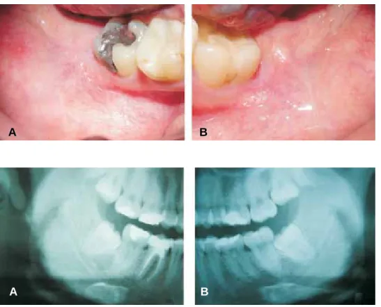
third molars. the patient informed of moderate pain. Clinically slight erithema of the mucosa in the right and left third molar area is observed (Figures 1A and 1B).
Radiographically, both third molars were classified as
class I, mesioangled with convergent roots with
www.medigraphic.org.mx
of Sanchez torres19 (Figures 2A and 2B). It is decidedto surgically remove both third molars under infiltration
local anesthesia.
ten minutes before procedure, approximately 20 cc of the patient’s blood were extracted, to obtain plasma rich in growth factors, as described in Anitua s proto-col. It was done in the following fashion: the patients blood was placed in sterile tubes with 3.8% sodium
citrate (vacutainer) as anticoagulant. Plasma was then centrifuged at 1800 RPM during 8 minutes (Model BTI PRGF system II). Plasma obtained was separated into
fractions through careful pipetting so as not to create
turbulences in obtained fractions. The first 500 micra
1 were of plasma poor in platelets. the following 500 micra 1 of plasma have a platelet count similar to the one found in peripheral blood. the fraction of plasma richest in platelets and growth factors are the 500 mi-cra 1 immediately above the red series. Once the pi-petting was achieved, clot formation was induced by adding 10% calcium chlor after approximately 5 to 8 minutes.20 On both right and left side perioral asepsis and antisepsis was achieved with the help of isodine, anesthetic block of lower dental nerve with articaine
and epinephrine (Medicaine 1/100). Approach was ini -tiated through the left side with the design of a
trian-gular flap. Once the flap was raised, osteotomy on the
tooth vestibular side was performed, to later carry out odontosection. the tooth was extracted in two section without any trouble. the alveolus was richly irrigated with saline solution. the second fraction of plasma rich in growth factors activated with 10% calcium chloride was used to irrigate the alveolus and it was completely
filled with a clot of plasma richer in growth factors. It was tightly sutured with 4-0 silk (Ethicon). The same
procedure was applied to the right side with the excep-tion of the plasma placement.
the patient is sent home with the indications specif-ic to this type of procedure: Clindamycin, taken orally, 300 mg every 6 hours and Ketocorolaco, 10 mg every 8 hours. A clinical follow up was observed examining
the patient on the third, fifth and seventh day after the
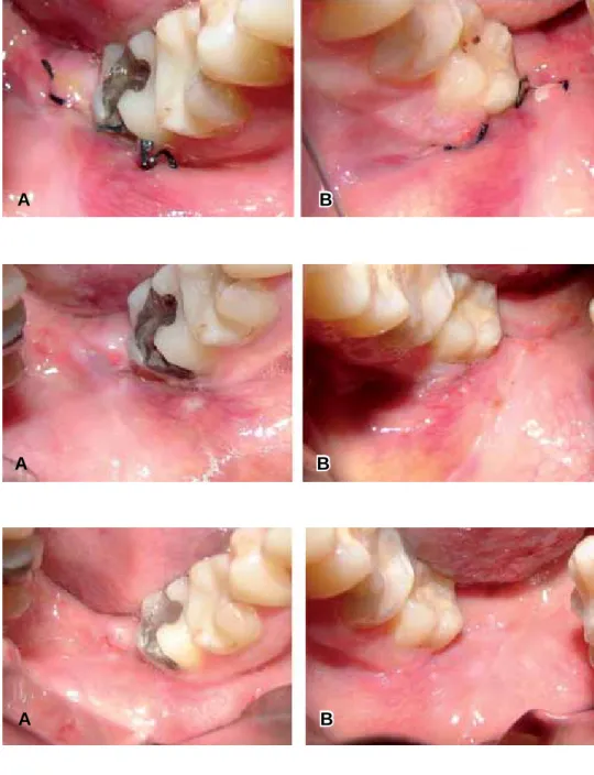
operation. On the third postoperative day clinical
ob-servation was of lesser extraoral inflammation on the left side, Intraorally, lesser inflammation and erythema
in the area was observed, along with improved epithe-lialization on the left side (Figures 3A and 3B). On the
fifth day it is decided to remove suture material from
both sides. On the left side, better epithelialization and less erithema of the wound is observed (Figures 4A and 4B). On the seventh postoperative day, there is a blatant difference in soft tissue regeneration in the left side when compared to the right one (Figures 5A
Figure 1. Region of the lower
third molar before treatment A) Right side B) Left side.
Figure 2. pretreatment
radio-graphic view A) Right side B) Left side.
A B
www.medigraphic.org.mx
and 5B). In all control visits, the patient the patientin-formed of lesser pain in the left side.
dIsCussIon
After surgery of impacted third molars, the patient feels psychologically affected in cases of slight bleed-ing, inflammation, pain, and limitations when open-ing the mouth. the patient feels uneasy about these symptoms. For these reasons, several methods have been suggested to reduce to the maximum these se-quels so often present in these cases. the following
have been recommended: use of corticosteroids, ice extraoral application in extraction sites to reduce
in-flammation, several analgesics and their combinations
to decrease pain. this case describes the evolution of the patient when plasma rich in growth factors is used
as an adjuvant to reduce this type of sequels.
Studies by Babbush21 and Mancuso22 have shown the ability of plasma rich in growth factors to reduce frequency of dry alveolus, and promote bone regener-ation in the socket. this plasma also reduces the for-mation of periodontal pockets or any pathological con-dition which compromises the distal face of the second
Figure 3. Region of the lower
third molar three days after treat-ment A) Right side B) Left side.
Figure 4. Region of the lower
third molar five days after treat -ment A) Right side B) Left side.
Figure 5. Region of the lower
third molar seven days after treatment A) Right side B) Left side.
A B
A B
www.medigraphic.org.mx
Este documento es elaborado por Medigraphic
molars. this is based on the fact that repair mecha-nisms of growth factors occur in bone regeneration and repair processes. Soft tissue repair mechanisms occur simultaneously, and these can easier be ob-served than the changes which take place in the bone. the advantages of applying plasma rich in growth fac-tors in third molar extraction sites, as in the case here described, is that upon intervening on the regeneration of the mucosa in that area, quality can be enhanced,
decreasing thus periodontal defects. Healthy flap re -generation depends on a design that must provide good blood supply, and on a periodontium with appro-priate osteogenic potential the osteogenic potential of the periostium can be altered by repeated pericoronitis episodes in the area. therefore we can rate this type of patients as candidates for the use of plasma rich in
growth factors after tooth extraction. These events jus -tify the exogenous application of growth factors to im-prove bone and soft tissue regeneration, as described in this case.
plasma rich in growth factors provide products which improve bone and soft tissue regeneration, both processes are expedited, the patient experi-ments less pain and swelling will decrease. No stud-ies were found in which a assessment was made of these two events after extraction of lower third molars, together with application of plasma rich in growth factors. In the present case, the patient re-ported less pain and swelling on the left side which received plasma rich in growth factors.
Methods to obtain plasma rich in growth factors and plasma rich in platelets vary. there are several tems to obtain plasma rich in platelets. In these sys-tems the amount of blood required varies, from 60cc
(SmartPrep, PCCS, Secquire, Acces, GPS, Magel
-lan) up to 450 cc (CATS). The most popular one is
perhaps the SmartpRep system. In this system, after
obtaining the patients blood it is first submitted to a
separation centrifugation, which separates red blood cells from white ones, platelets and plasma. plasma poor in platelets is separated putting it through a sec-ond centrifugation , called concentration centrifugation which separates and compacts platelets, white blood cells and a small portion of red cells. A new separation of plasma is performed, thus plasma rich in platelets sets in the bottom of the container. Once obtained, the plasma rich is platelets is activated with a combination of 10% calcium chloride and bovine thrombine.
Although Marx14 reports good results with plasma rich in platelets, in this case it was decided to use plas-ma rich in growth factors for the following reasons. the cost of equipment required for its elaboration is much lower. the amount of blood required to obtain plasma
rich in growth factors is lower than the amount of blood required in systems to obtain platelet rich plasma. Some systems for obtaining platelet rich plasma, the
process takes up to 32 minutes (PCCS). SmartPRep
requires the shortest period, 15 minutes. the protocol is similar to the one for obtaining plasma rich in growth factors. Controversy has also arisen concerning the use of bovine thrombine, for some cases have been reported where antithrombine antibodies have been detected with their use. It has been suggested that plasma rich in platelets may cause Kreutzfeldt-Jakob disease. this theory has been disclaimed on the basis that the vector causing the disease is a protein present only in the nervous cells of humans, cats, lambs and other animals, and bovine thrombine is derived
exclu-sively from the blood when submitted to a purification process. Regrettably, this is a difficult and expensive
process. In this particular case, a difference was ob-served in the healing of lower third molar extraction sites when applying plasma rich in growth factors. this quality of plasma rich in growth factors has been ex-ploited in several disciplines. In cosmetic surgery, it has been used to reduce swelling, ecchymosis and to accelerate regeneration of soft tissues.23 In dermato-logical surgery, it has been applied to obtain faster re-generation with less visible scars after excision of skin lesions.24 the patient informed of lesser pain on the side where plasma had been applied. Clinically bet-ter soft tissue regeneration and reduce swelling was found on the same side. All these facts lead us to be-lieve that application of plasma rich in growth factors
in third molar extraction sites can be an adjuvant for a
better regeneration of bone and soft tissues in shorter periods and at the same time decreasing common se-quels in this type of procedures, enabling the patient to return to his daily routines in shorter time.
ConClusIons
Experience gained with this case leads us to sug-gest that the use of plasma rich in growth factors can
benefit the postoperative period of patients after surgi -cal removal of lower third molars.
referenCes
1. White Rp, Shugars DFA, Shafer DM. Recovery after third mo-lar surgery: clinical and health-related quality of life outcomes. J Oral Maxillofac Surg 1983; 41: 706-710.
2. Liedholm R, Knutsson K, Lysell, Rohlin M. Mandibular third mo-lars: Oral surgeons assessment of the indications for removal. Br J Oral Maxillofac Surg 1999; 37: 443-450.
3. National Institute of Health. Consensus development conference for removal of third molars. J Oral Surg 1980; 38: 235-236.
www.medigraphic.org.mx
and secondary closure of the surgical wound after removal ofim-pacted mandibular third molars: a comparative study. Int J Oral Maxillofac Surg 2005; 34: 52-57.
9. Korbendau JM, Korbendau X. Clinical success in impacted third molar extraction. Quintessence Books, 2002.
10. Milloro M. Peterson’s Principles of Oral and Maxillofacial Sur-gery. BCDecker Inc., 2004.
11. tayapongsak p, O’Brien DA, Monteiro CB, Arceo-Diaz LL.
Au-tologous fibrin adhesive in mandibular reconstruction with par -ticulate cancellous bone and marrow. J Oral Maxillofac Surg 1994; 52: 161-166.
12. Matras H. The use of fibrin glue in oral and maxillofacial surgery.
J Oral Maxillofac Surg 1982; 40: 617.
13. Whitman DH, Berry RL, Green DM. platelet gel: An autolo-gous alternative to fibrin glue with applications in oral and maxillofacial surgery. J Oral Maxillofac Surg 1997; 55: 1294-1299.
14. Marx RE, Carlson ER, Eichstaedt RM, Schimmele SR, Strauss JE, Georgeff KR. platelet-rich plasma growth factor enhance-ment for bone grafts. Oral Surg Oral Med Oral Pathol Oral Radiol Endod 1998; 85: 638-646.
15. Marx RE, Garg AK. Dental and craniofacial applications of plate-let-rich plasma. Quintessence Books, 2005.
16. Anitua E. plasma rich in growth factors: preliminary results of use in the preparation of future sites for implants. Int J Oral Max-illofac Implants 1999; 14: 529-535.
21. Babubush CA. The use of PRP in conjunction with other bone
graft material: Allograft, alloplast, senograft. Presented at the 2nd Symposium on Platelet-Rich Plasma (PRP) & Its Growth Fac-tors, San Francisco, 2003: 23-26.
22. Mancuso J, Bennion JW, Hull MJ, Winterholler BW. platelet rich plasma: A preliminary report in routine impacted third molar sur-gery and the prevention af the alveolar osteitis. J Oral
Maxillofa-cial Surgery 2003; 61 (suppl 1).
23. powell Dm, Chang E, Farrior EH. Recovery from deep-plane rhytidectomy following unilateral wound treatment with autolo-gous plateket gel: A pilot study. Arch Facial Plat Surg 2001; 3: 245-250.
24. Adler SC, Kent KJ. Enhacing wound healing with growth factors. Facial Plast Surg Clin North Am 2002; 10: 129-146.
Address correspondence:
Amaury de Jesús Pozos Guillén MD
Facultad de Estomatología,
Universidad Autónoma de San Luis potosí. Av. Dr. Manuel Nava Num. 2,
Zona Universitaria, 78290; San Luis potosí, SLp México.
Phone: 52 (444)8262357 X 106 Fax: 52 (444)8139743.