Biological Research
versión impresa ISSN 0716-9760Biol. Res. vol.45 no.1 Santiago 2012
http://dx.doi.org/10.4067/S0716-97602012000100004
Biol Res 45: 27-31, 2012
RESEARCH ARTICLES
Chronic ethanol consumption in mice does not induce
DNA damage in somatic or germ cells, evaluated by the
bone marrow micronucleous assay and the dominant
lethal mutation assay
Manuel F Ellahueñe1*, L Patricia Pérez-Alzola2 and M Isabel Olmedo1
1 Unidad de Biodiversidad Acuática, Centro Nacional del Medio Ambiente (CENMA),
Universidad de Chile, Santiago, Chile.
2 Universidad Andrés Bello, Facultad de Ciencias Biológicas, República 252, Santiago,
Chile.
ABSTRACT
Although alcohol is known to be a carcinogen for humans, ethanol-genotoxicity studies are incomplete. Ethanol seems not to be a bacterial mutagen, but the results are conflicting in rodent assays. We investigate the genotoxicity in the bone marrow micronucleus (MN) test and in the dominant lethal mutation (DLM) assay using two long-term ethanol exposure protocols. In the MN test, mice consumed three doses (5, 10 and 15% v/v) for 32 weeks. MN induction was compared to two control groups of 5- and 38-week-old mice (the ages of the treated mice when the treatment was initiated and when they were killed, respectively). For the three groups treated with ethanol there was no significant increase in MN induction as compared to the first control group, but observed MN frequencies were significantly lower than in the 38-week-old control group. This suggests a protective effect against genotoxic damage caused by aging, probably due to ethanol action as a hydroxyl radical scavenger.
In the DLM assay, male mice drank ethanol at 15% or 30% (v/v) for 20 weeks. In both groups the number of dead implants was similar to the control, but there was a
significant reduction in total implants, indicating a pre-implantation loss.
INTRODUCTION
Chronic alcoholism is a mayor public health issue around the world. Consumption of alcohol has been related to cardiovascular diseases (Friedman, 1998), hepatic effects (Lieber, 1985), brain toxicity (Harper, 1998), and increased incidence of esophagus, larynx and oral cavity cancers. The International Agency for Research on Cancer (IARC, 1998) has found that there is sufficient evidence for the carcinogenicity of alcoholic beverages in humans, and has classified alcoholic beverages as Group I carcinogens, although the mechanism of ethanol carcinogenicity is still not known (Kayani and Parry, 2010).
There are many studies about the genotoxic potential of ethanol, some of which have shown chromosomal effects in lymphocytes of alcoholics, like sister chromatid
exchanges (reviewed in Obe and Anderson, 1987), induction of chromosomal
aberrations (López et al., 2001) and increased incidence of aneuploidy (Kucheira et al., 1986), suggesting that alcoholism may cause chromosome damage in humans.
The genotoxicity testing of ethanol was first reviewed by an expert group of the International Commission for the Protection against Environmental Mutagens and Carcinogenesis (ICPEMC) (Obe and Anderson, 1987). No conclusion was reached about the effects of ethanol in relation to genetic damage. In 1995, the UK Department of Health's Committee on Mutagenesis of Chemicals in Food, Consumer Products and the Environment reviewed the evidence for the mutagenecity of ethanol, acetaldehyde and alcoholic beverages. The Committee agreed that the consumption of alcoholic
beverages does not present any significant concern with respect to their mutagenic potential.
In 2001, Phillips and Jenkinson (Phillips and Jenkinson, 2001) made a review of the available information on the genotoxicity of ethanol, concluding that the data derived from studies using standard genotoxicity methods are incomplete. They reported that there is clear evidence that ethanol is not a bacterial or mammalian mutagen, but the results of some in vivo rodent assays are conflicting. The reported tests for
chromosome aberrations in vivo are all negative, a minority of micronucleus tests have given positive results, dominant lethal assays are divided between positive and
negative, and there is some limited evidence that high doses of ethanol can induce sister chromatid exchanges (SCE) and aneunogenic effects.
More recently, in vitro micronucleous (MN) tests with human TK6 cells showed non-MN induction with ethanol exposure up to 1.6% (v/v) (Bryce et al., 2007), while the cytokinesis blocked micronucleous assay (CBMN) revealed a dose dependent increase in the mean frequency of binucleated cells with MN with 0.8, 1.0 and 2.0% (v/v) of ethanol (Kayani and Parry, 2010)
The present study investigated the genotoxic potential of ethanol by using long-term exposure protocols to simulate chronic alcoholism in a mouse micronucleus bone marrow test and a mouse dominant lethal mutation assay.
Chemical Reagents
Ethanol (CAS N° 6417-5) was obtained from Merck Chemical Co. (Germany).
Animals and treatments
CF1 mice were obtained from the National Health Institute of Chile. They were kept with water and pellets (mouse chow Kimber, Chile) ad libitum, with a controlled temperature (24°C) and humidity (40 - 50%) and a 12-hour light-dark cycle. During the study animals were kept under the same environmental conditions, except that ethanol treated groups received ad libitum an aqueous ethanol solution instead of water. Since mice do not like the taste of ethanol, we first began the ethanol treatment with a very low dose, which was daily increased to 5, 10, 15 or 30% (v/v)
Micronucleus Assay
For chronic ethanol treatment, three groups of male mice (n=6) drank an aqueous solution of ethanol with concentrations of 5, 10 or 15% (v/v) from 5 to 38 weeks of age. Control animals (n=6) received only water for the same period of time. The mouse bone marrow micronucleus assay was performed as previously reported
(Ellahueñe et al., 1994). For each animal, 2000 polychromatic erythrocytes (PCE) were scored, and cytotoxicity was measured as the ratio of PCE/NCE
(normochromatic erythrocytes) in 200 total erythrocytes. The results were analyzed statistically by the Mann-Whitney U test, with the level of significance set at a = 0.05.
Dominant Lethal Mutation Assay
Two groups of male mice (n=10) were exposed to 15% or 30% (v/v) of ethanol in the drinking water, as the only choice of liquid, from 10 to 30 weeks old. The control group (n=10) received only water for the same period of time. After the exposure interval, the ethanol solution was replaced by water in order to have minimal effects on mating performance. Each male was separately caged with two virgin females; each morning the females were examined for the presence of the vaginal plug, mated females were replaced and non-mated females were kept in cages. This mating procedure was performed for three consecutive days. The mated females were killed by cervical dislocation 12 days after the vaginal plug was observed and each uterus was removed and examined for the number and status of all implantation sites. The numbers of total live and dead implants were scored. The percentage of induced dominant lethal
mutations (DLM) was calculated as:
The results were analyzed statistically by the student's t test, with the level of significance set at a = 0.05.
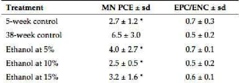
Table I shows the micronucleus bone marrow test results. There were no significant increases in induced MNPCEs at any of the ethanol doses (5, 10, or 15% v/v) as compared to the 5-week-old control group. In addition, no differences were observed among the three doses. However, there were significant differences in induced MN between the two control groups, 2.7 ± 1.2 in 5-week-old mice and 6.5 ± 3.0 in 38-week-old mice. As well, the MN frequencies observed in the three ethanol treated groups were all significantly lower than the values observed in the 38-week-old control group.
The PCE/NCE ratio was within the normal range (>0.1) for both the treatment and control groups, showing no cytotoxic effect of ethanol ingestion on the cell population.
Dominant lethal mutation results are summarized in Table II. The number of total implants per pregnant female is similar for both ethanol doses, and significantly lower than the number observed in the control group. The mean numbers of living embryos per pregnant female also decreased in both ethanol treated groups, but not to
statistically significant levels. No differences were observed for the mean number of dead implants, and the percentage of DLM was low and similar for both ethanol doses.
DISCUSSION
Alcohol abuse greatly increases the risk of different malignancies, including cancer, but the mechanisms by which ethanol could be a carcinogenic or co-carcinogenic agent remain unknown. The available data from studies on ethanol using standard
genotoxicity methods are incomplete and inconclusive (Phillips and Jenkinson, 2001). Nevertheless, some studies have shown that chronic alcoholism may cause
when ethanol was administered in drinking water at 5% for 10-30 days (Balansky et al., 1993) or at 10% and 20% for 3 to 7 weeks (Tates et al., 1980). As well, a non-significant increase in chromosomal aberration frequency was observed at 20% of ethanol administered for 30 days (Tavares et al., 2001). These negative results could be the consequence of the short exposure time period (30 days), or the small number of animals (Tates et al., 1980). However, if ethanol per se is neither carcinogenic nor mutagenic, it could act as an enhancer for carcinogenicity. In this way it has been suggested that the ability of ethanol to induce CYP2E1 (Guegerich et al., 1994) could be the mechanistic basis of ethanol for enhancement of genotoxicity and
carcinogenicity in mixtures containing carcinogens, such as alcoholic beverages (which contain urethane, and probably other known carcinogens). In support of this
suggestion, alcoholic beverages, such as tequila and brandy, were demonstrated to be more genotoxic with the sister chromatid exchange test in mouse bone marrow cells than was ethanol itself (Pina Calva and Madrigal-Bujaidar, 1993).
In this work, we investigated the possible genotoxic effects of chronic ethanol
exposure, so mice were drinking ethanol for a longer period of time than that used in the other cited in vivo ethanol studies. In the mouse bone marrow micronucleus assay (Table I), we observed that all ethanol doses tested did not increase the MNEPC frequency as compared to that in control animals. Because it has been reported that the MNEPC frequency increases in mice with age (Sato, 1995, Dass et al., 1997), we used two control groups, one 5 weeks old, the age at which the treated mice began drinking ethanol, and the other 38 weeks old, the age at which the treated mice were killed after drinking ethanol. Surprisingly, we observed that the MNEPC frequency was significantly lower in the three ethanol-treated groups compared to the 38-week-old control group, suggesting an ethanol protective effect against genotoxic damage caused by aging. This low MN frequency observed in our experiment could not be ascribed to an ethanol cytotoxicity effect, because the EPC/ENC ratio is within the normal range (> 0.1) in all of the treated and control animals (Table I).
Ethanol has previously been reported as a genotoxic protective agent. Different injected ethanol doses have been shown to reduce the induction of mouse MNEPC by urethane (Choy et al., 1995). The same authors also demonstrated that ethanol delays urethane genotoxicity for 12 hours (Choy et al., 1996).
Other authors have reported a radioprotective effect of ethanol. The addition of 10 mM ethanol reduced X-ray-induced chromosome aberrations in human lymphocytes in vitro, while ethanol was less effective in protection from carbon-induced chromosome aberrations. Since densely ionizing radiation produces lesions through direct action, while other ionizing radiation, like X-rays or g-rays, induces DNA lesions mostly by indirect action where free radicals play an important role, the authors concluded that ethanol protects DNA from X rays by scavenging hydroxyl (OH) radicals (Monobe and Ando, 2002). The same authors found that in mice that are given 1 ml of 5.5% ethanol orally 30 min before whole body irradiation, chromosome aberrations in spleen cells were significantly reduced by ethanol for g-ray irradiation, but not for carbon-ion irradiation (Monobe et al., 2003). These results may confirm the hypothesis that ethanol acts as a free radical scavenger. In human lymphocytes, ethanol showed a protective effect for hydrogen peroxide-induced DNA damage in vitro (Greenrod and Fenech, 2003), also apparently by acting as a free radical scavenger.
demonstrated that antioxidants could markedly decrease the levels of ethanol induced DNA single-strand breaks in mouse brain cells (Guo et al., 2007)
The primary site of ethanol absorption is the gastrointestinal tract. Only 2% - 10% of the total ethanol ingested is eliminated by the kidney, and the rest is mainly oxidized in the liver (Lieber, 1997). Ethanol oxidation occurs in three places in hepatocytes, by different pathways: (a) in the cell cytoplasm by alcohol dehydrogenase; (b) in the endoplasmic reticulum by the microsomal ethanol oxidation system (MEOS); and (c) in the peroxisomes by catalase (Burim et al., 2004). Each of these three oxidation
processes produces specific metabolites.
The first phase of ethanol biotransformation involves its oxidation to acetaldehyde, the main and primary metabolite of ethanol. Acetaldehyde is a highly reactive compound that can interact with DNA, forming DNA adducts of acetaldehyde like those observed in peripheral white blood cells of alcohol abusers (Fang and Vaca, 1995), or DNA strand breaks (Singh and Khan, 1995) and DNA cross-links in cultured human
lymphocytes (Blasiak et al., 2000). Thus, the high levels of acetaldehyde accumulated during ethanol metabolism could be responsible for the positive genotoxic effects of ethanol reported in some papers. According to many authors, ethanol does not possess genotoxic potential and the observed ethanol genotoxicity is only due to acetaldehyde. Nevertheless, Kayani and Parry (2010) have shown that both ethanol and
acetaldehyde can produce significant increases in MN induction, establishing that ethanol-MN induction is mainly through an aneugenic mechanism, while acetaldehyde does the same through a clastogenic effect. Different factors could regulate the rates of alcohol and acetaldehyde metabolism. One of these factors is alcohol
deshydrogenase (ADH) and acetaldehyde deshydrogenase (ALDH) polymorphisms, both enzymes being primarily responsible for the amount of acetaldehyde generated. There could be a relationship between polymorphisms of ethanol-induced metabolism genes and alcoholism (Chen et al., 2009), and an effect has been observed of drinking alcohol and ADH/ALDH polymorphism on DNA damage, as measured by the alkaline comet assay (Weng et al., 2010). Thus, human polymorphisms of these enzymes could explain the different effects of the ethanol consumed by alcoholics.
Chronic ethanol consumption leads to its oxidation by MEOS, where P4502E1 is the main component of this system, generating an adaptive increase of ethanol
months of abstinence in comparison to the frequency at the beginning of an intensive treatment program (Huttner et al., 1999).
In germ cells, the dominant lethal mutation assay (Table II) gave no evidence of a significant increase in post-implantation lethality, while a moderate but significant reduction in mean total implants was observed, indicating pre-implantation loss. These results are similar to those reported by Rao et al, who found a significant reduction in mean total implants in a Swiss strain, but not in CBA mice after acute ethanol
treatment (Rao et al., 1994). Thus, our results exclude the possibility that chronic ethanol exposure could induce germinal chromosome mutations in mice.
Finally, our results show that chronic treatment with ethanol does not induce genotoxic damage in somatic or germinal mouse cells evaluated by the micronucleus or the dominant lethal mutation assays. This suggests that ethanol could have a protective effect on age-related genotoxic damage, presumably due to free radical scavenging by ethanol, although further studies are required to confirm this effect and to elucidate the underlying mechanism.
REFERENCES
FRIEDMAN H (1998) Cardiovascular effects of alcohol in Recent Developments in Alcoholism, Volume 14: The Consequences of Alcoholism. Plenum Press: New York, New York. [ Links ]
LIEBER C (1985) Alcohol and the liver metabolism of ethanol, metabolic effects and pathogenesis of injury. Acta Med Scand Suppl 703: 1-55. [ Links ]
HARPER C (1998) The neuropathology of alcohol-specific brain damage, or does alcohol damage the brain? J Neuropathol Exp Neurol 57: 101-110. [ Links ]
IARC (1988) IARC Monographs on the Evaluation of Carcinogenic Risk to Humans, no 44, Alcohol Drinking, IARC, Lyon, 1-378. [ Links ]
KAYANI MA, PARRY JM (2010) The in vitro genotoxicity of ethanol and acetaldehyde. Toxicology in Vitro 24: 56-60. [ Links ]
OBE G, ANDERSON D (1987) Genetic effects of ethanol. Mutat Res 186: 177-200. [ Links ]
LÓPEZ MC, ROUBICEK M, ARZENO M (2001) Chronic alcohol ingestion and
chromosomal aberrations. A popular study in Mar del Plata, Argentina. J. Basic Applied Genetics 14(1): 1-4. [ Links ]
KUCHEIRA K, TANEJA N, MOHAN D (1986) Chromosomal aberrations and sister chromatid exchanges in chronic male alcoholics. Indian J Med Res 83: 417-421. [ Links ]
BRYCE SM, BEMIS JC, AVLASEVICH SL, DERTINGER SD (2007) In vitro micronucleous assay scored by flow cytometry provides a comprehensive evaluation of cytogenetic damage and cytotoxicity. Mutat Res 630: 78-91. [ Links ]
ELLAHUEÑE MF, PÉREZ-ALZOLA LP, ORELLANA-VALDEBENITO M, MUÑOZ C, LAFUENTE-INDO N (1994) Genotoxic evaluation of eugenol using the bone marrow micronucleus assay. Mutat Res 320: 175-180. [ Links ]
BURIM RV, CANALLE R, TAKAHASHI CS, TAVARES DC, MARTINELLI AC, SAKAMOTO-HOJO ET (2004) Clastogenic effect of ethanol in chronic and abstinent alcoholics. Mutat Res 560: 187-198. [ Links ]
MAFFEI F, FIMOGNARI C, CASTELLI E, STEFANINI, GF, FORTI GC, HRELIA P (2000) Increased cytogenetic damage detected by FISH analysis on micronuclei in peripheral lymphocytes from alcoholics. Mutagenesis 15(6): 517-523. [ Links ]
DE ALMEIDA REIS SR, SADIGURSKY M, ANDRADE MGS, SOARES LP, DO ESPIRITO SANTO AR, VILAS BOAS DS (2002) Efeito genotoxico do etanol em células da mucosa bucal. Pesqui Odontol Bras 16(3): 221-225. [ Links ]
BALANSKY RM, BLAGOEVA PM, MIRCHEVA ZI, FLORA S (1993) Coclastogenicity of ethanol with cigarette smoke in rat erythroblasts and anticlastogenicity in alveolar macrophages. Cancer Lett 72: 183-189. [ Links ]
TATES AD, VOGEL N, NEUTEBOOM I (1980) Cytogenetic effects in hepatocytes, bone marrow cells and blood lymphocytes of rats exposed to ethanol in the drinking water. Mutat Res 70: 285-288. [ Links ]
TAVARES DC, CECCHI AO, JORDAO JR AA, VANNUCCHI H, TAKAHASHI CS (2001) Cytogenetic study of chronic ethanol consumption in rats. Teratogen Carcin Mut 21:361-368. [ Links ]
GUENGERICH FP, SHIMADA T, YUN CH, YAMAZAKI H, RANEY KD, THEIR R, COLES B, HARRIS TM (1994) Interactions of ingested food, beverage, and tobacco components involving human cytochrome P4501A2, 2A6, 2E1, and 3A4 enzymes. Environ Health Perspect 102 Suppl 9: 49-53. [ Links ]
PINA CALVA A, MADRIGAL-BUJAIDAR E (1993) SCE frequencies induced by ethanol, tequila, and brandy in mouse bone marrow cells in vivo. Toxicol Lett 66: 1-5. [ Links ]
SATO S (1995) Effect of aging on spontaneous micronucleus frequencies in peripheral blood of nine mouse strains: the results of the 7th collaborative study organized by CSGMT/JEMS.MMS. Mutat Res 388 (1-6): 51-57. [ Links ]
CHOY WN, BLACK W, MANDAKAS G, MIRRO EJ, BLACK HE (1995) A pharmacokinetic study of ethanol inhibition of micronuclei induction by urethane in mouse bone marrow erythrocytes. Mutat Res 341(4): 255-263. [ Links ]
CHOY WN, MANDAKAS G, PARADISIN W (1996) Co-administration of ethanol
transiently inhibits urethane genotoxicity as detected by a kinetic study of micronuclei induction in mice. Mutat Res 367(4): 237-244. [ Links ]
MONOBE M, ANDO K (2002) Drinking breer reduces radiation-induced chromosome aberrations in human lymphocytes. J Radiat Res 43: 237-345. [ Links ]
MONOBE M, KOIKE S, UZAWA, A, ANDO K (2003) Effects of beer administration in mice acute toxicities induced by X rays and carbon ions. J Radiat Res 44: 75-80. [ Links ]
GREENROD W, FENECH M (2003) The principal phenolics and alcoholic components of wine protect human lymphocytes against hydrogen peroxide - and ionizing radiation-induced DNA damage in vitro. Mutagenesis 18: 119-126. [ Links ]
GUO L, SUN B, JANG JY, ZHAO YQ, DONG YX, SPRANGER MI, WU CK (2007) Direct in vivo evidence of protective effects of grape seeds procyanidin fractions and other antioxidants against ethanol- induced oxidative DNA damage in mouse brain cells. J Agric Food Chem 55: 5881-5891. [ Links ]
LIEBER CS (1997)Ethanol metabolism, cirrosis and alcoholism. Clin Chim Acta 257: 59-84. [ Links ]
FANG JL, VACA CE (1997) Detection of DNA adducts of acetaldehyde in peripheral white blood cells of alcohol abusers. Carcinogenesis 18(4): 627-632. [ Links ]
SINGH NP, KHAN A (1995) Acetaldehyde: genotoxicity and cytotoxicity in human lymphocites. Mutat Res 337(1): 9-17. [ Links ]
BLASIAK J, TRZECIAK A, MALECKA-PANAS E, DRZEWOSKI J, WOJEWÓDZKA M (2000)
In vitro genotoxicity of ethanol and acetaldehyde in human lymphocytes and the gastrointestinal tract mucosa cells. Toxicol In Vitro 14: 287-295. [ Links ]
CHEN Y-C, PENG G-S, WANG M-F, TSA T-P, YIN S-J (2009) Polymorphism of ethanol-ethanol metabolism genes and alcoholism: correlation of allelic variation and
pharmacokinetic and pharmacodinamic consequences. Chem Biol Interact 178: 2-7. [ Links ]
WENG H, WENG Z, LU Y, NAKAYAMA K, MORIMOTO K (2010) Effect of alcohol-drinking behavior and ADH1B and ALDH2 polymorphisms on basal DNA damage in human mononuclear cells as determined by the comet assay. Mutat Res 701: 132-136. [ Links ]
HUTTNER E, MATTHIES U, NIKOLOVA T, EHRENREICH H (1999) A follow-up study on chromosomal aberrations in lymphocytes of alcoholics during early, medium and long-term abstinence. Alcoholism: Clin & Exp Res 23(2): 344. [ Links ]
RAO UN, ARAVINDAKSHAN M, CHAUHAN PS (1994) Studies on the effect of ethanol on dominant lethal mutations in Swiss, C57BL6 and CBA mice. Mutat Res 311(1): 69-76. [ Links ]
Received: June 1, 2011. In revised form: September 13, 2011. Accepted: September 14, 2011.
* Corresponding author: Manuel Ellahueñe, Centro Nacional del Medio Ambiente (CENMA), Av. Larraín 9975, La Reina, Santiago, Chile, Phone: (562)-2994151, FAX: (562) 2751688, E-mail: [email protected]
© 2014 Sociedad de Biología de Chile
Canadá 253, piso 3º, Dpto. F.
PO Box 16164
Santiago - Chile
Tel.: (56-2) 22093503
Fax: (56-2) 22258427