videos in clinical medicine
summary pointsThe following text summarizes information provided in the video.
Pulmonary-Artery Catheterization
Christopher R. Kelly, M.D., and LeRoy E. Rabbani, M.D.
From New York–Presbyterian Hospital and Columbia University Medical Center, New York. Address reprint requests to Dr. Kelly at the Department of Medicine, New York– Presbyterian Hospital/Colum bia Univer sity Medical Center, 177 Fort Washington Ave., Rm. 6C12, New York, NY, 10032, or at [email protected].
DOI: 10.1056/NEJMvcm1212416 N Engl J Med 2013;369:e35.
Copyright © 2013 Massachusetts Medical Society. Overview
The modern flow-directed pulmonary-artery catheter, also known as the Swan–Ganz catheter, was first described in the Journal 43 years ago.1 The inflatable balloon at its tip permitted catheterization at the bedside, and the subsequent addition of a thermistor and infusion ports permitted measurement of cardiac output with the use of thermodilution.2
Indications
Pulmonary-artery catheterization aids the diagnosis and management of numerous cardiovascular illnesses, including pulmonary hypertension, cardiogenic shock, mixed shock states, cardiac tamponade, and mechanical complications of ST-segment elevation myocardial infarction (e.g., right ventricular infarction, ventricular septal rupture, and papillary-muscle rupture). It is also part of the standard evalu-ation of patients being considered for heart or lung transplantevalu-ation.
Although pulmonary-artery catheterization had historically been the standard of care for all critically ill patients, data from randomized, controlled trials have shown that it offers no clear benefits to patients with septic shock,3 acute respiratory distress syndrome,3,4 or acute decompensated heart failure.5 Similarly, pulmonary-artery catheterization offers no clear benefits in the routine treatment of patients
undergoing high-risk surgery.6 In such populations, cardiac function and volume
status should instead be assessed through noninvasive means when possible, in-cluding physical examination, measurement of B-type natriuretic peptide levels, echocardiography, measurement of variations in pulse pressure during respiration, and sonographic assessment of the diameter of the inferior vena cava during res-piration. In patients with acute respiratory distress syndrome, treatment deter-mined on the basis of central venous pressure, which can be obtained with a simple central venous catheter, yields outcomes equivalent to those of treatment
determined on the basis of pulmonary-capillary wedge pressure.4
Contraindications
Absolute contraindications include right-sided endocarditis, tumors, or masses, since the catheter may dislodge tissue into the pulmonary artery. Relative contrain-dications include severe coagulopathy and thrombocytopenia, either of which may complicate sheath insertion. Caution should be exercised in patients with left bundle-branch block, in whom catheter passage may induce complete heart block, and in patients with right-sided valve disease (e.g., tricuspid regurgitation), which makes catheter passage more difficult.
Equipment
e35(2)
To insert the introducer sheath, you will need gauze, sterile saline flushes, lido-caine, a 10-cc syringe, a 25-gauge needle, an 18-gauge introducer needle, a guide-wire, a scalpel with a number 11 blade, the introducer sheath itself with an inter-nal obturator, sutures, a needle driver, scissors, and an antibiotic-impregnated adhesive dressing. The use of ultrasonography greatly facilitates location of a central vein and is recommended. To use the probe during cannulation, you will need a sterile sleeve for the probe and conduction jelly.
To insert the pulmonary-artery catheter, you will need the catheter itself, a plastic sleeve, sterile saline flushes, appropriate tubing with stopcocks, and an electronic pressure monitor, preferably one capable of displaying multiple tracings at the same time.
The pulmonary-artery catheter (Fig. 1) is 110 cm long and 5 to 8 French in diameter, depending on the features and design. All catheters have a distal port, typically yellow, that connects to the catheter tip. Most catheters also have a proximal port, typically blue, that connects to a lumen 30 cm from the tip. Larger catheters usually have an accessory infusion port, typically clear or white, that also terminates approximately 30 cm from the tip.
The balloon at the catheter tip can be inflated by injecting air into the pink (or, in some cases, red) port; use only the syringe that is supplied with the catheter, since it will fill only to the balloon’s capacity. To reduce mechanical trauma during insertion, the inflated balloon should completely encircle the catheter tip. Make sure that the balloon is fully inflated whenever the catheter is advanced and fully deflated whenever it is withdrawn.
Most catheters contain a thermistor wire that terminates near the tip and per-mits measurement of cardiac output with the thermodilution technique. Some catheters also have additional features, such as oximeters and pacing wires, with their own specialized ports.
n engl j med 369;25 nejm.org december 19, 2013 A
B
C
D
E
AUTHOR: FIGURE: ARTIST:
OLF: Issue date:
AUTHOR, PLEASE NOTE: Figure has been redrawn and type has been reset.
Please check carefully. Kelly
1 of 3 ts
xx-xx-13 xx-xx-13
Figure 1. Standard Pulmonary-Artery Catheter.
The pulmonaryartery catheter is generally 110 cm long and 7 to 8 French in diameter. An airfilled syringe (A) is used to inflate the balloon at the catheter tip (inset). An accessory infusion port (B) is present in most catheters and connects to a lumen 30 cm from the catheter tip (arrow). The distal port (C) connects to a lumen at the catheter tip and is used to measure all pressures during catheter insertion. The proximal port (D) connects to an additional lumen 30 cm from the catheter tip and is used to monitor right atrial pressures once the catheter tip is in the pulmonary artery. A thermis tor wire extends from the catheter tip to an electronic connector (E) and is used to measure cardiac output by means of thermodilution.
Because the catheter tip may induce ventricular arrhythmias, a defibrillator and transvenous pacemaker should be available at all times. If fluoroscopy is used, each operator must wear a lead apron and thyroid guard. The patient should be shielded in such a way that the chest is not covered and sheath insertion is not hindered.
Preparation
After obtaining informed consent from the patient, review a preoperative checklist to confirm the identity and condition of the patient, the procedure to be performed, and the availability of all required equipment.
Place the patient in the supine position, and select a central vein for cannula-tion. The right internal jugular and left subclavian veins are preferred because the curvature of the catheter facilitates passage from these sites to the pulmonary artery. When cannulating an internal jugular or femoral vein, use ultrasonography to confirm the location and patency of the vein. (Cannulation of the subclavian vein generally relies on anatomical landmarks, although techniques incorporating
ultrasound guidance have recently been described.7)
Wash your hands with antimicrobial solution and don the sterile garments and sterile gloves. Sterilize the patient’s skin with chlorhexidine, and then place the drape. Insert the ultrasound probe into the sterile sleeve. Flush the introducer sheath and pulmonary-artery catheter with sterile saline, and inflate the balloon to confirm that there are no air leaks. Slide the pulmonary-artery catheter through its plastic sleeve.
Procedure
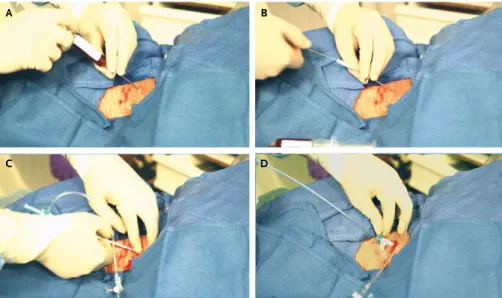
Insert the introducer sheath using the modified Seldinger technique (Fig. 2). Use the 25-gauge needle to infiltrate the skin and subcutaneous tissue with lidocaine. Next, advance the 18-gauge needle into the vein while applying negative pressure to the syringe. Use ultrasonography to directly visualize needle entry.
A B
C D
AUTHOR: FIGURE: ARTIST:
AUTHOR, PLEASE NOTE: Kelly
2 of 3 ts
Figure 2. Sheath Insertion with the Modified Seldinger Technique.
e35(4)
Once dark-red, nonpulsatile blood is aspirated, remove the syringe and insert the guidewire through the needle. Use the scalpel to stab the skin adjacent to the needle, and then remove the needle. While holding the guidewire to ensure that it remains accessible and does not embolize, insert the sheath and internal obtura-tor over the guidewire until the hub fills the wound. Remove the obturaobtura-tor and guidewire from the sheath, and then attach a sterile flush to the port to ensure brisk flow.
Attach the distal port of the pulmonary-artery catheter to the main pressure monitor. Place the catheter tip level with the patient’s heart, and set the pressure to zero. Orient the catheter so that its curvature follows its expected path, and then insert it into the sheath. Advance the catheter to 15 cm (i.e., halfway between the first two thin marks), at which point its tip will lie outside the sheath, and then inflate the balloon.
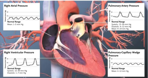
Continue to advance the catheter until a right-atrial-pressure waveform is trans-duced. The distance to the right atrium is typically 15 to 20 cm from an internal jugular or subclavian vein, and approximately 40 to 50 cm from a femoral vein. The right atrial waveform has several identifiable components (Fig. 3): an a wave, which indicates atrial contraction; an x descent, which indicates atrial relaxation; a small c wave, which indicates closure of the tricuspid valve; a v wave, which in-dicates passive atrial filling during right ventricular systole; and a y descent, which indicates passive atrial emptying following the opening of the tricuspid valve. Instruct an assistant to write down the mean right atrial pressure.
Advance the catheter another 5 to 10 cm until a right-ventricular-pressure wave-form is transduced (Fig. 3). This sinusoidal wavewave-form contains a swift upstroke and downstroke, representing ventricular systole, and a slower upstroke, repre-senting passive ventricular filling during diastole followed by right atrial
contrac-n econtrac-ngl j med 369;25 nejm.org december 19, 2013
Pulmonary-Artery Pressure
Normal Range
Systolic: 15–30 mm Hg Diastolic: 4–12 mm Hg Mean: 9–19 mm Hg
Pressure (mm Hg)
20
10
0
Pulmonary-Capillary Wedge Pressure
Normal Range
Mean: 4–12 mm Hg
Pressure (mm Hg)
20
10
0
Right Ventricular Pressure
Normal Range
Systolic: 15–30 mm Hg Diastolic: 1–7 mm Hg
Pressure (mm Hg)
20
10
0
Right Atrial Pressure
Normal Range
Mean: 1–5 mm Hg
Pressure (mm Hg)
20
10 a
a
d c v
y x 0
Figure 3. Pressure Waveforms in the Right Heart and Pulmonary Artery.
The rightatrialpressure waveform is notable for an a wave, which represents atrial contraction; an
x descent, which represents atrial relaxation and contains a small c wave, corresponding to tricuspid
valve closure; a v wave, which represents passive atrial filling during ventricular systole; and a y de scent, which represents passive atrial emptying during ventricular diastole. The rightventricular pressure waveform is notable for an a wave, which represents atrial systole, followed by a large upstroke and a large downstroke, which represent ventricular contraction and relaxation, respec tively. During diastole, the pressure slowly increases as the ventricle passively fills. The pulmonary arterypressure waveform features a swift upstroke and downstroke, with the addition of a dicrotic notch (d), representing pulmonicvalve closure; an overall increase in diastolic pressure as compared with the right ventricle; and a progressive decrease in pressure during diastole. The pulmonary capillary wedge pressure waveform is similar in appearance to the rightatrialpressure waveform.
tion. Instruct an assistant to write down the systolic and diastolic right ventricular pressures.
Advance the catheter another 5 to 10 cm until a pulmonary-artery-pressure waveform is transduced (Fig. 3). This waveform also contains a systolic-pressure waveform, but it is distinguished from the right ventricular waveform by a progres-sive decrease rather than an increase in pressure during diastole, an overall in-crease in diastolic pressure (so long as the pulmonic valve is functional), and a dicrotic notch (representing closure of the pulmonic valve). Instruct an assistant to write down the systolic, diastolic, and mean pulmonary-artery pressures.
Advance the catheter until the waveform indicating pulmonary-capillary wedge pressure is transduced (Fig. 3). This waveform is similar to the right atrial wave-form, except that greater variation may be noted during respiration. Instruct your assistant to write down the mean pressure at the end of expiration, whether the patient is breathing spontaneously or receiving mechanical ventilation. Although positive end-expiratory pressure may affect measurements of pulmonary-capillary wedge pressure, the effect is typically negligible when the positive end-expiratory pressure is less than 10 cm of water.
Once all measurements have been completed, deflate the balloon and confirm the reappearance of a pulmonary-artery-pressure waveform. If this waveform does not reappear, slowly withdraw the catheter until it does.
Aspirate blood from the distal port to measure the mixed venous oxygen satu-ration (Mvo2, also known as the pulmonary-artery saturation [Svo2]). Record the
cardiac output by connecting the thermistor to a computer and then injecting a saline bolus through the proximal port into the right atrium. On a plot of tem-perature against time, the area under the curve is inversely proportional to the cardiac output. Repeat the measurement of cardiac output until at least three consistent results have been obtained, and then calculate the average of the results.
Note the final position of the catheter. Confirm that the balloon has deflated. Fasten the plastic sleeve to the sheath, which will secure the catheter and may re-duce the risk of infection. Suture the sheath to the skin and apply adhesive dressing.
Aftercare
Obtain a portable chest radiograph to evaluate the position of the catheter and to rule out a pneumothorax. To minimize the risk of perforation or infarction, make sure that the catheter tip does not extend more than 4 or 5 cm beyond the midline.
With the catheter in place, the right atrial and pulmonary-artery pressures can be monitored continuously from the proximal and distal ports, respectively. The balloon can be periodically reinflated to reassess pulmonary-capillary wedge pres-sure but should always be deflated afterward. If you are able to meapres-sure the pul-monary-capillary wedge pressure when the balloon is only partially inflated, then the catheter has been inserted too far distally in the pulmonary artery. Withdraw the catheter until full balloon inflation is needed to measure the pulmonary-capillary wedge pressure.
Problems and Complications
n engl j med 369;25 nejm.org december 19, 2013 e35(6)
In each instance, the balloon should be deflated and the catheter withdrawn approximately 10 cm. The balloon can then be reinflated and the catheter read-vanced. If repeated attempts prove fruitless, the procedure should be attempted again with fluoroscopic guidance.
Common early complications include ventricular arrhythmias and right bundle-branch block, which are generally self-limiting. Complete heart block may occur in patients with preexisting left bundle-branch block. In rare cases, the guidewire may embolize and become inaccessible, or the catheter may become knotted in one of the cardiac chambers, preventing withdrawal. In either instance, a vascular surgeon or interventional radiologist must be consulted to ensure safe extraction.
Air embolism may occur if the catheter ports are not properly flushed with saline before the procedure or if the saline-filled tubing becomes disrupted. Manifestations include dyspnea, chest pain, tachycardia, hypotension, and in some cases, an acute increase in right heart pressures. In cases of air embolism, place the patient in the Trendelenburg position to limit the outflow of air from the right ventricle, and administer high-flow supplemental oxygen to reduce the nitrogen content of the blood and thereby promote the reabsorption of air. In severe cases, hyperbaric oxygen therapy is required.
Pulmonary-artery perforation is a rare but dangerous event that occurs in
ap-proximately 1 in 3000 patients.8 Risk factors include older patient age, prolonged
balloon inflation, pulmonary hypertension, and systemic anticoagulation. Symptoms include hemoptysis, hypoxemia, and shock. If immediate action is not taken, the risk of death is high. Keep the balloon inflated to limit further bleeding. Intubate the patient with a dual-lumen endotracheal tube, and then place the patient in the lateral decubitus position, with the affected side down. Immediately consult a thoracic surgeon and an interventional radiologist.
Later complications include pulmonary infarction, catheter-related infection, and thrombosis. To reduce the risk of pulmonary infarction, make sure that the catheter tip is positioned such that full inflation of the balloon is required to measure pulmonary-capillary wedge pressure and that the balloon is deflated after pulmonary-capillary wedge pressure measurements have been completed.
Interpretation
Normal pressures and waveforms are shown in Figure 3. The measured pressures can be used to calculate cardiac output, systemic vascular resistance, and pulmo-nary vascular resistance, as shown in Table 1.
Specific changes in chamber pressures and waveforms are typical of various
conditions.9 In pericardial tamponade, for example, high pericardial pressures
equalize diastolic pressure throughout the heart, and the pressure waveforms ap-pear abnormal. In the right atrium, the x descent apap-pears exaggerated because of the drop in pericardial pressure during ventricular systole, whereas the y descent appears blunted because the pericardial pressure remains constant during ven-tricular diastole.
Hemodynamic data can also help to clarify the cause of shock. A patient with cardiogenic shock has a low cardiac output, which raises filling pressures in the left atrium (creating high pulmonary-capillary wedge pressure). The systemic
vas-cular resistance rises to offset the changes in mean arterial pressure, and Mvo2
declines as tissues increase their oxygen extraction to compensate for decreased oxygen delivery.
A patient in the early stages of distributive shock (e.g., septic shock) has a pathologic reduction in systemic vascular resistance that results in functional
teriovenous shunting, which, in combination with sepsis-induced mitochondrial
dysfunction, causes a high Mvo2. Cardiac output increases to offset the changes
in mean arterial pressure. Pulmonary-capillary wedge pressure may initially be normal, but if adequate fluid resuscitation does not occur, pulmonary-capillary wedge pressure and cardiac output will fall.
A patient with a normal heart in hypovolemic shock (e.g., massive hemorrhage) has inadequate ventricular filling, which results in low pulmonary-capillary wedge pressure and cardiac output. As in cardiogenic shock, there are compensatory
changes in systemic vascular resistance and in Mvo2.
Summary
Pulmonary-artery catheterization can be safely performed at the bedside and yields a wealth of hemodynamic data. Although recent studies indicate that this proce-dure should not be performed routinely in critically ill patients, it remains invalu-able in the diagnosis and management of a wide range of cardiovascular illnesses. Because fatal complications can occur, however, the procedure should be performed only when its results are expected to aid clinical management.
Dr. Kelly reports receiving payment for the development of educational presentations from Hitachi Aloka. No other potential conflict of interest relevant to this article was reported.
Disclosure forms provided by the authors are available with the full text of this article at NEJM.org. We thank Josh Gramling and Kevin Maloney of Visual Health Solutions.
References
1. Swan HJ, Ganz W, Forrester J, Marcus H, Diamond G, Chonette D. Catheterization of the heart in man with use of a flow-directed balloon-tipped catheter. N Engl J Med 1970;283:447-51.
2. Forrester JS, Ganz W, Diamond G, McHugh T, Chonette DW, Swan HJ. Thermodilution cardiac output determi-nation with a single flow-directed cathe-ter. Am Heart J 1972;83:306-11.
3. Richard C, Warszawski J, Anguel N, et al. Early use of the pulmonary artery catheter and outcomes in patients with shock and acute respiratory distress syn-drome: a randomized controlled trial. JAMA 2003;290:2713-20.
4. The National Heart, Lung, and Blood Institute Acute Respiratory Distress Syn-drome (ARDS) Clinical Trials Network. Pulmonary-artery versus central venous catheter to guide treatment of acute lung injury. N Engl J Med 2006;354:2213-24.
5. Binanay C, Califf RM, Hasselblad V, et al. Evaluation study of congestive heart failure and pulmonary artery catheteriza-tion effectiveness: the ESCAPE trial. JAMA 2005;294:1625-33.
6. Sandham JD, Hull RD, Brant RF, et al. A randomized, controlled trial of the use of pulmonary-artery catheters in high-risk surgical patients. N Engl J Med 2003; 348:5-14.
7. Fragou M, Gravvanis A, Dimitriou V, et al. Real-time ultrasound-guided sub-clavian vein cannulation versus the land-mark method in critical care patients: a prospective randomized study. Crit Care Med 2011;39:1607-12.
8. Kearney TJ, Shabot MM. Pulmonary artery rupture associated with the Swan-Ganz catheter. Chest 1995;108:1349-52.
9. Davidson CJ, Bonow RO. Cardiac cath-eterization. In: Libby P, Bonow RO, Mann DL, Zipes DP, eds. Braunwald’s heart dis-ease: a textbook of cardiovascular medi-cine. 8th ed. Vol. 1. Philadelphia: Saunders, 2007:439-63.
Copyright © 2013 Massachusetts Medical Society.
Table 1. Calculation of Cardiac Output and Vascular Resistance with the Use of Fick’s Method.*
Value Equation Normal Values
Cardiac output (liters/min)
CO = CO2 a − Cv which can be simplified to
CO = 1.36 × Hgb × (SaOo2
2 − Svo2) × 10
4.8–7.3 for an average
adult
Cardiac index (liters/min/m2)
CI = BSACO
2.8–4.2
Systemic vascular resistance (dyn · sec · cm−5)
SVR = MAP − RACO × 80 700–1600
Pulmonary vascular resistance
(dyn · sec · cm−5) PVR =
PA − PCWP CO × 80
20–130
* In the equation for cardiac output (CO), O2 denotes oxygen consumption (ml/min), Ca the
oxy-gen content of arterial blood, Cv the oxygen content of venous blood, 1.36 the oxygen-carrying