Significance of ROS in oxygen sensing in cell systems with
sensitivity to physiological hypoxia
Constancio Gonzalez *, Gloria Sanz-Alfayate, M. Teresa Agapito,
Angela Gomez-Nin˜o, Asuncio´n Rocher, Ana Obeso
Departamento de Bioquı´mica y Biologia Molecular y Fisiologı´a,Facultad de Medicina,
Instituto de Biologı´a y Gene´tica Molecular(IBGM),Uni6ersidad de Valladolid y CSIC,47005Valladolid,Spain Accepted 28 August 2001
Abstract
Reactive oxygen species (ROS) are oxygen-containing molecular entities which are more potent and effective oxidizing agents than is molecular oxygen itself. With the exception of phagocytic cells, where ROS play an important physiological role in defense reactions, ROS have classically been considered undesirable byproducts of cell metabolism, existing several cellular mechanisms aimed to dispose them. Recently, however, ROS have been considered important intracellular signaling molecules, which may act as mediators or second messengers in many cell functions. This is the proposed role for ROS in oxygen sensing in systems, such as carotid body chemoreceptor cells, pulmonary artery smooth muscle cells, and erythropoietin-producing cells. These unique cells comprise essential parts of homeostatic loops directed to maintain oxygen levels in multicellular organisms in situations of hypoxia. The present article examines the possible significance of ROS in these three cell systems, and proposes a set of criteria that ROS should satisfy for their consideration as mediators in hypoxic transduction cascades. In none of the three cell types do ROS satisfy these criteria, and thus it appears that alternative mechanisms are responsible for the transduction cascades linking hypoxia to the release of neurotransmitters in chemoreceptor cells, contraction in pulmonary artery smooth muscle cells and erythropoietin secretion in erythropoietin producing cells. © 2002 Elsevier Science B.V. All rights reserved.
Keywords:Carotid body, signal transduction; Control of breathing, O2sensing; Mediators, erythropoietin; Mediators, ROS; Oxygen,
cellular sensing; Signal transduction, carotid body, erythropoietin cell, pulmonary vasculature
www.elsevier.com/locate/resphysiol
1. Cell systems with sensitivity to physiological hypoxia
The appearance of O2 on a previously anaero-bic earth, forced living organisms to develop an-tioxidant defense systems. Organisms that evolved tolerance to O2also developed mechanisms to use this new element for metabolic transformations
* Corresponding author. Tel.:+34-983-423-089; fax:+ 34-983-423-588
E-mail address:[email protected](C. Gonzalez).
and efficient energy production by using electron transport chains with O2 as the final electron acceptor. The development of efficient and secure energy production systems allowed the appearance of multicellular organisms. The acquisition of O2 and nutrients by diffusion from the surrounding aqueous medium was succeeded in multicellular organisms by the appearance of a circulatory system, a system of tubes to carry and to distribute O2 and nutrients among all cells of the living
organism. Evolution later provided an oxygenating or respiratory system capable of supplying O2to the
fluid moving in the circulatory system, and the moving fluid itself evolved to blood to gain in capacity to transport O2. All these acquisitions
provided living organisms with the independence necessary to abandon water and to move freely on land. Nevertheless, full acquisition of terrestrial freedom was only possible by the simultaneous appearance of homeostatic mechanisms. Some of those homeostatic mechanisms match O2supply to
whole-body energetic needs, by monitoring O2
levels in the body, and altering the overall function of the respiratory and circulatory systems, as well as the capacity of blood to transport O2.
There are two main systems regarding O2
homeostasis. They conform two functional loops with their origin in the O2-monitoring cells, the
chemoreceptor cells of the carotid body (CB) and the erythropoietin (EPO)-producing cells (Richalet, 1997; Gonzalez, 1998). These two cell types are receptor – effector units, which detect arterial blood O2 levels and respond with a secretory response. The functional loop originated in the CB works as follows: Chemoreceptors cells detect arterial blood PO2 and become activated when arterial PO2
de-creases; they release neurotransmitters which acti-vate the sensory nerve endings of the carotid sinus nerve, whose central projections end in the nucleus of the tractus solitarius in the medulla. Integration of the carotid sinus nerve input in the respiratory control centers of the brain stem results in an increased activity of the respiratory muscles, with increased ventilation and increased alveolar and arterial blood PO
2 (Gonzalez et al., 1992, 1994).
EPO-producing cells, which in adult animals are mainly located in the kidney, also detect arterial blood O2 levels and are activated by hypoxia to
release EPO; this glycoproteic hormone reaches the bone marrow and activates erythropoiesis. An increase in the red blood cell mass and ultimately in the capacity of the blood to transport O2follow (Jelkmann, 1992). These two systems probably are aided in maintaining the homeostasis of O2by the
pulmonary artery smooth muscle cells (PASMC), which, as the other cell two types, monitor oxygen levels and react to hypoxia with a contractile response leading to vasoconstriction and lung blood flow redistribution which would ameliorate the oxygenation of blood (Marshall et al., 1994). We have previously defined physiological hypoxia and these three cell types as endowed with sensitiv-ity to physiological hypoxia (Gonzalez, 1998).
These three cell systems, contributing to the homeostasis of O2, share other properties. They have a very low threshold to hypoxia, becoming activated at an arterial PO
2 of :70 mmHg and
increasing their effector activity with the intensity of hypoxia. In contrast, the response to hypoxia in most cells is a decrease in their activity (Hochachka et al., 1997) and as the hypoxic threshold for this behavior is higher, a more intense hypoxia is needed to elicit it. The different behavior of the cell types involved in homeostatic loops versus general cells of the organism allows to predict that the detection of O2 levels (i.e. (O2-sensing), if linked to O2
-con-sumption rate, should be associated to an increase of O2-consumption in the case of the cells involved in homeostatic loops and to a decrease in the rest of the cells. The increase in O2-consumption during hypoxia in the former cells is an absolute require-ment to support the increased activity they exhibit in these circumstances; the decrease in O2
-con-sumption during hypoxia in the rest of the cells is inferred from the near shutdown of all energy-re-quiring processes. Alternatively, O2-sensing could
be independent of O2-consumption rate in both
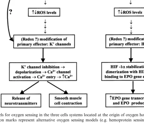
in-Fig. 1. Redox models for oxygen sensing in the three cells systems located at the origin of oxygen homeostatic loops. The top lateral arrows with question marks represent alternative oxygen sensing models (e.g. hemoprotein sensing) not discussed in this article.
creases glucose oxidation to CO2 in an ouabain-dependent manner (Obeso et al., 1989b, 1993).
An alternative way of stating the notions ex-pressed in previous paragraphs is to say that in higher organisms there are a few cell systems devoted to maintaining O2 levels in the entire
organism which appear to have a unique mecha-nism for O2-sensing. The uniqueness of this O2
-sensing mechanism refers to its threshold and to its ability to become coupled to and to activate cell processes demanding an energy supply. This article will consider the redox models of O2
-sens-ing. We shall discuss the available information in favor and against the participation of oxygen reactive species (ROS) in O2-sensing and the pos-sible significance of ROS as coupling factors be-tween hypoxia and the effector machinery of the cells involved in the homeostasis of oxygen (Fig. 1).
2. Free radicals, oxygen reactive species (ROS) and allied molecules
2.1. Primary free radicals
Free radicals are chemically defined as atoms or molecules that have one or more unpaired elec-tron; they usually are very reactive (Punchard and Kelly, 1996; Halliwell and Gutteridge, 1999). The primary processes that give rise to free radicals in biological systems are divided into two main cate-gories. Due to the high water content of living organisms, the cleavage of the chemical bonds of water by high-energy radiation (water radiolysis) constitutes an important source of free radicals according to the reaction:
H2OH(hydrogen radical)
Ultraviolet light and even visible light in the presence of some sensitizer molecules can simi-larly produce homolytic scission of hydrogen per-oxide in the skin to generate hydroxyl radicals (H2O22OH). More important, however, (at least quantitatively) is the genesis of free radicals by reactions involving the transference of elec-trons, and among them the most important are those reactions in which a single electron is trans-ferred to a molecule of O2to form the superoxide
radical, O2−(Davies and Dean, 1997). Phagocytes
possess an enzymatic complex containing FAD and a specific b-type cytochrome capable of tak-ing an electron from NADPH and transferrtak-ing it to O2 to form O2−. This is probably the only
reaction in which O2−is a beneficial product; this
radical and other ROS formed from it are used by phagocytic cells to kill bacteria. There are, how-ever, many unwanted processes producing O2−as
a byproduct, some of which are briefly com-mented here. There are auto-oxidation reactions in which several biologically important molecules such as tyrosine, catecholamines and tryptophan slowly reduce O2 to form O2−. O2− in turn is
capable of further oxidizing those molecules and a cascade of reactions might be started with poten-tially deleterious effects. Microsomal enzymes in-volved in reactions of oxidation and hydroxylation of many types of molecules (alco-hols, barbiturates, antibiotics, steroid hormone synthesis and metabolism, etc.), which use cy-tochrome P450 as an electron transport system, can also generate O2−. Superoxide radical is also
formed in the transport of O2 by hemoglobin.
Although essentially all O2binding to hemoglobin
(and myoglobin) does not imply the oxidation of Fe2+ in the heme moiety, there is, however, a
certain degree of delocalization of an electron when O2 is bound and thereby the appearance of
some kind of intermediate structure that can be written as an apparent resonant ‘equilibrium’: hemo-Fe2+– O
2lhemo-Fe3+– O2−. Usually, the
Fe2+ of heme group keeps the delocalized
elec-tron and hemoglobin releases O2, but occasionally (3%/day) the electron is kept by O2 and hemoglobin then releases O2−, the Fe2+ of the
heme group is transformed into Fe3+ and
methemoglobin is formed. In situations of tissue
injury, especially in the processes of ischemia-reperfusion, the enzyme xanthine dehydrogenase which oxidizes hypoxanthine and xanthine to uric acid using NAD+ as acceptor of electrons, is
modified by oxidative or proteolytic processes and converted into xanthine oxidase, which is capable of transferring the electrons of the purines to O2
to form O2−. However, there is no doubt that
quantitatively speaking the most important source of O2− is the mitochondrial electron transport
chain, where univalent reduction of O2 may take
place in some steps of the overall transport system (Fig. 2). In the two places where O2− can be
produced (complex I and ubiquinone pool) there are electron shuttle molecules (flavin adenin mononucleotide and ubiquinone), which can ac-cept one or two electrons. When they acac-cept a single electron they become free radical molecules themselves capable of slipping the electron to molecular O2. It is estimated that a small
percent-age (B5%) of the total O2 consumed by
mito-chondria at normal PO2 is converted to O2−.
To complete this overview on radicals it re-mains to say some words on reactive nitrogen species. Nitric oxide or nitrogen monoxide (NO) is a free radical as it has one unpaired electron. It is a gas molecule capable of freely diffusing through cell membrane systems. NOis physiolog-ically synthesized by a family of different nitric oxide synthases from the amino acidL-arginine. It
binds to Fe2+ of heme groups, and in fact many
of its physiological actions are mediated by the soluble guanylate cyclase, which is a hemoprotein that synthesizes cGMP, although cyclase indepen-dent physiological effects have been described (Archer et al., 1994; Bolotina et al., 1994; Ohkuma and Katsura, 2001). Due to its ability to react with heme groups, NO can inhibit cy-tochrome oxidase competitively with O2 (Boveris
et al., 2000). In body fluids NOreacts with O2to
give the more reactive radical nitrogen dioxide (2NO+O22NO2), but largely it is oxidized to
nitrite ion (4NO+O2+2H2O4NO2−) or reacts
with the heme groups of oxyhemoglobin to form metahemoglobin and NO3− (nitrate). In fact, the
Fig. 2. General schema of the mitochondrial respiratory chain illustrating the loci where O2−can be generated. The schema also
shows the most commonly used inhibitors of the respiratory chain, which when acting downstream of the loci of production of O2−
would increase, while those acting upstream will inhibit O2−production.
2.2. Secondary free radicals and other reacti6e species
Once generated, these three main free radicals (OH, O2− and NO) can undergo an enormous
variety of reactions with other molecules giving rise to new radicals or to species, which are oxidized or reduced. When new radicals are formed they are capable of undergoing further reactions originating chain reactions until one of the radicals reacts with a molecule (scavenger) to generate an unreactive compound. This wide-spread reactivity is the basis for the toxic effects of free radicals. For example, in their reactions with membrane lipids, OH abstract an atom of hydrogen from polyunsaturated fatty acids form-ing H2O plus a modified fatty acid which is a
carbon radical species readily reacting with molec-ular O2. The incorporation of O2by the fatty acid
carbon radical species transforms it into a peroxyl radical (RCOOCOOH) which in turn is capa-ble of abstracting another hydrogen atom from a nearby polyunsaturated fatty acid, thereby spreading out the alteration of lipids;
its replication or transcription. In sum, OHis so reactive that it will react in the very same place where it is produced and disappear immediately as such entity, but at the same time new reactive molecules would appear which would spread the effects. The question in the context of the present article is if there exists any mechanism in the cells capable of circumscribing the action of OHfor it to act as the signaling molecule during hypoxia as Kietzmann et al. (2000) suggest. Whatever the mechanism(s) it should be based in two premises: first, the production of OHshould be located in a very restricted area of the cell; this restricted area must be coincident with the location of the cell machinery that initiates the hypoxic transduc-tion cascade (for example, the O2-sensitive K+
channels or hypoxia inducible factor 1a, HIF-1a), and second, the ‘OH-receptor molecule’ itself (e.g. the O2-sensitive K+ channels or HIF-1a)
should act as a very effective OH scavenger to avoid the widespread action of secondary reactive molecules with the inevitable loss of capacity to focus the hypoxic signal.
The O2−is considerably much less reactive than
OH. It can be protonized to form HO2
(hy-droperoxyl radical) which has a greater reducing activity and which in addition is freely permeable to membranes owing to is lack of charge; both characteristics make HO2an important free
radi-cal in spite of the fact that its concentration in biological systems is 1/100 – 400 that of O2−. In
addition, hydroperoxyl radical is involved in the spontaneous dismutation of O2− to H
2O2 and
molecular O2. This reaction, which involves the
reduction of one O2− and the oxidation of
an-other to molecular O2, occurs through the
forma-tion of hydroperoxyl radical according to the equations:
H−+O
2
−HO
2;
HO2+O2−+H+H2O2+O2
Superoxide radical can donate one electron to transition metals transforming Fe3+, Cu2+ and
Mn3+ into Fe2+, Cu+ and Mn2+, respectively,
and then it acts as a reducing agent, but it can also act as an oxidizing agent reversing the reac-tion. These reducing/oxidizing reactions are very
important because the enzymes responsible for the cellular elimination of O2−, the superoxide
dismu-tases (cytosolic or CuZn superoxide dismutase and mitochondrial or manganese superoxide dis-mutase), are metalloproteins containing those metals and through cycles of reduction and oxida-tion of their metals accelerate enormously the disappearance of O2−. Other important reactions
of O2− include oxidation of ascorbate and the
catechol ring to form ascorbate and diphenol radicals and H2O2. Another reaction of interest is
that of O2− with hypoclorous acid (formed in
turn by the action of myeloperoxidase on H2O2+
Cl−HOCl+OH−) to yield O
2, Cl− and OH.
Summarizing, we can state that up to this point most of O2−ends up as H
2O2due to spontaneous
and enzyme catalized dismutation. To some ex-tent, we may say that H2O2 is the final common path of most free radicals in the organism.
However, H2O2 is harmful to the cells. It
be-longs to a category of molecules collectively called reactive oxygen species (ROS), which include free radicals. H2O2 and other ROS such as
hypoclor-ous acid or peroxynitrite (NO+O2−ONOO−;
see below) have in common with free radicals being more reactive than molecular O2, but unlike
free radicals they do not have unpaired electrons. The toxicity of H2O2 would explain the existence
of specific enzymatic systems to destroy it, includ-ing catalases (2H2O22H2O+O2) and peroxi-dases, the most important of which is glutathione peroxidase (H2O2+2GSHGSSG+2H2O; GSSG is back reduced to GSH by the action of the NADPH based glutathione reductase). The toxicity of H2O2 is not exerted directly unless it is
applied at high concentrations (\10 mm for many cells), but by the OHit can generate upon reaction with transition metals by the Fenton reaction (Fe2++H
2O2 Fe3++
OH+OH−) or upon homolytic fission by the
action of ultraviolet light. We already have exam-ined the widespread damaging effects of OH (Fig. 3).
Nitric oxide (NO) is also readily reactive with many other radicals. For example, it can react rapidly with superoxide to yield peroxynitrite (NO+O2−ONOO−) and with hydroxyl
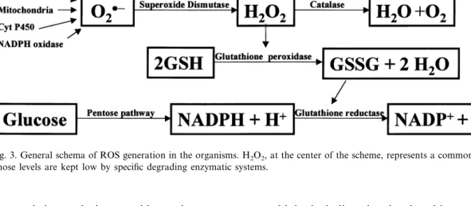
Fig. 3. General schema of ROS generation in the organisms. H2O2, at the center of the scheme, represents a common final product
whose levels are kept low by specific degrading enzymatic systems.
Peroxynitrites and nitrous acid are nitrogen reac-tive species capable of reacting with SH groups of proteins and with iron/sulfur electron transport-ing proteins of mitochondria. As we have already mentioned, NOcan react in a reversible manner with cytochrome oxidase, thereby competing with O2; this might be a mechanism to control the
oxidation rate of the energy-yielding compounds (nutrients). However, mitochondria must harmo-nize this potentially reversible regulating mecha-nism of NOwith its ability to react with O2−to
form peroxynitrite and through it to produce irreversible damaging effects on electron trans-porting proteins. Peroxynitrite can interact with tyrosine residues in a reaction catalyzed by super-oxide dismutase to form the very stable 3-nitroty-roxine, changing the overall charge of the residues of tyrosine present in proteins and making it difficult for 3-nitrotyrosine containing proteins to assemble into multimeric units or to interact with other proteins (Beckman and Koppenol, 1996) and to participate in signaling cascades, by alter-ing the efficiency of tyrosine phosphorylation. The great stability of 3-nitrotyrosine makes the mea-surement of its level a good index of nitric oxide/
peroxynitrite in biological systems. Peroxynitrite and other nitrogen reactive species can react with
thiols, including that in glutathione, to form ni-trosothiols. Special interest has recently been given to the nitrosylation of one cysteine residue in the ryanodine receptor/channel, which seems to occur in a PO2-dependent manner (Eu et al.,
2000). When sarcoplasmic vesicles are exposed to ambient PO2 (:150 mmHg) six to eight thiol
residues per ryanodine receptor become oxidized, the oxidation reverting upon exposure to PO2 of
10 mmHg, which is in the range of that physiolog-ically encountered in the muscle; on switching back to ambient PO2, reoxidation of those thiol
residues takes place. These cysteine residues will constitute, according to (Eu et al., 2000), an O2 -sensor. It is conceptually important to realize that this potential O2-sensor needs to be coupled to a
redox system, which must be present in the sar-coplasmic reticulum vesicles, and which must be responsible for switching back and forth from the oxidized to the reduced state. Only the reduced form of the channel is susceptible for physiologi-cal modulation by NO, which does so by nitrosy-lating one of the reduced cysteine residues, or in other words, the redox state of the channel set by PO2 determines the capacity to respond to (or the
3. ROS as mediators of the hypoxic transduction cascade
3.1. Criteria to consider ROS mediators of a physiological cellular response
Following classical studies on messengers such as neurotransmitters (McLennan, 1963) and the most recent article by Lander (1997) on ROS in signal transduction, it is possible to put forward a set of criteria as requirements to consider ROS as true second messengers with the capacity to con-trol cellular functions under physiological condi-tions. A first criterion should be that the putative ROS molecule acting as messenger or mediator of the hypoxic response must increase or decrease during hypoxia; a given relationship (modulation can modify that putative relationship) should exist between the ROS level change, the intensity of hypoxia and the strength of the hypoxic response. The second criterion would be that the cellular mechanism responsible for the change in the level of ROS molecule, putative messenger, needs to be identified. The third criterion would be the defini-tion of the primary target of the ROS as well as the definition of the change produced by the ROS molecule on its target. The possibility exists that the primary target of the putative messenger ROS molecule is itself the effector(s) of the cell re-sponse or on the contrary, it is possible that the primary target of ROS is a first link of a chain or cascade of events coupling the change in the ROS levels to the effector(s) of the cell response; in this latter case the entire cascade should be defined. The fourth criterion would be to define the cellu-lar responses elicited by the effector(s). The fifth criterion would be the definition of mechanisms responsible for the inactivation of ROS signal. Obviously if the ROS species increase during hy-poxia we should be looking for ROS degrading mechanisms, but if they decrease we should be looking for the enzyme or cells process leading to the restoration of normal ROS levels. In any case, if a given stimulus acts via a change in ROS levels, the mechanisms for the elimination of those ROS should be induced by prolonged pre-sentation of the stimulus. The sixth criterion (pharmacological) would be that the manipulation
of the ROS levels, whether by reagents that mod-ulate glutathione levels or that directly scavenge ROS, should alter the cell response in a pre-dictable and reproducible manner; a sixth b or seventh criterion would be that overexpression or silencing of the expression of the ROS scavenging enzymes should alter the cell response in a pre-dictable and reproducible manner.
3.1.1. First criterion: the signal ROS le6els during hypoxia
The very first question that should be addressed is if hypoxia increases or decreases ROS in gen-eral or if there is a unique ROS species whose production follows a particular behavior during hypoxia. The classical view is that the rate of production of ROS changes in direct relation to the tissue PO
2 (Chance et al., 1979; Archer et al.,
1986, 1989). In fact, this view is generally ac-cepted at present (Archer et al., 1993; Gorlach et al., 1994; Fandrey et al., 1994, 1997; Halliwell and Gutteridge, 1999; Kietzmann et al., 2000; Gnaiger et al., 2000). However, quite recently Schumacker and coworkers (Chandel et al., 1998; Duranteau et al., 1998; Chandel and Schumacker, 2000; Chandel et al., 2000) have found the opposite: hypoxia produces an increase in the cellular rate of ROS production which takes place at the level of mitochondria and which is proportional to the level of hypoxia. Chandel et al. (1998) claim that their measurements using dichlorofluorescein fluorescence, which apparently measures preferen-tially H2O2, are the correct ones. However there are many measurements using different methods (see Chance et al., 1979) in which it has been shown that hypoxia decreases the rate of produc-tion of ROS, the decrease being attributed to a decrease of production in mitochondria as well as in microsomal and peroxisomal enzymes. Archer et al. (1993) and Paky et al. (1993) using lucigenin chemiluminescence, which apparently measures preferentially O2− (Halliwell and Gutteridge,
their production if ROS are measured with dichlorofluorescein (Chandel et al., 1998). Inter-estingly, both dyes yielded an inhibition of ROS generation under rotenone and diphenileneiodo-nium (DPI) (Cross et al., 1990; Ehleben et al., 1997; Chandel et al., 1998; Duranteau et al., 1998); rotenone and DPI also decreased lucigenin chemiluminescence (Archer et al., 1993, 1999). However, while antimycin A increased dichlor-ofluorescein fluorescence (Chandel et al., 1998) it decreased lucigenin chemiluminescence (Archer et al., 1993).
With this panorama it is difficult to make a meaningful formulation of the putative ROS molecule(s), which are signals for the hypoxic transduction cascade. We do not know if the signal should be a particular ROS species that decreases during hypoxia, a general decrease in the ROS levels, or if on the contrary, we should be searching for something that increases during hypoxia. We would like to quote (Semenza, 1999, 2000) when he writes that ‘the direct measure-ments of ROS are so demanding that they gener-ate data supporting opposing views’. However, we want to make explicit some thoughts on the prob-lem, being aware that experimental data might contradict the opinions expressed here. When we consider the general mechanisms for ROS produc-tion presented in previous paragraphs, the intu-itive notion would be that the rate of production of ROS in cells should be directly proportional to the PO
2 of the cells. Reactions of auto-oxidation
as well as oxidations driven by enzymes and the acceptance by molecular O2of electrons leaking at
the level of the quinone pool in mitochondria (see Fig. 2) would decrease during hypoxia, when PO2
is diminished. At the same time, however, it would appear that the generation of O2− at the
quinone pool level would depend on the rate of flow of electrons through the respiratory chain, i.e. on the respiratory rate. As stated in the first section of this article, cell systems involved in the general homeostasis of O2 are activated by
hy-poxia, this activation requiring an increase in cell respiration to support cell and homeostatic loop function (Obeso et al., 1993, 1997). Thus, it could be expected that in these specialized cells hypoxia produces an increase in electron flow during an
ample range of hypoxia (from :70 to 20 mmHg of arterial PO
2), and thereby, an increase in the
electron leakage and mitochondrial formation of O2− is conceivable in those situations (see Fig. 3
in Hoshi and Heinemann, 2001) when hypoxia is intense enough a compromise will occur and in spite of the high affinity of cytochrome oxidase for O2, it will be unable to transfer enough
elec-trons to O2 and the respiratory rate and the
genesis of ATP will drop. In the case of the CB in vivo this metabolic compromise occurs at arterial PO2:20 mmHg; below this PO2 the CB is unable
to adequately signal the level of hypoxia and chemoreceptor action potential frequency de-creases, instead of increasing (see Fidone and Gonzalez, 1996). The majority of the cells of the organism will behave during hypoxia as the CB below the compromise PO
2: all O2
−-generating
processes including that of mitochondria would decrease as the intensity of hypoxia increases. To state this in a different manner, it would appear that a differential response could be expected: in most cells of the organism hypoxia would tend to decrease ROS production while in CB chemore-ceptor cells, in EPO-producing cells and in smooth muscle cells of the pulmonary arteries hypoxia would tend to increase ROS production at least in those ranges of hypoxia compatible with life. In contrast, Schumacker and co-workers measured an increase in ROS production in car-diomyocytes and hepatoma cells (EPO-producing) which is comparable in magnitude and in PO
2
dependence in both cell types (Chandel et al., 1998; Duranteau et al., 1998) and Fandrey et al. (1994) measured a decrease in the rate of produc-tion and release of H2O2 to the incubating
solu-tion by EPO-producing hepatoma cells precisely in the range of PO2 where they secrete EPO.
In sum, the first criterion is not settled, we do not know whether hypoxia increases or decreases ROS levels.
3.1.2. Second criterion:cell mechanisms responsible for the change in ROS production during hypoxia should be defined
Fan-drey et al., 1994; Kietzmann et al., 2000; Weir and Archer 1995; Archer et al., 1993, 2000; Fu et al., 2000) have been considering that a NADPH ox-idase similar or identical to that present in phago-cyte cells would be responsible for that decrease. The main findings to propose NADPH oxidase as the mechanism for the decrease in ROS produc-tion during hypoxia were: (1) the microspec-trophotometric description in the CB of a cytochrome bgss, (2) the microspectrophotometric observation that hypoxic activation of the CB was paralleled by a reduction of FAD and NADP+
and (3) the observation that DPI (an inhibitor of NADPH oxidase, and in general of all flovoen-zymes) was able to activate the CB chemorecep-tors’ sensory discharge and to occlude the response to hypoxia. At the same time, DPI com-pletely eliminated the fluorescent signal emitted by rhodamine 123 attributed to H2O2 (Cross et al., 1990). These initial observations were soon followed by the immunocytochemical demonstra-tion of all the subunits of NADPH oxidase in the CB (Kummer and Acker, 1995), although more recent studies have shown that most of the intra-glomic immunostaining is localized in macrophages infiltrating the CB tissue (Dvo-rakova et al., 2000).
As discussed elsewhere (Gonzalez, 1998), this oxidase has a Km for O2 (5 – 30 mm or 3 – 18
mmHg) that would make reasonable the proposi-tion that at the arterial PO
2 threshold for the
activation of the homeostatic loops the rate of O2− formation by NADPH oxidase could start
falling below Vmax. There are, however, several observations and considerations that in our opin-ion make untenable the proposal that the decrease in the activity of NADPH oxidase, and thereby the decrease in ROS generated by it, are the mediators or messengers linking the decrease of PO2 to the cellular effectors. For example,
NADPH is a multimeric enzyme with two mem-brane-bound and several cytoplasmic subunits, and to become active they need to be assembled in the membrane (Dang et al., 2001). The question therefore arises: what is the mechanism promoting the assembling of the subunits and of keeping the enzyme permanently active in resting normoxic conditions? Also, does the proposed hypoxic
inhi-bition imply disassembling of the enzyme? Taking into account that K+ channels are early effectors
(may be the first effectors) of the hypoxic trans-duction cascade in chemoreceptor cells and in PASMC, and that they respond to low PO
2 in
isolated membrane patches of rabbit and rat chemoreceptor cells (Ganfornina and Lo´pez-Barneo, 1991; Riesco et al., 2001), how can we explain any activity and any back and forth change in activity of the enzyme in the inside-out patches perfused with saline at different PO2? In
addition, NADPH oxidase does not react with CO (Gabig et al., 1982; Iizuka et al., 1985) and yet CO, at concentrations just enough to compen-sate the decrease in PO2, is capable of preventing
the inhibition of the K+ channels produced by
hypoxia (Lo´pez-Lo´pez and Gonzalez, 1992), the hypoxic activation of chemoreceptor activity (Lahiri et al., 1993) and the hypoxic vasoconstric-tion of the lung circulavasoconstric-tion (Tamayo et al., 1997), implying that the cell system where O2 is sensed
has an affinity for CO comparable to that for O2
itself (Fig. 4).
Returning to the observations supporting NADPH oxidase involvement in O2 sensing, it
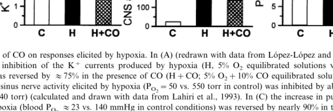
Fig. 4. Effects of CO on responses elicited by hypoxia. In (A) (redrawn with data from Lo´pez-Lo´pez and Gonzalez, 1992) it can be seen that the inhibition of the K+ currents produced by hypoxia (H, 5% O
2 equilibrated solutions vs. C, 20% O2 in control
conditions) was reversed by:75% in the presence of CO (H+CO; 5% O2+10% CO equilibrated solutions). In (B) the increase
in the carotid sinus nerve activity elicited by hypoxia (PO2=50 vs. 550 torr in control) was inhibited by about 70% by CO (O2=50
torr+CO=140 torr) (calculated and drawn with data from Lahiri et al., 1993). In (C) the increase in pulmonary arterial pressure elicited by hypoxia (blood PO2:23 vs. 140 mmHg in control conditions) was reversed by nearly 90% in the presence of CO (blood
PO2:23 mmHg and PCO:56 mmHg) (redrawn with data from Tamayo et al., 1997).
membrane bound subunit) exhibit a normal re-sponse to hypoxia measured as Ca2+ transients in
chemoreceptor cells or as ventilatory response (Roy et al., 2000).
The situation in the pulmonary circulation is comparable. NADPH oxidase is present in PASMC and its inhibition with DPI fully blocked the hypoxic vasoconstriction (Thomas et al., 1991; Grimminger et al., 1995; Thompson et al., 1998) and decreased lucigenin chemoluminiscence (Archer et al., 1999). Contrary to the hypothesis that hypoxia inhibits NADPH oxidase (see Weir and Archer, 1995), these findings are compatible with the opposite notion, i.e. NADPH oxidase, if involved at all on hypoxic vasoconstriction, should be activated by hypoxia (and not inhib-ited) so that its inhibition by DPI abolishes the vasoconstrictor response. Consistent with this idea, Marshall et al. (1996) observed that hypoxia does indeed increase the activity of NADPH ox-idase. However, NADPH oxidase does not appear to be involved at all on the genesis of hypoxic pulmonary vasoconstriction, because knock out animals for the neutrophil oxidase exhibited a normal hypoxic pulmonary vasoconstriction (Archer et al., 1999, 2000). Even further, the data of Archer et al. (1993, 1999, 2000) appear to exclude ROS as mediators of the hypoxic pul-monary vasoconstriction. Thus, DPI and rotenone decreased lucigenin chemoluminiscence
and, as just stated, DPI inhibits the hypoxic constriction, but rotenone per se produced a vaso-constrictor response, which was greater in null animals for NADPH oxidase. Both agents, DPI and rotenone, would inhibit complex I at mito-chondrial level and reduce ROS production, and rotenone produces pulmonary vasoconstriction while DPI suppresses it. To further complicate the interpretations it has been found that in isolated PASMC DPI per se increased Ca2+
i and elicited a
contractile response in an ample range of concen-trations, and in myocytes contracted by hypoxia DPI at 1 – 5 mm inhibited the contraction but did not reduce the Ca2+
i (Zhang et al., 1997),
indicat-ing that DPI has side-effects down in the trans-duction cascade probably unrelated to NADPH oxidase inhibition (as appears to be the case in the CB, see above). Previously, it had been shown that DPI per se inhibited K+ and Ca2+ currents
in a non-specific manner in PASMC (Weir et al., 1994) and carotid body chemoreceptor cells (Wy-att et al., 1994).
would decrease the activity of the enzyme and ROS levels would decrease, this decrease being a critical link between low PO
2and the transcription
of the EPO gene. However, Gleadle et al. (1995) showed that DPI per se applied in a range of concentrations from 0.3 to 5 mM did not activate EPO production in normoxic hepatoma cells; on the contrary, DPI inhibited in a dose-dependent manner hypoxia-induced, but not CoCl2-induced,
EPO transcription. Obviously these findings argue against a role of NADPH oxidase inhibition in the genesis of the hypoxic response, otherwise DPI in normoxic conditions should mimic hy-poxia and in hypoxic conditions should be ineffec-tive or potentiate the effect of hypoxia. Regarding the lack of effect of DPI on CoCl2-induced EPO transcription the interpretation is difficult because as already stated CoCb decreases ROS production according to Fandrey et al. (1997) and increases the production according to Chandel et al. (1998). The latter authors found that the production of ROS induced by CoCl2 is of unknown, but
ex-tramitochondrial origin, and in any case insensi-tive to DPI. Finally, the participation of NADPH was questioned by Ratcliffe et al. (1995) arguing that EPO deficiency was not a trait in granuloma-tous patients which lack one or other subunit of NADPH oxidase and therefore the functional en-zyme, and in fact, it was demonstrated by Wenger et al. (1996) that B-cell lines derived from normal and from granulomatous patients expressed vas-cular endothelial growth factor under hypoxic conditions and under CoCl2 treatment in a very similar manner (the upregulation of vascular en-dothelial growth factor and EPO genes expression is considered to be under similar control); even further, reconstitution of the NADPH oxidase activity in granulomatous cells by transfecting the absent genes did not alter the sensitivity to hy-poxia suggesting that H2O2 levels are not critical
to control the HIF-1a-dependent expression of vascular endothelial growth factor.
However, in keeping with the NADPH model, Fandrey et al. (1997) reported that desferrioxam-ine (an iron chelator which also increases EPO gene expression) and CoCl2decreased the produc-tion of ROS (as determined by dihydrorhodamine 123 fluorescence) as hypoxia does, therefore, they
proposed that this diminution of ROS was the common factor for the three situations to increase EPO production. Even further, Fandrey et al. (1994) have previously found that addition of H2O2 to the incubation medium (at concentra-tions greater than 100 mM) reduced or abolished the production of EPO induced by hypoxia, and in the 1997 article (Fandrey et al., 1997) they reported that desferrioxamine antagonized the ac-tion of H2O2concluding that H2O2acted via OH
generated by a Fenton reaction. These and other observations are in the most recent formulation of the model linking NADPH oxidase and hypoxic activation of gene expression. In this model the inhibition of the oxidase in hypoxia leads to a decrease in H2O2 and as a consequence to a decrease in OH in perinuclear areas of the cell which ultimately would cause a change from oxi-dized to reduced state of the transcription factors triggering the transcription of hypoxia regulated genes (Kietzmann et al., 2000). As already stated, it is hard for us to imagine a localized production of OH. Overall, however, this notion of a dimin-ished rate of ROS production during hypoxia would fit the observations of Huang et al. (1996) who showed that hypoxia inducible factor-1a (HIF-1a; this factor would be the primary target or the primary effector of hypoxia in EPO-pro-ducing cells; see Fig. 1) which in normoxic condi-tions is degraded in the proteosome, is stabilized during hypoxia by a reduction process which al-lows it to reach the nucleus and to bind to the EPO gene enhancer to activate the rate of tran-scription. In fact Huang et al. (1996) reported that pre-exposure of hepatoma cells to H2O2prevented
Table 1
Lack of positive correlation between ROS levels and hypoxic responses
Carotid body EPO-cells Lung circulation ROS levels
Stimulates
Hypoxia Decrease/increase Stimulates Vasoconstriction
Rotenone Decrease Stimulates Inhibits hypoxic response Vasoconstriction DPI Decrease Stimulates 0 in normoxia inhibits hypoxic response Inhibits hypoxic response
Stimulates 0 in normoxia, 0 in hypoxia
Increase/decrease Vasoconstriction
Antimycin A
Cyanide (Azide) Increase Stimulates 0 in normoxia, 0 in hypoxia Vasoconstriction
chemoreceptors) whose cells possess O2 -modu-lated K+ channels and are positive to NADPH
oxidase, DPI mimics hypoxia on K+ currents
(Youngson et al., 1993; Fu et al., 1999) and the response to hypoxia of the cells is lost in knock outs for NADPH oxidase (Fu et al., 2000). These finding would suggest that NADPH oxidase is linked to O2-sensing in these cells. Moreover, very
recently several isoforms of NADPH oxidase have been cloned (e.g. Zhu et al., 1999; Geiszt et al., 2000) one of them named renox with a location in the renal cortex compatible with a possible func-tion in the control of EPO-gene (Geiszt et al., 2000).
The proponents of the theory that ROS in-crease during hypoxia (Chandel et al., 1998, 2000; Duranteau et al., 1998; Chandel and Schumacker, 1999, 2000) state that it takes place at the level of mitochondria (Fig. 2). Superoxide itself or H2O2
(product of O2− dismutation) would reach the
cytosol to stabilize HIF-la by promoting its oxi-dized form (Chandel et al., 2000; just the opposite to the claim given by Huang et al., 1996; see above). We have already speculated that in cells involved in homeostatic loops we should probably expect an increase in the production of ROS due to increased flow of electrons in the respiratory chain. Schumacker and co-workers propose that the increase in ROS they found would be due to a certain degree to the reduced state of the entire respiratory chain produced by the inadequate functioning of cytochrome oxidase imposed by hypoxia; this extra reduced state of the entire respiratory chain (in comparison to normoxia) would favor the reduced state of quinones and the leakage of electrons to form O2−. We would tend
to believe that in cells involved in oxygen
homeo-static loops this would occur at more extreme hypoxias due to the high affinity of cytochrome oxidase on the one hand (see Gnaiger et al., 2000) and due to the high blood flow of the organs wherein reside the cells forming part of the O2
homeostatic loops, on the other.
A different question is whether increased ROS levels could be unequivocally considered media-tors of the hypoxic responses. The answer is no (Table 1). Thus, hypoxia, antimycin A and azide (and therefore cyanide) increase ROS levels (Duranteau et al., 1998) and only hypoxia in-creases the rate of transcription of EPO gene (Tan and Ratcliffe, 1991; Pugh et al., 1991; Firth et al., 1994). Rotenone and DPI, which decrease the levels of ROS, inhibit the hypoxic increase in EPO-gene transcription, but cyanide and an-timycin A did not. In conjunction these data indicate that ROS levels per se are not related to EPO gene transcription, and that the energy metabolism of the cells is similarly unrelated to EPO gene transcription (during hypoxia, the three poisons, rotenone, cyanide and antimycin A should dramatically decrease ATP/ADP+
rotenone and antimycin A which decreased luci-genin chemiluminescence increased pulmonary artery pressure and cyanide which increased chemiluminescence also increased pulmonary arte-rial pressure (Archer et al., 1993); obviously, ac-cording to Chandel and coworkers only rotenone should produce a decrease in ROS the rest of the situations would increase them, and yet all experi-mental maneuvers increased pulmonary artery pressure.
3.1.3. Third criterion. The primary target for the increased or decreased le6els of ROS should be identified.The change produced in the target by the signal (change in ROS le6els) should be defined. The mechanisms of target/effector coupling should also be defined
We do not know the identity of the primary ROS targets in the hypoxic transduction cascade. Therefore, we will equate the primary target of ROS with the first known or suspected effector on the cells which are K+channels in the case of CB
chemoreceptor and PASMC and HIF-1a in the case of EPO-producing cells (see Fig. 1). The recognition that release of catecholamines induced by hypoxia in the CB chemoreceptor cells, as well as that induced by high extracellular K+, was
inhibited by dihydropyridine antagonists ofL-type Ca2+ channels (Obeso, 1984) suggested to us that
hypoxia must depolarize chemoreceptor cells. This observation prompted us to develop a primary culture of rabbit CB dissociated cells to study electrophysiologically chemoreceptor cells. In agreement with previous data of our laboratory (Obeso, 1984; Almaraz et al., 1986; Rocher et al., 1988), using the patch clamp technique in the isolated preparation of rabbit chemoreceptor cells we demonstrated that these cells were excitable cells with Na+, K+ and Ca2+ voltage-dependent
channels, and more importantly, that a transient component of the outward directed K+ current
was reversibly inhibited by hypoxia (Lo´pez-Barneo et al., 1988; Lo´pez-Lo´pez et al., 1989); recent studies have identified the K+ channel
supporting this transient (O2-sensitive current as a putative member of the Kv.4 family (Pe´rez-Garcı´a et al., 2000). Following this initial finding, the presence of O2-modulated K+ currents in
chemoreceptor cells has been recognized in all the species studied. In the rat chemoreceptor cells two types of O2-modulated K+ channels have been
described; a Ca2+-dependent K+ current (Peers,
1990) carried out by the maxi-K+ (Wyatt and
Peers, 1995; Lo´pez-Lo´pez et al., 1997) and a leak voltage-independent current (Buckler, 1997, 1999), recently identified as TASK-1 channel (Buckler et al., 2000). In the cat CB chemorecep-tor cells, K+ current sensitive to hypoxia is a
delayed rectifier-like current (Chou and Shirahata 1996). In PASMC, the original description of Demodulated KT channels was made on freshly dispersed canine fibers, and it was found that the KT current sensitive to hypoxia was Ca2+
-depen-dent (Post et al., 1992). Soon after, Yuan et al. (1993) found that in primary cultures of rat PASMC the current sensitive to hypoxia was of the Ca2+ independent delayed rectifier type
(Yuan et al., 1993). In more recent studies (e.g. Yuan et al. 1998; Archer et al., 1998; Patel et al., 1997; Osipenko et al., 2000) several Kv channels have been identified in PASMC and it would appear that those channels most likely to carry the O2-sensitive current would be the homomeric
Kv1.2, Kv1.5, the Kv2.1, the Kv3.1 and the het-eromeric Kv1.2/Kv1.5 and Kv2.1/Kv9.3 (see Pe´rez-Garcı´a and Lo´pez-Lo´pez, 2000; Archer et al., 2000).
In accord with criterion 3, experimental find-ings must clarify to what extent, if any, the O2 modulated channels are sensitive to redox changes, and if so, to determine if those redox changes are likely to be produced by hypoxia. Fast inactivating channels supporting macro-scopic currents like those sensitive to hypoxia in rabbit chemoreceptor cells are susceptible to mod-ulation by redox agents including H2O2,
K+ channels decreases in isolated patches on
lowering PO
2 (Ganfornina and Lo´pez-Barneo,
1991; Pe´rez-Garcı´a et al., 1999) in a reversible manner, and to our knowledge, O2 at the ranges of pressures we are dealing with (10 – 150 mmHg), cannot oxidize or reduce thiol or other functional groups in the proteins (except for some autoxida-tion of tyrosine and tryptophan; Halliwell and Gutteridge, 1999). In addition, it is necessary to postulate that a redox system capable of transfer-ring the electrons back and forth to the channel protein is present in the isolated membrane patches (see the above comments on the article by Eu et al., 2000). Furthermore, CO is able to prevent or to reverse the action of hypoxia on K+
currents (i.e. CO behaves as O2; Lo´pez-Lo´pez and Gonzalez, 1992) and yet there are no cell mecha-nisms by which the highly unreactive CO at the concentrations used can substitute O2 to increase the rate of ROS production. Maxi-K+ channels,
which are O2-modulated in rat chemoreceptor
cells, are also sensitive to modulation by redox reagents like those mentioned above for the tran-sient K+ channels, but the direction of those
modulations are not always the expected ones (Thuringer and Findlay, 1997; Liu et al., 1999; Soh et al., 2001; Riesco et al., 2001; but see Tang et al., 2001). Therefore, the notion of hypoxia acting through reduction of sulfhydryl or other chemical groups of the channel proteins suscepti-ble to redox modulation seems untenasuscepti-ble, espe-cially if we consider that CO is able to reverse the effect of hypoxia in isolated membrane patches of rat chemoreceptor cells increasing the opening probability of maxi-K+ when it is decreased by
hypoxia (Riesco et al., 2001). In this regard, it should be mentioned that in a recent study from our laboratory it was possible to clearly distin-guish between the action of reducing agents and the action of low PO2: we observed that
dithio-threitol was able to modify some kinetic proper-ties of transfected shaker and shaker/b1.2 and Kv4.2/b1.2 channels, while low PO2 only
modu-lated the latter channels (Pe´rez-Garcı´a et al., 1999). In other words, it is possible that reducing agents alter the conducting properties of ion chan-nels, but that does not necessarily mean that hypoxia alters those properties, and if hypoxia
alters them, it may be that the direction of the alteration could be different. The same type of arguments can be made regarding the regulation of K+ channels by redox agents versus hypoxic
regulation in PASMC: in general the modification of the currents by reducing agents corresponds to that of hypoxia (see Archer et al., 2000) but the sensitivity to hypoxia of Kv3.1 in isolated patches (Osipenko et al., 2000)1 brings the same
consider-ations just mentioned in the case of the CB chemoreceptor cells.
The identification of HIF-1a as the primary effector in EPO producing cells occurred as a result of coordinated studies of several groups. Transgenic mice carrying a 4 kb fragment of the human erythropoietin gene which contained the 5 exons and 4 introns of the intact gene, a 0.4 kb of the 5% end and a 0.7 kb of the 3% end exhibited hypoxic induction of the human gene as bleeding resulted in increased levels of human EPO mRNA in the liver of the mice; in addition, several trans-genic mice derived from different independent cell lines were polycythemic (see Semenza 1994). The findings indicated that the EPO-gene fragment contained hypoxia regulated elements. The de-scription by Golberg and co-workers of two hu-man hepatoma cell lines exhibiting hypoxia regulated expression of EPO (Goldberg et al., 1987, 1988) allowed an ample series of trasfection experiments with reporter genes which fed to the discovery of the 3’ enhancer region with the abil-ity to bind proteins (e.g. Pugh et al., 1991). A search aimed at identifying the protein factors binding to the 3% enhancer culminated with the
1In our whole-cell patch recordings, CO was used by gas
bubbling of the perfusing solutions. The gas mixtures con-tained: 20% O2/80% N2, in control conditions; 5% O2/95% N2,
in hypoxic conditions, and; 5% O2/10% CO/85% N2, during
CO application. This concentration of CO prevented or re-versed the effect of 5% O2by:70%. With this O2/CO ratio,
identification of HIF-1 (Wang and Semenza, 1993a,b) as a factor induced by hypoxia, capable of binding to the 3’ enhancer region of EPO and other hypoxia-regulated genes and capable of causing an increase in their rate of transcription. Thereafter, it was demonstrated that in fact HIF-1 was a heterodimer composed of HIF-HIF-1a, whose levels are regulated by low PO2, and by the
consti-tutively expressed HIF-1b [also known as ARNT (aryl hydrocarbon receptor nuclear translocator), as AhR (aryl hydrocarbon receptor) and as dioxin receptor], the dimerization was shown to be neces-sary, not for the translocation of both compo-nents to the nucleus, but for the stable association of HIF-1 within the nuclear compartment (see Gassmann et al., 2000). Additional studies demonstrated that HIF-1a as well as HIF-1b mRNA levels, transcription and translation rates were not modified by hypoxia. Since HIF-1a protein increased during hypoxia it implied that in normoxia the protein is degraded and that hy-poxia prevents the degradation (Huang et al., 1996).
Therefore, the question we should make at this point is how the change in ROS levels produces such stabilization of HIF-1a to prevent its degra-dation. Huang et al. (1996) showed that the levels of HIF-1a, which are increased by hypoxia, de-creased soon if intact cells were re-exposed to normoxia (21% O2), but exposure of cell extracts to the same normoxic levels of O2 did not alter the binding of HIF-1a to DNA. In other words, for high PO
2to destabilize HIF-1athe integrity of
the cells is necessary. In additional experiments it was shown that H2O2 (0.1 – 1.0 mM) resulted in a
dose-dependent decrease in the EPO mRNA ex-pression induced by hypoxia which runs parallel to a decrease in HIF-1abinding to DNA and to a decrease in HIF-1a protein, without modification of HIF-1a mRNA levels or translation rate. It was also observed that extracts from hypoxic cells treated with H2O2 showed a normally high
bind-ing capacity to DNA. Those findbind-ings appear to indicate that the binding of HIF-1a to DNA is controlled primarily at the level of HIF-1a protein. In addition, Huang et al. (1996) also studied some of the modifications of HIF-1a which might alter its binding to DNA. They
found that oxidizing sulfhydryl reagents added to cell extracts inhibited the binding in a dose-depen-dent manner, this inhibition was reversible with reducing thiol reagents except when sulfhydryl alkylating agents were used. In addition, in a cotransfection experiment using a construct ex-pressing thioredoxin under the control of the con-stitutive cytomegaloviral promoter and enhancer, along with another construct expressing luciferase under the control of the SV40 promoter and EPO-enhancer, they observed that overexpression of thioredoxin resulted in a potentiation of the expression of luciferase both in normoxic and hypoxic conditions, such potentiation appeared to be under the control of the HIF-1 binding because transfection with a mutated EPO enhancer, which was unable to bind HIF-1a, caused the loss of the response. As a whole the results would indicate that H2O2, like normoxia, produces modifications in the HIF-1a molecule which force it to be directed to degradation, but which does not im-pede its binding to DNA. Alternatively, hypoxia promotes changes in HIF-1a, which prevent its degradation, leading to accumulation of the tran-scription factor; the changes induced by hypoxia appear to be redox dependent because they can be mimicked by thioredoxin and counteracted by normoxia and H2O2; a similar conclusion was
attained by Srinivas et al. (1999). An alternative interpretation to the findings of Huang et al., is that hypoxia (or the decrease of ROS) or the overexpression of thioredoxin produced changes in the degrading enzymes of the proteosoma which make them incapable of degrading a nor-mal (or reduced) HIF 1a.
However, in a series of experiments Chandel et al. (2000) found essentially the opposite: hypoxia and CoCl2, but not desferriexamine, increased
also the expression of HIF-1a. Neither maneuver decreased the expression of the reporter gene or decreased the levels of HIF-1a promoted by des-ferrioxamine. Addition of H2O2andtert -butylhy-droperoxide to normoxic cells promoted an increase of HIF-la protein as well as an increased expression of the reporter gene; these maneuvers were ineffective in cells overexpressing catalase. A phosphatase inhibitor and an inhibitor of phos-phatidyl inositol 3-kinase abolished the stabiliza-tion of HIF-1a induced by hypoxia, CoCl2 and
desferrioxamine and a proteasome inhibitor was able to reverse those effects; because treatment of the cells with H2O2 was unable to prevent the
effects of the phosphatase and kinase inhibitors, the authors concluded that ROS (including H2O2) act to stabilize HIF-1a at a step prior to the action of the phosphatases and kinases. Desfer-rioxamine would produce the stabilization of HIF-1aby a mechanism independent of ROS and would also act at a step prior to the phosphatases and kinases. Curiously, antimycin A, which in-creased ROS, did not induce the expression of the reporter gene or accumulation of HIF-1a. Chan-del et al. (2000) state that it is because this mito-chondrial inhibitor causes a smaller increase in ROS levels than hypoxia. However, it is difficult to understand how hypoxia can produce a more reduced state at the quinone pool than antimycin (see Fig. 2). In addition, antimycin A was applied in normoxia implying that the concentrations of O2 to accept the electrons from semiquinone are much higher than in hypoxia (20% O2 vs. 1.5% in their experiments) and, therefore, the formation of O2−should be facilitated (Chance et al., 1979).
The controversy is obvious: to Huang et al. (1996) it is the decrease of ROS levels which causes the stabilization of HIF-1aand to Chandel et al. (2000) it is the increase of ROS levels that does the same. Therefore, and in keeping with the statement of the third criterion, we do not know if we should be looking for a reduction or an oxida-tion of HIF-1aas responsible for its stabilization. An additional aspect that we feel forced to restate at this point (which is seldom taken into account; see above) is that in the proposed signalling by ROS the subcellular localization of the ROS sig-nal at the target place should be taken into
con-sideration, otherwise a ‘true’ signaling would not exist, Finally, we agree with Halliwell and Gutte-ridge (2000) when they state regarding signaling by ROS: ‘‘one must be wary: cells in culture are usually hyperoxic and they lack many of the antioxidants they use in vivo…Cells adapt to live in culture. It is not impossible that, with tumor cell lines in particular, adaptations to favor growth may occur by using radicals to trigger pathways that are triggered by other means in vivo’’.
3.1.4. Fourth criterion. Definition of the cellular responses elicited by the effector acti6ated by the changes in ROS le6els
At this point in the article it is obvious that we can define the cellular responses elicited by the stimulus, i.e. hypoxia. However, we have serious difficulties in defining the cellular effects produced by the ROS level changes occurring during hy-poxia because we do not know what those changes are. And even if we knew the changes in ROS levels produced during hypoxia we do not know if they are cause or consequence of activa-tion of the effector and cellular responses.
Hypoxia in CB chemoreceptor cells and PASMC reduces the opening probability of the O2regulated K+channels (see criterion 3) leading
to cell depolarization, activation of voltage oper-ated Ca2+ channels and release of
neurotransmit-ters in chemoreceptor cells or contraction in smooth muscle cells (see Fig. 1). In criterion 3 we have already indicated that sulfhydryl reagents cause changes in the opening probability or am-plitude of the currents that can be compatible with a decrease or an increase in ROS levels. Referring specifically to ROS, there are many studies in which H2O2 has been applied directly
and its effects on several types of K+ channels
established. For example, Vega-Saenz de Miera and Rudy (1992) showed that H2O2 augmented
the current carried by transient type K+ channels
(like those O2-sensitive channels in rabbit
chemoreceptor cells) without affecting other K+
-butylhydroper-oxide activate maxi-K+ (Hayabuchi et al. 1998;
Shin et al., 2000; Guo et al., 2000; Barlow et al., 2000). All these data would favor the notion that hypoxia decreases ROS, and thereby hypoxia would decrease the activity of K+ channels and
depolarization would occur. However, there are studies in which it has been found that H2O2
inhibits K+ channels. That is the case for
maxi-K+ (DiChiara and Reinhart, 1997; Tang et al.,
2001). These studies would be in agreement with the notion that hypoxia increases ROS levels and thereby causes inhibition of K+ channels, cell
depolarization and increased cell activity.
When we consider the effects of H2O2 on the
overall response of the system, the study of Os-anai et al. (1997) in the intact CB is very illustra-tive as they concluded that maneuvers aimed at altering endogenous H2O2, including exogenous H2O2application, did not provide evidence for its supposedly critical role in O2 chemoreception. On
the contrary, in the pulmonary circulation tert -butylhydroperoxide, H2O2 and ROS generating
systems (i.e. perfusion of the lungs with xanthine plus xanthine oxidase) clearly inhibited hypoxic pulmonary vasoconstriction (Archer et al., 1986; Mohazzab and Wolin, 2000). In the case of EPO-producing cells we have commented in previous paragraphs on the apparently dual actions of H2O2since on the one hand it inhibited
EPO-pro-duction induced by hypoxia (Fandrey et al., 1994) and on the other hand it augmented the stabiliza-tion of HIF-1a and promoted the expression of a reporter gene under the control of the EPO en-hancer (Chandel et al., 2000). It should also be mentioned that Ueno et al. (1988) working with renal carcinoma EPO-producing cells and Ndele et al. (1996)) working with HEP 3B cells found that H2O2 and ROS generating systems increased
EPO production while ROS scavengers have the opposite effects.
3.1.5. Fifth criterion. The mechanism(s)
responsible for the inacti6ation of the ROS signal should be defined
Obviously we should consider here both possi-bilities: that the signal is a decrease and that the signal is an increase in ROS levels. In the first case, if a decrease in ROS levels is considered to
be the result of a decrease in the activity of NADPH oxidase, the signal would disappear, i.e. normal ROS levels will be recovered, when PO2
3.1.6. Sixth criterion. Manipulation of ROS le6els by alteration of glutathione le6els or by the direct action of ROS sca6engers should produce the predicted change in the cell response
O6erexpression and elimination of the enzymes sca6enging ROS should produce the predicted change in the cell responses.
The interpretation of the data resulting from the manipulation of ROS and/or glutathione levels is not always straightforward due to the multiple targets of ROS. For example, the activation or mimicking of glutathione peroxidase (e.g. by ad-ministration of ebselen) would decrease the GSH/
GSSG ratio, just as a situation in which there is an increase in the rate of ROS production, but con-trary to that situation ebselen will produce a decrease in the levels of H2O2. Since the decrease in the GSH/GSSG ratio would tend to increase the opening probability of K+ channels, and
thereby to cause membrane hyperpolarization (and vasodilation, in the case of pulmonary circu-lation), and the decrease of H2O2 would tend to
reduce the activity of guanylate cyclase, and thereby to inhibit Ca+ dependent K+ channels
(and to produce vasoconstriction, in the case of pulmonary circulation), the overall effect could be very complex and difficult to define (see Wolin et al., 1999; Mohazzab and Wolin, 2000). In addi-tion, and at least in the case of the lung, ROS can modify the metabolism of arachidonic acid-derived metabolism in such a manner that an increase in ROS levels would augment the produc-tion of vasoconstrictor metabolites (Seeger et al., 1986; Sanderud et al., 1993; see Wolin et al., 1999). Experiments in the CB directly focused on this last criterion have been performed in our labora-tory very recently. We have measured GSH and GSSG levels in calf CB in normoxia, hypoxia and after treatment of the organs with N -acetylcys-teine, a precursor of glutathione and direct scav-enger of ROS and measured the release of catecholamines from chemoreceptor cells in those experimental situations (Obeso et al., 2000b). N -acetylcysteine would mimic a situation in which there is a decreased production of ROS and would oppose a situation of increased rate of ROS production. In other words,N-acetylcysteine would mimic hypoxia, if we accept that hypoxia
decreases ROS levels, and would oppose hypoxia if we accept that it augments ROS levels. We found that hypoxia (PO
2:46 mmHg) did not
alter the absolute levels or the GSH/GSSG quo-tients. N-acetylcysteine increased absolute glu-tathione levels by primarily increasing GSH; therefore, it increased the GSH/GSSG ratio. Yet
N-acetylcysteine did not modify the basal nor-moxic or hypoxia-induced release of cate-cholamines suggesting that ROS are not critical in setting the hypoxic response.
In the case of the pulmonary circulation, Archer et al. (1986) showed that diamide (an agent that reacts with GSH and oxidizes it to GSSG) reduced the pressor response to hypoxia. Diamide would mimic a situation in which there is an increased production of ROS: it will cause a decrease in GSH/GSSG ratio and therefore the cellular power for scavenging of ROS leading to an increase in ROS levels. Both the decrease in GSH/GSSG ratio and the increase in ROS levels would tend to oppose the vasoconstriction pro-voked by hypoxia (see above). In agreement with the expected actions, pretreatment of the animals with N-acetylcysteine prevented the effects of di-amide (Archer et al., 1986). In isolated perfused lungs, suplementation of the superfusates with catalase and superoxide dismutase neutralized the inhibition on hypoxic pulmonary vasoconstriction produced by ROS generating systems (xanthine/
significance of ROS levels on hypoxic pulmonary vasoconstriction.
The data related to this criterion in EPO-pro-ducing cells are likewise inconclusive. In dis-cussing the stabilization of HIF-1a (criterion 3) we have focused on some discrepancies that might relate to this criterion. For example, we have mentioned that according to Huang et al. (1996) overexpression of thioredoxin potentiated the ex-pression of a reporter gene under the control of EPO enhancer implying stabilization of HIF-1a. On the other hand, Chandel et al. (2000) reported that overexpression of catalase prevented stabi-lization of HIF-1a. A similar controversy exists regarding H2O2. Thus Fandrey et al. (1994)
re-ported that H2O2inhibited the production of EPO induced by hypoxia and that catalase reversed the inhibition produced by H2O2. On the contrary, Ueno et al. (1988), Ndele et al. (1996) (see also Chandel et al., 2000) reported that catalase inhib-ited the EPO production stimulated by H2O2and
by ROS-generating systems glucose/glucose ox-idase and xanthine/xanthine oxidase.
4. Conclusion
With the available information we do not know if hypoxia increases or decreases ROS levels, and therefore we do not know if the putative role of ROS, i.e. to inhibit K+ channels in the CB
chemoreceptor cells and in PASMC and to stabi-lize HIF-1a in EPO-producing cells, is the result of the reduction or the oxidation of the K+
channel proteins or HIF-1a. The uncertainties remain when we consider the effects of agents that increase or decrease ROS levels on the responses of CB chemoreceptor cells, PASMC and EPO-producing cells. There are several mitochondrial poisons, which have opposite effects on the rate of ROS production and levels, and yet all activate chemoreceptor cells and PASMC in normoxia and potentiate their hypoxic response. Similarly, there are mitochondrial agents that can either decrease or increase ROS levels, yet they fail to alter normoxic EPO production, implying that neither the decrease nor the increase in ROS levels mimic hypoxia. The panorama is not clarified by
the use of agents or experimental maneuvers that alter the rate of ROS scavenging. There are re-ports indicating that ROS scavenging enzymes or drugs tend to mimic hypoxia and reports indicat-ing the opposite, both, in PASMC and in EPO-producing cells. In the CB chemoreceptor cells the scavenging of ROS did not alter the normoxic output of the cells nor their hypoxic response. In sum, the available information on O2-sensing/
hy-poxic transduction does not support an unequivo-cal role for ROS in those cellular processes. The possibility exists that many of the contradictory findings, which have been obtained in tumor cell lines, represent peculiarities of particular clones used in different laboratories. In any case, it should always be kept in mind that the metabolic properties of tumor cells might differ substantially from the parental normal cells. In designing ex-periments, it should also be remembered that maneuvers aimed at manipulating ROS can do more than simply alter ROS levels: special care should be taken in selecting the potentially pleiotropic ROS scavengers and ROS mimetics.
Acknowledgements
Supported by Spanish DGICYT Grant PB97/
0400. Thanks to Professor Benito Herreros for critical reading.
References
Acker, H., 1994a. Cellular oxygen sensors. Ann. New York Acad. Sci. 718, 3 – 10.
Acker, H., 1994b. Mechanisms and meaning of cellular oxygen sensing in the organism. Respir. Physiol. 95, 1 – 10. Acker, H., Xue, D., 1995. Mechanisms of O2 sensing in the
carotid body in comparison with other O2-sensing cells.
News Physiol. Sci. 10, 211 – 215.
Almaraz, L., Gonzalez, C., Obeso, A., 1986. Effects of high potassium on the release of H-dopamine from the cat carotid body in vitro. J. Physiol. London 379, 293 – 307. Anichkov, S.V., Belen’kii, M.L., 1963. Pharamacology of the
Carotid Body Chemoreceptors. MacMillan, New York, USA.