Association of plasma visfatin with hepatic and systemic
inflammation in nonalcoholic fatty liver disease
Halil Genc,* Teoman Dogru,* Muammer Kara,* Serkan Tapan,** Cemal Nuri Ercin,* Cengizhan Acikel,*** Yildirim Karslioglu,**** Sait Bagci*
* Department of Gastroenterology. ** Department of Medical Biochemistry. *** Department of Epidemiology. **** Department of Pathology. Gulhane School of Medicine, Ankara, Turkey.
ABSTRACT
Background. Visfatin is a proinflammatory and insulin-mimetic adipokine contributing to whole body glucose and lipid metabolism. Studies to date are conflicting regarding the relationship between visfatin and non-alcoholic fatty liver disease (NAFLD). The aim of the present study was to evaluate the relationship of circulating visfatin with NAFLD. Material and methods. The study included 114 NAFLD patients and 60 healthy non-diabetic controls. Plasma visfatin, adiponectin, tumor necrosis factor alpha (TNF-α) and interleukin-6 (IL-6) levels were measured by ELISA. High sensitive C-reactive protein (hsCRP) levels were measured by immunoturbidimetric fixed rate method. Insulin sensitivity determined by homeostasis model assessment (HOMA-IR) index. Results. TNF-α, IL-6 and hsCRP levels were higher and, Adiponectin levels were lower in NAFLD group when compared to healthy controls (p < 0.001, for all). However, no difference was found regarding to visfatin levels between two groups. Different histologic subgroups of NAFLD had a significantly higher TNF-α, IL-6 and hsCRP, and lower adiponectin levels than those with controls (p < 0.001, for all). On the other hand, no statistically significant difference was found regarding to visfatin levels among different histologic groups. Visfatin was found to be negatively correlated with TNF-α (r = -0.236, p = 0.011) in NAFLD group. However, no association was found between visfatin and histological findings. Conclusion. Our findings show that plasma visfatin levels are not altered in the early stages of NAFLD. However, it is inversely associated with TNF-α. These findings suggest a role for visfatin in protection against liver injury in this widespread disease.
Key words. Adiponectin. Tumor necrosis factor alpha. Interleukin-6.
Correspondence and reprint request: Halil Genc, M.D.
Department of Gastroenterology, Gulhane School of Medicine, Tevfik Saglam Caddesi, 06018, Etlik, Ankara, Turkey Ph.: +90 312 3044052. Fax: +90 312 3044000, E-mail: [email protected]
Manuscript received: December 01, 2012. Manuscript accepted: January 23, 2013.
INTRODUCTION
Non-alcoholic fatty liver disease (NAFLD) is a cli-nicopathological entity which is characterized by the presence of fat droplets in the hepatocytes without alcohol consumption, representing a spectrum of he-patic injuries, ranging from pure fatty infiltration (simple steatosis: SS) to inflammation (non-alcoholic steatohepatitis: NASH), fibrosis, and cirrhosis.1 It is also the hepatic manifestation of the metabolic
syndrome (MetS), with insulin resistance as the main pathogenetic mechanism.2,3 Adipose tissue acts as a store of energy and an active endocrine organ. Adipokines (adipocytokines) –agents secreted prima-rily by adipocytes–modulate lipid and glucose meta-bolism and insulin sensitivity.4 In addition to their well established role in controlling adipose tissue physiology, adipokines have been shown to be invol-ved in regulation of the inflammatory response, an-giogenesis and fibrogenesis. As a result, adipokines together with insulin resistance seem to play a dis-tinct role in the pathogenesis of NAFLD.5,6
maturation and inhibits neutrophil apoptosis. Vis-fatin enhances activation of leukocytes, synthesis of adhesion molecules and production of proinflamma-tory cytokines. Visfatin expression is upregulated in a variety of acute and chronic inflammatory diseases including rheumatoid arthritis, sepsis, acute lung injury, inflammatory bowel disease and psoriasis.7-9
The relationships between visfatin and NAFLD have recently been examined.10,11,12 However, the available information on visfatin is still too little and controversial leaving our understanding of this adipokine and its physiological and pathophysiologic roles in NAFLD in its infancy. The aim of the present study was to evaluate the relationships of plasma visfatin with both pro- and anti-inflamma-tory adipocytokines and liver histology in this clinically relevant condition.
MATERIAL AND METHODS
Study population
From January 2008 to December 2011, we enrolled 114 consecutive, male patients with biopsy-proven NAFLD. All patients were recruited in the outpatient clinic and were on an unrestricted dieta-ry regimen. Body weight was stable during the 3 months preceding the study. No participants had any clinical evidence of cancer, cirrhosis, overt nephropathy or cardiovascular disease. Type 2 diabetes (T2D) was excluded by oral glucose tolerance test in NAFLD group according to the American Diabetes Association classification.13 Subjects with hypertension were also excluded. None of them was taking any medications known to affect circulating visfatin and inflammatory biomarkers such as lipid-lowering medications, metformin or thiazoli-dinediones.
NAFLD diagnosis was based on chronic elevation of transaminases (1.5 times the upper normal value for 3 months or longer), absence of hepatitis B and C virus markers, absence of autoantibodies indicative of autoimmune hepatitis or celiac disease, absent or negligible alcohol consumption (< 20 g/day), and bright liver at ultrasound scanning. In all patients, diagnosis was confirmed by liver biopsy.
The control group, recruited from hospital staff members and relatives, consisted of 60 apparently healthy male volunteers with normal liver ultraso-nography and normal liver function tests. Before in-clusion, all the study and control subjects underwent careful physical examination and detailed
laboratory investigations to exclude any condition that may interfere with glucose tolerance and inflam-mation. The study was approved by the local ethics committee of Gulhane School of Medicine and all participants gaved their written informed consent to study, which was conducted according to the Helsinki Declaration.
Clinical and laboratory data
All participants provided a medical history and underwent a clinical examination. The weight and height of the participants were measured with a calibrated scale after the patients had removed their shoes and any heavy clothing. Body mass index (BMI) was computed as body weight/(height2). Waist circumference (WC) was measured as the mid-point between the lower costal margin and the level of the anterior superior iliac crests.
For biochemical analyses, all blood samples were collected from an antecubital vein, between 08.00 and 09.00 a.m. after an overnight fasting. The sam-ples were centrifuged for 5 min at 4,000 rpm, aliquo-ted and immediately frozen at -80 ºC for analyses until examination. All samples were run in the same assay. Fasting plasma glucose (FPG), total choleste-rol (TC), triglyceride (TG), and high-density lipo-protein cholesterol (HDL-C) levels were measured by the enzymatic colorimetric method with Olympus AU2700 auto analyzer using reagents from Olympus Diagnostics (GmbH, Hamburg, Germany). Low den-sity lipoprotein cholesterol (LDL-C) was calculated by Friedewald’s formula.14 HsCRP level was deter-mined in serum by immune turbidimetric fixed rate method by Olympus AU-2700 autoanalyzer (Ham-burg, Germany). Intra-assay coefficient of variation (CV) and inter-assay CV were 5.8 and 3.1%, respec-tively. The minimum detectable concentration for hsCRP was 0.07 mg/L.
The serum basal insulin level was measured in duplicate by the chemiluminescence’s method using reagents from Roche Diagnostics (Mannheim, Ger-many). Insulin resistance was calculated by modi-fied homeostasis model assessment of insulin resistance (HOMA-IR), with the following formula: HOMA-IR = fasting plasma insulin (µU/mL) x fas-ting plasma glucose (mg/dL)/405. HOMA-IR was ori-ginally reported by Matthews, et al.,15 and this index has been shown to be well correlated with the results of the euglycemic–hyperinsulinemic clamp method to determine insulin resistance.16
Pharmaceuticals, Belmont, CA, USA). The minimum detectable concentration for visfatin was 2.25 ng/mL. Intra-assay CV and inter-assay CV were 5 and 12%, respectively. Plasma adiponectin levels were determined by ELISA (Human Adiponectin ELISA Kit, Sissach, Switzerland). The minimum detectable concentration for adiponectin was 0.6 ng/mL. Intra-assay CV ranged from 2.35 to 4.66%, while inter-assay CV ranged from 5.7 to 6.72% for adiponectin. Plasma TNF-α and IL-6 levels were determined by ELISA (Human TNF-α High Sensitivity ELISA and Human IL-6 High Sensitivity ELISA, eBioscience, Vienna, Austria). The minimum detectable concen-tration for TNF-α and IL-6 were 0.13 pg/mL and 0.03 pg/mL, respectively. The calculated overall intra-assay CV for TNF-α and IL-6 and were 8.5 and 4.9%, while the calculated overall inter-assay CV for TNF-α and IL-6 were 9.8 and 6.0%, respecti-vely. Measurements were carried out using ELISA plate reader Bio-Tek Synergy HT (Biotek Instru-ments Inc., Winooski, VT, USA).
Liver histology
The histological diagnosis of NAFLD was esta-blished by the study pathologist, blinded to subjects’ details. Liver biopsy specimens were scored using the semiquantitative classification of Kleiner, et al.17 Patients were subdivided into three histological
groups: SS (steatosis in the absence of inflammation and ballooning hepatocyte degeneration), borderline NASH (steatosis with minimal, rare inflammation, and hepatocyte ballooning), and NASH (steatosis with inflammation and hepatocyte ballooning, often with fibrosis). In brief, the degree of steatosis, liver injury, and inflammatory activity were scored using an 8-point scale (steatosis 0–3; lobular inflam-mation 0–3; ballooning hepatocyte degeneration 0–2). The stage of fibrosis was scored using a 6-point scale (1a, b =mild (1a)/moderate (1b) zone 3 perisinus-oidal fibrosis; 1c =portal fibrosis only; 2 =zone 3 and portal/periportal fibrosis; 3 = bridging fibrosis; 4 = cirrhosis).
Statistical analysis
The SPSS software package version 15.0 for Win-dows (SPSS, Chicago, IL) was used for statistical evaluation. Data are presented as the mean ± stan-dard deviation (SD) and median (min–max). Kolmo-gorov–Smirnov test was used to determine the distribution characteristics of variables, and Levene’s test was used to evaluate the equality of va-riance. Differences between groups were tested for significance by Mann-Whitney U test and indepen-dent sample t-test. Analysis of covariance was per-formed to evaluate the effect of BMI on visfatin levels in NAFLD group. The relationship between
Table 1. The characteristics of the subjects with NAFLD and controls.
Parameter NAFLD (n = 114) Control (n= 60) p
Age (years) 32.00 (20.00-45.00) 29.00 (21.00-43.00) 0.012* BMI (kg/m²) 28.20 (22.00-38.00) 24.00 (18.50-28.70) < 0.001* WC (cm) 98.00 (32.00-126.00) 88.00 (73.00-100.00) < 0.001* Glucose (mg/dL) 94.00 (64.00-122.00) 84.00 (63.00-107.00) < 0.001* Insulin (µU/L) 14.02 (2.57-53.78) 6.22 (2.21-23.91) < 0.001* HOMA-IR 3.09 (0.53-13.68) 1.19 (0.34-5.79) < 0.001* AST (IU/mL) 48.00 (15.00-139.00) 20.00 (9.00-38.00) < 0.001* ALT (IU/mL) 103.00 (19.00-330.00) 19.50 (6.00-40.00) < 0.001* GGT (IU/mL) 59.00 (19.00-455.00) 21.00 (11.00-58.00) < 0.001* TC (mg/dL) 203.42 ± 44.65 180.53 ± 29.43 < 0.001** LDL-C (mg/dL) 124.67 ± 35.11 111.12 ± 25.98 0.010** HDL-C (mg/dL) 40.44 ± 6.47 44.95 ± 7.01 < 0.001** TG (mg/dL) 168.00 (26.00-617.00) 92.00 (37.00-359.00) < 0.001* Visfatin (ng/mL) 13.66 ± 2.35 13.33 ± 2.73 0.416** TNF-α (pg/mL) 10.94 ± 4.57 1.59 ± 0.74 < 0.001** IL-6 (pg/mL) 0.39 (0.09-1.43) 0.11 (0.08-0.81) < 0.001* hsCRP (mg/L) 1.95 (0.37-21.80) 0.77 (0.15-4.00) < 0.001* Adiponectin (µg/mL) 3.97 (1.01-14.38) 7.09 (0.80-25.81) < 0.001*
variables was analyzed by Spearman’s rho correla-tion. P values < 0.05 were considered statistically significant.
RESULTS
Clinical and laboratory data of the patients and healthy controls included in the study are summari-zed in table 1. The NAFLD subjects have higher BMI, WC, FPG, TC, LDL-C, TG, fasting plasma in-sulin, HOMA-IR, ALT, AST and GGT than those of control subjects (p < 0.05 for all). In contrast, the control subjects have higher HDL cholesterol levels than patients. In addition, TNF-α, IL-6 and hsCRP levels were higher and adiponectin levels were lower in NAFLD group when compared with those of healthy controls (p < 0.001, for all). However, no difference was found regarding visfatin plasma levels between two groups.
NAFLD patients were subsequently divided into SS (n = 31), borderline NASH (n = 35) and NASH (n = 48) groups. Groupwise comparisons showed that NASH, borderline NASH and SS patients have a significantly higher TNF-α, IL-6 and hsCRP, and lower adiponectin plasma levels than those with controls (p < 0.001, for all). On the other hand, no statistically significant difference was found regar-ding to visfatin levels among different histologic subgroups (Table 2).
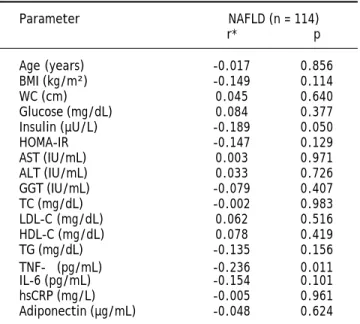
We further analyzed our results searching for pos-sible interactions between visfatin and other parame-ters studied. Visfatin levels were found to be negatively correlated with TNF-α (r = -0.236, p = 0.011) in NAFLD group. However, visfatin was not associated with all the other parameters included in the study (Table 3). Moreover, no association was found between visfatin and histological findings (Table 4). Visfatin levels were also not found to be associated with NAFLD subgroups even after adjust-ment for BMI by covariance analysis (p = 0.49).
Because of the well-known relationship of visfatin with MetS,18 we aimed to search the possible asso-ciation of this molecule with MetS in subjects with NAFLD. But, according to the National Cholesterol Education Program19 only 13 patients had MetS, so we couldn’t analyze this relationship.
DISCUSSION
There are conflicting reports in the literature regarding the relationship between visfatin, an adi-pocytokine with proinflammatory and immunomo-dulating properties, and NAFLD. It has been Table 2.
The characteristics of the subjects with NAFLD subgroups and controls.
Parameter
Control (n = 60)
NASH (n = 48)
SS (n = 31)
Borderline NASH (n = 35)
p1 * P2 * p3 * Age (years) 29.00 (21.00-43.00) 31.00 (21.00-43.00) 33.00 (21.00-45.00) 32.00 (20.00-42.00) 0.105 0.011 0.068 BMI (kg/m²) 24.00 (18.50-28.70) 28.20 (23.00-33-00) 28.40 (25.00-37.00) 28.20 (22.00-38.00) < 0.001 < 0.001 < 0.001 WC (cm) 88.00 (73.00-100.00) 100.00 (86.00-123.00) 98.00 (74.00-116.00) 98.50 (86.00-126.00) < 0.001 < 0.001 < 0.001 Glucose (mg/dL) 84.00 (63.00-107.00) 94.00 (70.00-122.00) 94.50 (71.00-118.00) 94.00 (64.00-120.00) < 0.001 0.001 < 0.001 Insulin (µU/l) 6.22 (2.21-23.91) 14.10 (2.60-54.10) 12.70 (7.00-53.80) 15.60 (5.80-41.80) < 0.001 < 0.001 < 0.001 HOMA-IR 1.19 (0.34-5.79) 3.30 (0.50-11.00) 3.0 (1.40-13.70) 3.10 (1.30-9.60) < 0.001 < 0.001 < 0.001 AST (IU/mL) 20.00 (9.00-38.00) 57.50 (27.00-118.00) 40.00 (23.00-89.00) 42.00 (15.00-139.00) < 0.001 < 0.001 < 0.001 ALT (IU/mL) 19.50 (6.00-40.00) 120.00 (55.00-286.00) 89.00 (35.00-142.00) 91.00 (19.00-330.00) < 0.001 < 0.001 < 0.001 GGT (IU/mL) 21.00 (11.00-58.00) 61.00 (30.00-455.00) 59.00 (19.00-409.00) 56.00 (20.00-154.00) < 0.001 < 0.001 < 0.001 TC (mg/dL)
180.53 ± 29.43
196.50 (83.00-304.00) 214.00 (151.00-338.00) 200.00 (99.00-320.00) 0.005 < 0.001 0.030 LDL-C (mg/dL) 111.12 ± 25.98 127.00 (28.00-221.00) 128.00 (76.00-219.00) 122.00 (45.00-198.00) 0.034 0.002 0.048 HDL-C (mg/dL) 44.95 ± 7.01 39.00 (29.00-56.00) 40.00 (28.00-52.00) 40.00 (27.00-55.00) 0.001 0.026 0.001 TG (mg/dL) 92.00 (37.00-359.00) 145.50 (26.00-472.00) 232.00 (44.00-617.00) 167.00 (49.00-579.00) < 0.001 < 0.001 < 0.001 Visfatin (ng/mL) 13.33 ± 2.73 14.00 (8.60-21.20) 13.2 (10.80-17.00) 14.30 (9.20-19.10) 0.162 0.402 0.307 TNF-α (pg/mL) 1.59 ± 0.74 10.70 (1.70-19.40) 10.70 (2.00-19.70) 10.70 (3.80-20.00) < 0.001 < 0.001 < 0.001 IL-6 (pg/mL) 0.11 (0.08-.81) 0.50 (0.10-1.40) 0.40 (0.10-1.30) 0.40 (0.10-0.90) < 0.001 < 0.001 < 0.001 hsCRP (mg/L) 0.77 (0.15-4.00) 2.20 (0.37-15.70) 1.70 (0.50-11.70) 15.60 (5.80-21.80) < 0.001 < 0.001 < 0.001 Adiponectin (µg/mL) 7.09 (0.80-25.81) 4.00 (1.00-14.40) 4.30 (2.50-11.10) 3.40 (1.60-7.50) < 0.001 0.001 < 0.001
*Mann Whitney U test (Statistically significant p value is <0.008 since Bonferroni correction). p1: control
vs.
NASH. p2: control
vs.
SS. p3: control
vs.
Borderline NASH. BMI: body mass index. WC: waist
cir-cumference. HOMA-IR: homeostasis model assessment for insulin resistance. AST: aspartate aminotransferase. ALT: alanine aminotr
ansferase. GGT: gamma-glutamyl transpeptidase. TC: total cholesterol.
LDL-C: low-density lipoprotein cholesterol. HDL-C: high-density lipoprotein cholesterol. TG: triglyceride.
TNF-α
reported that circulating visfatin levels were signifi-cantly increased in subjects with NAFLD when com-pared to both lean and obese healthy controls. In addition, visfatin levels decreased markedly when NASH was diagnosed. However, it was still signifi-cantly higher than in both lean and obese healthy subjects.20 Aller, et al. found that serum visfatin level was not related to steatosis grade and did not differ between groups with low and high-grade stea-tosis in overweight and obese patients with NAFLD. On the other hand, visfatin levels were related to the severity of portal inflammation and predicted the presence of portal inflammation. However, there was no association between visfatin and grade of lo-bular inflammation in NAFLD.12 In another study, Gaddipati, et al. showed that visceral visfatin in NASH were significantly lower than SS. Moreover, the visceral visfatin levels were higher in non-NA-FLD subjects. The low level of visceral visfatin was independent of BMI and IR. On the basis of these findings, the authors pointed to the protective role of visfatin in NAFLD.21 A study by Dahl, et al. showed that liver visfatin expression and its serum level were markedly decreased in patients with NAFLD, with no difference between SS and NASH.11 In the present study, we did not find any difference regarding circulating visfatin levels in subjects with NAFLD when compared to healthy controls. In addition, no difference was found regarding visfatin plasma levels among the different histological groups of NAFLD.
We suggest some possible explanations for the lack of relationship between visfatin and NAFLD observed in our study. Firstly, when the above men-tioned studies were analyzed separately, it can be seen that most of the patients with NAFLD had metabolic confounders like severe obesity, T2D and hypertension. It has been reported that visfatin levels may be affected from these metabolic risk factors.12,20,22 Secondly, some part of these subjects was also using the medications related to these metabolic problems. Various drug treatments for T2D, hypertension, and hyperlipidemia with thiazo-lidinediones, statins, angiotensin converting enzyme inhibitors, biguanide, and insulin therapy have been shown to alter adipokine levels measured in this cohort and thus blurred the distinction between his-tological groups.23-26 On the other hand, there are also contradictory findings regarding the effect of these confounders on circulating visfatin concentra-tions.27-29 We suggest that the lack of difference regarding visfatin levels in our groups could be dependent on the selection criteria of our study
Table 4. Relationship between visfatin and histological
fin-dings.
Visfatin p
Steatosis
Grade 1 13.16 ± 1.87 0.335* Grade 2 13.95 ± 2.14
Grade 3 14.16 ± 2.95
Ballooning degeneration
Grade 0 13.27 ± 1.63 0.073* Grade 1 14.09 ± 2.49
Grade 2 13.03 ± 2.36
Lobular inflammation
Grade 0 13.27 ± 1.63 0.737* Grade 1 14.09 ± 2.49
Grade 2 13.03 ± 2.36
Fibrosis
Stage 0 13.74 ± 2.77 0.811* Stage 1 13.71 ± 2.04
Stage 2 14.01 ± 2.44 Stage 3 14.86 ± 0.36
*Kruskal Wallis test.
Table 3. Correlations between visfatin and other variables in
NAFLD group.
Parameter NAFLD (n = 114)
r* p
Age (years) -0.017 0.856 BMI (kg/m²) -0.149 0.114 WC (cm) 0.045 0.640 Glucose (mg/dL) 0.084 0.377 Insulin (µU/L) -0.189 0.050 HOMA-IR -0.147 0.129 AST (IU/mL) 0.003 0.971 ALT (IU/mL) 0.033 0.726 GGT (IU/mL) -0.079 0.407 TC (mg/dL) -0.002 0.983 LDL-C (mg/dL) 0.062 0.516 HDL-C (mg/dL) 0.078 0.419 TG (mg/dL) -0.135 0.156 TNF-α (pg/mL) -0.236 0.011 IL-6 (pg/mL) -0.154 0.101 hsCRP (mg/L) -0.005 0.961 Adiponectin (µg/mL) -0.048 0.624
population, as we excluded subjects with T2D, hy-pertension and severe obesity (BMI ≥ 40 kg/m2). The abnormalities in glucose and blood pressure that can potentially affect circulating visfatin concentrations are frequently accompanied by NAFLD. Therefore, we think that including only the subjects free from these confounding factors is an important feature of the present investigation.
Visfatin has been suggested as an inflammatory cytokine that is produced and released by the adipose tissue derived macrophages.30,31 It promotes B-cell maturation and inhibits neutrophil apoptosis,32,33 and enhances activation of leukocytes, synthesis of adhesion molecules and production of proinflamma-tory cytokines.9,34 In CD14+ monocytes, visfatin en-hanced the production of IL-1b, TNF-α, and especially of IL-6.30 On the other hand, visfatin expression in adipocytes was found to be downregulated by IL-6.35 Visfatin was also reported to be expressed in hepato-cytes and inhibited apoptosis of hepatohepato-cytes in vitro. The antiapoptotic effect of visfatin in hepatocytes in-volved enzymatic synthesis of nicotinamide adenine dinucleotide.31 Plasma visfatin was found to be nega-tively correlated to the grade of necro-inflammatory activity in patients with chronic hepatitis C, sugges-ting that visfatin may be a regulator of the inflamma-tory process in this condition.36 Because hepatocyte apoptosis is an important feature of chronic hepatitis, downregulation of visfatin in advanced inflammatory processes suggests a hepatoprotective role for visfa-tin. Circulating visfatin levels were significantly de-creased in liver cirrhosis compared with healthy controls, presumably owing to decreased hepatic expression and production. The different under-lying etiologies of liver cirrhosis had no significant impact on plasma visfatin levels or on hepatic visfa-tin production. Patients in the early clinical stages of cirrhosis –child class A liver cirrhosis– already had decreased plasma visfatin levels that were, however, significantly higher than those of pa-tients with child class B or C liver cirrhosis.37 In the present study, we did not find any association of visfatin with liver histology in NAFLD. On the other hand, visfatin levels were correlated negatively and significantly with TNF-α in our cohort. In light of the above data, we suggest that visfatin may have a protective role against hepatocyte injury during the ongoing inflammatory process in early stages of NAFLD. Hence, in agreement with this hypothesis, a similar protective effect of visfatin against hepato-cyte injury was also described in NAFLD.38 Moreover, TNF-α levels in visceral adipose tissue were shown to be inversely associated with visceral
visfatin levels, suggesting that TNF-α down regula-tes visfatin expression in NAFLD.21 These observa-tions may additionally support a protective role of visfatin against the liver injury in this clinically relevant condition.
The present study has several limitations. Firstly, because of the small number of patients and the strict inclusion criteria, the findings obtained are not representative for all subjects with NAFLD. But we think that the design of our study was a require-ment for the goals to achieve. Secondly, all partici-pants were men, and that it remains to be determined if these results are similar also in wo-men. Lastly, LDL-C levels were calculated by Friedewald’s formula. Although this formula is usually recommended for this purpose because of its low costs, accessibility and ease of use, it has seve-ral limitations.
In conclusion, the results of this study show that plasma visfatin levels are not altered in the early stages of NAFLD. However, it is inversely associated with TNF-α. Our findings further support a role for visfatin in protection against liver injury in this widespread disease. More studies in larger population are however needed to integrate these results.
ABBREVIATIONS
• IL-6: interleukin-6.
• BMI: body mass index.
• BMI: body mass index.
• FPG: fasting plasma glucose.
• HDL-C: high-density lipoprotein cholesterol.
• HOMA-IR: insulin sensitivity determined by
ho-meostasis model assessment index.
• hsCRP: high sensitive C-reactive protein.
• MetS: metabolic syndrome.
• NAFLD: non-alcoholic fatty liver disease.
• NAMPT: nicotinamide phosphoribosyltransferase.
• NASH: non-alcoholic steatohepatitis.
• PBEF-1: pre–B-cell colony-enhancing factor 1.
• SS: simple steatosis. • T2D: type 2 diabetes. • TC: total cholesterol. • TG: triglyceride.
• TNF-α: tumor necrosis factor alpha.
DECLARATION OF INTEREST
FINANCIAL DISCLOSURE
This work was supported by a grant from the Re-search Center of Gulhane Medical School (2010/49).
ACKNOWLEDGMENT
None.
REFERENCES
1. Bellentani S, Marino M. Epidemiology and natural history of non-alcoholic fatty liver disease (NAFLD). Ann Hepatol 2009; 8: 4-8.
2. Almeda-Valdés P, Cuevas-Ramos D, Aguilar-Salinas CA. Me-tabolic syndrome and non-alcoholic fatty liver disease. Ann Hepatol 2009; 8: 18-24.
3. Chávez-Tapia NN, Uribe M, Ponciano-Rodríguez G, Medi-na-Santillán R, Méndez-Sánchez N. New insights into the pathophysiology of nonalcoholic fatty liver disease. Ann Hepatol 2009; 8: 9-17
4. Marra F, Bertolani C. Adipokines in liver diseases. Hepato-logy 2009; 50: 957–69.
5. Bertolani C, Marra F. The role of adipokines in liver fibro-sis. Pathophysiology 2008; 15: 91-101.
6. Polyzos SA, Kountouras J, Zavos C. Nonalcoholic fatty liver disease: the pathogenetic roles of insulin resistance and adipocytokines. Curr Mol Med 2009; 9: 299-314.
7. Bemdt J, Kloting N, Kralisch S, Kovacs P, Fasshauer M, Schon MR, Stumvoll M, et al. Plasma visfatin concentrations and fat depot-specific mRNA expression in humans. Diabetes 2005; 54: 2911-6.
8. Imai S. Nicotinamide phosphoribosyltransferase (Nampt): A link between NAD biology, metabolism, and diseases. Curr Pharm Des 2009; 15: 20-8.
9. Luk T, Malam Z, Marshall JC. Pre-B cell colony-enhancing factor [PBEF]/visfatin: visfatin novel mediator of innate immunity. J Leukoc Biol 2008; 83: 804-16.
10. Kukla M, Ciupinska-Kajor M, Kajor M, Wylezol M, Zwirska-Korczala K, Hartleb M, Berdowska A, et al. Liver visfatin expression in morbidly obese patients with nonalcoholic fatty liver disease undergoing bariatric surgery. Pol J Pa-thol 2010; 61: 147-53.
11. Dahl TB, Haukeland JW, Yndestad A, Ranheim T, Gladhaug IP, Damås JK, Haaland T, et al. Intracellular nicotinamide phosphoribosylotransferase protects against hepatocyte apoptosis and is down-regulated in nonalcoholic fatty liver disease. J Clin Endocrinol Metab 2010; 95: 3039-47. 12. Aller R, de Luis DA, Izaola O, Sagrado MG, Conde R,
Velas-co MC, Alvarez T et al. Influence of visfatin on histopatho-logical changes of non-alcoholic fatty liver disease. Dig Dis Sci 2009; 54: 1772-7.
13. Diagnosis and Classification of Diabetes Mellitus. ADA posi-tion statements. Diabetes Care 2011; 34: 62-9.
14. Friedewald WT, Levy RI, Fredrickson DS. Estimation of the concentration of lowdensity lipoprotein cholesterol in plasma, without use of the preparative ultracentrifuge. Clin Chem 1972; 18: 499-502.
15. Matthews DR, Hosker JP, Rudenski AS, Naylor BA, Trea-cher DF, Turner RC. Homeostasis model assessment: insu-lin resistance and beta-cell function from fasting plasma glucose and insulin concentrations in man. Diabetologia 1985; 28; 412-9.
16. Bonora E, Targher G, Alberiche M, Bonadonna RC, Saggiani F, Zenere MB, Monauni T, et al. Homeostasis model assess-ment closely mirrors the glucose clamp technique in the assessment of insulin sensitivity: studies in subjects with various degrees of glucose tolerance and insulin sensitivi-ty. Diabetes Care 2000; 23: 57-63.
17. Kleiner DE, Brunt EM, Van Natta M, Behling C, Contos MJ, Cummings OW, Ferrell LD, et al. Design and validation of a histological scoring system for nonalcoholic fatty liver di-sease. Hepatology 2005; 41: 1313-21.
18. Zhong M, Tan HW, Gong HP, Wang SF, Zhang Y, Zhang W. Increased serum visfatin in patients with metabolic syn-drome and carotid atherosclerosis. Clin Endocrinol [Oxf] 2008; 69: 878-84.
19. Grundy SM, Cleeman JI, Merz CN, Brewer HB Jr, Clark LT, Hunninghake DB, Pasternak RC, et al. National Heart, Lung, and Blood Institute; American College of Cardiology Foundation; American Heart Association. Implications of recent clinical trials for the National Cholesterol Educa-tion Program Adult Treatment Panel III guidelines. Circula-tion 2004; 110: 227-9.
20. Jarrar MH, Baranova A, Collantes R, Ranard B, Stepanova M, Bennett C, Fang Y, et al. Adipokines and cytokines in non-alcoholic fatty liver disease. Aliment Pharmacol Ther 2008; 27: 412-21.
21. Gaddipati R, Sasikala M, Padaki N, Mukherjee RM, Sekaran A, Jayaraj-Mansard M, Rabella P, et al. Visceral adipose tissue visfatin in nonalcoholic fatty liver disease. Ann He-patol 2010; 9: 266-70.
22. Gunes F, Akbal E, Cakir E, Akyurek O, Altunbas M, Ozbek M. Visfatin may be a novel marker for identifying stages of essential hypertension in advanced age patients. Intern Med 2012; 51: 553-7.
23. Derosa G, Maffioli P, Ferrari I, Palumbo I, Randazzo S, Fo-gari E, D’Angelo A, et al. Different actions of losartan and ramipril on adipose tissue activity and vascular remode-ling biomarkers in hypertensive patients. Hypertens Res 2011; 34: 145-51.
24. Ozkaya M, Cakal E, Ustun Y, Engin-Ustun Y. Effect of metformin on serum visfatin levels in patients with polycystic ovary syndrome. Fertil Steril 2010; 93: 880-4.
25. Ibáñez L, López-Bermejo A, Díaz M, Enríquez G, Valls C, de Zegher F. Pioglitazone [7.5 mg/day] added to flu-tamide-metformin in women with androgen excess: additional increments of visfatin and high molecular weight adiponectin. Clin Endocrinol [Oxf] 2008; 68: 317-20.
26. Storka A, Vojtassakova E, Mueller M, Kapiotis S, Haider DG, Jungbauer A, Wolzt M. Angiotensin inhibition stimula-tes PPARgamma and the release of visfatin. Eur J Clin In-vest 2008; 38: 820-6.
27. Dogru T, Sonmez A, Tasci I, Yilmaz MI, Erdem G, Erturk H, Bingol N, et al. Plasma visfatin levels in young male pa-tients with uncomplicated and newly diagnosed hyperten-sion. J Hum Hypertens 2007; 21: 173-5.
28. Erdem G, Dogru T, Tasci I, Bozoglu E, Muhsiroglu O, Tapan S, Ercin CN, et al. The effects of pioglitazone and metfor-min on plasma visfatin levels in patients with treatment naive type 2 diabetes mellitus. Diabetes Res Clin Pract 2008; 82: 214-8.
29. Dogru T, Sonmez A, Tasci I, Bozoglu E, Yilmaz MI, Genc H, Erdem G, et al. Plasma visfatin levels in patients with newly diagnosed and untreated type 2 diabetes mellitus and im-paired glucose tolerance. Diabetes Res Clin Pract 2007; 76: 24-9.
30. Curat CA, Wegner V, Sengene’s C, Miranville A, Tonus C, Buse R, Bouloumié A. Macrophages in human visceral adi-pose tissue: increased accumulation in obesity and a source of resistin and visfatin. Diabetologia 2006; 49: 744-7.
31. Dahl T, Ranheim T, Holm S, Berge R, Aukrust P, Halvorsen B. Nicotinamide phosphoribosyltransferase and lipid accu-mulation in macrophages. Eur J Clin Invest 2011; 41: 1098-104.
32. Samal B, Sun Y, Stearns G, Xie C, Suggs S, McNiece I. Clo-ning and characterization of the cDNA encoding a novel human pre-B cell colony-enhancing factor. Mol Cell Biol 1994; 14: 1431-37.
33. Jia SH, Li Y, Parodo J, Kapus A, Fan L, Rotstein OD, Mars-hall JC. Pre-B cell colony-enhancing factor inhibits neutro-phil apoptosis in experimental inflammation and clinical sepsis. J Clin Invest 2004; 113: 1318-27.
34. Moschen AR, Kaser A, Enrich B, Mosheimer B, Theurl M, Nie-deregger H, et al. Visfatin, an adipocytokine with
proinfla-mmatory and immunomodulating properties. J Immunol 2007; 178: 1748-58.
35. Kralisch S, Klein J, Lossner U, Bluher M, Paschke R, Stumvoll M, Fasshauer M. Interleukin-6 is a negative regulator of visfatin gene expression in 3T3–L1 adi-pocytes. Am J Physiol Endocrinol Metab 2005; 289: 586-90.
36. Kukla M, Zwirska-Korczala K, Gabriel A, Waluga M, Warakomska I, Berdowska A, Rybus-Kalinowska B, et al. Visfatin serum levels in chronic hepatitis C patients. J Vi-ral Hepat 2010; 17: 254-60.
37. de Boer JF, Bahr MJ, Boker KH, Manns MP, Tietge UJ. Plas-ma levels of PBEF/Nampt/ visfatin are decreased in pa-tients with liver cirrhosis. Am J Physiol Gastrointest Liver Physiol 2009; 296: 196-201.