Acute kidney injury in critically ill cirrhotic patients: a review
Jimena Muciño-Bermejo,* Raúl Carrillo-Esper,* Misael Uribe,* Nahum Méndez-Sánchez** Intensive Care Unit and Liver Research Unit, Medica Sur Clinic & Foundation, Mexico City, Mexico.
ABSTRACT
Acute kidney injury (AKI) is an important marker of morbidity and mortality in critically ill cirrhotic pa-tients. The most common causes of AKI in cirrhotic patients include prerenal or hepatorenal syndrome (HRS). Diagnosis of AKI may be delayed by the lack of clinical, biochemical, and radiological markers with proven sensitivity and specificity in cirrhotic patients. In this review, we discuss the epidemiology, patho-physiology, diagnosis, and therapies for AKI in cirrhotic patients admitted to an intensive care unit (ICU).
Key words. Renal failure. Hepatorenal syndrome. Intensive Care Unit.
Correspondence and reprint request: Nahum Méndez-Sánchez, M.D., PhD. Liver Research Unit. Medica Sur Clinic & Foundation
Puente de Piedra, Núm. 150. Col. Toriello Guerra, Del. Tlalpan CP 14050, México City. Ph.: (+52 55) 5424-7200, Ext. 4215 Fax: (+5255) 5666-4031
E mail: [email protected]
Manuscript received: December 02, 2011. Manuscript accepted: January 14, 2012.
INTRODUCTION
Acute kidney injury (AKI) is characterized by a sudden drop in the glomerular filtration rate (GFR), extracellular fluid and acid-base disorders, and the failure of the kidney to excrete nitrogenous waste products.1,2 The association between liver and kid-ney disease was well documented over 100 years ago when, in 1877, the German pathologist, Frerichs, described the clinical association of oliguria, ascites, and normal kidney histology.3
The combination of liver disease and renal dys-function can occur as a result of systemic conditions that affect both the liver and the kidney, although primary disorders of the liver complicated by renal dysfunction are much more common.4 Renal failure secondary to liver dysfunction is generally prerenal and unaccompanied by alterations in renal histology, although intrinsic renal abnormalities can further complicate acute or chronic liver disease. Postrenal acute renal failure develops rarely in chronic liver disease.5
Hepatorenal syndrome (HRS) is a unique form of functional renal failure that may complicate advan-ced liver disease, hepatic failure, or portal
for more than 3 months, whereas acute-on-chronic kidney disease is defined as an increase of serum creatinine > 50% above the baseline, or a rise in serum creatinine of ≥ 26.4 mmol/L (0.3 mg/dL) within 48 h, in patients with cirrhosis who have a GFR < 60 mL/min for more than 3 months. HRS ty-pes 1 and 2 represent specific forms of acute and chronic kidney disease, respectively.4
EPIDEMIOLOGY
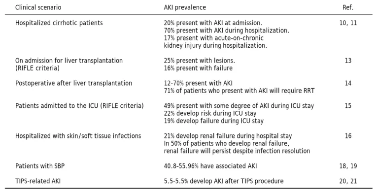
AKI is a common event in cirrhotic patients, al-though its exact prevalence is unknown and it va-ries widely in clinical settings (Table 2).
• Renal failure is the fifth leading cause of hospita-lization in cirrhotic patients and its incidence in hospitalized patients is high.9
• In hospitalized cirrhotic patients, 20% present with AKI on admission10 and as many as 70% de-velop AKI during hospitalization. Of these hospi-talized cirrhotic patients, 17% present with acute-on-chronic kidney failure and 13% present with de novo acute renal failure.11 In patients with ascites, there is a 23.6% probability of deve-loping AKI during the first year after the first episode of ascites.12
• According to the Risk, Injury, Failure, Lesion, and End-stage renal disease (RIFLE) criteria, 25% of patients admitted to receive liver transplanta-tion develop kidney lesions and 16.7% experience kidney failure.13 In the postoperative period after liver transplantation, the AKI rate varies between 12 and 70%, and 71% of this subpopulation will require renal replacement therapy (RRT).14 • AKI is the third leading cause of ICU admission
in cirrhotic patients. Regardless of the ICU ad-mission diagnosis, 49% of patients develop some degree of AKI during their ICU stay (22% develop risk and 19% failure, according to the RIFLE cri-teria).15
• In a 6-year retrospective study of patients with cirrhosis and skin or soft tissue infection, 21.7% developed renal failure after hospital admission Table 1. Diagnostic criteria for hepatorenal syndrome.
Modified criteria for the diagnosis of hepatorenal syndrome.
1. Cirrhosis with ascites.
2. Serum creatinine > 1.5 mg dL-1. 3. Absence of shock.
4. Absence of hypovolemia (no improvement in renal function after two days of diuretic withdrawal and volume expansion with albumin doses of 1 g kg-1 d-1). 5. No ongoing or recent treatment with nephrotoxic
drugs.
6. Absence of intrinsic renal disease (proteinuria < 0.5 g d-1; urine RBCs < 50 HPF-1; normal renal US).
Table 2. Prevalence of AKI among cirrhotic patients in different clinical settings.
Clinical scenario AKI prevalence Ref.
Hospitalized cirrhotic patients 20% present with AKI at admission. 10, 11
70% present with AKI during hospitalization. 17% present with acute-on-chronic
kidney injury during hospitalization.
On admission for liver transplantation 25% present with lesions. 13
(RIFLE criteria) 16% present with failure
Postoperative after liver transplantation 12–70% present with AKI 14
71% of patients who present with AKI will require RRT
Patients admitted to the ICU (RIFLE criteria) 49% present with some degree of AKI during ICU stay 15 22% develop risk during ICU stay
19% develop failure during ICU stay
Hospitalized with skin/soft tissue infections 21% develop renal failure during hospital stay 16 In 50% of patients who develop renal failure,
renal failure will persist despite infection resolution
Patients with SBP 40.8–55.96% have associated AKI 18, 19
and renal failure was persistent, despite the reso-lution of the infection.16 Of patients with bacte-rial infections other than spontaneous bactebacte-rial peritonitis (SBP), i.e., infections of the urinary tract and biliary tract, pneumonia, cellulitis, bac-teremia of unknown origin, 36.3% developed re-nal dysfunction, and 37.9% of rere-nal dysfunction events were irreversible.17
• Among the SBP patients, 40.8-55.96% presented with some degree of renal injury; of these, 57.37% of the AKI episodes were transient, 19.67% had a steady course, and 22.95% had a progressive course.18,19
• In a 6-year single-center retrospective analysis including 34 patients treated with transjugular intrahepatic portosystemic shunts (TIPS), 5.8% of patients20 presented with AKI as a procedure-related complication. This result is similar to that obtained in another single-center 6-year sur-vey of 128 patients treated with TIPS for variceal hemorrhage, where the procedure-related acute renal failure rate was 5.5%.21
The most common causes22 of AKI in cirrhotic pa-tients are prerenal failure (32%), and acute tubular necrosis (35%). HRS types 1 and 2 represent 20 and 6% of AKI cases in hospitalized cirrhotic patients, respectively.
In patients admitted to the hospital with decom-pensated cirrhosis, 39% present with AKI on admis-sion, and the leading etiologies are bacterial infections (40%), hypovolemia (32%), parenchymal kidney disease (15%), HRS type 2 (9%), and HRS type 1 (12%).23
Risk factors for developing AKI in cirrhotic pa-tients include variceal bleeding, PTH, sepsis seconda-ry to SBP, drug toxicity (including aminoglycosides and contrast media), and HRS.24
PATHOPHYSIOLOGY
In cirrhotic patients, AKI is related to circulatory disturbances such as diminished peripheral vascular
resistances because of splanchnic vasodilation trig-gered by PTH accompanied by an increase in endo-genous vasodilators such as nitric oxide (NO) and endogenous cannabinoids (Figure 1).25 In patients with advanced cirrhosis, there is also vasoconstric-tor system activation, including the renin-angioten-sin-aldosterone axis, the sympathetic nervous system, and arginine hypersecretion triggered by no-nosmotic stimulus. This compensatory mechanism promotes sodium and free water retention, which fa-vors ascites formation. In fact, AKI is uncommon in patients without ascites or edema (Table 3).
• PTH is a functional hemodynamic intrarenal di-sorder characterized by high-level stimulation of the renin-angiotensin-aldosterone system (RAAS) and high levels of vasopressin, as well as local hyperproduction of nitric oxide and other vasodi-lators including adrenomedullin and glucagon, which are strong arterial vasodilators. This arte-rial vasodilation is dominant in the splanchnic territory where it causes a reduction in the effi-cient arterial volume (EAV), which lowers mean arterial pressure.
• The reduced EAV triggers the stimulation of RAAS by vasopressin production, leading to re-nal cortical vasoconstriction. Initially, these me-chanisms have an adaptive function that reestablishes the EAV and arterial pressure. The-re is a rise in plasma volume because of water and sodium retention, and systemic arterial vaso-constriction (except in the splanchnic arteries) results from the stimulation of V1 vascular re-ceptors by vasopressin, which increases the car-diac output and maintains peripheral vascular resistance.26
In the early stages of PTH, an increase in car-diac output maintains peripheral vascular resis-tance, where the same initially adaptive mechanisms cause pathology in the more advan-ced stages of cirrhosis. Ascites is a consequence of excessive renal sodium and water retention be-cause of RAAS stimulation. Dilutional
hypona-Table 3. Hemodynamic changes during different stages of cirrhosis.
Compensated Diuretic-responsive Diuretic-resistant HRS
cirrhosis ascites ascites
Splanchnic/systemic arterial vasodilation Normal/+ + ++ +++
Effective circulating volume Normal - - -
-Renin, aldosterone, vasopressin, norepinephrine Normal + ++ +++
Renal sodium retention/plasma volume + ++ +++ ++++
tremia also occurs as a consequence of V2 renal tubular receptor stimulation by vasopressin and renal selective retroresorption of water,27 and HRS follows extreme renal vasoconstriction. • In the later stages, the EAV and arterial
pressu-re apressu-re pressu-reduced and natriuria drops dramatically. The retained water and sodium is distributed mainly in the venous visceral territory and the initial EAV cannot be reestablished. The visceral capillary pressure increases and, in the presence of hypoalbuminemia, this leads to peritoneal transudation of fluid and hyperproduction of vis-ceral lymph, which results in ascites formation. • Dilutional hyponatremia, ascites, and HRS are
different manifestations of the same pathogenic axis, which may be considered parts of a conti-nuous clinical spectrum. A second renal injury (sepsis, hypovolemia, or nephrotoxic drugs) can trigger AKI. During SBP, aerobic Gram-negative bacteria are translocated from the intestine to
produce an inflammatory response in the perito-neum, which is accompanied by monocyte activa-tion, the production of proinflammatory cytokines and supplementary nitric oxide, overex-pression of Toll-like receptors, and the activation of nuclear factor kappa B (NF-κB) and interleu-kin-6.28
Nonsteroidal anti-inflammatory drugs may cause AKI in cirrhotic patients, because their renal function depends greatly on prostaglandins.29 In cirrhotic patients, AKI may coexist with renal in-juries derived from cirrhosis etiologies, such as HCV- or HBV-associated glomerular disease and diabetic nephropathy in nonalcoholic steatohepa-titis.4
• HRS type I, which presents as an acute renal fa-ilure, or type II, which presents as refractory as-cites, manifests when renal vasodilator mechanisms are overwhelmed by vasoconstrictor mechanisms and supplementary vasoconstrictors Figure 1. Pathophysiology of circulatory abnormalities and renal failure during cirrhosis. Portal hypertension triggers splan-chnic vasodilation and the compensatory activation of the vasoconstrictor system. In the early stages, increases in the cardiac output and plasma volume may restore the effective arterial blood volume, although the sustained activation of vasoconstrictor systems leads to ascites formation, abnormal renal autoregulation, and eventual renal failure in the later stages. A second re-nal injury, such as hypovolemia or sepsis, may accelerate the progression of rere-nal failure.
Renal failure Increased intrahepatic vascular resistance.
Obstruction to portal flow: Portal hypertension. Bacterial translocation.
Baroreceptor activation:
Activation of sodium retaining vasoconstrictor systems: -Atrial natriuretic peptide.
-RAAS axis.
Abnormal renal autoregulation
-Argumented sensitivity to vasoconstrictors -Diminished capacity to excreee solute-free water. -Hyponatremia.
-Diminished GRF. Diminished renal vasodilation
Augmented renal vasoconstriction Tachycardia
Increased plasma volume Increased
cardiac output
Cirrhotic cardiomyopathy
Restoration of effective arterial blood volume
Low effective arterial blood volume Splachnic arterial vasodilation
Nitric oxide
(adenosine, thromboxane A2, endothelin-1, cys-teinyl leukotriene, and neuropeptide Z) become active. There is a rise in the plasma level of endo-thelins (because of the effect of endotoxinemia) and a significant rise in angiotensin II and intra-renal vasoconstrictors as a consequence of a rai-sed production of thromboxanes, leukotrienes, and adenosine. There is also a reduced produc-tion of intrarenal vasodilators such as prosta-glandins and kallikrein (an imbalance in the kinin–kallikrein equilibrium).30
DIAGNOSIS
One of the main problems in the evaluation of re-nal function in cirrhotic patients is that most rere-nal biomarkers are not sensitive or specific for the diag-nosis of AKI in cirrhotic patients.
Prognostic scoring systems such as model of end-stage liver disease (MELD) have shown that serum creatinine is an important prognostic bio-marker in cirrhotic patients, but its sensitivity and specificity is poor because it overestimates the re-nal function in patients with low muscle mass and 67% of cirrhotic patients are malnourished.31,32 The decreased hepatic production of creatinine re-sults in lower serum creatinine levels. Hyperbiliru-binemia, ketonic bodies, hyperglycemia, and hyperuricemia may also interfere with serum crea-tinine measurements.33,34 In summary, serum crea-tinine levels below 1 mg/dL cannot exclude renal failure in cirrhotic patients.
In healthy patients, inulin clearance is the gold standard for assessing the GFR because it is fully filtered and not secreted or reabsorbed in the kid-ney. Other inulin-like polyfructoses such as iohexol and iothalamate have also been used in GFR assess-ments, with or without radiolabels. However, inu-lin, iohexol, and iothalamate have not been validated as renal function markers in cirrhotic patients.35,36 In this population, cystatin C levels and/or Doppler ultrasonography are preferred when assessing renal function:
• Cystatin C is a low molecular weight protein that is produced in all nucleated cells at a constant rate, and it is eliminated by glomerular filtra-tion. After filtration, it is reabsorbed and catabo-lized by tubular epithelial cells. Compared with creatinine, the serum cystatin C level does not depend on sex, age, or lean muscle mass, and its measurement is unaffected by hyperbilirubine-mia. The sensitivity of cystatin C in the
diagno-sis of AKI is equal in both cirrhotic and noncirr-hotic patients when a cut-off point of 1.25 mg/dL is used.35
• Renal Doppler ultrasonography is useful for eva-luating renal vascular resistance during HRS diagnosis because it can detect important vaso-constrictions. An elevated vascular resistance in-dex (VRI) predicts deterioration in the renal function of cirrhotic patients with a normal crea-tinine value. An elevated VRI is a marker of a greater risk of postoperative renal dysfunction in orthotropic liver transplantation (OLT) candida-tes, even if VRI and GFR are not correlated.36
Neutrophil gelatinase-associated lipocalin (NGAL) is a small protein expressed by neutro-phils and various epithelia, including the renal proximal tubules. NGAL was initially proposed as a marker of infections and certain adenocarcino-mas, but it is now apparent that its early and dramatic increase in the urine after renal injury may make it a useful marker. Tissue release of NGAL may be induced by inflammatory processes that affect the epithelium, proximal and distal airways, and some neoplastic lineages. In the renal tubules, NGAL mRNA expression increases a few hours after renal injury.37 Plasma NGAL is freely filtered in the glomerulus, and the vast majority of NGAL is reabsorbed via endocytosis in the proximal renal tubules. Thus, an increase in uri-nary NGAL excretion can occur only because of proximal tubule injury or the increased de novo synthesis of NGAL. Acute kidney injury results in a dramatic increase in NGAL mRNA expression in the lungs and liver. NGAL is an acute phase reac-tant that may be released by neutrophils, macro-phages, and other immune cells, and so plasma concentrations of NGAL have been proposed as an AKI biomarker.38 The serum NGAL levels of cirr-hotic patients may be a sensitive novel marker for low GFR (< 50 mL/min).39
TREATMENT
• Albumin. HRS is one of the main indicators ofl u albumin use in cirrhotic patients. The actual crite-ria for HRS are: the presence of cirrhosis and as-cites, a decrease in GFR (or serum creatinine > 1.5 mg/dL), absence of shock or nephrotoxic drug use, unchanged renal function after 48 h of diure-tic suspension, expansion of the plasma volume with albumin (1 g/kg/d), and the absence of renal parenchymal disease. However, the role of albu-min is not limited to its diagnosis in HRS. The treatment of type 1 HRS is focused on terlipressin in conjunction with albumin. The beneficial effect of albumin on renal function and systemic hemo-dynamics is related to plasma volume expansion and to a vasoconstrictor effect, mainly in the peri-pheral blood circulation. The American Associa-tion for the Study of Liver Diseases guidelines on HRS indicate that the use of albumin in conjunc-tion with vasoactive drugs should be considered, especially in patients where liver transplantation is indicated.41 Moreover, albumin infusion at the time of diagnosis reduces the incidence of type 1 HRS and hospital mortality in patients with spon-taneous bacterial peritonitis.42
• Vasoconstrictors. Vasoconstrictors counteracta n t i the characteristic splanchnic vasodilation seen in advanced cirrhosis. The most commonly used drugs are:
a . TTerlipressin. A vasopressin analogue thatp i may be administered at doses of 4-6 mg/d (1 mg every 4-6 h). The dose can be doubled to 2 mg every 4-6 h if the basal creatinine level does not decrease by 25% on the third day of treatment. Terlipressin treatment should be continued until the serum creatinine level falls to < 1.5 mg/dL. The likelihood of any im-provement attributable to terlipressin treatment is practically zero if no clinical improvement is obtained after 2 weeks of its treatment. Terlipressin treatment is associa-ted with an improved cardiac and urinary output, lower serum creatinine and renin le-vels, and mean arterial pressure improvement. The use of terlipressin improves the 15-day survival rate in patients with HRS type 1 but not in those with HRS type 2. Ischemic events may affect the heart, mesenteries, and fingers in as many as 12% of patients during terli-pressin treatment, which is contraindicated if peripheral artery disease is present.43
b . N rNoradrenaline infusion at a rate of 0.5-3r n l e mg/kg/h together with intravenous albumin is
as effective as terlipressin plus albumin in im-proving the renal and circulatory function of patients with HRS.44
.
c . MMidodrine. A selective alpha-1 adrenergicd nr agonist, taken together with octreotide (a so-matostatin analogue) is more effective than dopamine in improving the GFR, lowering serum creatinine levels, and increasing the urinary volume of patients with HRS type 1. The combined use of midodrine and octreotide reduces mortality rates in this group of pa-tients.45 The combined use of octreotide, mido-drine, and albumin also benefits renal function and 1-month survival rates of pa-tients with HRS type 1 and HRS type 2.46,47
A recent pooled analysis of clinical trials assessed the therapeutic response to vasoconstrictors in 21 HRS studies. This pooled analysis included clinical studies that tested terlipressin, midodrine, octreoti-de, and noradrenaline, as vasoconstrictors and it as-sessed the mean arterial pressure, serum creatinine level, urinary output, and plasma renin activity at baseline and throughout the treatment. This analy-sis concluded that there was an increase in mean ar-terial pressure during vasoconstrictor therapy in patients with HRS irrespective of the vasoconstric-tor used, and this was associated with improved kid-ney function. However, this association was lower in patients from cohorts treated with alpha-1-adre-nergic agonists compared with cohorts treated with vasopressin.48
demonstrated whether either modality is supe-rior for the cirrhotic patient.53,54
•• Transjugular intrahepatic portosystemicTransjugular intrahepatic portosystemicaa juju a a n n pa i r o s epa i r o s e cc
u t P
shunt (TIPS).u t P
shunt (TIPS). TIPS has been reported to im-prove the renal function of patients who receive this treatment because of refractory ascites, va-riceal bleeding, refractory hepatic hydrothorax, or presurgical portal decompression. TIPS leads to a significant improvement in serum creatini-ne within 1 week and sustaicreatini-ned improvement (mean 0.18 mg/dL) 3 months after the procedu-re in patients with baseline cprocedu-reatinine, although not in patients with a basal creatinine above 2 mg/dL.55 A single-center retrospective study found that patients with baseline creatinine le-vels of < 1.2 mg/dL experienced no significant improvement in serum creatinine levels after TIPS, whereas patients with a baseline creatini-ne of 1.2-1.9 mg/dL showed significant improve-ments in their creatinine levels. However, there was no significant difference in their MELD sco-res before and after the procedure. Finally, pa-tients with baseline creatinine levels > 2 mg/dL showed a marked improvement in their creatini-ne levels and MELD score after TIPS. In pa-tients with stage 2, 3, or 4 chronic kidney disease, GFR was significantly improved after TIPS, and patients with stage 2 or 4 chronic ki-dney disease also displayed a marked improve-ment in their MELD score after TIPS. The biggest improvement in renal function was seen in patients where TIPS was indicated by refrac-tory ascites or variceal bleeding. The improved renal function was not related to the portosyste-mic gradient decrease after TIPS.56
•• Liver transplantation.Liver transplantation. By definition, patientsv v n pln lp t nt n with HRS have advanced hepatic failure, which means they are candidates for liver transplanta-tion. The long-term survival of liver transplant recipients is approximately 65%. In the absence of sepsis, HRS should be considered a semi-ur-gent indication for liver transplantation. In cases where liver transplantation is available, other therapies such as systemic vasoconstrictors, he-modialysis, albumin-based liver replacement sys-tems, and TIPS should be viewed as a bridge to transplantation.57 The 3-year survival rates are slightly higher in patients without HRS before transplantation (100%) compared with those with HRS at the time of transplantation. Thus, it is recommended that HRS be treated to achieve normal serum creatinine levels before transplan-tation.
PROGNOSIS
Renal failure is a serious event with a poor prog-nosis in patients with cirrhosis. The development of AKI during hospitalization for acute upper gastroin-testinal bleeding or sepsis is strongly associated with a poor outcome. The in-hospital mortality of patients with type 1 HRS is 75.2%. In addition to the prognostic factors related to the degree of liver failure (i.e., MELD and the Child–Pugh score), a systemic inflammatory response has independent prognostic value in this setting.58
A systematic literature review of the impact of AKI on cirrhotic patients, which included 74 studies (43 prospective and 31 retrospective), showed that the mortality of cirrhotic inpatients was 67% in tho-se with AKI at the time of admission but 25% in those without AKI, with the mortality odds ratio be-ing 7.7 in the AKI group.59
• Mortality rates of the AKI group at 1, 3, and 12 months were 58, 69, and 63%, respectively. After excluding studies based on ICU populations, the global mortality of cirrhotic patients with AKI was 63% at 1 month, 55% at 3 months, 77% at 6 months, and 63% at 12 months.
• The diagnostic criteria used to define AKI also affected the clinical prognosis and only 62 of the 74 studies defined “renal failure”. The median overall mortality was 65% when renal failure was defined but 70% when it was not. GFR was used to define AKI in four studies (79 patients) and creatinine clearance in one (46 patients). In three studies (359 patients), renal failure was de-fined according to the RIFLE classification, and HRS was defined in 25 studies (1184 patients). • Several thresholds were reported for serum
crea-tinine in the 24 studies where it was used as a diagnostic criterion for AKI, i.e., 1.3 mg/dL (one study, 56 patients), 1.5 mg/dL (17 studies, 907 patients), 2 mg/dL (six studies, 269 patients), and 2.26 mg/dL (one study, 26 patients). In stu-dies that used a cut-off of 1.5 mg/dL serum crea-tinine, the median overall mortality was lower than when a higher cutoff was used (55 vs. 77%; p = 0.08) or when HRS was defined (55 vs. 70%; p = 0.01). This confirmed the relationship bet-ween mortality and the severity of renal failure as defined by an elevated creatinine level.
MELD score, hepatic encephalopathy, sepsis, ventilator dependency, treatment with octreotide and midodrine, and treatment with terlipressin. • Multivariate analyses were performed to assess
prognostic factors in 33 studies (5,568 patients). The Child–Pugh score/class, MELD, and age were associated with a poor prognosis.
• Renal failure was used as an independent prog-nostic factor (categorical variable) in 59% of stu-dies, whereas continuous variables that were used to assess the severity of renal failure, inclu-ding serum creatinine, serum urea, and GFR, were not independent prognostic factors.
The AKI-associated mortality of cirrhotic hospita-lized patients varied according to the clinical set-ting, as follows:
• Patients with type 1 HRS had an 80% mortality rate at 2 weeks, while only 10% survived at 3 months. Patients with HRS type 2 had an avera-ge survival of 3 months. The MELD score was an independent prognostic factor in this popula-tion. The median survival rates of patients with HRS type 2 and a MELD score < 20 points was 8 months, whereas patients with a MELD score > 20 points had an average survival rate of 1 month.60
• In patients with acute renal failure that was not attributable to HRS, the mortality rates at 1 and 12 months were 41 and 36%, respectively. In pa-tients admitted to ICU, the mortality rates at 1 and 3 months were 81 and 84%, respectively. • In patients with SBP and AKI, the overall
morta-lity rate was 9% with a 3-month mortamorta-lity rate of 46%, whereas in patients with infectious diseases other than SBE the overall mortality rate was 3% with a 1-year mortality rate of 12%.
• The prognosis of AKI also varied according to its etiology. In a prospective study of 562 consecuti-ve patients with cirrhosis and renal failure, the 3-month mortality rate was 15% with HRS, 31% with infection-associated AKI, 46% with hypovo-lemia-associated AKI, and 73% with parenchymal nephropathy.61
• In a retrospective study of 138 cirrhotic patients admitted to an ICU in a teaching hospital in France, the 6-month survival rate was 59% and the in-hospital survival rates of patients requi-ring vasopressors or mechanical ventilation were 20 and 33%, respectively. The in-hospital morta-lity rate among patients requiring RRT was 31%.62
CONCLUSIONS
AKI is common among cirrhotic patients and up to 49% of patients who require hospitalization be-cause of decompensated cirrhosis present with AKI during hospitalization. In cirrhotic patients, AKI diagnosis may be delayed because conventional mar-kers of renal function overestimate GFR, such as the serum creatinine levels. Cystatin C and renal ul-trasonography are preferred in the evaluation of re-nal function during the AKI diagnostic workup of cirrhotic patients. Initially, the treatment of AKI in cirrhotic patients should be directed toward the co-rrection of reversible causes, circulatory anomalies such as splanchnic vasodilation and compensatory vasoconstrictor mechanisms of vasoconstriction, in-cluding RAAS activation and sympathomimetic sta-tes. HRS is advanced hepatic failure and it should be considered a semi-urgent indication of liver trans-plantation.
ABBREVIATIONS
• AKI: acute kidney injury.K : • HRS: hepatorenal syndrome. • ICU: intensive care unit.CU • GFR: glomerular filtration rate. • PTH: portal hypertension.H • IAC: International Ascites Club.C: • ADQI: acute dialysis quality initiative.D : • CKD: chronic kidney disease.
• RIFLE: risk, injury, failure, lesion, and end-stage renal disease criteria.
• SBP: spontaneous bacterial peritonitis.BP:
• TIPS: transjugular intrahepatic portosystemicI shunt.
• RAAS: renin-angiotensin-aldosterone system.A • EAV: efficient arterial volume.AV
• VRI: vascular resistance index.R
• MELD: Model of End-stage Liver Disease.E D:
• NGAL: neutrophil gelatinase-associatedG
lipocalin.
• RRT: renal replacement therapy.R • OLT: orthotropic liver transplantation.
REFERENCES
1. Thadhani R, Pascual M, Bonventre JV. Acute renal failure.
N Engl J Med 1996; 334: 1448-60.
2. Singri N, Ahya SN, Levin ML. Acute renal failure. JAMA
2003; 289: 747-51.
3. Ng CKF, Chan MHM, Tai MHL, Lam CWK. Hepatorenal syn-drome. Clin Biochem Rev 2007; 28: 11-7.
clas-sification system of renal dysfunction in patients with ci-rrhosis. Gut 2011; 60: 702-9.
5. Betrosian AP, Agarwal B, Douzinas EE. Acute renal dys-function in liver diseases. World J Gastroenterol 2007; 13: 5552-9.
6. Slack AJ, Wendon J. The liver and kidney in critically ill pa-tients. Blood Purif 2009; 28: 124-34.
7. Anon. Hepatorenal syndrome or hepatic nephropathy? Lan-cet 1980; 315: 801-2.
8. Salerno F, Gerbes A, Ginès P, Wong F, Arroyo V. Diagnosis, prevention and treatment of hepatorenal syndrome in ci-rrhosis. Gut 2007; 56: 1310-8.
9. Cholongitas E, Senzolo M, Patch D, Kwong K, Nikolopoulou V, Leandro G, Shaw S, et al. Risk factors, sequential organ failure assessment and model for end-stage liver disease scores for predicting short term mortality in cirrhotic pa-tients admitted to intensive care unit. Aliment Pharma-col Ther 2006; 1: 883-93.
10. García-Tsao G, Parikh C, Viola A. Acute kidney injury in ci-rrhosis. Hepatology 2008; 48: 2064-77.
11. Warner NS, Cuthbert JA, Bhore R, Rockey DC. Acute kid-ney injury and chronic kidkid-ney disease in hospitalized pa-tients with cirrhosis. J Investig Med 2011; 59:1244-51. 12. Montoliu S, Ballesté B, Planas R, Álvarez MA, Rivera M,
Mi-quel M, Masnou H, et al. Incidence and prognosis of diffe-rent types of functional renal failure in cirrhotic patients with ascites. Clin Gastroenterol Hepatol 2010; 8: 616-22. 13. Tinti F, Umbro I, Giannelli V, Merli M, Ginanni Corradini S, Nofroni I, et al. Acute renal failure in liver transplant reci-pients: role of pretransplantation renal function and 1-year follow-up. Transplant Proc 2011; 43: 1136-8.
14. Biagioni E, Cavazzuti I, Busani S, Trevisan D, Zavatti L, Ferrari E, Massimo G, et al. Acute renal failure and renal replacement therapy in the postoperative period of or-thotopic liver transplant patients versus non elective ab-dominal surgery patients. Transplant Proc 2011; 43: 1145-7.
15. Cholongitas E, Calvaruso V, Senzolo M, Patch D, Shaw S, O’Beirne J, Burroughs AK. RIFLE classification as predicti-ve factor of mortality in patients with cirrhosis admitted to intensive care unit. J Gastroenterol Hepatol 2009; 24: 1639-47.
16. Pereira G, Guevara M, Fagundes C, Solá E, Rodríguez E, Fernández J, Pavesi M, et al. Renal failure and hyponatre-mia in patients with cirrhosis and skin and soft tissue in-fection. A retrospective study. J Hepatol 2012; 13 [In press].
17. Kim JH, Lee JS, Lee SH, Bae WK, Kim NH, Kim KA, Moon YS. Renal dysfunction induced by bacterial infection other than spontaneous bacterial peritonitis in patients with ci-rrhosis: incidence and risk factors. Gut Liver 2009; 3: 292-7.
18. Perdomo Coral G, Alves de Mattos A. Renal impairment af-ter spontaneous bacaf-terial peritonitis: incidence and prog-nosis. Can J Gastroenterol 2003; 17: 187-90.
19. Jung ES, Lee JS, Kim MH, Kim NH, Kim KA, Moon YS. Renal dysfunction after spontaneous bacterial peritonitis in ci-rrhosis: incidence and risk factors. Korean J Gastroente-rol 2006; 48: 401-7.
20. Goykhman Y, Ben-Haim M, Rosen G, Carmiel-Haggai M, Oren R, Nakache R,Szold O, et al. Transjugular intrahepa-tic portosystemic shunt: current indications, patient se-lection and results. Isr Med Assoc J 2010; 12: 687-91. 21. Gaba RC, Omene BO, Podczerwinski ES, Knuttinen MG,
Cot-ler SJ, Kallwitz ER, Berkes JL, et al. TIPS for treatment of variceal hemorrhage: clinical outcomes in 128 patients at
a single institution over a 12-year period. J Vasc Interv Radiol 2012; 23: 227-35.
22. Wong LP, Blackley MP, Andreoni KA, Chin H, Falk RJ, Klem-mer PJ. Survival of liver transplant candidates with acute renal failure receiving renal replacement therapy. Kidney Int 2005; 68: 362-70.
23. Bittencourt PL, de Carvalho GC, de Andrade Regis C, Kalil JR, Cerqueira LA, Barbosa DS, da Silva Nery M, et al. Cau-ses of renal failure in patients with decompensated cirrho-sis and its impact in hospital mortality. Ann Hepatol 2012; 11: 90-5.
24. Moreau R, Lebrec D. Acute renal failure in patients with cirrhosis: perspectives in the age of MELD. Hepatology
2003; 37: 233-43.
25. Ginès P, Schrier RW. Renal failure in cirrhosis. N Engl J Med 2009; 361: 1279-90.
26. Olteanu D, Lupu D. The kidney in cirrhosis with portal hy-pertension. J Med Life 2010; 3: 175-7.
27. Ginès P, Wong R, Milutinovic S, Ruiz L, Olteanu D. Effects of Satavaptan (SR 121463B), a selective vasopressin V2 re-ceptor antagonist, on serum sodium concentration and as-cites in patients with cirrhosis and hyponatraemia. J Hepatol 2006; 44; 204-13.
28. Wiest R, Garcia-Tsao G. Bacterial translocation (BT) in ci-rrhosis. Hepatology 2005; 41: 422-33.
29. Salerno F, Badalamenti S. Drug induced renal failure in ci-rrhosis. In: Ginès P, Arroyo V, Rodés J, Schrier W (eds.). Ascites and Renal Dysfunction in Liver Disease. 2nd. ed. Malden, MA: Blackwell; 2005, p. 372-82.
30. Cardenas A, Ginès P. Hepatorenal Syndrome: Current Con-cepts. In: De Franchis R (ed.). Portal hypertension IV. Proceedings of the 4th International Consensus Workshop on Methodology and Treatment. Blackwell; 2004.
31. Huisman EJ, Trip EJ, Siersema PD, van Hoek B, van Erpe-cum KJ. Protein energy malnutrition predicts complicatio-ns in liver cirrhosis. Eur J Gastroenterol Hepatol 2011; 23: 982-9.
32. Guglielmi FW, Panella C, Buda A, Budillon G, Caregaro L, Clerici C, Conte D, et al. Nutritional state and energy balance in cirrhotic patients with or without hypermeta-bolism. Multicentre prospective study by the ‘Nutritional Problems in Gastroenterology’ Section of the Italian So-ciety of Gastroenterology (SIGE). Dig Liver Dis 2005; 37: 681-8.
33. Badiou S, Dupuy AM, Descomps B, Cristolead JP. Compari-son between the enzymatic vitro assay for creatinine de-termination and three other methods adapted on the Olympus analyzer. J Clin Lab Anal 2003; 17: 235-40. 34. Cholongitas E, Marelli L, Kerry A, Senzolo M, Goodier DW,
Nair D, Thomas M, et al. Different methods of creatinine measurement significantly affect MELD scores. Liver Transplant 2007; 13: 523-9.
35. Cholongitas E, Shusang V, Marelli L, Nair D, Thomas M, Patch D, Burns A, et al. Review article: renal function assess-ment in cirrhosis-difficulties and alternative measure-ments. Aliment Pharmacol Ther 2007; 26: 969-78. 36. Francoz C, Glotz D, Moreau R, Durand F. The evaluation of
renal function and disease in patients with cirrhosis. J He-patol 2010; 52: 605-13.
37. Hvidberg V, Jacobsen C, Strong RK, Cowland JB, Moestrup SK, Borregaard N. The endocytic receptor megalin binds the iron transporting neutrophil-gelatinase-associated li-pocalin with high affinity and mediates its cellular uptake.
FEBS Lett 2005; 579: 773-7.
gelatinase-as-sociated lipocalin as a novel early urinary biomarker for is-chemic renal injury. J Am Soc Nephrol 2003; 14: 2534-43. 39. Gerbes AL, Benesic A, Vogeser M, Krag A, Bendtsen F,
Mø-ller S. Serum neutrophil gelatinase-associated lipocalin-a sensitive novel marker of renal impairment in liver cirrho-sis? Digestion 2011; 84: 82-3.
40. Madan K, Mehta A. Management of renal failure and asci-tes in patients with cirrhosis. Int J Hepatol 2011; 79: 2-7. 41. Alves de Mattos A. Current indications for the use of albu-min in the treatment of cirrhosis. Ann Hepatol 2011: 10: S15-S20.
42. Arroyo V, Fernandez J. Pathophysiological basis of albumin use in cirrhosis. Ann Hepatol 2011; 10: S6-S14.
43. Proulx NL, Akbari A, Garg AX, Rostom A, Jaffey J, Clark HD. Measured creatinine clearance from timed urine co-llections substantially overestimates glomerular filtration rate in patients with liver cirrhosis: a systematic review and individual patient meta-analysis. Nephrol Dial Trans-plant 2005; 20: 1617-22.
44. Alessandria C, Ottobrelli A, Debernardi-Venon W, Todros L, Cerenzia MT, Martini S, Balzola F, et al. Noradrenalin vs. terlipressin in patients with hepatorenal syndrome: a prospective, randomized, unblinded, pilot study. J Hepa-tology 2007; 47: 499-505.
45. Angeli P, Volpin R, Gerunda G, Craighero R, Roner P, Me-renda R, Amodio P, et al. Reversal of type 1 hepatorenal syndrome with the administration of midodrine and oc-treotide. Hepatology 1999; 29: 1690-7.
46. Skagen C, Einstein M, Lucey MR, Said A. Combination treatment with octreotide, midodrine, and albumin impro-ves survival in patients with type 1 and type 2 hepatore-nal syndrome. J Clin Gastroenterol 2009; 43: 680-5. 47. Esrailian E, Pantangco ER, Kyulo NL, Hu KQ, Runyon BA.
Octreotide/Midodrine therapy significantly improves re-nal function and 30-day survival in patients with type 1 hepatorenal syndrome. Dig Dis Sci 2007; 52: 742-8. 48. Velez JC, Nietert PJ. Therapeutic response to
vasocons-trictors in hepatorenal syndrome parallels increase in mean arterial pressure: a pooled analysis of clinical trials.
Am J Kidney Dis 2011; 58: 928-38.
49. Wong LP, Blackley MP, Andreoni KA, Chin H, Falk RJ, Klem-mer PJ. Survival of liver transplant candidates with acute renal failure receiving renal replacement therapy. Kidney Int 2005; 68: 362-70.
50. Davenport A, Bouman C, Kirpalani A, Skippen P, Tolwani A, Mehta RL, Palevsky PM. Delivery of renal replacement the-rapy in acute kidney injury: what are the key issues? Clin J Am Soc Nephrol 2008; 3: 869-75.
51. Davenport A. Renal replacement therapy in the patient with acute brain injury. Am J Kidney Dis 2001; 37: 457-66.
52. Davenport A, Will EJ, Davidson AM. Improved cardiovascu-lar stability during continuous modes of renal replacement therapy in critically ill patients with acute hepatic and re-nal failure. Crit Care Med 1993; 21: 328-38.
53. Davenport A, Will EJ, Davison AM. Effect of renal replace-ment therapy on patients with combined acute renal and fulminant hepatic failure. Kidney Int Suppl 1993; 41: S245-S251.
54. Gonwa TA, Wadei HM. The challenges of providing renal re-placement therapy in decompensated liver cirrhosis. Blood Purif 2012; 20: 144-8.
55. Aggarwal A, Mitchell JE, Hanouneh I, Atreja A, Zein N. The effect of transjugular intrahepatic portosystemic shunt on renal function in patients with liver cirrhosis. Gas-troenterology 2011; 140: S958.
56. Anderson CL, Saad WE, Kalagher SD, Caldwell S, Sabri S, Turba UC, Matsumoto AH, et al. Effect of transjugular in-trahepatic portosystemic shunt placement on renal func-tion: a 7-year, single-center experience. J Vasc Interv Radiol 2010; 21: 1370-6.
57. Restuccia T, Ortega R, Guevara M, Ginès P, Alessandria C, Ozdogan O, Navasa M, et al. Effects of treatment of hepa-torenal syndrome before transplantation on posttrans-plantation outcome. A case-control study. J Hepatology
2004; 40: 140-6.
58. Thabut D, Massard J, Gangloff A, Carbonell N, Francoz C, Nguyen-Khac E, Duhamel C, et al. Model for end-stage liver disease score and systemic inflammatory response are major prognostic factors in patients with cirrhosis and acute functional renal failure. Hepatology 2007; 46: 1872-82.
59. Fede G, D’Amico G, Arvaniti V, Tsochatzis E, Germani G, Georgiadis D, Morabito A, et al. Renal failure and cirrhosis: a systematic review of mortality and prognosis. J Hepatol
2011; 10: 1-9.
60. Wadei HM, Gonwa TA. Hepatorenal syndrome in the inten-sive care unit. J Intensive Care Med 2011; 2: 1-3.
61. Martín-Llahí M, Guevara M, Torre A, Fagundes C, Restuc-cia T, Gilabert R, Solá E, et al. Prognostic importance of the cause of renal failure in patients with cirrhosis. Gas-troenterology 2011; 140: 488-96.