Analysis of host responses and fitness in different pandemic H1N1 (2009) influenza virus in mice and ferrets
164
0
0
Texto completo
(2) María Montoya González investigadora del Centre de Recerca en Sanitat Animal (CReSA) y del Instituto de Investigación Agroalimentaria (IRTA) y Dolores Jaraquemada Pérez de Gúzman profesora catedrática de inmunología de la Universitat Autònoma de Barcelona e investigadora del Institut de Biotecnologia i Biomedicina (IBB),. Certifican: Que la memoria titulada, “Analysis of host responses and fitness in different pandemic H1N1 (2009) influenza virus in mice and ferrets” presentada por Pamela Analia Martinez Orellana, ha sido realizada bajo su supervisión y tutoria en la Universitat Autònoma de Barcelona y Centre de Recerca en Sanitat Animal y que es apta para la optención del grado de Doctor en Inmunología.. Para que conste a los efectos oportunos, firman el presente certificado en Bellaterra (Barcelona), 21 de abril de 2014.. Directora: María Montoya González. Tutora: Dolores Jaraquemada Pérez de Gúzman. 2.
(3) PhD studies presented by Pamela Analia Martinez Orellana were financially supported by a Pre-Doctoral grant from the Spanish Ministry of Science and Innovation (Ministerio de Ciencia e Innovación, MICINN), and Instituto de Salus Carlos III through “programa de investigación en la nueva gripe A H1N1”. This work was partially supported by the GR09/0023, GR09/0021, GR09/0040 and GR09/0039 projects funded by the Spanish Government (MICINN and ISCIII).. 3.
(4) INDEX 4.
(5) INDEX OF CONTENTS. 1. SUMMARY/RESUMEN. 11. 2. LIST OF ABBREVIATIONS. 18. 3. INTRODUCTION. 24. I. II.. III.. Introduction to influenza. 24. Influenza viruses. 25. i. Classification and antigenic types. 25. ii.. Structure of the influenza virus. 25. iii.. Genetics of the influenza virus. 26. iv.. Host receptors. 27. Immune response to Influenza infection i. Innate immunity ii.. IV.. 28 28. Cells involved on innate immune response to influenza virus infection. 29. iii.. Adaptive immune system. 30. iv.. Humoral immunity. 31. v.. Cellular immunity. 32. Pathogenesis and clinical signs during Influenza Infection. 34. i. Genes involved on influenza A pathogenicity. V.. and virulence. 34. Influenza pandemics. 36. i. Pandemics in the XX century ii.. 36. 2009 Swine influenza pandemic: the first pandemic of XXI century. 39. iii.. Origin of pandemic A (H1N1) 2009 virus. 39. iv.. Epidemiology of 2009 pandemic. 41. v.. Virulence markers pdmH1N1 2009 virus. 43. vi.. Pathogenesis and clinical signs during pdmH1N1 2009 Infection. vii.. 43. Clinical risk factors of pdmH1N1 2009 virus Infection. 44 5.
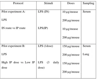
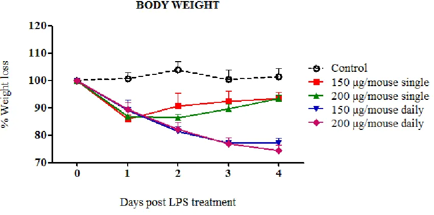
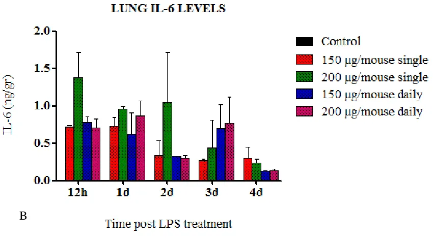
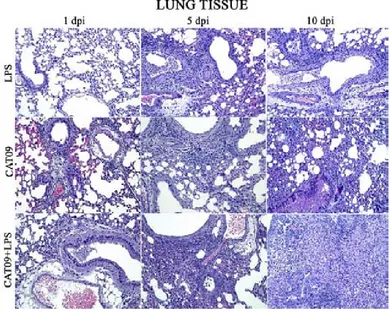
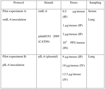
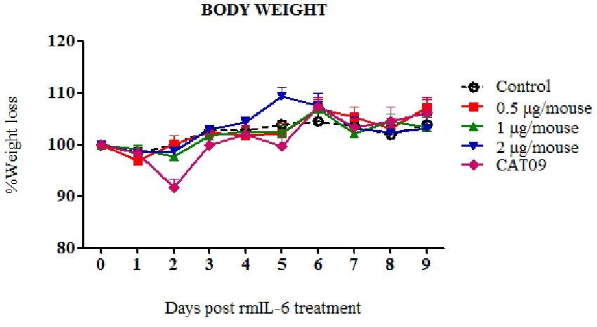
(6) viii.. Immunopathogenesis during pdmH1N1 2009 influenza infection. VI. VII.. 46. Oseltamivir resistance (OsR). 50. Animal models for influenza studies. 51. 4. HYPOTHESIS AND GENERAL OBJECTIVES. 56. 5. FIRST STUDY: Role of inflammation in influenza A pdmH1N1 2009 virus I. II. III.. 57. Introduction. 58. Hypothesis and specific objectives. 59. Materials and methods. 60. i. Cell lines. 60. ii. Viral Load. 60. iii. pdmH1N1 2009 Catalonian virus. 61. iv. Ethics statement. 61. v. Mice treatment and infection. 61. vi. Sampling. 62. vii. IL-6 by Enzyme-Linked Immunosorbent Assay (ELISA) 62 viii. Determination of viral load in tissues. IV.. 62. ix. Histopathology. 62. x. Statistical analysis. 63. Results. 64. i. Mice Pilot experiments. 64. 1. Pilot experiment A: Route of LPS administration. 64. a. IL-6 secretion on serum and lung. 66. 2. Pilot Experiment B: LPS dose a. IL-6 secretion on serum and lung. 67 69. ii. Role of LPS-derived inflammation on pdmH1N1 2009 virus infection. 71. iii. Virus Replication. 72. iv. IL-6 concentrations on serum and lung. 73. v. Histopathology. 75 6.
(7) 6. SECOND STUDY: Role of IL-6 on pdmH1N1 2009 virus infection in mice I. II. III.. IV.. 77. Introduction. 78. Hypothesis and specific objectives. 79. Materials and methods. 80. i. Cell line and viral preparation. 80. ii. pdmH1N1 Catalonian virus. 80. iii. IL-6 plasmid. 80. iv. In vitro plasmid IL-6 transfection. 81. v. Immunofluorescence assay. 82. vi. Ethics staments. 82. vii. Mice treatment and infection. 82. viii. Sampling. 82. ix. IL-6 detection by ELISA. 82. x. IL-10 detection by ELISA. 82. xi. Determination of viral load in tissues. 83. xii. Hemagglutination Inhibition (HI) Assay. 83. xiii. Histopathology. 83. xiv. Statistical analysis. 83. Results. 84. i. Pilot experiments. 84. ii. Pilot experiment A: rmIL-6 inoculation. 85. a. Virus Replication. 86. b. IL-6 expression on rmIL-6-inoculated mice and on CAT09 infected-mice iii. In vitro pIL-6 transfection 1. IL-6 production on Vero transfected cells. 86 88 88. 2. IL-6 production on supernatants of pIL-6 transfected-cells iv. Pilot experiment B: in vivo IL-6 plasmid transfection. 90 91. v. Role of IL-6 on pdmH1N1 2009 virus infection in C57BL6 mice. 92 7.
(8) 1. Mice and pdmH1N1 2009 infection. 92. 2. Viral replication. 94. 3. Antibody response. 95. 4. IL-6 concentrations on serum and lung. 96. 5. IL-10 concentrations on serum and lung. 97. 6. Histopathology. 99. 7. THIRD STUDY: In vitro and in vivo studies on pdmH1N1. I. II. III.. IV.. 2009-oseltamivir resistant virus in mice. 102. Introduction. 103. Hypothesis and specific objectives. 104. Materials and methods. 105. i.. Cell line and virus propagation. 105. ii.. Oseltamivir resistance viruses. 105. iii.. In vitro infection. 105. iv.. In vivo infection. 106. i. Ethics statement. 106. ii. Mice infection and sampling. 106. iii. Cytoquine detection by ELISA. 106. iv. Determination of viral load in tissues. 106. v. Hemagglutination Inhibition (HI) Assay. 106. vi. Histopathology. 106. vii. Statistical analysis. 106. Results i.. 107 In vitro viral growth of oseltamivir-sensitive and resistant pdmH1N1 2009 viruses. 107. ii.. R6 and R7 infection in mice. 109. iii.. Virus Replication. 111. iv.. Antibody response. 112. v.. IL-6 levels on OsR virus infected mice. 113. vi.. IL-10 levels on OsR virus infected mice. 114. vii.. Histopathology. 116. 8.
(9) 8. FOURTH STUDY: pdmH1N1 2009 influenza infection in. I. II. III.. IV.. ferrets from a mild and fatal case. 118. Introduction. 119. Hypothesis and specific objectives. 120. Materials and methods. 121. i.. Cell line and virus propagation. 121. ii.. Viral Load. 121. iii.. Virus. 121. iv.. Ethics statement. 121. v.. Animals and infection. 123. vi.. Clinical score. 123. vii.. Sampling. 124. viii.. Blood collection. 125. ix.. Acute phase proteins. 125. x.. Determination of viral load in tissues. 125. xi.. Hemagglutination Inhibition (HI) Assay. 125. xii.. Histopathology and Immunohistochemistry. 126. xiii.. Statistical analysis. 126. Results. 127. i.. Clinical score. 127. ii.. Clinical observations on pdmH1N1 2009 infected ferrets. 128. iii.. Acute phase proteins (APP). 130. iv.. Antibody response. 132. v.. Viral load. 133. vi.. Histopathology and Immunohistochemistry. 135. 9. DISCUSSION. 139. 10. CONCLUSIONS. 151. 11. OTHER PUBLICATIONS. 154. 12. REFERENCES. 156. 9.
(10) SUMMARY/RESUMEN 10.
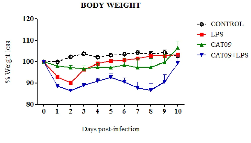
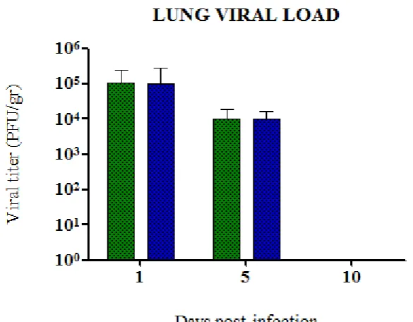
(11) 1. SUMMARY. Influenza is a worldwide public health concern, being one of the most common infectious diseases and a highly contagious airborne pathology. From April 2009, a new influenza A H1N1 virus with swine origin gave rise to the emergence of worldwide outbreaks which was subsequently declared as a pandemic situation. Nowadays pdmH1N1 2009 virus continues on circulation and generally triggers mild and selflimiting infections. Nevertheless, a small percentage of the patients require hospitalization and specialized attention in Intensive Care Units (ICUs). Noteworthy, in ICU patients an increased proinflammatory cytokine production has been identified. This observation would suggest the hypothesis that the heterogeneity in the outcome of pdmH1N1 2009 influenza virus infection could be due not only to differential fitness/virulence of the diverse circulating pandemic virus strains but also to the host immune environment that may contribute to severe respiratory pathogenesis, probably by an exacerbated immune response associated to hypercytokinemia. To test such hypothesis the work was divided in four studies describing the experiments performed in mice and ferrets with pdmH1N1 2009 viruses isolated during the 2009 outbreak from patients that showed mild to severe disease in order to analyse pathological features of the infection. Experiments in chapters 5 and 6 were conducted in order to evaluate whether high levels of proinflammatory cytokines and in particular IL-6, might affect host immune responses and the clinical course of infection. Mice were treated with LPS or mice expressing with high levels of IL-6 were infected with pdmH1N1 2009 (CAT09) simultaneously. In the case of LPS exposure, results showed that clinical signs and weight loss were directly influenced by LPS in CAT09 infected. However, no differences in viral load of lungs from infected mice were detected upon LPS exposure, indicating that LPS treatment was not affecting viral replication in vivo. IL-6 secretion upon LPS exposure correlated with body weight loss and higher pathology.. 11.
(12) The role of IL-6 in influenza infection was addressed by inducing IL-6 in mice prior CAT09 infection. Again, IL-6 levels correlated with weight loss. Surprisingly, viral replication was not affected by high levels of IL-6 since viral load did not exhibit significant differences when both infected groups were compared, although high level of IL-6 in infected animals correlated with sooner viral clearance than the CAT09 infected animals. A strong antibody response was detected in infected animals, being only CAT09 infected mice without IL-6 treatment, the ones with the highest hemagglutinin inhibiting titers. IL-10 levels correlated with IL-6 levels in serum and lungs in the first days after infection. Finally, histopathological lesions were more severe in mice with high levels of IL-6 and CAT09 infected. From the onset of the 2009 pandemic, oseltamivir resistance (OsR) mutations have been described on circulating pdmH1N1 2009 viruses. In chapter 7, in vitro and in vivo experiments in which two strains (R6 and R7) of OsR pandemic virus were compared. There were kinetics differences in both virus in vitro that finally were reflected in the pathogenesis of infection in vivo. The results obtained in the in vitro analysis showed different fitness in viral replication in the virus studied in comparison with a oseltamivir sensitive virus (F), being F>R6>R7. On the following in vivo experiment, both OsR strains produced a fatal outcome although with different magnitude and kinetics, R6infected group experimented a 40% of lethality and R7-group a 20% at 4 dpi. However, at 7 dpi the percentatge of survival was a 50% in both OsR-infected groups. Viral replication detected in lungs from OsR-infected groups had higher but not statistically different values for R7 than for R6. There was a strong antibody response at 14 dpi on both infected groups for each virus but no cross- reactive antibodies. Interestingly, high levels of IL-6 were detected in serum from R7-mice with significant differences. Surprisingly, levels of IL-6 in lungs of R6, R7 and control animals were similar at all time-point with no statistical differences. Serum and lung IL-10 had also slightly higher values in R7-mice when compared with controls at 3 and 5 dpi respectively. Histopahological findings showed more severe lesions on R7-mice at 5 dpi.. 12.
(13) To analyze possible virulence differences in viral fitmess, ferrets were infected with two contemporary pdmH1N1 2009 viruses from two patients without known co-morbid conditions, one that became fatal (F) while the other only showed mild (M) respiratory disease. These were studied in chapter 8. Ferrets developed different degree of clinical signs severity that did not correlate with the origin of the virus used in the infection, exhibiting severe (S) or non severe (NS) pathology. A significant decrease in body weight was detected in S animals compared to NS animals at 4 to 7 dpi. Clinical progress of the infection correlated directly with histopathological findings. The analysis of the acute phase proteins showed that the concentrations of haptoglobin (HP) and serum amyloid A (SAA) increased in both groups after 2 dpi. Virus titres in all tissues were higher in ferrets belonging to S group when compared to ferrets belonging to NS group at 4 dpi. Animals infected with both virus showed a strong hemagglutinin inhibiting antibody response in sera to both viruses at 10 and 14 dpi. Ferrets with a severe progress of the clinical infection showed slightly higher antibody responses and higher viral titers after infection.. 13.
(14) RESUMEN. La gripe es un problema de salud pública en todo el mundo, siendo una de las enfermedades infecciosas más comunes y una patología de las vías aéreas altamente contagiosa. Desde abril de 2009, un nuevo virus de influenza A H1N1, con origen porcino dio lugar a la aparición de brotes en todo el mundo siendo declarada posteriormente como una situación de pandemia. Hoy en día el virus pdmH1N1 de 2009 continúa en circulación, generalmente provocando infecciones leves y autolimitadas. Sin embargo, un pequeño porcentaje de pacientes requieren hospitalización y atención especializada en la Unidad de Cuidados Intensivos (UCI). Es digno de mención comentar que se ha identificado un aumento en la producción de citoquinas proinflamatorias en pacientes de UCI. Esta observación sugiere la hipótesis de que la heterogeneidad en el resultado de la infección por virus de la gripe pdmH1N1 de 2009 podría ser debido no sólo a la diferencia entre fitness /virulencia de las diversas cepas de virus pandemico circulantes, sino también por el medio ambiente inmune del huésped que pueden contribuir a la patogénesis respiratoria grave, probablemente por una respuesta inmune exacerbada asociada a hipercitoquinemia. Para probar esta hipótesis el presente trabajo se divide en cuatro estudios que describen los experimentos realizados en ratones y hurones con los virus pdmH1N1 de 2009 aislados durante el brote de 2009 de pacientes que mostraron grados de enfermedad desde leve a severa con el fin de analizar las características patológicas de la infección. Los experimentos en los capítulos 5 y 6 se llevaron a cabo con el fin de evaluar si los altos niveles de citoquinas proinflamatorias y, en particular IL-6, podrían afectar a la respuesta inmune del huésped y al curso clínico de la infección. Los ratones fueron tratados con LPS o ratones que expresaron altos niveles de IL-6 fueron infectados con el virus pdmH1N1 de 2009 (CAT09) simultáneamente. En el caso de la exposición a LPS, los resultados mostraron que los signos clínicos y la pérdida de peso fueron directamente influenciadas por LPS en los animales infectados con CAT09. Sin embargo, no se detectaron diferencias en la carga viral de los pulmones de los ratones infectados con exposición de LPS, lo que indica que el tratamiento con LPS no estaba 14.
(15) afectando a la replicación viral in vivo. La secreción de IL-6 después de la exposición a LPS correlacionó con pérdida de peso corporal a mayor grado de patología. El papel de la IL-6 en la infección por influenza se abordó mediante la inducción de IL6 en ratones antes de ser infectados con CAT09. Una vez más, los niveles de IL-6 se correlacionaron con la pérdida de peso. Sorprendentemente, la replicación viral no fue afectada por altos niveles de IL-6 debido a que la carga viral no mostró diferencias significativas cuando se compararon los dos grupos infectados, aunque los animals infectados con alto nivel de IL-6 aclararon el virus antes que los animales infectados con CAT09. Se detectó una fuerte respuesta de anticuerpos en los animales infectados, siendo los ratones infectados sólo con CAT09 sin tratamiento con IL-6, los que obtuvieron los mayores títulos de inhibición de hemaglutinación. IL-10 correlacionó con los niveles de IL-6 en el suero y en los pulmones en los primeros días después de la infección. Finalmente, las lesiones histopatológicas fueron más graves en los ratones con altos niveles de IL-6 e infectados con CAT09. Desde el inicio de la pandemia de 2009, mutaciones de resistencia a oseltamivir (OsR) se han descrito en virus circulante pdmH1N1 de 2009. En el capítulo 7 estudios in vitro e in vivo en el que se compararon dos cepas (R6 y R7) de virus pandémico OsR. Hubo diferencias in vitro en la cinética de ambos virus que finalmente se reflejó en la patogénesis de la infección in vivo. Los resultados obtenidos en el análisis in vitro mostraron diferencias en el fitness en la replicación viral de los virus estudiados respecto a un virus sensible a oseltamivir (F), siendo F > R6 > R7. En el siguiente experimento in vivo, ambas cepas OsR produjeron un resultado fatal aunque en diferente magnitud y cinética; el grupo infectado con R6 experimentó un 40 % de letalidad y el grupo R7 un 20 % a los 4 dpi. Sin embargo, a 7 dpi el porcentaje de supervivencia fue de un 50 % para ambos grupos infectados con virus OsR. La replicación viral detectada en los pulmones de los grupos infectados con virus OsR obtuvo los valores no estadísticamente diferentes para R7 que para R6. Hubo una fuerte respuesta de anticuerpos a los 14 dpi en ambos grupos infectados para cada virus, pero no anticuerpos de reacción cruzada. Curiosamente, se detectaron altos niveles de IL-6 en el suero de los ratones R7 con diferencias significativas. Sorprendentemente, los niveles de IL-6 en los pulmones de R6, R7 y animales control fueron similares a todos los tiempos sin diferencias estadísticas. Niveles de IL-10 de suero y pulmón mostraron 15.
(16) valores ligeramente más altos también en ratones R7 en comparación con los controles a los 3 y 5 dpi, respectivamente. Hallazgos histopatológicos mostraron lesiones más severas en ratones R7 a 5 dpi.. Para analizar las posibles diferencias de virulencia, se infectaron hurones con dos virus pdmH1N1 de 2009 de dos pacientes sin condiciones comórbidas conocidas, uno con consecuencia fatal (F), mientras que el otro sólo mostró enfermedad respiratoria leve (M). Este estudio está comprendido en el capítulo 8. Los hurones desarrollaron diferente grado de severidad de signos clínicos que no se correlacionaron con el origen del virus utilizado en la infección, exhibiendo patología severa (S) o no severa (NS). Se detectó una disminución significativa en el peso corporal en los animales S en comparación con los animales NS a 4 a 7 dpi. La evolución clínica de la infección se relaciono directamente con los hallazgos histopatológicos. El análisis de las proteínas de fase aguda mostró que las concentraciones de haptoglobina (HP) y amiloide A sérico (SAA) incrementaron en ambos grupos después de 2 pi. Los títulos de virus en todos los tejidos fueron más altos en los hurones pertenecientes al grupo S en comparación con los hurones pertenecientes al grupo de NS a 4 dpi. Los animales infectados con ambos virus mostraron una fuerte inhibición de la hemaglutinina de la respuesta de anticuerpos en el suero de ambos virus a 10 y 14 dpi. Los hurones con un progreso severo de la infección clínica mostraron respuestas de anticuerpos ligeramente más altos y los títulos virales más altos después de la infección.. 16.
(17) LIST OF ABBREVIATIONS. 17.
(18) 2. LIST OF ABBREVIATIONS. A A/ CastillaLaMancha/RR5661/2009- M A/Baleares/RR6121/2009- R6 A/CastillaLaMancha/RR5911/2009- F A/Catalonia/63/2009- CAT09 A/Madrid/RR7495/2011- R7 Acute phase proteins- APP Alveolar macrophages- Mf Antibody-dependent cell-mediated cytotoxicity- ADCC Antigen presenting cells- APC Avidin-biotin-peroxidase- ABC. B Biosafety level 3- BSL3 Bovine serum albumin- BSA Broncheo alveolar lavage- BAL. C C57BL6/JOlaHsd- C57BL6 Centers for Disease Control and Prevention- CDC Chronic obstructive pulmonary disease- COPD Conventional DCs- cDCs Cytophatic effect- CPE Cytotoxic T lymphocytes- CTL. D Days post infection- dpi Dendritic cells- DCs Diaminobenzidine tetrahydrochloride- DAB Dulbecco's Modified Eagle Medium- DMEM 18.
(19) E Enzyme-Linked Immunosorbent Assay- ELISA. F Fetal bovine serum- FBS. G Granulocyte colony-stimulating factor- G-CSF Granzymes- Gr. H Haematoxylin eosin- HE Haptoglobin- Hp Hemagglutination inhibition- HAI Hemagglutinin- HA Highly pathogenic avian influenza- HPAI Human immunodeficiency virus- HIV. I Influenza virus- IV Instituto de Salud Carlos III- ISCIII Intensive care unit- ICU Interferon alpha- IFN-a Interferon delta: IFN-δ Interferon gamma- IFN-γ Interferon gamma-induced protein 10- IP-10 Interleukin 2- IL-2 International Organization of Epizooties- OIE Intranasally- IN Intraperitoneally- IP Intravenously- IV Ion channel- M2 19.
(20) K Knock out- KO. L Lipopolysaccharide- LPS. M Macrophage inflammatory proteins 1 beta- MIP-1β Madin-Darby Canine kidney- MDCK Major histocompatibility complex- MHC Matrix protein- M1 Minimum Essential medium Eagle- MEM Monocyte chemoattractant protein-1- MCP-1 Multiplicity of infection- MOI Murine IL-6- mIL-6 N National Center for Biotechnology Information- NCBI National Influenza Centre- NIC Natural cytotoxicity receptors- NCR Natural killer cells- NK Neuraminidase inhibitors- NAIs Neuraminidase- NA Nitric Oxide Synthase 2- NOS2 NOD-like receptor family pryin domain containing 3- NLRP3 Non severe- NS Non-structural protein 1- NS1 Nuclear export protein- NEP; also known as NS2 Nucleoprotein- NP. O Oseltamivir resistance- OsR. 20.
(21) P Pandemic A (H1N1) 2009- pdmH1N1 Pathogen-associated marker pattern- PAMP Pattern-recognition receptors- PRRs Phosphate Buffer Saline- PBS Plaque Forming Unit- PFU Plasmacytoid DCs- pDCs Plasmid psDNA3.1+-mIL6- pIL-6 Polymerase complex- PB1, PB2 and PA. R Real time polymerase chain reaction- RT-PCR Recombinant mouse IL-6- rmIL-6 Regulatory T cells- Tregs Retinoic acid inducible gene-I- RIG-I. S Serum amyloid A- SAA Severe- S Sialic acid- SA Specific pathogen free- SPF Swine IV- swIV. T T cells- Tregs T helper 17- Th17 T helper- Th Tissue Culture Infective Dose- TCID50 Toll like receptors- TLRs Tuberculosis- TB Tumor Necrosis Factor alpha- TNF- α. 21.
(22) V Vascular endothelial growth factor- VEGF Viral RNA- vRNA. W White blood cells- WBC Wild type- WT. 22.
(23) INTRODUCTION. 23.
(24) 2. INTRODUCTION. I.. Introduction to influenza. Before Influenza virus (IV) took his place as etiological agent, the term influenza was established in Italy in the XV century. The concept derived from the former Italian expression ex influentia colesti used to refer to its mysterious origin in which the stars influenced the course of the disease. Previously, in 412 BC Hippocrates, the father of medicine, described a flu-like disease for the first time at Perinthus in North Greece 1. Influenza is one of the most common infectious diseases and a highly contagious airborne pathology with potentially fatal outcomes. It is characterized by symptoms that include fever, headache, cough, nasal congestion, sneezing, and whole-body aches 2. Despite the availability of inactivated vaccines derived from the current circulating strains, every year large segments of the human population are affected by influenza infection, because of frequent natural variation of the hemagglutinin (HA) and neuraminidase (NA) envelope proteins of the virus. This variation allows the virus to escape neutralization by preexisting circulating antibody in the blood stream, present as a result of either previous natural infection or immunization 2. IVs are unique in their ability to cause both recurrent annual epidemics and more serious pandemics that spread rapidly and may affect all or most age-groups. The size of epidemics and pandemics, and their relative impact, reflects the interplay between the extent of antigenic variation of the virus, the amount of protective immunity in populations, and the relative virulence of the viruses 3.. 24.
(25) II.. Influenza viruses i. Classification and antigenic types. The IV belongs to the family of Orthomyxoviridae, defined by viruses that have a negative-sense, single-stranded, and segmented RNA genome. IVs are divided into types A, B, and C on the basis of variation in the nucleoprotein antigen. In types A and B the HA and NA antigens undergo genetic variation, which is the basis for the emergence of new strains; type C is antigenically stable 4.The influenza A viruses are further subdivided on the basis of antigenic differences between the HA and NA surface proteins. There are now 18 different HA (H1 to H18) and 11 different NA (N1 to N11) subtypes for influenza A viruses, being the recently designated as H18N11 novel influenza A virus described in a flat faced fruit bat (Artibeus planirostris) from Peru5.. ii. Structure of the influenza virus. IVs are spherical or filamentous enveloped particles 80 to 120 nm in diameter. The helically symmetric nucleocapsid consists of a nucleoprotein (NP) and a multipartite genome of single-stranded antisense RNA in seven or eight segments (Figure 1). The envelope carries the HA attachment protein and the NA. The virus binds to host cells via HA. Transcription and nucleocapsid assembly take place in the nucleus. Progeny virions are assembled in the cytoplasm and bud from the cell membrane, killing the cell. IV genome comprises eight viral RNA (vRNA) segments. Each segment of the genome encodes a single virus polypeptide: PB2, PB1, PA, HA, NP, NA, M1, or NS1. 6, 7. .. Transcripts of the M1 and NS1 genes produce M2 and NS2 as splicing variants 8 . Until then, it was thought that the influenza viral genome encode 10 viral proteins in total. However, further studies of the IV genome identified exception of a segment that encodes two proteins by alternative splicing. In 2001, a viral protein, PB1-F2, was discovered as a second polypeptide made from the PB1 mRNA 9. Later, a third major polypeptide PB1-N40 was also identified as synthesized from the PB1 mRNA 10. More recently, the novel influenza A virus protein PA-X was discovered. 11. and in 2013. Muramoto and colleages 12 identified small proteins produced from the PA segment and 25.
(26) identified the translation initiation codons of these proteins on the PA mRNA by use of mutational analysis; the nature of these proteins has remained unclear. Currently, the identification of novel influenza virus proteins is receiving considerable interest and influenza segment PA has now been shown to encode as many as four proteins, PA, PAX, PA-N155 and PA-N182. 12. . All the studies together demonstrated that the eight. segments of the influenza viral genome can encode up to 16 proteins.. Figure 1. Influenza A virus particle. Schematic representation of Influenza A virus particle and gene segments. The influenza genome consists of eight single-stranded RNAs. The non-structural proteins and/or newly identified proteins with unknown function are depicted in the rectangles. The hemagglutinin (HA), neuraminidase (NA), and M2 proteins are inserted into the host-derived lipid envelope. The matrix (M1) protein underlies the lipid envelope. A nuclear export protein (NEP/NS2) is also associated with the virus. The viral RNA segments are coated with nucleoprotein and are bound by the polymerase complex. Imagen adapted from Schrawen et al 13.. iii. Genetics of the influenza virus. Genetic evolution of IV is given by key elements such as his segmented nature coupled to the error-prone RNA polymerase transcription and replication of the viral genome. Therefore, IV through the accumulation of mutation (antigenic drift) and/or reassortment (antigenic shift) oftentimes resulted in enhanced pathogenesis and expanded host range 2.. 26.
(27) Antigenic drift is caused by point mutations and it is defined as the minor gradual antigenic changes in the HA or NA protein. Influenza displays a high mutation rate due to the error-prone nature of the viral polymerase, so mutant viruses are easy to isolate 7. Mutations on the human virus HA or NA amino acid sequence occur at a frequency of less than 1% per year. Nonetheless, antigenic drift variants can cause epidemics and often prevail for 2–5 years before being replaced by a different variant 2. The antigenic sites of HA are all located in HA1 at or near the top of the molecule and mostly are found in protein loops. Similarly, antigenic drift has been found for NA and the sites of antigenic drift mapped to specific regions of the NA atomic structure 2. An important mutation that may occur in some IV strains is the H275Y mutation on the NA protein which confers to the virus Oseltamivir resistance (OsR). Antigenic shift, as a consequence of antigenic shift, IV develops a “reassortment” which is the switching of individual viral RNA gene segments during mixed infections with different IVs. Viruses resulting from such genetic exchanges are called reassortant viruses. Although reassortment occurs for influenza A, B, and C viruses, it does not occur among the different types. 2,7. . Antigenic shift leads to high infection rates in an. immunologically naïve population and is the cause of influenza pandemics through the introduction of a new human virus 2. The emergence of new pandemic strains of influenza A virus usually results from such a reassortment. There is a great deal of evidence for the reassortment of RNA segments between human and animal viruses in vivo and among human viruses in nature 2.. iv. Host receptors. One of the important factors conducting the tissue or cellular tropism of the virus is the specific binding of its surface glycoprotein HA to sialic acid (SA) receptors on the target cell surface. 4, 14. . HA binds to SA possessing either α2,3- or α2,6-linkage to. galactose. Interestingly, receptor preferences are thought to define host range 15. For example, in avian species α2,3-linked receptors are most abundant in the digestive tract, the primary site of influenza infection in avians, in human both α2,6- and α2,3- linked SA can be found in cells within the respiratory tract, but in different locations: α2,6linked SA are preferentially expressed in cells in the human upper respiratory tract, whereas α2,3-linked receptors are found in cells deeper in the lungs 16. Ferrets develop 27.
(28) similar clinical signs than human after infection with influenza virus, likely in part because the distribution of α2,6- and α2,3-linked SA receptors in the ferret respiratory tract resembles that observed in humans. 17,18. . Recently, it has been shown that α2,6-. linked SA receptors are more abundant than α2,3-linked receptors throughout the ferret respiratory tract 19. Moreover, the presence of α2,6- and α2,3-linked SA receptors in swine tracheal epithelial cells allows transmission of both avian and human viruses to pigs. 20. . This. supports the hypothesis that these animals can serve as a mixing vessel for the reassortment of novel influenza A viruses and their subsequent transmission to humans.. III.. Immune response to Influenza infection. When a infectious pathogen as influenza virus invade a host, the immune system response with the innate immune system that provides an immediate, but non-specific response followed by and adaptive immune response. On a second encounter, the immune system improved its recognition of the pathogen and adapts its response to defence the host against the pathogen. Both, innate and adaptive immune responses will be described in more detail on the following pages, a schematic picture adapted from Crisci et al. 2012 21 describe both immune responses in Figure 2.. i. Innate immunity. The innate immune system forms the first line of defense against influenza virus infection. It consists of components (e.g. mucus and collectins) that aim to prevent infection of respiratory epithelial cells. In addition, rapid innate cellular immune responses are induced that aim at controlling virus replication 22. Influenza A virus infection is sensed by infected cells via pattern-recognition receptors (PRRs) that recognize viral RNA, the main pathogen-associated marker pattern (PAMP) of influenza A viruses. The PRRs are toll like receptors (TLRs), retinoic acid inducible gene-I (RIG-I) and the NOD-like receptor family pryin domain containing 3 (NLRP3) protein 23. TLR7 binds single-stranded viral RNA (especially in plasmacytoid dendritic cells) and TLR3 and RIG-I bind double-stranded viral RNA (in most other infected 28.
(29) cells). Studies performed in mice showed that signalling of these receptors leads to production of proinflammatory cytokines and type I interferons 24.. ii.. Cells involved on innate immune response to influenza virus infection. Alveolar macrophages (Mφ). Once macrophages become activated in the lungs during IV infection, they phagocytose (apoptotic) influenza virus-infected cells to limit viral load 25,26. Activated macrophages also spread Nitric Oxide Synthase 2 (NOS2) and Tumor Necrosis Factor alpha (TNF-α) two molecules that have been identified as contributors to influenza virus induced pathology. 27. . These two distinct and competing functions of alveolar macrophages in. the immune response against influenza virus infection emphasize the importance of a balanced response.. Dendritic cells (DC). Dendritic cells (DC) play an important role as professional antigen-presenting cells during an influenza infection. The conventional DCs (cDCs), monitor the airway epithelial lumen to detect and opsonise (neutralized) virions and apoptotic bodies from infected cells but can also be infected themselves. 22. . Mice experiments demonstrated. that DC present influenza virus derived antigens, the immuno-peptides (epitopes), by Major histocompatibility complex (MHC) class I or class II molecules to activate a T cells response 28. Other DC subtype that has been investigated are the plasmacytoid DCs (pDCs), highly specialized in sensing viruses cell that readily secreting Interferon alpha (IFN-α) after exposure to swine IV (swIV) 29.. 29.
(30) Natural killer cells (NK). NK are important effector cells of the innate immune response. They can recognize antibody-bound influenza virus infected cells and lyse these cells, a process called antibody dependent cell cytotoxicity (ADCC). These cells can recognize influenza virus-infected cells with their cytotoxicity receptors (NCR) NKp44 and NKp46. Upon binding to the IV HAs the receptors trigger the human NK cell to lyse the infected cell 30. .. γδ T cells. Myeloid cells such as monocytes, Mφ, neutrophils, and myeloid DCs, clearly display innate characteristics, while lymphoid lineage B and αβ T cells represent the classical adaptive response. γδ T cells, however, display characteristics of both. γδ T cells, while sharing αβ T cell functions, also perform immune surveillance of an innate character and are the only major set of tissue-resident T cells 31 The antiviral activities of γδ T cells have been demonstrated in different models. 32. . In. the mouse model, γδ T cells were shown to contribute to recovery from influenza pneumonia 33, but no data are available on the contribution of γδ T cells at early stages of influenza virus infections. Activated mouse γδ T cells showed profound cytotoxicity against hemagglutinin (H1 or H3) expressing target cells in a non–major histocompatibility complex–restricted manner 34. In human, γδ T cells could efficiently kill macrophages infected with human (H1N1) and avian (H9N2 and H5N1) demonstrating the antiviral activity and the capacity of this cells to inhibit virus replication against influenza A viruses 35.. iii.. Adaptive immune system. The adaptive immune system forms the second line of defense against influenza virus infection. It consists of humoral and cellular immunity mediated by virus-specific antibodies and T cells respectively (Figure 2). 30.
(31) iv.. Humoral immunity. IV infection induces virus-specific antibody responses36. Presences of specific antibodies that recognize HA and NA have been correlated with protective immunity when the efficacy of current human influenza vaccines was tested. 37. . The HA-specific. antibodies provide protection when they are in direct correlation with the virus that causes the infection. HA-specific antibodies are capable to neutralize the virus by binding to the HA, inhibiting virus attachment and entry in the host cell of elderly and adults vaccinated individuals. 38. . Also, antibodies to the NA have protective potential.. By binding NA, antibodies do not directly neutralize the virus but they inhibit enzymatic activity that finally limits virus spread. Furthermore, NA-specific antibodies also facilitate ADCC and also may contribute to clearance of mice virus-infected cells 39. . NP is an important target for protective T cells. Also, NP-specific antibodies in mice. and human may contribute to protection against influenza virus infection 40. The leading antibody isotypes in the influenza specific humoral immune response are IgA, IgM and IgG. Mucosal or secretory IgA antibodies are produced locally and transported along the mucus of the respiratory tract. They can afford local protection from infection in airway epithelial cells. These antibodies are also able to in vitro neutralize IV intracellularly 41. Serum IgAs are produced rapidly after IV infection in human patients and the presence of these antibodies is indicative of a recent IV infection. 42. . Serum. antibodies of the IgG subtype predominantly transudate into the respiratory tract and afford long-lived protection on IV infected children 43. In mice, IgM antibodies initiate complement mediated neutralization of influenza virus and are a hallmark of primary infection 44.. 31.
(32) Figure 2. Immunity during influenza virus infection. The innate immune system forms the first line of defence against influenza infection. In the course of the innate immune response, cells like macrophages, dendritic cells, natural killer and γδT cells are recruited with the objective of controlling and blocking virus replication and dissemination. These cells secrete different types of chemical mediators such as cytokines that will activate the T cells and induces their differentiation or elicit an adaptive response with the production of specific IV-antibodies responses. Adapted from Crisci et al., 2013 21.. v.. Cellular immunity. Upon infection with IV, CD4+ T cells, CD8+ T cells and regulatory T cells (Tregs) are induced. CD4+ T cells play an important role in the immune response to this pathogen through the secretion of antiviral cytokines, and by providing help to CD8+T cells and B cells by promoting the development of immunological response of mice 45,46. CD4+ T cells also participate directly in viral clearance through the secretion of antiviral cytokines 45,47. CD4+ T cells are activated after recognizing virus-derived MHC class IIassociated peptides on Antigen presenting cells (APC) that also express co-stimulatory molecules. In human immunodeficiency virus (HIV) infected patients, it was observed that some CD4+ T cells display cytolytic activity to infected cells 48. However, the most important phenotype of these cells is T helper (Th) cells. Naive CD4+ cells can differentiate into T helper 1 (Th1) cells that are characterized by the production of Interferon gamma (IFN-γ), Interleukin 2 (IL-2) and TNF-α. Alternatively, antigen signalling in the presence of IL-4 induces the naive CD4+ cell population to develop into Th2 effectors secreting IL-4, IL-5 and IL-1349. Viral infections are known to predominantly induce Th1 or Type 1 immunity that promotes the activation of CD8+ T 32.
(33) cells and macrophage functions and drives B cell differentiation. In addition, regulatory T cells (Tregs) and T helper 17 (Th17) cells have been identified that regulate the cellular immune response to influenza virus infection. In contrast, in an inflammatory environment, Th17 cells improve T helper responses by producing IL-6 which inhibits Treg function 50. On the other hand, cytotoxic CD8+ T cells move to respiratory sites where virus replication is localized to eliminate infected cells. The main function of virus-specific CD8+ T cells is that of cytotoxic T lymphocytes (CTL). Upon influenza virus infection these cells are activated in the lymphoid tissues and recruited to the site of infection. There, they recognize and eliminate influenza virus infected cells and thus prevent production of progeny virus. Their lytic activity is mediated by the release of perforin and granzymes (Gr) (e.g. GrA and GrB). Perforin permeabilizes the membrane of the infected cells and subsequently Grs enter the cell and induce apoptosis. Recently, in human and mice experiments it was shown that even in the absence of GrA and GrB influenza virus-specific CTL were able to lyse target cells in vivo 51. Furthermore, CTL produce cytokines that improve antigen-presentation by stimulating MHC expression and that display antiviral activity. Post-infection virus-specific CTL are found in the lymphoid organs and in circulation. The reactivity and affinity of the memory CTL during a secondary infection depends on the co-stimulation they received during their initial differentiation phase 52. Human CTL induced by IV infection are mainly directed against NP, M1 and PA proteins 53,54. These proteins are highly conserved and therefore CTL responses display a high degree of cross-reactivity, even between different subtypes of influenza A virus.. 33.
(34) IV.. Pathogenesis and clinical signs during Influenza Infection. Influenza in adults and adolescents typically presents with an abrupt onset of fever and chills, accompanied by headache and sore throat, myalgias, malaise, anorexia, and a dry cough. Fever (38–40°C) peaks within 24 h of onset and lasts 1–5 days. Physical signs include the appearance of being unwell, hot and moist skin, flushed face, injected eyes, hyperaemic mucous membranes, and a clear nasal discharge. Although several of the symptoms of influenza are common to all age-groups, a review of published reports of influenza in children, adults, and elderly adults shows that the proportion of patients in whom these complaints are noted varies by age 55.. i. Genes involved on influenza A pathogenicity and virulence. HA, plays a critical role in adaptation to certain hosts through its affinity for receptors differentially expressed between species. 15,20. . HA receptor preference appears to affect. transmission by controlling the anatomical site of viral replication. The presence of HA is initially expressed as a precursor of HA0 and then cleaved into HA1 and HA2, forming a disulfide bond-linked complex. Structural data show that a loop structure exists in the cleavage site between HA1 and HA2, and this flexible loop is crucial for the efficient cleavage of HA0 56. Cleavage susceptibility of HA0 correlates well with the pathogenicity of highly pathogenic avian influenza (HPAI) viruses in poultry. 57. . In. mice, the multi-basic cleavage site was required for H5N1 virulence and viral spread to the mouse brain following intranasal infection 58. PB2, numerous substitutions within the PB2 subunit have been shown to alter host range and virulence. H5N1 viruses with a PB2 E627K mutation cause a lethal, systemic infection in mice, but become nonpathogenic for mammals if this residue remains a glutamic acid. 58. . A PB2 K627E mutation reduces transmission of human IVs in the. guinea pig, presumably because of reduced replicative ability in the upper respiratory tract. 59. . A PB2 D701N mutation is also associated with increased influenza virus. virulence in mammals by increases viral replication. 60. . Interestingly, pdmH1N1 2009. have neither the PB2 627K nor the 701N mutations that are associated with high pathogenicity, although second-site compensatory mutations in PB2 (590S and 591R) have been identified 61. 34.
(35) PB1-F2, various studies have suggested that this protein plays an important role in virulence of primary IV infection and in promoting secondary bacterial infection. 62. .. PB1-F2 was shown to contribute to the pathogenicity of the 1918, 1957, and 1968 pandemic strains, as well H5N1 HPAI viruses. 63,64. . When an S66N mutation was. incorporated into the PB1-F2 proteins of H5N1 and 1918 viruses, they became attenuated in mice. 63. . In addition, viruses with an N66S mutation caused increased. disease severity, lung titers, and cytokine production in mice. 62. . Interestingly,. pdmH1N1 2009 express only a truncated, 11–amino acid PB1-F2 protein, although introduction of PB1- F2, either with 66N or 66S, into recombinant pdmH1N1 2009 virus did not substantially enhance its virulence in mice or ferrets or predispose mice to secondary bacterial infection with Streptococcus pneumonia 65. NS1, prevents activation of transcription factors that induce IFN-β by blocking recognition of influenza PAMPs through RIG-I 65,66. The NS1 proteins of H5N1 HPAI viruses are associated with the induction of proinflammatory cytokines in the infected host 66. Likewise, an H5N1 P42S change results in a substantial increase in virulence in the mouse model and reduced levels of IFN-αβ production in vitro 67. L103F and I106M mutations in the NS1 of H5N1 viruses, which increase NS1 binding to the cellular premRNA processing protein cleavage and increase in vitro viral replication, presumably by suppressing expression of IFN-α/β mRNAs 68. NA, optimal influenza virus replication requires a functional balance between HA sialic acid binding affinity and receptor destroying, enzymatic activity of NA 69. This balance can be perturbed by a number of events, such as reassortment, introduction into a novel host, and antiviral therapy. The earliest human isolates of the 1957 H2N2 pandemic viruses paired an NA with preference for α2,3-linked substrates with an HA that bound well to α2,6- SA receptors. Over the years, this N2 gradually acquired the ability to cleave both α2,3 and α2,6 linkages, adapting to meet the receptor specificity of the HA 70. . This likely provided a selective advantage by allowing progeny virions to be released. more efficiently from the cell surface. Antiviral drugs can also influence the adaptation of influenza viruses.. 35.
(36) V.. Influenza Pandemics. Pandemics are rare events that occur every 10–50 years and cause a colossal loss of human lives. The lack of experience of the human immune system to identified and solved an influenza infection from a newly strain, could turn a seasonal IV in a pandemic virus with the efficient and sustained ability to be transmitted human-tohuman and finally, spreading globally. Over the human health history, historical record have been preserved as an evidence as how a pandemic of influenza could affect human population not only due to the high morbidity and mortality of the disease, but also for the high societal costs reflected on absenteeism, reduction and even paralysis of many sectors as schools or businesses, saturation of health services by the large number of patients that need medical attention and in the worst, loss of a family`s primary breadwinner. It is impossible to know with certainty the first time an IV infected humans or when the first influenza pandemic occurred. However, many historians have speculated that the year 1510 a.d. (500 years ago) marks the first recognition of pandemic influenza while there are historical records describing a pandemic outbreak of influenza-like disease in Europe. Mentioned influenza-like disease was characterized by a “gasping oppression” with cough, fever, and a sensation of constriction of the heart and lungs began to spread, apparently everywhere 71.. i. Pandemics in the XX century. During the last XX century, three pandemics of influenza affected the human population. The involved influenza subtypes were the followings: H1N1 (1918-1919), H2N2 (1957-1963) and H3N2 (1968-1969) 1. Each subtype was originated as a consequence of reassortment. In order of appearance, the first one was the so called “Spanish” influenza in 1918 and 1919, with an HA related to those of swine viruses or H1 subtype viruses. Viruses of this subtype circulated until 1957, when viruses of the H2N2 subtype (Asian strains) were isolated. The H2 subtype HA has little or no crossreactivity with the H1 HA. In addition to containing an H2 HA, the Asian strains had a new NA (N2). For 11 years, the H2N2 strains of influenza virus spread and changed 36.
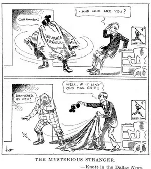
(37) until the next pandemic in 1968 with the introduction of a new H3 subtype (Hong Kong strains). These drastic antigenic changes came about from the reassortment of previously circulating human viruses and IVs of animal origin.. The H1N1pandemic of 1918–1919 “Spanish influenza”: on 1918 the world witnessed the worst IV outbreak in recorded history; the named “Spanish influenza” (“Spanish Flu”), publicity and sanitary strategies to try to control dissemination could not stop virus spread (Figure 3). The virus was spread all over the global population in three waves between 1918 and 1919 carrying 20 million to 50 million of human lives was especially dangerous to young adults. 73. 72. . It. with unusually high numbers of deaths in. young and healthy people aged 15 to 35 years 74. It has been estimated that about 25 per cent of the world’s population was infected. During this period, development of “Spanish Flu” spread was strongly influenced by the First World War (1914-1918). In fact, global dissemination and severity were directly linked to the war and the movement of troops 75. Studies focusing on the HA protein have found that the HA gene contributed to efficient viral replication and high virulence of the 1918 virus in mice 26,76. . Recently, the genome of the 1918 pandemic IV was completely sequenced 77, and. the virus was reconstructed using reverse genetics. 26. . The reconstructed 1918 virus. caused a highly pathogenic respiratory infection in mice. 26. and macaque models that. culminated in acute respiratory distress and a fatal outcome 78. It was shown that mice vaccinated with the monovalent pdmH1N1 2009 vaccine were completely protected in a lethal challenge model with the 1918 influenza virus. Because the 2009 pandemic H1N1 virus contains the HA gene derived from the classical swH1N1 lineage, it is antigenically very similar not only to classical swH1N1 viruses, but also to the 1918 virus. 73. . Also, ferrets immunised with DNA vaccines encoding proteins of the original. 1918 H1N1 pandemic virus exhibited protective cross-reactive immune responses against infection with a 1947 H1N1 virus and a recent 1999 H1N1 virus. 79. .. Consequently, seroepidemiologic studies had demonstrated cross-protective immunity in the population, primarily in people >60 years 80.. 37.
(38) Figure 3. The Mysterious Stranger: a cartoon in the Dallas News during the 1918–1919 influenza pandemic. Source: How to fight Spanish influenza. Literary Digest 1918 Oct 12;59:13. The cartoon is attributed to Knott of the Dallas News 81.. The H2N2 pandemic of 1957-1963 “Asian influenza”: it was named after the first identification in Guizhou, a province in south-central China. Investigations to elucidate the origin of the Asian circulation strain, placed the novel pandemic H2N2 as an avianhuman reassortant. Unlike “Spanish Flu”, the principal target of this virus was centred on the elderly population but also on infants, with about 1 to 2 million of deaths worldwide. 82. . Although the proportion of people infected was high, the illness was. relatively mild compared to the Spanish flu. In December of 1957 when it was believed controlled, a second wave struck at the beginning of 1959, to suddenly disappear given rise to the next pandemic. 83,84. . H2N2 stopped circulating in the human population in. 1968. However, strains of H2 subtype still continue to circulate in birds and occasionally in pigs and they could be reintroduced into the human population through antigenic drift or shift. 38.
(39) The H3N2 pandemic of 1968 “Hong Kong influenza”: the last pandemic of the XX century was a new of Asian origin. Influenza A viruses of the H3 subtype caused the 1968 Hong Kong pandemic, the HA gene being introduced into humans following a reassortment event with an avian virus. 85. . The pandemic began in Hong Kong and. deaths hovered all over the world. It is believed that 1 to 2 million of people died. 83. .. Since 1968, H3N2 has been one of the most prevalent seasonal influenza virus circulating in human and swine population 86. In a cross-reactive immunity experiment, it was demonstrated that a DNA vaccine, based on the HA and NA of the 1968 H3N2 pandemic virus, induced cross-reactive immune responses against a recent 2005 H3N2 virus challenge in ferrets 79.. ii.. 2009 Swine influenza pandemic: the first pandemic of XXI century.. The first pandemic of the XXI century have been originated by an swine-origin influenza A H1N1 virus, characterized by a novel combination of gene segments “triple reassortant”, that had not been identified among human or swIV. As mentioned before, pigs are considered logical candidates for reassortment because they can be infected by either human or avian viruses as they possess both SA receptor (SAα2,6 and SAα2,3) in the cells of the respiratory system. 20. . In addition, pigs are known to be involved more. frequently in interspecies transmission of influenza A viruses than other animals 87.. iii.. Origin of pandemic A (H1N1) 2009 virus. On April 15 and April 17, 2009, the first two human cases of influenza caused by a new influenza strain were confirmed. A 10-year-old children from southern California was the first infected identified; two days later, the Centers for Disease Control and Prevention (CDC) confirmed a second case of infection with the same virus in a 9-yearold girl from an adjacent county in California. 88. . During the subsequent 2 weeks,. additional cases of infection with this new virus were detected in Mexico, California, Texas, and other states. 88. . That unique combination of gen segments had not been 39.
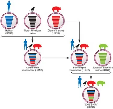
(40) previously indentified. The CDC distributed information confirmed that these cases were caused by the same new swine strain of influenza A (H1N1) virus, also CDC described that the generation of the circulating strain derived from a triple reassortment of human, swine and avian viruses. 89. . Phylogenetic analyses of pdmH1N1 2009 virus. isolates revealed a great homogeneity of genomic sequences. The virus was antigenically distinct from human seasonal influenza viruses but genetically related to three viruses that circulated in pigs. The pdmH1N1 2009 virus has therefore inherited virus gene segments of all three sources: swine, human and avian origin. 90,91. . CDC. released the genomic sequences of vRNAs from 6 swine flu isolates from California and Texas on 29 April 2009. 88. . The samples of the infected patients were genetically. analysed and the NA (N1) and M genes of the pdmH1N1 2009 virus were shown to have different origins, from the “avian-like” Eurasian swine H1N1 lineage, which emerged in Europe in 1979 after reassortment between a classical swine and an avian H1N1 virus. 92. . The virus then spread through Europe and Asia. 93. , displacing the. classical swine H1N1 virus from Europe and generating new reassortants in swine with different influenza A viruses of human origin 94. Finally, the PA, PB1 and PB2 genes of the pdmH1N1 2009 virus are from the North American H3N2 “triple-reassortant” lineage, which was first isolated from pigs in America in 1998 in which it showed unusual pathogenicity. 95. . The pdmH1N1 2009 virus has therefore inherited virus gene. segments of all three sources: swine, human and avian origin (Figure 4).. 40.
(41) Figure 4. Diagrammatic representation of the evolutionary history of pdmH1N1virus. Reassortment of North American swine H3N2 and H1N2 triple-reassortant viruses (of North American avian, human [H3N2], and classical swine [H1N1] origin) with Eurasian avian-like swine viruses (H1N1) resulted in the pdmH1N1 2009 virus. Each gene segment of avian, human, or swine origin corresponds to a characteristic feature on the surface of the schematic viral particle. Adapted from Tscherne et al 96.. iv.. Epidemiology of A (H1N1) 2009 pandemic. The emergence of the pdmH1N1 2009 influenza virus in humans in early April came as a total surprise. The pdmH1N1 2009 strain quickly spread worldwide through humanto-human transmission. On December 30th, 2009 the number of countries that reported laboratory-confirmed pdmH1N1 2009 virus cases in humans was 208 and more than 214 on April 18th, 2010. 97. . Studies have shown that pdmH1N1 2009 virus was. circulating in the environment three months prior to the outbreak 98. 41.
(42) The transmissibility of the pdmH1N1 2009 virus in house of infected patients was lower than that seen in past pandemics 99. The mean time between the onset of symptoms in a patient case and the onset of symptoms in the house contact infected by that patient was 2.6 days (2.2–3.5). A characteristic feature of the pdmH1N1 2009 was that it disproportionately affected children and young adults as compared to the older age groups 100. In most countries, the majority of pdmH1N1 2009 virus cases have occurred in younger age groups, with the median age estimated to be 12–17 years in Canada, USA, Chile, Japan and the UK. On 2009, of the 272 patients with pdmH1N1 2009 virus infection who were hospitalized in the USA in a three month period, 45% were under the age of 18 years, whereas only 5% were 65 years of age or older 101. This age distribution suggested partial immunity to the virus in older population. 102. .. This hypothesis was supported by subsequent studies which showed that 33% of humans over 60 years of age had cross-reacting antibodies to pdmH1N1 2009 virus by hemagglutination-inhibition test and neutralization tests 88,92,103. It should be noted that while the highest rate of severe disease leading to hospitalization has been in patients less than 5 years of age, the highest case fatality rate was recorded in the 50–60-year old population 97. More than 3 years after the emergence of the 2009 pdmH1N1 virus, the associated global mortality remains unclear. Of 18.500 laboratoryconfirmed pdmH1N1 2009 virus-associated deaths identified during April, 2009, to April, 2010 worldwide, less than 12% were reported from Africa and southeast Asia, although these regions are home to more than 38% of the world's population. 104. . Spain. was one of the first European countries to inform pandemic influenza cases105. Nowadays, epidemiological survilance of the pdmH1N1 2009 virus indicated that we are in a postpandemic period which does not mean that the pdmH1N1 2009 virus has gone away, in fact this pathogen is still on circulation.. 42.
(43) v.. Virulence markers pdmH1N1 2009 virus. As well as the pathogenicity, the virulence of the IVs can be measured using parameters of morbidity and mortality within animal models.. IV pathogenicity is considered. multigenic and it is determined by the variety of genes within a particular IV strain within a specific host. 54,91. . Genetic mutations in IV proteins, including HA and NA 54,. NS1 106 and PB1-F2 63, and the polymerase complex, occur during viral host adaptation and result in enhanced virulence. The particular genetic mutations related to specific characteristics can enhance various aspects of the viral life cycle, including virus binding and entry, genome transcription and translation, virion assembly and release, and evasion of innate immune responses have been identified (Table 1) 96.. Table 1. Influenza genes involved on pathogenicity and virulence. Adapted from Tscherne et al, 2011 96.. vi.. Pathogenesis and clinical signs during pdmH1N12009 infection. In general terms, pdmH1N1 2009 virus infection is mostly a mild, self-limiting upper respiratory tract illness with (or for some patient groups, without) fever, cough and sore throat, myalgia, malaise, chills, rhinorrhea, conjunctivitis, headache and shortness of breath. Up to 50% of patients present with gastrointestinal symptoms including diarrhea and vomiting. The spectrum of clinical presentation varies from asymptomatic cases to. 43.
(44) primary viral pneumonia resulting in respiratory failure, acute respiratory distress, multi-organ failure and death 101. The pdmH1N1 2009 virus replicates in the cells of the upper and lower respiratory tract, the incubation period appears to range from 2 to 7 days, but most patients probably shed virus from day 1 before the onset of symptoms through 5–7 days after. 107. . The median. period during which the virus could be detected with the use of real time polymerase chain reaction (RT-PCR) in quarantined patients was 6 days (range 1–17), whether or not fever was present 108.. vii.. Clinical risk factors of pdmH1N1 2009 virus infection. Influenza infection in immunocompromised hosts may prolong the illness longer than normal, and the virus may replicate for an extended period of time. Approximately, one quarter to one half of patients with pdmH1N1 2009 virus infection who were hospitalized or died had no reported coexisting medical conditions. 109,110. . Underlying. conditions that are associated with complications from seasonal influenza also are risk factors for complications from pdmH1N1 2009 virus infection. Chronic obstructive pulmonary disease (COPD), asthma, cardiovascular disease, hypertension and diabetes have been reported as risk factors for critical illness following infection with the pdmH1N1 2009 virus 101,111,112. In addition, during the 2009 pandemic, pregnancy 113,114 obesity. 114,115. and smoking. 111. were identified as risk factors for severity. Therefore,. basal immune alterations and/or the presence of a previous pro-inflammatory state favoured by the presence of medical conditions may impact the normal development of specific immune responses against the pdmH1N1 2009 virus, increasing the risk of developing severe forms of the infection 116. The patients in the high risk groups need to be cared more and treated at priority as compared to low risk groups to prevent the loss of life.. 44.
(45) Pregnancy: It is linked to a number of induced changes in the immune system of the mother, aimed at tolerating the fetus. In pregnant women, the balance between pro- and anti-inflammatory factors seems to be crucial. 117. . In addition, changes in the peripheral. levels of immune mediators such as Interferon delta (IFN-δ), TNF, Vascular endothelial growth factor (VEGF), Granulocyte colony-stimulating factor (G-CSF), eotaxin, and Monocyte chemoattractant protein-1(MCP-1) may impact the proper performance of the immune response. 118. . Although these changes in the immune system are not fully. understood, it is believed that they may increase the severity of some infections. 117,119. .. Previous 1918 and 1957 pandemics reported as well as the risk to the pregnant woman, the risks to the fetus; increased rates of miscarriage, stillbirth, and premature birth 3. The increased mortality detected in pregnant female mice infected with the pdmH1N1virus was associated with increased infiltration of neutrophils and macrophages in the lungs of these animals. Also, pregnant mice showed higher levels of chemokines and proinflammatory cytokines, lower respiratory epithelial regeneration and poorer fetal development than nonpregnant mice. 120,121. . Although pregnant women represent only 1. to 2% of the population, among patients with pdmH1N1 2009 virus infection, they have accounted for up to 7 to 10% of hospitalized patients,101 6 to 9% of ICU patients, 111 and 6 to 10% of patients who died 122,123.. Pulmonary complications: Primary viral pneumonia is associated with a high mortality rate. It begins within 24 h of the onset of febrile illness with a dry cough that later becomes productive of bloody sputum accompanied by tachypnoea, diffuse fine rales, progressive cyanosis, and respiratory failure 3. IVs can lead to an acute exacerbation of chronic bronchitis in people with chronic obstructive pulmonary disease or cystic fibrosis and to wheezing in patients with asthma. 124. . Results of pdmH1N1. 2009 virus experiments with cell cultures and mouse models have reported that a high level of cytokines could itself prevent the development of and appropriated immune response against viruses, affecting dendritic cell function along with HLA-II mediated antigen presentation125,126. In a mouse model of COPD, animals infected with influenza virus showed an exacerbated inflammatory response to infection. 127. . In humans,. previous studies demonstrated that chronic respiratory diseases such as COPD and asthma during a pdmH1N1 2009 influenza infection are characterized by a basal predisposition to the release of inflammatory mediators 128. 45.
(46) Metabolic complications: Among patients with severe or fatal cases pdmH1N1 2009 virus infection as severe obesity or morbid obesity has been reported at factors of higher pathogenicity than in the general population. 111,122. . In addition to the risks associated. with obesity, such as cardiovascular disease or diabetes, immunological alterations in the obese may contribute to its role as a risk factor in pdmH1N1 2009 infection. 129. .. Obese mice infected with the pdmH1N1 2009 virus exhibited significant higher morbidity and mortality compared to non-obese mice. 73. . Adipocytes have structural. similarities with the immune cells and perform certain functions related to them, such as the release of inflammatory mediators. Furthermore, differentiation of macrophages in the adipose tissue is conditioned by the metabolic environment and immune cells in turn are able to control lipid and glucose metabolism, suggesting an immune metabolic axis. Then, a chronic caloric excess could interfere with the mechanisms of the immune response 130.. viii.. Immunopathogenesis during pdmH1N1 2009 influenza infection. The immune system is designed to protect and maintain homeostasis and the ability of an organism to adapt to the environment. Therefore, it plays a key role in viral clearance, as explained in the section “Immune response to Influenza infection” (section III). Human autopsy studies of pdmH1N1 2009 infected patients have pointed out the contribution of excessive acute inflammatory responses to death following severe influenza infection, including influx of innate cells into the lungs and overproduction of cytokines (Figure 5) and chemokines that culminate in life-threatening pulmonary immunopathology 131.. 46.
(47) Hypercytokinemia: a first report published in December 2009 revealed that severe disease caused by the pdmH1N1 2009 virus was characterized by the presence of high systemic levels of cytokines, chemokines and other immune mediators from the early stages of the disease 125,132,133. Infection by the influenza pdmH1N1 2009 virus induced the secretion of antiviral defense-related chemokines (interferon gamma-induced protein 10 (IP-10)), macrophage inflammatory proteins 1 beta (MIP-1β), MCP-1 and IL-8). These chemotactic molecules mobilize T lymphocytes, monocytes, macrophages and neutrophils to the site of infection to fight the infection. 134. . However, an accumulation. of these cells may contribute to inflammatory-mediated damage to the infected tissue. Infected patients also exhibited elevated levels of other pro-inflammatory immune mediators that stimulate T Th1, IFN-δ, TNF-α, IL-15, IL-12p70. On the other hand, Th1 cytokines may, as chemokines, contribute to tissue injury. Studies on cytokine profiles also revealed elevation of two Th17 related cytokines (IL-9, IL-6) in the early course of the severe cases of pneumonia caused by pdmH1N1 2009 virus. However, a beneficial role of IL-17 in lethal influenza has been previously proposed. 132,133. . Additionally, G-. CSF, which has been described as interfering with the synthesis of IL-17. 135. has been. reported to be directly associated with the risk of death in critically ill patients. Regarding IL-6, there is a fairly broad consensus in the literature that this cytokine could be a potential biomarker for severe pdmH1N1 2009 infection, in both human and in mouse studies 125,132,136,137. Elevated systemic levels of IL-6 were strongly associated with ICU admission and with fatal outcomes. Furthermore, in animal and clinical studies, global gene expression analysis indicated a pronounced IL-6-associated inflammatory response. 137,138. . In addition, IL-6 has been implicated in the cytokine. storm following avian influenza A H5N1 and in severe acute respiratory syndrome infection. 139. infection. 140. . It has also been associated with severe cases of seasonal influenza. . Cases of severe pandemic influenza disease were also marked by high. levels of two immunomodulatory cytokines (IL-10 and IL-1ra). Hypercytokinemia persisted in the most severe cases, which could have perpetuated the inflammatory damage and, in consequence, the respiratory failure observed in these patients (Figure 5) 133,138. Similarly, other clinical studies demonstrated that high plasma levels of IL-6, IL-8 and MCP-1 correlated with the extent and progression of pneumonia 141. . The most severe cases also showed persistent viral shedding, again indicating poor. control of viral replication. 47.
(48) Antibodies reponse: one of the most striking features of the 2009 pandemic was the low proportion of elderly individuals infected by the new virus, compared to seasonal influenza 101,111,112. In addition, severe illness caused by the new variant predominated in young patients, with 90% of deaths occurring in patients <65 year old. 136,142. which is. contrary to the normal trend in seasonal influenza. It is believed that adults born after 1956 have suffered previous exposures to antigenically related influenza viruses, developing in consequence cross-reactive antibodies with the ability to recognize the 2009 strain. 80,143. . Studies on antibody prevalence show the presence of cross-reacting. antibodies in as much as in 33% of the over-60 population. 144. . This result is consistent. with the fact that young adults admitted to the ICU during the 2009 pandemic lacked protective antibodies in the early stages of the disease, as revealed by hemagglutination inhibition (HAI) and micro-neutralization assays. 132,138. . However, the absence of early. HAI and neutralization activity was the rule in young patients, independent of disease severity and outcome. It is important to note that most of these critical patients were able to mount specific antibody responses against the pandemic virus, regardless of severity 138. This suggests that factors other than the development of specific antibodies contribute to the pathogenesis of severe pandemic influenza.. Cellular immune responses: cross-reactive T CD4+ Th lymphocytes and T CD8+ CTLs established by vaccination campaigns or natural infection by the seasonal influenza A virus have been reported to contribute to clearance of the pdmH1N1 virus from the lungs145,146. Even in the absence of protective antibody responses, individuals vaccinated against seasonal influenza A may still benefit from pre-existing crossreactive memory CD4+ T cells thus reducing their susceptibility to the influenza pdmH1N1 2009 virus 146. T CD4+ effector cells are essential for virus clearance, but in turn, they may contribute to the hypercytokinemia observed in the most severe cases caused by influenza pdmH1N1 2009 virus infection. In fact, a study in a murine model demonstrated that depletion of T cells prevented immunopathology, although with decreased viral clearance. 147. . In turn, CD8+ T cells are known to release cytotoxic. molecules (granzyme and perforin) and antiviral cytokines (TNF-α and IFN-δ), which are essential for mediating the elimination of infected cells. A small report on human autopsy tissues documented diffuse alveolar damage, haemorrhage and necrotizing 48.
(49) bronchiolitis in the lungs of patients who died from influenza pdmH1N1 2009 virus infection. Immunohistological examination revealed an aberrant immune response associated with marked expression of TLR3 and IFN-δ and a large number of CD8+ T cells and Gr B+ cells within the lung tissue 148, highlighting the role of cellular immune responses in the immunopathology of pdmH1N12009 influenza infection. T cells from influenza pdmH1N1 2009 infected patients presenting with a severe clinical course have been described as resulting in impaired effector cell differentiation and as failing to respond to mitogenic stimulation. 126. severe acute phase of the infection. . In addition, T cell anergy is observed during the 126. . The adaptive immune response of pdmH1N1. 2009 virus infected patients has been reported to be characterized by decreases of CD4lymphocytes and of B-lymphocytes and by increases in T-regulatory lymphocytes. 149. .. The latter cells may suppress the development of specific responses against the virus. Critical pandemic influenza illnesses coursed with lower expression in the white blood cells (WBC) of a group of genes key to the development of antigen presentation and adaptive immune response 138. Deficiencies in the cellular immunity occurred in severe cases of influenza pdmH1N1 2009 infection could help explain the poor control of the virus observed in these patients, and the increased risk of these patients to suffer from bacterial over-infections.. 49.
Figure
+7
Documento similar
The results indicate that the virus isolated from the fatal case replicates faster, induces higher levels of cytokines in human alveolar lung epithelial cells and is more pathogenic
In the “big picture” perspective of the recent years that we have described in Brazil, Spain, Portugal and Puerto Rico there are some similarities and important differences,
The faculty may have uploaded some file with the complete timetable by subjects.. 7) Scroll down and click on the academic course. You will find all the classes grouped by
Bartoli et al., 2015b; Thakur et al., 2016) and provides the first evidence of the WHOP region (from woody host and Pseudomonas) contribution to virulence and fitness of Psv NCPPB
In the preparation of this report, the Venice Commission has relied on the comments of its rapporteurs; its recently adopted Report on Respect for Democracy, Human Rights and the Rule
The expression ((having become common since the spring)) -the French original reads ((etant devenues populaires des le printems»- refers to the spring of 1708 and is
For this reason, we studied the induction of different innate immune molecules at early stages of infection, in different lung anatomic compartments, aiming to discern the cytokine
Government policy varies between nations and this guidance sets out the need for balanced decision-making about ways of working, and the ongoing safety considerations
