Otras secciones de este sitio:
☞ ☞ ☞ ☞
☞ Índice de este número
☞ ☞ ☞ ☞
☞ Más revistas
☞ ☞ ☞ ☞
☞ Búsqueda
Others sections in this web site:
☞ ☞ ☞ ☞
☞ Contents of this number ☞
☞ ☞ ☞
☞ More journals ☞
☞ ☞ ☞ ☞ Search Artículo:
Risk factors for liver fibrosis progression in patients with chronic hepatitis C
Copyright © 2003: Mexican Association of Hepatology ANNALS OF HEPATOLOGY
Number 1 January-March 2003 Volume 2
M. de Torres et al. / Risk factors for liver fibrosis progression in patients with chronic hepatitis C 5
edigraphic.com
Annalsof Hepatology
Abstract
The major hepatological consequence of HCV infection is the progression to cirrhosis and its potential compli-cations. Several factors have been clearly shown to be associated with fibrosis progression rate: duration of infection, age, male sex, consumption of alcohol, HIV coinfection and low CD4 count. As age and duration of infection increases, the risk of fibrosis increases and the impact of treatment (IFN) decreases. In conclusion, fi-brosis progression has a progressive acceleration, sex, age and consumption of alcohol are strongly involved in this progression ; the possibility to assess with non-agressive biochemical markers the fibrosis stage will probably allow in the future to identify other factors related to fibrosis progression.
Key words: Chronic hepatitis C, liver fibrosis pro-gression, cirrhosis.
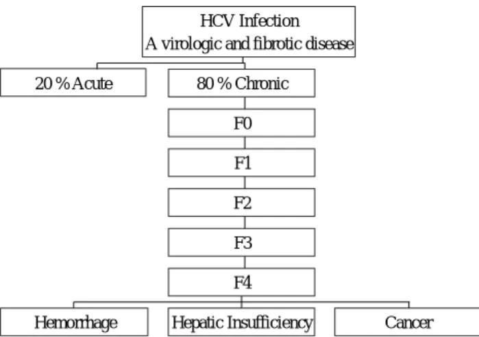
Mortality associated with chronic hepatitis C is usual-ly due to the development of cirrhosis and its potential complications: haemorrhage, hepatic insufficiency and primary liver cancer.1-6
Current understanding of HCV infection has been ad-vanced by the concept of liver fibrosis progression7,8
(Fig-ures 1-3). Fibrosis is the deleterious but variable conse-quence of chronic inflammation. It is characterized by the deposition of extra-cellular matrix component leading to the distortion of the hepatic architecture with impairment of liver microcirculation and liver cell functions. HCV is usually only lethal when it leads to cirrhosis, the last stage of liver fibrosis. Therefore, an estimate of fibrosis progres-sion represents an important surrogate endpoint for evalua-tion of the vulnerability of an individual patient and for as-sessment of the impact of treatment on natural history.
Concise Review
Risk factors for liver fibrosis progression in
patients with chronic hepatitis C
Mercedes de Torres,1 Thierry Poynard1
1Service d’Hépato-Gastroentérologie Groupe Hospitalier Pitié-Salpêtrière.
Address for correspondence: Thierry Poynard, M.D., Ph. D.
Service d’ Hépato-Gastroentérologie Groupe Hospitalier Pitié-Salpêtriére 47-83, Boulevard del’ Hôpital 75651 Paris Cedex 13
Tel: 33 1 42 16 10 02-Fax: 33 1 42 16 14 27 E-mail: [email protected]
Fibrosis stages and necro-inflammatory activity grades
Activity and fibrosis are two major histologic features of chronic hepatitis C which are included in different pro-posed classifications.9-12 One of the few validated scoring
systems is called the METAVIR scoring system.11,12 This
system assesses histologic lesions in chronic hepatitis C using two separate scores, one for necro-inflammatory grade (A for Activity) and another for the stage of fibrosis (F). These scores were defined as follows; Stages of fibro-sis (F) (Figure 1): F0 = no fibrofibro-sis, F1 = portal fibrofibro-sis without septa, F2 = portal fibrosis with rare septa, F3 = nu-merous septa without cirrhosis, F4 = cirrhosis. Grade for activity (A): A0 = no histologic activity, A1 = minimal ac-tivity, A2 = moderate acac-tivity, A3 = severe activity. The degree of activity was assessed by integration of the sever-ity of the intenssever-ity of both piecemeal (periportal) necrosis and lobular necrosis as described in a simple algorithm.12
The intra- and inter-observer variations of this METAVIR scoring system are lower than those of the widely used Kn-odell scoring system.9,10 For METAVIR fibrosis stages
there is an almost perfect concordance (kappa = 0.80) among pathologists. The Knodell scoring system has a non-linear scale. There is no stage 2 for fibrosis (range 0-4) and the activity grade ranges from 0 to 18 with the sum of periportal necrosis, intralobular and portal inflammation grades. The modified Histological Activity Index is more detailed with 4 different features and continuous grades and the modified fibrosis staging includes 6 stages.
Activity grade, which represents the necrosis fea-ture, is not a good predictor of fibrosis progression.7 In
fact fibrosis alone is the best marker of ongoing fibro-genesis.13 So far there is no study demonstrating
clear-ly that activity grades are predictive of fibrosis pro-gression independently of fibrosis stage.14 Fibrosis
Annals of Hepatology 2(1) 2003: 5-11
6
edigraphic.com
The dynamic view of fibrosis progressionFibrosis stage summarizes the vulnerability of a patient and is predictive of the progression to cirrhosis7 (Figure 2).
There is a strong correlation for fibrosis stages, almost lin-ear, with age at biopsy and duration of infection. This corre-lation was not observed between activity grades.
Because of the informative value of fibrosis stage there is an interest for clinician to assess the speed of the fibrosis progression. The distribution of fibrosis progression rates suggests the presence of at least 3 populations: one popula-tion of “rapid fibrosers”, a populapopula-tion of “intermediate fi-brosers” and one population of “slow fifi-brosers” (Figure 3). Therefore the expressions of a mean (or median) fibrosis progression rate per year (stage at the first biopsy/ duration of infection) and of a mean expected time to cirrhosis does not signify that the progression to cirrhosis is universal and inevitable. Using the median fibrosis progression rate, in un-treated patients, the median expected time to cirrhosis is 30 years; 33% of patients has an expected median time to cir-rhosis of less than 20 years and 31% will progress to cirrho-sis in more than 50 years, if ever (Figure 3).
Limitations of any estimate of fibrosis include (i) the dif-ficulty in obtaining paired liver biopsies, (ii) the necessity for large numbers of patients to achieve statistical power and (iii) the sample variability in fibrosis distribution. Because the time elapsed between biopsies is relatively short (usually between 12 to 24 months), the number of events, (transition from one stage to another) is rare. Therefore the compari-sons between fibrosis progression rates requires a large sam-ple size to observe significant differences. The slope of pro-gression is difficult to assess because there is no large data-base with several biopsies. Therefore the real slope is currently unknown and even if there is a linear relationship between stages and age at biopsy or duration of infection, other models are possible.15 Furthermore liver biopsy has its
own limit to assess liver fibrosis. Although it is considered as the gold standard to score fibrosis, its value is limited by sample variability. At least a 15-mm length biopsy is man-datory to accurately assess fibrosis.
Factors associated with fibrosis progression
Factors associated and not associated with fibrosis are summarized in Table I. Several factors have been clearly shown to be associated with fibrosis progression rate:4,7,16-18
duration of infection, age, male gender, consumption of alco-hol, HIV coinfection and low CD4 count. The progression from infection to cirrhosis depends strongly on sex and age.4,7
Age
The role of ageing in fibrosis progression could be re-lated to higher vulnerability to environmental factors, es-pecially oxidative stress, to reduction in blood flow, in mitochondria capacity, or in immune capacities.19
Figure 1. The METAVIR Fibrosis staging system F0 is normal liver (no fibrosis). F1 = portal fibrosis. F2 = few septa. F3 = many septa. F4 = cirrhosis.
Fibrosis progression: F0-F4
F1
F3
F2
F4
Central VeinFew Septa Portal tract fibrosis
Numerous Septa
Cirrhosis
Figure 2. The model of fibrosis progression from infection to compli-cations estimated key numbers of HCV natural history from literature and our database: The median time from infection (F0) to cirrhosis (F4) is 30 years. The mortality at 10 years for cirrhosis is 50%. The transition probability per year from non-complicated cirrhosis to each of the complications is around 3%.
20 % Acute
Hemorrhage Hepatic Insufficiency Cancer F4
F3 F2 F1 F0 80 % Chronic
HCV Infection A virologic and fibrotic disease
Figure 3. Progression of liver fibrosis in patients with chronic hepati-tis C. Using the median fibrosis progression rate, in untreated patients, the median expected time to cirrhosis is 30 years (Intermediate fibro-ser). 33% of patients have an expected median time to cirrhosis of less than 20 years (Rapid fibroser). 31% will progress to cirrhosis in more than 50 years, if ever (Slow fibroser).
Progression of liver fibrosis
0 1 2 3 4
0 10 20 30 40 50
F METAVIR
Duration in years
Rapid
Intermediate
Slow fibroser
edigraphic.com
The effect of age on fibrosis progression is soimpor-tant that it is impossible to assess any rate of fibrosis without taking into account the age at infection.7,32,57,67
The estimated probability of progression per year for men aged between 61 and 70 years was 300 times greater than that for men aged between 21 and 40 years.4
In patients infected before 20 years of age, there were either very slow or no events during the first 30 years. In those aged 20 to 30 years and those 30 to 40 years, there was a clear increase of the slopes after 30 years of infec-tion. In those aged 40 to 50 years, there was a clear crease in slopes versus younger ages after 10 years of in-fection. And after 50 years of age, there were steep rates of fibrosis progression for all stages of fibrosis,61 (Figure 6).
Recently we have assessed the potential of Markov mod-elling for quantifying fibrosis progression in HCV patients according to cofactors and assessing treatment impact, even for data as sparse as two biopsies per patient. Fibrosis gression was modelled as a time-homogeneous Markov pro-cess through three stages: F0+F1, F2 and F3+F4. Data from 287 patients (including them 236 with known time of infec-tion) who had had two biopsies were used to build the mod-el, which was applied separately to patients receiving inter-feron alpha (IFN) (n = 185) and untreated patients (n = 102). Age and duration of infection were found to be significant independent cofactors of progression.
Gender
In the male gender, there was an increase in the slope of fibrosis progression compared to females for F3 and F4, independent of age at infection and of alcohol con-sumption. Differences were greater after duration of 20 years of infection. However the role of body mass index as a confounding variable as well as metabolic factors must be investigated.57,63
The female gender is associated to 10 times less rapid progression to cirrhosis than male whatever the age.18
Oestrogen modulates fibrogenesis in experimental injury. Oestrogen blocks proliferation and fibrogenesis by stellate cells in primary culture. Oestrogen could be modifying the expression of transforming growth factor and other soluble mediators. Recently, a study has been done to evaluate the influence of pregnancies, oral contraceptives, menopause,
and hormone replacement therapy on liver fibrosis pro-gression in HCV-infected women. Through multivariate analyses, the rate of fibrosis progression was higher in post-menopausal (p = 0.05) and nulliparous (p = 0.02) women, and was associated with high histology activity in-dex (p < 0.001). Prior use of oral contraceptives had no significant influence. Among post-menopausal women, the rate of fibrosis progression (± SE) was lower in women who received hormone replacement therapy as compared to untreated patients (0.099 ± 0.016 vs 0.133 ± 0.006 METAVIR units/year; p = 0.02), and was similar to that of pre-menopausal women (0.093 ± 0.012 METAVIR units/ year; p = NS).58
Alcohol
The role of alcohol consumption has been established for daily doses greater than 50 grams per day7,16
progres-sion after 10 years of infection to stages of fibrosis F2, F3 and F4. For lower doses there are discordant results with even preliminary studies suggesting a protective effect of very small doses. Alcohol consumption is difficult to quantify and conclusions must be prudent. However it seems from these studies that influence of alcohol is inde-pendent from other factors, weaker compared with age, and is exerted only at toxic levels of intake.
HIV coinfection
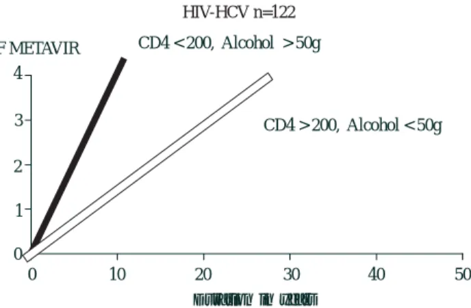
Several studies have demonstrated that patients coin-fected with HCV and HIV have a faster fibrosis progres-sion rate than controls even before the occurrence of marked decline in CD4 cell count63 and after taking into
account age, sex and alcohol consumption17,20 (Figure
4a). An HIV-infected patient with less than 200 CD4 cells/µL and drinking more than 50 g of alcohol daily has a median expected time to cirrhosis of 16 years versus 36 years for an HIV-infected patient with more than 200 CD4 cells/µL, drinking 50 g or less of alcohol daily (Fig-ure 4b).
We identified by multivariate analysis identified 4 in-dependent predictors of progression to cirrhosis: absence of protease inhibitor therapy (relative risk [RR] = 4.74, 95% confidence interval [CI], 1.34-16.67), heavy alcohol Table I. Factors associated or not with fibrosis progression.
Associated in uni and multivariate analysis Not sure Not associated
Age at infection Necrosis Last serum viral load Duration of infection Inflammation Genotype
Age at biopsy Hemochromatosis heterozygote Mode of infection Consumption of alcohol > 50 g per day Cigarette consumption DR antigens HIV coinfection Steatosis Liver viral load CD4 count < 200/mL Body Mass Index HCV-HVR1 complexity Male gender Moderate alcohol consumption
Annals of Hepatology 2(1) 2003: 5-11
8
edigraphic.com
consumption (> or = 50 g daily) (RR = 4.71, 95% CI,1.92-11.57), low CD4 cell count (< 200/microL) (RR = 2.74, 95% CI, 1.17-6.41), and age at HCV contamination (> or = 20 years) (RR = 2.37, 95% CI, 1.04-5.38). This study suggested that protease inhibitor therapy might not accelerate progression to HCV-related cirrhosis. Further-more, chronic use of antiretroviral therapy containing PI together with reduction of alcohol consumption and maintenance of high CD4 count could have a beneficial impact on liver fibrosis progression in HIV/HCV coin-fected patients.64
Viral factors
Viral factors such as genotype, viral load at the time of the biopsy, and quasi species are not associated with fi-brosis.7,21,22 There are very few studies for the following
factors and more studies with high sample size are need-ed: fluctuations of HCV RNA, intrahepatic cytokines pro-files, HLA class genotype, C282Y heterozygote hemo-chromatosis gene mutation, and cigarette consumption. About genotypes, there was no statistically significant overall difference between HCV genotypes in the inci-dence of cirrhosis at 20 years. There was clearly no asso-ciation between genotype 1 (1b or 1a) with fibrosis pro-gression, even at 40 years. In fact, slower slopes in pa-tients infected with genotype 1b in comparison to genotype 3 was observed. The role of specific steatosis related to genotype 3 is suspected for this increase in fi-brosis progression.69 HCV viral load had no effect.
Be-cause it is impossible to perform a longitudinal study with repeated liver biopsies and repeated viral load determina-tions, we do not know if viral load varies36 and if a high
viral load at the beginning of infection could be
predic-tive of fibrosis thereafter. However, even for F1 in the early years, there was no difference in the slopes accord-ing to the viral load.57
Smoking
About hepatotoxicity of cigarette smoke a recently study has shown that smoking was related to increased fibrosis and activity scores in age-adjusted (P = 0.09 and P = 0.05 respectively) and multivariate analyses (P = 0.03 and P = 0.04 respectively).65
Risk of fibrosis in patients with normal transaminases
Patients with repeated normal serum transaminases ac-tivity have lower fibrosis progression rate than matched control patients with elevated transaminases23 (Figure 5).
However there is still 15 % of these patients with moder-ate or high fibrosis progression rmoder-ates. Therefore, we rec-ommend assessing fibrosis stage, by liver biopsy or bio-chemical markers68 in these PCR positive patients. If the
patient has septal fibrosis or portal fibrosis with a high fi-brosis rate a treatment should be considered.
Histological activity
Histological activity is not always associated with fibro-sis progression.7,14,47,57 Differences in slopes were
graphi-cally observed between grade A0 and higher grades only after duration of 20 years of infection. Sampling error and intra-observer discordances are possible causes of false negative results. Fibrosis stage is always a better predictor of fibrosis progression than the activity grade.7
Progression of liver fibrosis in patients HCV and HIV positive
0 1 2 3 4
0 10 20 30 40 50
F METAVIR
Duration in years HIV n=122
Matched n=122
Benhamou Y et al Hepatology 1999;30:1054-8 Benhamou Y et al Hepatology 1999;30:1054-8
Figure 4a. Progression of liver fibrosis among patients coinfected by HCV and HIV. There is a significant increase of fibrosis progression rate among HIV in comparison to matched controls infected by HCV alone.
Figure 4b. Progression of liver fibrosis among patients coinfected by HCV and HIV. There is a very significant increase of fibrosis progre-ssion rate among patients with CD4 < 200 per mm3 and drinking more than 50g of alcohol per day.
Progression of liver fibrosis in coinfection
HIV-HCV n=122
0 1 2 3 4
0 10 20 30 40 50
F METAVIR
Duration in years CD4 < 200, Alcohol > 50g
CD4 > 200, Alcohol < 50g
edigraphic.com
Mode of infectionIn study with big sample size, mode of infection and fibrosis progression. We observed significantly more pa-tients without cirrhosis at 20 years among intravenous (IV) drug users (95%) in comparison to transfused pa-tients (89%) which was probably related to a younger age at infection.57
Steatosis and metabolic factors
Steatosis occurs in more than 50% of patients with chronic hepatitis C and is associated with increased he-patic fibrosis. In many of these patients the pathophysiol-ogy of steatosis appears to be the same as for patients with non-alcoholic fatty liver disease-that is, related to visceral adiposity and obesity.62 This suggest that
increas-ing body mass index has an important role in the patho-genesis of steatosis in the chronic hepatitis C. Weight loss in patients with chronic hepatitis C may be associated with a reduction in steatosis and abnormal liver enzymes and an improvement in fibrosis, despite the persistence of the virus. Weight reduction may provide an important ad-junct treatment strategy for patients with chronic hepatitis
C.66,67 Recently we have confirmed that genotype 3 HCV
infection was associated with significantly more steatosis than other genotype, with lower cholesterol levels and that this “viral” steatosis disappeared in sustained re-sponders. Therefore any studies on steatosis in patients with chronic hepatitis C must separate the genotype 3 population from other patients.
Impact of treatment on fibrosis progression
Better knowledge of factors associated with fibrosis progression has permitted to better assess the impact of treatment on fibrosis progression.
According to the Markov age-dependent modelling, a ten-year increment in duration of infection increased the risk of progression by 32% for IFN-treated patients and by 51% for untreated patients. The course of a series of 1000 IFN-treated and 1000 untreated patients was simu-lated over 5 years according to the initial stage of fibrosis and age and duration of infection at diagnosis. IFN treat-ment decreased the risk of progression to F3+F4 by a fac-tor of 4.8, for subjects aged 40 years, infected for 10 years, and in F0+F1 at diagnosis. As age and duration of infection increased, the risk of fibrosis increased and the impact of IFN treatment decreased.
We pooled individual data from 3010 naïve patients with pre-treatment and post-treatment biopsies from 4 randomized trials.63 Ten different regimens combining
standard interferon, PEG interferon, and ribavirin were compared. The impact of each regimen was estimated by the percentage of patients with at least 1 grade improve-ment in the necrosis and inflammation (METAVIR score), the percentage of patients with at least 1 stage worsening in fibrosis METAVIR score, and by the fibro-sis progression rate per year. Necrofibro-sis and inflammation improvement ranged from 39% (interferon 24 weeks) to 73% (PEG 1.5 µg/kg + ribavirin >10.6mg/kg/day; P < 0.001). Fibrosis worsening ranges from 23% (interferon 24 weeks) to 8% (PEG 1.5 µg/kg + ribavirin >10.6mg/ kg/day; P < 0.001). All regimens significantly reduced the fibrosis progression rates in comparison to rates be-Figure 5. Progression of liver fibrosis in patients HCV PCR positive
with repeated normal transaminases ALT. There was a significant re-duction of fibrosis progression rate in comparison to matched controls with abnormal transaminases ALT.
Progression of liver fibrosis in patients infected by HCV with normal ALT
0 1 2 3 4
0 10 20 30 40 50
F METAVIR
Duration in years
Normal ALT Matched elevated ALT
Adapted from Mathurin et al, Hepatology 1998; 27:868-72
Figure 6. Probability of fibrosis progression to F4 according to age at infection. A total of 2313 patients were included in the F4 analysis including 729, 165, and 32 patients still at risk at 20, 30 and 40 years infection duration, respectively. Whatever the stage, there were hig-her probabilities of fibrosis progression according to the age at infec-tion (p < 0.001). First line represents 754 patients infected before the age of 21 years. Second line represents 851 patients infected between 21 to 30 years. Third line represents 348 patients infected between 31 to 40 years. Fourth line represents 211 patients infected between 41 to 50 years. Fifth line represents 149 patients infected after the age of 50 years.
0.00 0.25 0.50 0.75 1.00
0 10 20 30 40
Progression to F4 according to age at infection
>50 years
41-50
31-40
21-30
<21
Annals of Hepatology 2(1) 2003: 5-11
10
edigraphic.com
fore treatment. The reversal of cirrhosis was observed in75 patients (49%) of 153 patients with baseline cirrhosis. Six factors were independently associated with the absence of significant fibrosis after treatment: baseline fibrosis stage (odds ration [OR] = 0.12; P < 0.0001), sustained viral response (OR = 0.36; P < 0.0001), age < 40 years (OR = 0.51; P < 0.001), body mass index < 27 kg/m2 (OR = 0.65; P < 0.001), no or minimal baseline
ac-tivity (OR = 0.70; P = 0.02), and viral load < 3.5 millions copies per milliliter (OR = 0.79; P = 0.03).
In conclusion, fibrosis progression is mostly regular from stage to stage, with progressive accelerations, and that the main factors associated with fibrosis progression are age and daily alcohol consumption equal to or greater than 50 gr. The progression of fibrosis begins to acceler-ate at 50 years of age, whacceler-atever the duration of infection. Metabolic factors as glucose intolerance, steatosis and overweight are probably important independent factors. The possibility to assess with non-aggressive biochemical markers68 (Bismut et www.biopredictive.com) the fibrosis
stages will probably allow in the future to identify other factors related to fibrosis progression.
References
1. W.H.O. Hepatitis C: global prevalence. Wkly Epidemiol Rec 1997; 72: 341-4.
2. Darby SC, Ewart DW, Giangrande PLF, et al. Mortality from liver cancer and liver disease in haemophilic men and boys given blood products contaminated with hepatitis C. Lancet 1997; 350: 1425-31. 3. El-Serag HB, Mason A. Rising incidence of hepatocellular
carci-noma in the United States. N Engl J Med 1999; 34: 745-50. 4. Deuffic S, Buffat L, Poynard T, Valleron AJ. Modeling the hepatitis
C virus epidemic in France. Hepatology 1999; 29: 1596-601. 5. Deuffic S, Poynard T, Valleron AJ. Correlation between HCV
preva-lence and hepatocellular carcinoma mortality in Europe. J Viral Hepa-titis 1999; in press.
6. Alter MJ, Kruszon-Moran D, Nainan OV et al. The prevalence of hepatitis C virus infection in the United States, 1988 through 1994. N Engl J Med 1999; 341: 556-562.
7. Poynard T, Bedossa P, Opolon P, for the OBSVIRC, METAVIR, CLINIVIR and DOSVIRC groups. Natural history of liver fibrosis progression in patients with chronic hepatitis C. Lancet 1997; 349: 825-32.
8. Sobesky R, Mathurin P, Charlotte F, et al. Modeling the impact of interferon alfa treatment on liver fibrosis progression in chronic hepa-titis C: a dynamic view. Gastroenterology 1999; 116: 378-386. 9. Knodell KG, Ishak KG, Black WC, et al. Formulation and
appli-cation of a numerical scoring system for assessing histological activity in asymptomatic chronic active hepatitis. Hepatology 1981; 1: 431-435.
10. Ishak K, Baptista A, Bianchi L, et al. Histological grading and stag-ing of chronic hepatitis. J Hepatol 1995; 22: 696-699.
11. The METAVIR cooperative group. Inter- and intra-observer varia-tion in the assessment of liver biopsy of chronic hepatitis C. Hepatology 1994; 20; 1: 15-20.
12. Bedossa P, Poynard T. An algorithm for the grading of activity in chronic hepatitis C. The METAVIR Cooperative Study Group. Hepatology 1996; 24: 289-93.
13. Paradis V, Mathurin P, Laurent A, et al. Histological features predic-tive of liver fibrosis in chronic hepatitis C infection. J Clin Pathol 1996; 49: 998-1004.
14. Yano M, Kumada H, Kage M, et al. The long term pathological evo-lution of chronic hepatitis C. Hepatology 1996; 23: 1334-40. 15. Datz C, Cramp M, Haas T, et al. The natural course of hepatitis C
virus infection 18 years after an epidemic outbreak of non-A, non-B hepatitis in a plasmapheresis centre. Gut 1999; 44: 563-67. 16. Wiley TE, McCarthy M, Breidi L, McCarthy M, Layden TJ. Impact
of alcohol on the histological and clinical progression of hepatitis C infection. Hepatology 1998; 28: 805-9.
17. Benhamou Y, Bochet M, Di Martino V, Charlotte F, Azria F, Coutellier A, et al. Liver fibrosis progression in human immunodeficiency vi-rus and hepatitis C vivi-rus coinfected patients. The Multivirc Group. Hepatology 1999; 30: 1054-8.
18. Bissell DM. Sex and Hepatic Fibrosis. Hepatology 1999; 29: 988-989. 19. Poynter ME, Daynes RA. Peroxysome proliferator-activated recep-tor a activation modulates cellular redox status, represses nuclear factor-kB signaling, and reduces inflammatory cytokine production in aging. J Biol Chem 1998; 273: 32833-41.
20. Pol S, Fontaine H, Carnot, F et al. Predictive factors for development of cirrhosis in parenterally acquired chronic hepatitis C: a compari-son between immunocompetent and immunocompromised patients. J Hepatol 1998; 29: 12-9.
21. De Moliner L, Pontisson P, De Salvo GL, et al. Serum and liver HCV RNA levels in patients with chronic hepatitis C: correlation with clinical and histological features. Gut 1998; 42: 856-60.
22. Roffi L, Ricci A, Ogliari C J, et al. HCV genotypes in Northern Italy: a survey of 1368 histologically proven chronic hepatitis C patients. J Hepatol 1998; 29: 701-6.
23. Mathurin P, Moussalli J, Cadranel JF, et al. Slow progression rate of fibrosis in hepatitis C virus patients with persistently normal alanine transaminase activity. Hepatology 1998; 27: 868-72.
24. Tong MJ, el-Farra NS, Reikes AR, Co RL. Clinical outcomes after transfusion-associated hepatitis C. N Engl J Med 1995; 332: 1463-6. 25. Consensus Statement. EASL International Consensus Conference on
Hepatitis C. J Hepatol 1999; 30: 956-961.
26. Pagliaro L, Peri V, Linea C, Camma C, Giunta M, Magrin S. Natural history of chronic hepatitis C. Ital J Gastroenterol 1999; 31: 28-44. 27. Kenny-Walsh E, for the Irish Hepatology Research Group. Clinical outcomes after hepatitis C infection from contaminated anti-D im-mune globulin. N Engl J Med 1999; 340: 1228-33.
28. Datz C, Cramp M, Haas T, Dietze O, Nitschko H, Froesner G, Muss N, et al. The natural course of hepatitis C virus infection 18 years after an epidemic out break of non-A, non-B hepatitis in a plasmapheris centre. Gut 1999; 44: 563-7.
29. Chossegros P, Pradat P, Bailly F, Chemello L, Sauleda S, Saracco G, Thursz M, et al. Natural history of chronic hepatitis C: fibrosis pro-gression is not linear. J Hepatol 1999; 30 (suppl 1): 52.
30. Observatoire National de l’Hépatite C. Groupe OBSVIRC. Observatoire national de l’hépatite C: bilan inital. Association Française pour l’Etude du Foie, Lyon 1994. (Abstract in: Gastroenterol Clin Biol 1995: 19-81).
31. Cacoub P, Poynard T, Ghillani P, Charlotte F, Olivi M, Piette JC, Opolon P, for the Multivirc Group. Extrahepatic manifestations in patients with chronic hepatitis C. Rheum Arthr 1999; In Press. 32. Poynard T, Marcellin P, Lee SS, et al. Randomised trial of interferon
alpha2b plus ribavirin for 48 weeks or for 24 weeks versus inter-feron alpha2b plus placebo for 48 weeks for treatment of chronic infection with hepatitis C virus. Lancet 1998; 352: 1426-32. 33. Mc Hutchison J, Gordon S, Schiff E, et al. Interferon alfa-2b alone
or in combination with ribavirin as initial treatment for chronic hepa-titis C. N Engl J Med 1998; 339: 1485-92.
34. The METAVIR cooperative group. Inter- and intra-observer varia-tion in the assessment of liver biopsy of chronic hepatitis C. Hepatology 1994; 20; 1: 15-20.
35. Bedossa P, Poynard T for the METAVIR cooperative study group. An algorithm for the grading of activity in chronic hepatitis C. Hepatology 1996; 24: 289-93.
edigraphic.com
37. Stuyver L, Rossau R, Wyseur A, et al. Typing of hepatitis C virusisolates and characterization of new subtypes using a line probe as-say. J Gen Virol 1993; 74: 1093-102.
38. Poynard T, Bedossa P, Chevalier M, Mathurin P, Lemonier C, Trepo C, Couzigou P, Payen JL, Sajus M, Costa JM, Vidaud M, Chaput JC, and the multicentre study group. A comparison fo three interferon alpha-2b regimens for the long-term treatment of chronic non A, non B hepatitis. New Engl J Med 1995; 332: 1457-62.
39. Fang JW, Albrecht JK, Jacobs S, Lau JY. Quantification of serum hepatitis C virus RNA. Hepatology 1999; 29: 997-8.
40. Sobesky R, Mathurin P, Charlotte F, et al. Modeling the impact of interferon alfa treatment on liver fibrosis progression in chronic hepa-titis C : a dynamic view. Gastroenterology 1999; 116: 378-386. 41. Deuffic S, Buffat L, Poynard T, Valleron AJ. Modeling the hepatitis C
virus epidemic in France. Hepatology 1999; 29: 1596-601. 42. Goodson JD, Taylor PA, Campion EW, et al. The clinical course of acute
hepatitis in the ederly patient. Arch Intern Med 1982; 142: 1485-8. 43. Wynne HA, Cope LH, Mutch E, et al. The effect of age upon liver
volume and apparent liver blood flow in healthy man. Hepatology 1989; 9: 297-301.
44. Nagel JE, Chrest JJ, Adler HH. Enumeration of T lymphocytes by monoclonal antibodies in young age and aged humans. J Immunol 1991; 191: 151: 599-603.
45. Makinodan T, Kay MMB. Age influence on the immune system. Adv Immunol 1980; 29: 287-91.
46. Poynter ME, Daynes RA. Peroxysome proliferator-activated recep-tor α activation modulates cellular redox status, represses nuclear factor-kB signaling, and reduces inflammatory cytokine production in aging. J Biol Chem 1998; 273: 32833-41.
47. Yano M, Kumada H, Kage M, et al. The long term pathological evo-lution of chronic hepatitis C. Hepatology 1996; 23: 1334-1340. 48. Roudot-Thoraval F, Bastie A, Pawlotsky JM, Dhumeaux D.
Epide-miological factors affecting the severity of hepatitis C virus-related liver disease: a French survey of 6.664 patients. The Study Group for the Prevalence and Epidemiology of Hepatitis C Virus. Hepatology 1997; 26: 485-90.
49. Bissell DM. Sex and Hepatic Fibrosis. Hepatology 1999; 29: 988-989 50. Yasuda M, Shimizu I, Shiba M, Ito S. Suppressive Effects of Estra-diol on Dimethylnitrosamine-Induced Fibrosis of the Liver in Rats. Hepatology 1999; 29: 719-727.
51. Ashcroft GS, Dodsworth J, van Boxtel E, et al. Estrogen accelerates cutaneous wound healing associated with an increase in TGF-beta1 levels. Nat Med 1997; 3: 1209-1215.
52. Wang YJ, Wang SS, Bickel M, Guenzler V, Gerl M, Bissell DM. Two novel antifibrotics, HOE 077 and Safironil, modulate stellate cell activation in rat liver injury: differential effects in males and females. Am J Pathology 1998; 152: 279-287.
53. Bentzen SM, Skoczylas JZ, Overgaard M, Overgaard J. Radiotherapy-related lung fibrosis enhanced by tamoxifen. J Nat Cancer Inst 1996; 88: 918-922.
54. Roffi L, Ricci A, Ogliari C, Scalori A, Minola E, Colloredo G, et al. HCV genotypes in Northern Italy: a survey of 1368 histologically proven chronic hepatitis C patients. J Hepatol 1998; 29(5): 701-6.
55. Pontisso P, Bellati G, Brunetto M, et al. Hepatitis C virus RNA pro-files in chronically infected individuals: do they relate to disease activity? Hepatology 1999; 29: 585-9.
56. Paradis V, Mathurin P, Laurent A, Charlotte F, Vidaud M, Poynard T, Hoang C, Opolon P, Bedossa P. Histological features predictive of liver fibrosis in chronic hepatitis C infection. J Clin Pathol 1996; 49: 998-1004.
57. Poynard T, Ratziu V, Charlotte F, Goodman Z, McHutchison J, Albrecht J. Rates and risk factors of liver fibrosis progression in patients with chronic hepatitis C. J Hepatol 2001; 34(5): 730-9. 58. Di Martino V, Lebray P, Moussalli J, Buffet C, Poynard T. Impact of
pregnancies, oral contraceptives and menopause on HCV-related liver fibrosis progression. Hepatology 2001; 34(suppl): 222ª.
59. Di Martino V, Rufat P, Boyer N, Renard P, Degos F, Martinot-Peignoux M, Matheron S, Le Moing V, Vachon F, Degott C, Valla D, Marcellin P. The influence of human immunodeficiency virus coinfection on chronic hepatitis C in injection drug users: a long-term retrospective cohort study. Hepatology 2001; 34(6): 1193-9. 60. Benhamou Y, Di Martino V, Bochet M, Colombet G, Thibault V, Liou
A, Katlama C, Poynard T. Factors affecting liver fibrosis in human immunodeficiency virus-and hepatitis C virus-coinfected patients: impact of protease inhibitor therapy. Hepatology 2001; 34(2): 283-7. 61. Pessione F, Ramond MJ, Njapoum C, Duchatelle V, Degott C, Erlinger S, Rueff B, Valla DC, Degos F. Cigarette smoking and hepatic lesions in patients with chronic hepatitis C. Hepatology 2001; 34: 121-125. 62. Ratziu V, Giral P, Charlotte F, Bruckert E, Thibault V, Theodorou I, Khalil L, Turpin G, Opolon P, Poynard T. Liver fibrosis in over-weight patients. Gastroenterology. 2000; 118(6): 1117-23. 63. Poynard T, McHutchison J, Manns M, Trepo C, Lindsay K, Goodman
Z, Ling MH, Albrecht J. Impact of pegylated interferon alfa-2b and ribavirin on liver fibrosis in patients with chronic hepatitis C. Gas-troenterology 2002; 122(5): 1303-13.
64. Hickman IJ, Clouston AD, Macdonald GA, Purdie DM, Prins JB, Ash S, Jonsson JR, Powell EE. Effect of weight reduction on liver histology and biochemistry in patients with chronic hepatitis C. Gut 2002; 51(1): 89-94.
65. Hourigan LF, MacDonald GA, Purdie D, Whitehall V, Shorthouse C, Clouston A, Powell EE. Fibrosis in chronic hepatitis C correlates significantly with body mass index and steatosis. Hepatology 1999; 29: 1215-1219.
66. Freeman AJ, Dore G, Law MG, Thorpe M, Overbeck JV, Lloyd AR, Marinos G, Kaldor JM. Estimating progression to cirrhosis in chronic hepatitis C virus infection. Hepatology 2001; 34: 809-816. 67. Deuffic-Burban S, Poynard T, Valleron AJ. Quantification of
fibro-sis progression in patients with chronic hepatitis C using a Markov model. J Viral Hepat 2002; 9: 114-22.
68. Imbert-Bismut F, Ratziu V, Pieroni L, Charlotte F, Benhamou Y, Poynard T. Biochemical markers of liver fibrosis in patients with hepatitis C virus infection: a prospective study. Lancet. 2001; 7; 357(9262): 1069-75.