Receptor 7 Variant Associated with Systemic Lupus
Erythematosus
Yun Deng1., Jian Zhao1., Daisuke Sakurai1
, Kenneth M. Kaufman2,3, Jeffrey C. Edberg4, Robert P. Kimberly4, Diane L. Kamen5, Gary S. Gilkeson5, Chaim O. Jacob6, R. Hal Scofield7,8,9, Carl D. Langefeld10, Jennifer A. Kelly7, Rosalind Ramsey-Goldman11, Michelle A. Petri12,
John D. Reveille13, Luis M. Vila´14, Graciela S. Alarco´n4, Timothy J. Vyse15, Bernardo A. Pons-Estel16 on behalf of the Argentine Collaborative Group", Barry I. Freedman17, Patrick M. Gaffney7, Kathy
Moser Sivils7, Judith A. James7,8, Peter K. Gregersen18, Juan-Manuel Anaya19, Timothy B. Niewold20, Joan T. Merrill21, Lindsey A. Criswell22, Anne M. Stevens23,24, Susan A. Boackle25, Rita M. Cantor26, Weiling Chen1, Jeniffer M. Grossman1, Bevra H. Hahn1, John B. Harley2,3, Marta E. Alarco´n-Riquelme7,27 on behalf of the BIOLUPUS and GENLES networks", Elizabeth E. Brown4,28, Betty P. Tsao1*
1Division of Rheumatology, University of California Los Angeles, Los Angeles, California, United States of America,2Division of Rheumatology and The Center for Autoimmune Genomics and Etiology, Cincinnati Children’s Hospital Medical Center, Cincinnati, Ohio, United States of America,3U.S. Department of Veterans Affairs Medical Center, Cincinnati, Ohio, United States of America,4Department of Medicine, University of Alabama at Birmingham, Birmingham, Alabama, United States of America,5Department of Medicine, Division of Rheumatology, Medical University of South Carolina, Charleston, South Carolina, United States of America,6Department of Medicine, Keck School of Medicine, University of Southern California, Los Angeles, California, United States of America,7Arthritis and Clinical Immunology Program, Oklahoma Medical Research Foundation, Oklahoma City, Oklahoma, United States of America,8Department of Medicine, University of Oklahoma Health Sciences Center, Oklahoma City, Oklahoma, United States of America,9U.S. Department of Veterans Affairs Medical Center, Oklahoma City, Oklahoma, United States of America, 10Department of Biostatistical Sciences, Wake Forest University Health Sciences, Wake Forest, North Carolina, United States of America,11Division of Rheumatology, Northwestern University Feinberg School of Medicine, Chicago, Illinois, United States of America,12Department of Medicine, Johns Hopkins University School of Medicine, Baltimore, Maryland, United States of America,13Department of Internal Medicine, University of Texas Health Science Center at Houston, Houston, Texas, United States of America,14Department of Medicine, University of Puerto Rico Medical Sciences Campus, San Juan, Puerto Rico,15Divisions of Genetics and Molecular Medicine and Immunology, King’s College London, London, United Kingdom,16Department of Medicine, Sanatorio Parque, Rosario, Argentina,17Department of Internal Medicine, Wake Forest School of Medicine, Winston-Salem, North Carolina, United States of America,18Robert S. Boas Center for Genomics and Human Genetics, Feinstein Institute for Medical Research, North Shore LIJ Health System, Manhasset, New York, United States of America,19Center for Autoimmune Diseases Research, Universidad del Rosario, Bogota, Colombia,20Division of Rheumatology and Department of Immunology, Mayo Clinic, Rochester, Minnesota, United States of America, 21Clinical Pharmacology Program, Oklahoma Medical Research Foundation, Oklahoma City, Oklahoma, United States of America,22Rosalind Russell Medical Research Center for Arthritis, Department of Medicine, University of California San Francisco, San Francisco, California, United States of America,23Division of Rheumatology, Department of Pediatrics, University of Washington, Seattle, Washington, United States of America,24Center for Immunity and Immunotherapies, Seattle Children’s Research Institute, Seattle, Washington, United States of America,25Division of Rheumatology, School of Medicine, University of Colorado Denver, Aurora, Colorado, United States of America,26Department of Human Genetics, University of California Los Angeles, Los Angeles, California, United States of America,27Centro de Geno´mica e Investigacio´n Oncolo´gica (GENYO), Pfizer–Universidad de Granada–Junta de Andalucia, Granada, Spain,28Department of Epidemiology, University of Alabama at Birmingham, Birmingham, Alabama, United States of America
Abstract
We previously reported that the G allele of rs3853839 at 39untranslated region (UTR) of Toll-like receptor 7 (TLR7) was associated with elevated transcript expression and increased risk for systemic lupus erythematosus (SLE) in 9,274 Eastern Asians [P= 6.5610210, odds ratio (OR) (95%CI) = 1.27 (1.17–1.36)]. Here, we conducted trans-ancestral fine-mapping in
13,339 subjects including European Americans, African Americans, and Amerindian/Hispanics and confirmed rs3853839 as the only variant within theTLR7-TLR8region exhibiting consistent and independent association with SLE (Pmeta= 7.5610211,
OR = 1.24 [1.18–1.34]). The risk G allele was associated with significantly increased levels of TLR7mRNA and protein in
peripheral blood mononuclear cells (PBMCs) and elevated luciferase activity of reporter gene in transfected cells.TLR739UTR
sequence bearing the non-risk C allele of rs3853839 matches a predicted binding site of microRNA-3148 (miR-3148), suggesting that this microRNA may regulateTLR7expression. Indeed, miR-3148 levels were inversely correlated withTLR7
transcript levels in PBMCs from SLE patients and controls (R2= 0.255,P= 0.001). Overexpression of miR-3148 in HEK-293 cells
led to significant dose-dependent decrease in luciferase activity for construct driven byTLR739UTR segment bearing the C allele (P= 0.0003). Compared with the G-allele construct, the C-allele construct showed greater than two-fold reduction of luciferase activity in the presence of miR-3148. Reduced modulation by miR-3148 conferred slower degradation of the risk G-allele containingTLR7transcripts, resulting in elevated levels of gene products. These data establish rs3853839 ofTLR7as a shared risk variant of SLE in 22,613 subjects of Asian, EA, AA, and Amerindian/Hispanic ancestries (Pmeta= 2.0610219,
Citation:Deng Y, Zhao J, Sakurai D, Kaufman KM, Edberg JC, et al. (2013) MicroRNA-3148 Modulates Allelic Expression of Toll-Like Receptor 7 Variant Associated with Systemic Lupus Erythematosus. PLoS Genet 9(2): e1003336. doi:10.1371/journal.pgen.1003336
Editor:Mark I. McCarthy, University of Oxford, United Kingdom
ReceivedOctober 15, 2012;AcceptedJanuary 8, 2013;PublishedFebruary 28, 2013
Copyright:ß2013 Deng et al. This is an open-access article distributed under the terms of the Creative Commons Attribution License, which permits unrestricted use, distribution, and reproduction in any medium, provided the original author and source are credited.
Funding:Support for this work was provided by the U.S. National Institutes of Health grants: R01AR043814 (BPT), R01AR057172 (COJ), R01AR043274 (KMS), R01AI063274 and RC2AR058959 (PMG), N01AR62277, R37AI024717, R01AR042460, and P20RR020143 (JBH), P01AI083194 (JBH, KMS, RPK, LAC, TJV, MEA-R, COJ, BPT, and PMG), P01AR49084 (RPK, JCE, EEB, RR-G, LMV, and MAP), R01AR33062 (RPK), P30AR48311 (EEB), P01AR052915 and U01AI090909 (JDR), K24AR002138, P60AR30692 and UL1RR025741 (RR-G), R01AR43727 (MAP), K08AI083790, L30AI071651, and UL1RR024999 (TBN), R01CA141700 and RC1AR058621 (MEA-R), R01AR051545-01A2 (AMS), P30AR053483, P30GM103510, U19AI082714, and U01AI101934 (JAJ), R21AI070304 (SAB), P60AR053308 and M01RR-00079 (LAC), P60AR049459 and UL1RR029882 (GSG and DLK). Additional support was provided by the Lupus Research Institute grant (BPT); the Alliance for Lupus Research grants (BPT, YD, KLM, TBN, LAC, and COJ); the Arthritis National Research Foundation Eng Tan Scholar Award (TBN and JZ); Charles Barkley Research Award (EEB); the Arthritis Research UK (TJV); the Federico Wilhelm Agricola Foundation Research grant (BAP-E); the Arthritis Foundation (AMS and PMG); Clinical and Translational Science Grant Numbers UL1RR025014-02 (AMS), UL1TR000165 (JCE), and UL1RR025005 (MAP) from the National Center for Advancing Translational Sciences (NCATS) and National Center for Research Resources (NCRR) component of the National Institutes of Health (NIH); Kirkland Scholar Award (LAC); Wake Forest University Health Sciences Center for Public Health Genomics (CDL); and UCLA Clinical and Translational Science Institute (CTSI) grants UL1RR033176 and UL1TR000124. The funders had no role in study design, data collection and analysis, decision to publish, or preparation of the manuscript.
Competing Interests:The authors have declared that no competing interests exist. * E-mail: [email protected]
.These authors contributed equally to this work.
"Memberships of the consortia are provided in the Acknowledgments.
Introduction
Systemic lupus erythematosus (SLE [OMIM 152700]) is a complex and heterogeneous autoimmune disease with a strong genetic component that is modified by environmental exposures. Although the detailed etiopathogenesis of SLE remains unknown, excessive innate immune activation involving toll-like receptors (TLRs, particularly TLR7/8/9) and type I interferon (IFN) has been recognized as an important pathogenic mechanism in the disease [1]. Therapeutics targeting the TLR/IFN pathway are in development for the treatment of SLE, with ongoing clinical trials investigating monoclonal antibodies against IFN-aand inhibitors for TLR7/TLR9 (reviewed in [2]). Recent genome-wide associ-ation (GWA) and follow-up studies have revealed the associassoci-ation of a number of polymorphic variants in genes encoding components of the TLR/type I IFN pathway with susceptibility to SLE (reviewed in [3,4]), providing insights at the molecular level to refine our understanding of this dysregulated pathway in the predisposition to SLE.
Our previous study identified a single nucleotide polymorphism (SNP), rs3853839, in the 39UTR of an X-linked geneTLR7to be associated with SLE in 4,334 cases and 4,940 controls of Eastern Asian descent [5], providing the first convincing evidence for the genetic contribution ofTLR7to human SLE. Individuals carrying the risk G allele exhibited increasedTLR7transcripts and a more robust IFN signature than non-risk C allele carriers [5]. In this study, by fine mapping theTLR7-TLR8region, we confirmed that the previously reported functional SNP rs3853839, located within a predicted binding site of miR-3148, was most likely responsible for observed association with SLE in three populations of non-Asian ancestry. We demonstrated a differential miR-3148 modulation explaining the effect of allelic variation at rs3853839 onTLR7expression.
Results
Confirmation of the association between rs3853839 and SLE susceptibility in European American, African
American, and Hispanic ancestries
We conducted genotyping and imputation for genetic variants covering ,80 kb of the TLR7-TLR8 region on Xp22.2. After
applying quality control measures, 41 genotyped SNPs and 57–75
imputed SNPs/INDELs (insertion-deletion) (varying among dif-ferent ancestries) were assessed for association with SLE in unrelated cases and healthy controls of European American (EA, 3,936 cases vs. 3,491 controls), African American (AA, 1,679 vs. 1,934) and Hispanic enriched for the Amerindian-European admixture (HS, 1,492 vs. 807) descent (Figure 1A).
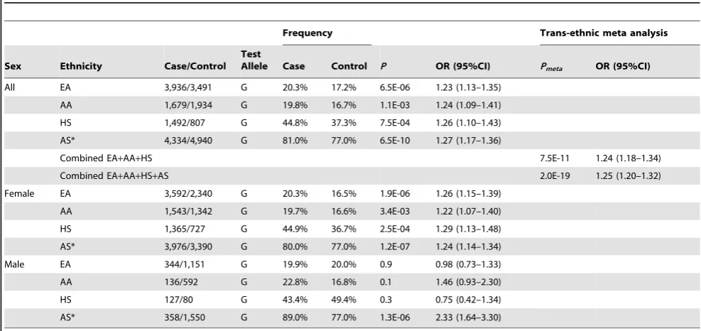
The strongest association signal was consistently detected at rs3853839 in the three ancestries, including EA (minor allele frequency of 20.3% in cases vs. 17.2% in controls,P= 6.561026,
OR [95%CI] = 1.23 [1.13–1.35]), AA (19.8% vs. 16.7%, P= 1.161023, OR = 1.24 [1.09–1.41]) and HS (44.8% vs. 37.3%, P= 7.561024, OR = 1.26 [1.10–1.43]) (Figure 1B, Table 1). After Bonferroni correction for multiple comparisons, the association of rs3853839 with SLE remained significant in EA and HS, and reached a nominal significance in AA. Combining the EA, AA and HS datasets, the meta-analysis P value of rs3853839 (Pmeta= 7.5610211, OR = 1.24 [1.18–1.34]) exceeded the commonly used threshold of 561028 for genome-wide
significance (Figure 1C, Table 1). Thus, the association of rs3853839 with SLE previously identified in Eastern Asians was confirmed in three non-Asian ancestries.
Only six other SNPs within a relatively small interval of 5 kb spanning from TLR7 39downstream to TLR8 intron 1 were consistently associated with SLE (P,0.05) in EA, AA and HS (Table S1), and remained significant trans-ancestral meta-analysis Pvalues after Bonferroni correction (5.561026#Pmeta#1.361026,
Taken together, we confirmed rs3853839 as the only SNP in the TLR7-TLR8region showing an independent association with SLE across all three non-Asian ancestries. A meta-analysis by combining all datasets of Asian and non-Asian ancestries showed compelling evidence of association with SLE at rs3853839 (Pmeta= 2.0610219, OR = 1.25 [1.20–1.32], Table 1). Given the
location of TLR7 at X chromosome, we examined the allelic association of rs3853839 separately by gender. Of note, the sex-specific association of rs3853839 with SLE previously detected in Asian men [5] was not replicated in non-Asian ancestries (Table 1).
Regulation of TLR7expression by rs3853839
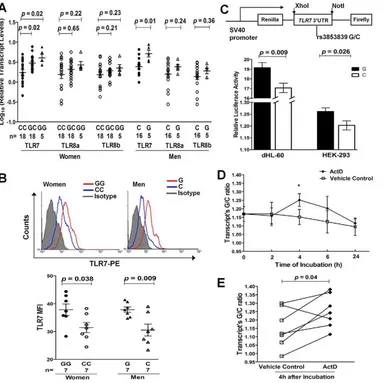
Given the convincing evidence for the trans-ancestral associa-tion of rs3853839 with SLE susceptibility, we then evaluated its effect on regulation of TLR7/8 expression. Messenger RNA (mRNA) levels of TLR7 and the two alternative TLR8 isoforms were measured by real-time PCR in PBMCs from healthy EA individuals (n = 62). TLR7 mRNA levels were significantly different among women (n = 41) carrying different genotypes of rs3853839 [P= 0.003, one-way analysis of variance (ANOVA)], in which the GG and GC carriers exhibited notably increasedTLR7 mRNA levels compared with the CC carriers [P= 0.02 for GG (n = 5) vs. CC (n = 18) and 0.02 for GC (n = 18) vs. CC, respectively, Student’s t test; Figure 2A) and the number of rs3853839 risk G allele was significantly correlated with increased TLR7mRNA levels (R2= 0.26,P= 861024, linear regression test). Consistently, male G allele carriers (n = 5) also had significantly higher TLR7 mRNA expression than male C allele carriers (n = 16) (P= 0.01, Figure 2A). There was no significant association of rs3853839 genotypes with mRNA levels of twoTLR8isoforms in either women or men (Figure 2A). No sex differences inTLR7 orTLR8mRNA levels were observed between individuals carrying the same genotype [GG women vs. G men:P= 0.41 (TLR7), 0.63 (TLR8a) and 0.50 (TLR8b); CC women vs. C men: P= 0.10 (TLR7), 0.91 (TLR8a) and 0.65 (TLR8b)]. These results were in
accordance with our previous observations in Chinese [5], supporting the importance of rs3853839 in regulating TLR7 rather thanTLR8gene expression.
We assessed the intracellular expression of TLR7 and TLR8 proteins by flow cytometry in PBMCs from 7 pairs of healthy women (GG vs. CC) and men (G vs. C), respectively. Of the 7 pairs of individuals in each gender, 4 pairs were of EA descent and 3 pairs were Asians. Compared with C allele carriers, G allele carriers had significantly higher TLR7 protein levels in PBMCs (P= 0.038 and 0.009 in women and men, respectively; Figure 2B), especially in CD19+
B cells and CD14+monocytes (Figure S2). No significant association
between rs3853839 genotypes and TLR8 protein levels was observed in either total PBMCs or in specific cell subsets (Figure S3).
We next performed luciferase reporter assays to further confirm the functional effect of rs3853839 on TLR7 expression. PCR-amplifiedTLR739UTR fragments with either the G or C allele of rs3853839 were cloned downstream of an SV40 promoter-driven Renilla luciferase gene in the psiCHECK-2 vector, which also contained a firefly luciferase gene to serve as an internal transfection normalization control (Figure 2C). Constructs were then transiently transfected into either HEK-293 or differentiated HL-60 (dHL-60, neutrophil-like cells) cells. After 24 hours, cell lysates transfected with the G-allele construct showed significantly higher luciferase activity than those transfected with the C-allele construct in both HEK-293 and dHL-60 cells (P= 0.026 and 0.009, respectively; Figure 2C). Taken together, consistent results fromex vivoandin vitrostudies indicated that the SLE-risk G allele of rs3853839 conferred elevated TLR7 expression at the both mRNA and protein level.
Allelic differences of rs3853839 inTLR7mRNA degradation rate
To explore the mechanism of rs3853839 in regulating TLR7 mRNA turnover, we assessed allelic difference in TLR7 mRNA degradation by pyrosequencing. We first determined the rs3853839 G/C allele ratio in genomic DNA (gDNA) and cDNA from healthy EA women (n = 7) carrying the GC genotype. The mean G/C allele ratio in cDNA was significantly higher than the theoretical ratio of 1 as detected in gDNAs (P= 0.02, Figure S4), indicating a higher expression of the G- than the C-allele containingTLR7transcripts in heterozygous PBMCs. The allelic specific expression analysis in EA was similar to our previous findings in Chinese [5], and confirmed the result of real-time PCR that the G allele of rs3853839 is associated with increasedTLR7 mRNA expression. Then, PBMCs were cultured in the absence or presence of the transcriptional inhibitor actinomycin D (ActD), and the G/C allele ratio in cDNA (normalized to that measured in gDNA) was determined after 0, 2, 4, 6, and 24 hours, respectively. As shown in Figure 2D and 2E, the G/C ratio in cDNA appeared to change over time when PBMCs were incubated with ActD and exhibited a statistical difference at the 4 hour point (P= 0.04), implicating slower degradation of the G than the C allele-containing TLR7 transcript in heterozygous PBMCs. The inhibitory effect of ActD on RNA synthesis was corroborated by a decrease in total TLR7mRNA level at increasing time points after the addition of ActD in PBMC aliquots measured by real-time PCR (Figure S5).
Alteration of microRNA–3148–mediated modulation of TLR7expression by rs3853839
MicroRNAs (miRNAs) that bind to target sequences located within the 39UTR of mRNAs by base pairing have been shown to result in accelerated mRNA turnover or translation repression [6]. Author Summary
Systemic lupus erythematosus (SLE) is a debilitating autoimmune disease contributed to by excessive innate immune activation involving toll-like receptors (TLRs, particularly TLR7/8/9) and type I interferon (IFN) signaling pathways. TLR7 responds against RNA–containing nuclear antigens and activates IFN-a pathway, playing a pivotal role in the development of SLE. While a genomic duplication ofTlr7 promotes lupus-like disease in the
Y-linked autoimmune accelerator (Yaa) murine model, the lack of common copy number variations atTLR7in humans led us to identify a functional single nucleotide polymor-phism (SNP), rs3853839 at 39 UTR of the TLR7 gene, associated with SLE susceptibility in Eastern Asians. In this study, we fine-mapped the TLR7-TLR8 region and con-firmed rs3853839 exhibiting the strongest association with SLE in European Americans, African Americans, and Amerindian/Hispanics. Individuals carrying the risk G allele of rs3853839 exhibited increasedTLR7 expression at the
both mRNA and protein level and decreased transcript degradation. MicroRNA-3148 (miR-3148) downregulated the expression of non-risk allele (C) containing transcripts preferentially, suggesting a likely mechanism for increased
TLR7 levels in risk-allele carriers. This trans-ancestral
Table 1.Association of rs3853839 with SLE in multiple ancestries.
Frequency Trans-ethnic meta analysis
Sex Ethnicity Case/Control Test
Allele Case Control P OR (95%CI) Pmeta OR (95%CI)
All EA 3,936/3,491 G 20.3% 17.2% 6.5E-06 1.23 (1.13–1.35)
AA 1,679/1,934 G 19.8% 16.7% 1.1E-03 1.24 (1.09–1.41)
HS 1,492/807 G 44.8% 37.3% 7.5E-04 1.26 (1.10–1.43)
AS* 4,334/4,940 G 81.0% 77.0% 6.5E-10 1.27 (1.17–1.36)
Combined EA+AA+HS 7.5E-11 1.24 (1.18–1.34)
Combined EA+AA+HS+AS 2.0E-19 1.25 (1.20–1.32)
Female EA 3,592/2,340 G 20.3% 16.5% 1.9E-06 1.26 (1.15–1.39)
AA 1,543/1,342 G 19.7% 16.6% 3.4E-03 1.22 (1.07–1.40)
HS 1,365/727 G 44.9% 36.7% 2.5E-04 1.29 (1.13–1.48)
AS* 3,976/3,390 G 80.0% 77.0% 1.2E-07 1.24 (1.14–1.34)
Male EA 344/1,151 G 19.9% 20.0% 0.9 0.98 (0.73–1.33)
AA 136/592 G 22.8% 16.8% 0.1 1.46 (0.93–2.30)
HS 127/80 G 43.4% 49.4% 0.3 0.75 (0.42–1.34)
AS* 358/1,550 G 89.0% 77.0% 1.3E-06 2.33 (1.64–3.30)
*AS: Previously published data in population of Eastern Asian descent (5).
[image:4.612.61.555.485.718.2]Abbreviation: AS, Eastern Asian; AA, African American; EA, European American; HS, Amerindian/Hispanics; OR, odds ratio; CI, confidence interval. doi:10.1371/journal.pgen.1003336.t001
Figure 1. Allelic associations of SNPs in theTLR7-TLR8region with SLE.(A) The genomic structure of theTLR7-TLR8region and the location of
all studied SNPs are indicated. (B) Association signals (2log10P) are plotted against the position of each SNP (based on GRch37/hg19) in European Americans (EA), African Americans (AA), and Hispanics (HS). Genotyped and imputed SNPs are depicted with circles and triangles, respectively. The diamond identifies theTLR739UTR SNP rs3853839. SNPs are highlighted using different colors according to their LD strength (r2) with rs3853839. (C) A trans-ancestral meta-analysis is conducted on 40 genotyped SNPs (circles) and 14 imputed SNPs (triangles) that are shared by the three ancestries (SNPs listed in Table S1) using fixed and random model, respectively. The dashed line represents the significance level of 561028.
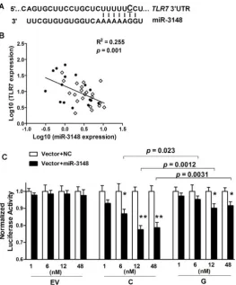
Single nucleotide change either within or around the sequence of miRNA target sites can potentially alter the base-pairing patterns and affect miRNA-mediated regulation [7,8]. The updated TargetScan database (Release 6.2; http://www.targetscan.org) indicates that rs3853839 is located within a binding site of miR-3148, where the non-risk allele (C), but not the risk allele (G), is predicted to match miR-3148 at the second position (Figure 3A). We hypothesized that the C to G variation of rs3853839 could reduce the binding and regulation incurred by miR-3148, therefore, leading to dysregulated TLR7 expression. We first showed that transcript levels of miR-3148 and TLR7 were inversely correlated in PBMCs from 16 patients with SLE and
[image:5.612.63.446.61.444.2]21 healthy controls (R2= 0.255,P= 0.001; Figure 3B), suggesting the possible regulation ofTLR7expression by miR-3148. Next, to verify whether allelic variation of rs3853839 affects the interaction of miR-3148 withTLR7 39UTR, psiCHECK-2 vectors containing TLR7 39UTR segment with either the C or G allele of rs3853839 were cotransfected with various doses of miR-3148 or nontarget control mimic into HEK-293 cells. As shown in Figure 3C, we observed significant dose-dependent miR-3148-mediated decrease in luciferase activity for the C-allele construct (P= 0.0003 over all miR-3148-treated C-allele vector groups, ANOVA test), but not for the G-allele construct (P= 0.14). Cotransfection with miR-3148 at a concentra-tion of 6, 12, and 48 nM, respectively, led to greater than two-fold Figure 2. The SLE-risk G allele of rs3853839 confers elevated TLR7 expression through slower mRNA degradation.(A) Association of rs3853839 genotypes withTLR7/8transcript levels in EA normal PBMCs. Each symbol represents an individual and horizontal lines indicate mean6
SEM values. (B) Association of rs3853839 genotypes with TLR7 protein levels in normal PBMCs. FACS histograms show the log MFI values plotted against the cell counts for PBMCs in individuals carrying G or C allele, compared with isotype control. Results are from one representative pair (GG or G vs. CC or C) of 7 in each gender group. MFI of TLR7 expression in PBMCs is graphically depicted. Each symbol represents an individual and horizontal lines indicate mean6SEM values. (C) Verification of the G allele conferring elevated expression of a luciferase reporterin vitro.TLR739UTR
segment bearing G or C allele of rs3853839 was cloned into the psiCHECK-2 reporter vector and luciferase activity was determined after 24 hours of transfection. Relative luciferase activity is Renilla/Firefly luciferase ratio. Data show the mean6SEM and are representative of cumulative data from four independent experiments. (D) The kinetics of the G/C allele ratio inTLR7transcripts from PBMCs of healthy heterozygous individuals (n = 7) in the absence or presence of actinomycin D (ActD). The G/C allele ratio obtained inTLR7transcripts was normalized to that measured from gDNA of the
same sample. Data are expressed as mean6SEM at each time point and representative of cumulative data from two independent experiments with seven healthy donors.*P
,0.05. (E) Summary of the G/C allele ratio inTLR7transcripts 4 hours after the addition of ActD or vehicle control (n = 7). Comparisons are between ActD and vehicle control cultures;P= 0.04; pairedttest. FACS, Fluorescence-activated cell sorter; MFI, mean fluorescence
intensity.
reduction of luciferase activity in the C-allele than the G-allele construct [reduction in C-allele vs. G-allele construct: 13.2% vs. 4.8%,P= 0.023 (6 nM); 22.5% vs. 9.9%,P= 0.0012 (12 nM); 21.4% vs. 8.5%,P= 0.0031 (48 nM)]. These data supported the bioinfor-matic prediction that miR-3148 directly targetsTLR739UTR and the C to G variation of rs3853839 within the binding site alters the inhibitory effect of miR-3148 on modulatingTLR7expression.
Discussion
Fine-mapping of the TLR7-TLR8 region with high-density genetic markers based on large scale genotyping and imputation confirmed SNP rs3853839 at TLR7 39UTR as the most likely causal variant responsible for the association of TLR7-TLR8 region with SLE in populations of EA, AA and HS ancestry. In accordance with our previous observation in Asians [5], we detected elevated TLR7 expression at both mRNA and protein levels in PBMCs from EA homozygous risk G allele carriers, as well as a higher level of the risk than the non-risk allele-containing TLR7transcripts in EA heterozygous PBMCs. The fact that two distinct ancestries share the same genotype-phenotype association implicates an important regulatory effect of rs3853839 on TLR7 expression. Toward this end, we have extended functional studies
showing slower degradation of the risk allele-containing TLR7 transcripts in heterozygous PBMCs and regulation of TLR7 expression by miRNA-3148 that targets 39UTR at the position of rs3853839. Finally, we showed that the presence of the risk G allele resulted in reduced suppression by miRNA-3148, suggesting a likely mechanism for increasedTLR7 expression in risk-allele carriers.
[image:6.612.62.326.60.378.2]The importance of TLR7 upregulation on mediating autoim-mune responses has been addressed in murine models of SLE. The Y-linked autoimmune accelerator (Yaa) modifier, suggested mainly due toTlr7gene duplication, provides a prime example of TLR7 dysregulation leading to autoreactivity and inflammatory pathol-ogy [9–11]. Increasing Tlr7 gene dosage via generation of transgenic mice results in development of systemic autoimmunity, the severity of which directly correlates with the degree of Tlr7 overexpression [12]. Increased Tlr7 gene dosage promotes autoreactive lymphocytes activation, dendritic cells proliferation, and secretion of proinflammatory cytokines and IFN-a[12], which in turn upregulates TLR7 expression, leading to a feedback loop exacerbating autoimmunity [13]. In patients affected with SLE, up-regulated expression of TLR7 mRNA has been reported in PBMCs and B cells [14,15]. Although a copy number variation (CNV) study in Mexican population showed increased TLR7 Figure 3. The SLE-risk G allele of rs3853839 displays reduced transcript modulation by miR-3148.(A) TargetScan’s predicted miR-3148-binding site inTLR739UTR. The C allele, rather than G allele of rs3853839 corresponds to the second base of this seed region. (B) Inverse correlation of miR-3148 andTLR7transcript levels in PBMCs from 16 patients with SLE (solid circles) and 21 controls (open diamonds). (C) HEK-293 cells were
cotransfected with empty reporter vector (EV), luciferase constructs driven byTLR739UTR segment containing either C or G allele of rs3853839 and increasing concentrations (1, 6, 12, and 48 nM) of miR-3148 or nontarget control (NC) mimics. Luciferase activity was determined 24 hours after transfection. Normalized luciferase activity is the Renilla/Firefly ratio of miR-3148-treated reporter vector compared with the same NC-treated reporter vector. Data show the mean6SEM and are representative of cumulative data from three independent experiments.P= 0.0003 over all miR-3148-treated C-allele vector groups, and not significant over all miR-3148-miR-3148-treated G-allele or empty vector groups (ANOVA test).P*= 0.02,P**,0.0001
copies in childhood-onset SLE patients [16], no evidence for common CNVs at theTLR7-TLR8region has been identified in individuals of diverse ancestries through our previous study by three independent methods including quantitative real-time PCR, PmeI pulsed-field gel electrophoresis and Southern blot [5], two recent studies using customized CGH platforms [17,18] as well as other studies listed in the Database of Genomic Variants (http:// projects.tcag.ca/variation; the latest version released in November 2010), suggesting that mutations similar to Yaa are not a frequent feature of human SLE. The current study identifying genetic variations conferred by a regulatory SNP inTLR7expression and SLE susceptibility suggests that murine models provide profound clues to human genetics if we look beyond the specific mutations identified in the relevant pathways.
Unlike our findings in Asians that both sexes showed association [5], the impact of rs3853839 on risk for SLE was only observed for women in the non-Asian datasets (Table 1). Given the low prevalence of SLE in men, it is often challenging to collect a large enough number of affected men in a given population. Under the assumption that the associated G allele confers genetic risk with an odds ratio of 1.26 in EA, 1.22 in AA and 1.29 in HS subjects (ORs were determined in female datasets), and consideringP,0.05 as the threshold of significance, the power estimate for female samples in each ancestry reaches more than 85%, whereas for male samples it is only 50% in EA, 19% in AA and 25% in HS dataset. Thus, there was clearly inadequate power to evaluate this association in AA and HS men. Despite a relatively robust sample size of EA men (344 SLE vs. 1,151 controls), a significantly higher G allele frequency was observed in male than female controls (20.0% vs. 16.5%, P= 0.005), contributing to the difficulty in assessing association with SLE in EA male subjects.
To our knowledge, the association of rs3853839 (or its tag SNP) with SLE has not been reported in four SLE GWA studies in European-derived populations [19–22] and three GWA studies in Asians [23–25]. According to the 1000 Genomes Project data, rs3853839 locates in a region with poor LD structure and cannot be tagged by any known SNP at the TLR7-TLR8 region with r2.0.65. The SNP rs850632, located at TLR7 39downstream, shows the strongest LD with rs3853839 in Europeans (r2= 0.38) and Asians (r2= 0.65). However, neither rs3853839 nor rs850632 has been included in predesigned commercial genotyping arrays of those GWA studies, resulting in the absence of associations. Even if rs3853839 was genotyped, the published GWA studies might have inadequate statistical power to capture its association in the initial discovery analyses [5].
Evidence of other TLR7 polymorphisms associated with SLE has been reported, including two intronic SNPs (rs179019 and rs179010) found in Japanese population [26] and an exonic SNP (rs179008) in individuals from Southern Brazil [27]. The reported associations were modest due to limited sample size of these studies (less than 400 cases and 450 controls), and none of them have been confirmed by the current fine-mapping study using a large collection of EA, AA and HS cases-controls (Table S1). TLR8 polymorphisms have been described in infectious diseases [28,29] with a genetic effect localized to a functional variant at exon 1 (rs3764880, Met1Val). The G allele of rs3764880, which abolishes a putative start codon within the alternative TLR8 transcript isoform a (Figure 1A), conferred a protective effect on suscepti-bility to pulmonary tuberculosis in Indonesian and Russian men [28], as well as on HIV disease progression in Germans [29]. Our data showed a significantly increased frequency of rs3764880-G allele in SLE than healthy controls in the three non-Asian datasets; however, its association with SLE was dependent on that ofTLR7 SNP rs3853839. Other variants at theTLR7-TLR8region showed
either weaker association than rs3853839 in trans-ancestral meta-analysis or association uniquely in EA or HS. Taken together, these data support rs3853839 as the most likely polymorphism associated with SLE shared by multiple ancestries. Although imputation facilitated our ability to capture common variants (MAF.1%), further refinement in genetic effects of rare variants (MAF,1%) is needed by deep sequencing of this locus, especially the intergenic region betweenTLR7andTLR8that was not well imputed in this study.
Variations in 39UTR regions may be important in gene regulation. To date, expression quantitative trait loci (eQTL) mapping has been widely used for characterization of SNPs that affect gene expression [30]. Although the TLR7 expression has been measured in previous whole-genome eQTL studies, currently only those using EBV-transformed lymphoblastoid cell lines of 1000 Genomes Project individuals provide publically available genotyping data of rs3853839. Based on the study by Stranger et al [31], we found that CG carriers of rs3853839 showed elevated TLR7 expression compared with CC carriers in YRI women (P= 0.012). In male individuals, the G allele of rs3853839 showed a trend of association with elevated TLR7 expression in CHB+JPT, CEU and YRI men, and the association was significant when combining all male data (P= 0.014). These findings are consistent with our results that rs3853839 alleles are associated with differentialTLR7expression.
An important finding of this study is that the SLE-associated variant rs3853839 confers a genetic effect on modulation ofTLR7 expression by an epigenetic factor miR-3148. Accumulating evidence suggests that miRNAs are fine tuners of TLR signaling pathways [32]. Regulation by miRNA may occur at various levels of TLR pathways by targeting adaptor molecules, downstream regulators and cytokines (reviewed in [32,33]). However, few studies point to TLR themselves (e.g. TLR2 and TLR4) being directly targeted by miRNAs [34,35]. Using algorithms from TargetScan, only the newly identified human miR-3148 [36], which is not evolutionarily conserved among mammals, is predicted to bind TLR7 39UTR sequences at the position of rs3853839. The inverse correlation of miR-3148 andTLR7levels in PBMCs, along with functional validation by reporter gene assay, confirms an inhibitory effect of miR-3148 on regulating TLR7 expression and allelic variation of rs3853839 affecting miRNA-mRNA interactions. Further study will focus on investi-gating miR-3148 expression patterns in specific immune cell types, assessing biological impacts of changes in miR3148-mediated TLR7 expression on downstream immune responses, and evaluating roles of other miRNAs that target sequences in the vicinity of rs3853839. Of interest, an unconventional role for miRNAs has been identified as endogenous activators for RNA-sensing receptors (TLR7/8) in a cell- or tissue-type specific manner [37,38]. Therefore, miRNA regulation in TLR7 signaling is more complicated than we expected and further functional studies showing the exact effects of miRNAs on TLR7 responses are warranted.
for amelioration of autoimmune diseases such as SLE where excessive TLR7 activation exists.
Materials and Methods
Ethics statement
Written informed consent was obtained from all study participants and each participating institution had Institutional Review Board (IRB) approval to recruit samples. The overall study was approved by the IRB of the Oklahoma Medical Research Foundation (OMRF).
Subjects
To test the association ofTLR7-TLR8with SLE, we used a large collection of case-control subjects from the collaborative Large Lupus Association Study 2 (LLAS2), including European American (4,248 cases vs. 3,818 controls), African American (1,724 cases vs. 2,024 controls), and Hispanic enriched for the Amerindian-European admixture (1,622 cases vs. 887 controls). African Americans included 286 Gullahs (155 cases vs. 131 controls), who are subjects with African ancestry. Cases were defined by meeting at least four of the 1997 American College of Rheumatology (ACR) revised criteria for the classification of SLE [39].
SNP genotyping and quality control
DNA samples were processed at the Lupus Genetics Studies Unit of OMRF. SNP genotyping was performed using an Illumina custom bead array on the iSCAN instrument for 47 SNPs covering theTLR7-TLR8region on Xp22.2 and 347 admixture informative markers (AIMs). SNPs meeting the following criteria were included in the association analysis: well-defined cluster scatter plots, SNP call rate .90%, minor allele frequency .1%, total proportion missing ,5%, P.0.05 for differential missing rate between cases and controls, and Hardy-Weinberg proportion (HWP) test with aP.0.01 in controls andP.0.0001 in cases.
Subjects with genotype missing rate.10% (due to low quality), shared identical by descent.0.4 or showing mismatch between the reported and estimated gender were removed. The global ancestry of each subject was estimated based on genotype of AIMs using principal components analysis [40] and ADMIXMAP [41], as described in another LLAS2 study [42], and then genetic outliers were removed.
Finally, a total of 13,339 unrelated subjects, including European Americans (EA: 3,936 cases vs. 3,491 controls), African Americans (AA: 1,679 vs. 1,934; composed of 92.5% of African Americans and 7.5% Gullahs) and Hispanics enriched for the Amerindian-European admixture (HS: 1,492 vs. 807), were analyzed for 41 genotyped SNPs ofTLR7-TLR8.
Imputation methods
Imputation was performed at 12.86–12.95 Mb on Xp22.2 using IMPUTE 2.1.2 [43], with SNP/INDEL genotypes of 381 Europeans, 246 Africans and 181 Americans from the 1000 Genomes Project (‘‘version 3’’ of the Phase 1 integrated data, March 2012 release) as references in imputation for our EA, AA and HS subjects, respectively. Imputed genotypes had to meet information score of.0.9, as well as the quality control criteria as described above. After imputation, we obtained an additional 75 variants for EA, 57 for AA and 63 for HS (the number varied based on LD structure) for further analysis.
Real-time PCR
Total RNA was purified with TRIzol reagent (Invitrogen) from PBMCs and reverse-transcribed into cDNA with Superscript II
Reverse Transcription kit (Invitrogen). The mRNA levels ofTLR7 (NM016562.3) andTLR8(isoform a: NM138636.4 and isoform b: AF246971.1) were measured by quantitative real-time PCR using TaqMan assays (TLR7 probe: Hs00152971_m1; TLR8 isoform a probe: Hs00607866_mH;TLR8 isoform bprobe: Hs00152972_ml, Applied Biosystems). All samples were run in triplicate. Relative expression levels ofTLR7andTLR8were normalized to the level of RPLP0, calculated by the 22DDCtmethod and Log10 transformed. The association of rs3853839 with mRNA levels ofTLR7orTLR8 was evaluated using ANOVA, Student’stand linear regression test. To examine the correlation of miR-3148 and TLR7 mRNA levels, total RNA enriched in small RNAs were isolated from PBMCs using mirVanaTM miRNA isolation kit (Invitrogen), followed by reverse transcription with TaqMan MicroRNA Reverse Transcription kit (Applied Biosystems; for detecting miR-3148) and Superscript II Reverse Transcription kit (Invitro-gen; for detectingTLR7), respectively. The miR-3148 level was quantified using Taqman MicroRNA Expression assay (Applied Biosystems), and the TLR7 level was measured using the same probe as described above. All samples were run in triplicate. Relative expression levels of miR-3148 andTLR7 were normal-ized to the level of snRNA U6 andRPLP0, respectively, calculated by the 22DDCt method and Log10 transformed. Association between transcript levels of TLR7and miR-3148 was evaluated using linear regression test.
Flow cytometry
Four-color flow cytometry was performed to investigate intracellular expression of TLR7 and TLR8 in PBMCs from healthy EA and Asian individuals who were homozygous for rs3853839 (7 pairs of G-allele vs. C-allele carriers in each gender group). Freshly isolated PBMCs were incubated with 2% pooled human serum to block nonspecific binding to Fccreceptors and then incubated with peridinin chlorophyll protein (PerCP)-conjugated anti-human CD3, allophycocyanin (APC)-(PerCP)-conjugated anti-human CD19 and phycoerythrin (PE)-conjugated or fluores-cein isothiocyanate (FITC)-conjugated anti-human CD14 (Milte-nyi Biotec) to identify T cell, B cell and monocyte subpopulations, respectively. For intracellular staining, PBMCs were fixed in Fixation buffer (R&D Systems) for 10 minutes at room temper-ature, washed twice in Permeabilization/Wash buffer (R&D Systems) and stained with PE-conjugated mouse anti-human TLR7 mAb (R&D Systems) and FITC-conjugated mouse anti-human TLR8 mAb (Imgenex) for 1 hour at room temperature. Background fluorescence was assessed using appropriate isotype-and fluorochrome-matched control antibodies. Cells were col-lected and analyzed by FACSCalibur flow cytometer equipped with the manufacturer’s software (CellQuest; BD Biosciences). Student’st test was used to compare protein levels of TLR7 or TLR8 in PBMCs from individuals of different genotypes.
Plasmid construction and luciferase reporter assay
HEK-293 (human embryonic kidney cell line) and HL-60 (human leukemic cell line) cells were obtained from the American Type Culture Collection (ATCC). HEK-293 cells were main-tained in Dulbecco’s modified Eagle’s medium supplemented with 10% FBS, seeded on a 24-well plate at a concentration of 26105 cells/well, and transiently transfected using Lipofectamine 2000 (Invitrogen) with 1mg of either rs3853839 G or C reporter
construct. HL-60 cells are predominantly a neutrophilic promy-elocyte (precursor) and can be induced to differentiate to neutrophil-like cells when grown in RPMI 1640 medium with 15% FBS plus 2 mM L-glutamine, 25 mM HEPES and 1.25% DMSO [44]. Differentiated HL-60 cells seeded on 24-well plates (26106 cells/well) were electroporated with 3mg of report
construct on a nucleofector device (Amaxa). The luciferase activity in total cell lysates was measured after 24 hours using a dual luciferase reporter assay system (Promega). Renilla luciferase activities were normalized to firefly luciferase activities. Each transfection was performed in quadruplicates and triplicates for HEK-293 and HL-60 cells respectively, and luciferase assays were repeated four times.
MicroRNA hsa-miR-3148 and nontarget control (NC) mimics were synthesized by Thermo Fisher Scientific. To test the effect of miR-3148, HEK-293 cells plated in 96-well plates were transiently cotransfected with 100 ng of each reporter construct (psiCHECK-2 empty vector, rs3853839-G or -C allele constructs) and increasing concentrations (1, 6, 12 and 48 nM) of miR-3148 or nontarget control mimic using Lipofectamine 2000 reagent (Invitrogen), and luminescence was measured 24 hours after transfection. Each transfection was performed in quadruplicates and repeated three times. Luciferase activity of reporter vectors was compared using Student’sttest.
Assessment of allelic difference in RNA degradation rate
PBMCs isolated from EA healthy women with the GC genotype of rs3853839 (n = 7) were cultured in the absence or presence of 5mg/mL ActD for 0, 2, 4, 6 and 24 hours. Using real-time PCR,
we detected a decrease in totalTLR7mRNA levels over time with ActD incubation, which confirmed the transcriptional inhibition by ActD and allowed for detection of allelic differences in mRNA degradation. The G/C allelic ratio in the cDNA and gDNA after treatment of PBMCs with or without ActD were determined by pyrosequencing and calculated using software PSQMA 2.1 (Biotage) as previously described [5]. The G/C allele ratio obtained in TLR7 transcripts was normalized to that measured from gDNA of the same sample. A paired t test was used to compare the mean G/C allele ratio inTLR7transcripts in PBMCs treated with ActD or vehicle control at each time point.
Statistical analysis
Associations of SNPs with SLE were assessed in each ancestral group under a logistic regression model adjusted for gender and the first three principal components estimated using AIMs. Conditional haplotype-based association tests were also performed by adjusting for gender and the first three principal components. The trans-ancestral meta-analysis was conducted on 40 genotyped and 14 imputed SNPs that were shared by the three ancestries with both a fixed and random-effects model. Homogeneity of odds ratios was evaluated using Cochrane’s Q test. For each SNP, if the Cochran’s Q test showed no evidence of genetic heterogeneity (P.0.05), a fixed-effects model was implemented; otherwise, a random-effects model was used. The Bonferroni correctedP-value threshold was adjusted to P,9.161024 on the basis of the
maximum number of tests across all populations (55 independent variants with r2,0.8). All analyses described above were
performed using PLINK v1.07. Pairwised LD values shown in Figure 1 and Figure S1 were calculated using Haploview 4.2. Other data were analyzed using GraphPad Prism 4.0 software. A Pvalue,0.05 was considered to be statistically significant.
Supporting Information
Figure S1 Conditional haplotype-based association tests among seven SNPs within TLR7-TLR8 region that show consistent association with SLE (P,0.05) in all three ancestral groups. (A) Trans-ancestry meta-analysis of 40 genotyped SNPs (circles) and 14 imputed SNPs (triangles) that are shared by the three ancestries using fixed and random model, respectively. The rectangle indicates the seven SNPs that show significant and consistent association with SLE in all three ancestral groups. Arrows identify the two strongest SNPs in the meta-analysis. The dashed line represents the significance level of 561028. (B, C, D) Pairwised LD values (r2) of the seven SLE-associated SNPs, their allelic P value andPvalue after conditioning on the SNP shown as ‘‘–’’ are depicted in EA, AA and HS ancestry, respectively. ND represents that these two SNPs are non-distinguishable in the conditional test. (TIF)
Figure S2 Representative dot plots and quantification of CD3+
TLR7+
T cells, CD19+
TLR7+
B cells and CD14+
TLR7+
monocytes in PBMCs from healthy women (A, B) and men (C, D) carrying G or C allele of rs3853839 (n = 7 pairs GG or G vs. CC or C in each gender group). Numbers in upper quadrants indicate mean percentages of double positive cells in PBMCs.
(TIF)
Figure S3 Fluorescence-activated cell sorter (FACS) analysis of TLR8 staining. (A) FACS histograms show the log MFI values plotted against the cell counts for PBMCs in individuals carrying either G or C allele of rs3853839. Results are from 1 representative pair (GG or G vs. CC or C) of 7 in each gender group. (B) MFI of TLR8 expression in PBMCs is graphically depicted. Each symbol represents an individual and horizontal lines indicate mean 6 SEM values. (C, D) Quantification of CD3+
TLR8+
T cells, CD19+
TLR8+
B cells and CD14+
TLR8+
monocytes in PBMCs from healthy women and men carrying G or C allele of rs3853839, respectively.
(TIF)
Figure S4 Higher G/C allele ratio in cDNAs than in gDNAs from PBMCs of seven healthy EA women heterozygous for rs3853839.
(TIF)
Figure S5 The kinetics ofTLR7mRNA levels in PBMCs after incubation with or without actinomycin D (ActD). PBMCs from heterozygous individuals (n = 7) were cultured in the absence or presence of 5mg/mL actinomycin D for the indicated time, and
thenTLR7mRNA levels were measured by RT-PCR normalized toRPLP0. Data are presented as mean6SEM at each time point and representative of two independent experiments with seven donors.
(TIF)
model was applied. Otherwise, a random effect model was used. The finally applied meta P value and OR for each SNP are highlighted in bold. Abbreviation: G, genotype; I, imputed; OR, odds ratio; –, missing data.
(DOC)
Acknowledgments
We thank all subjects for participation in this study. We thank Hui Wu and Erika Magdangal for help with DNA preparation and organization, and Nan Shen and Shujun Wang for assistance with psiCHECK-2 plasmids construction.
The BIOLUPUS network is composed of Johan Frostega˚rd, MD, PhD (Huddinge, Sweden), Lennart Truedsson, MD, PhD (Lund, Sweden), Enrique de Ramo´n, MD PhD (Ma´laga, Spain), Jose´ M. Sabio, MD, PhD (Granada, Spain), Marı´a F. Gonza´lez-Escribano, PhD (Sevilla, Spain), Javier Martin, MD, PhD (Granada, Spain), Norberto Ortego-Centeno (Granada, Spain), Jose´ Luis CAllejas MD (Granada, Spain), Julio Sa´nchez-Roma´n, MD (Sevilla, Spain), Sandra D’Alfonso, PhD (Novara, Italy), Sergio Migliarese MD (Napoli, Italy), Gian-Domenico Sebastiani MD (Rome, Italy), Mauro Galeazzi MD (Siena, Italy), Torsten Witte, MD, PhD (Hannover, Germany), Bernard R. Lauwerys, MD, PhD (Louvain, Belgium), Emoke Endreffy, PhD (Szeged, Hungary), La´szlo´ Kova´cs, MD, PhD (Szeged, Hungary), Carlos Vasconcelos, MD, PhD (Porto, Portugal) and Berta Martins da Silva, PhD (Porto, Portugal).
The members of GENLES Network are Hugo R. Scherbarth, Pilar C. Marino, Estela L. Motta, Susana Gamron, Cristina Drenkard, Emilia Menso, Alberto Allievi, Guillermo A. Tate, Jose L. Presas, Simon A. Palatnik, Marcelo Abdala, Mariela Bearzotti, Alejandro Alvarellos, Francisco Caeiro, Ana Bertoli, Sergio Paira, Susana Roverano, Cesar E. Graf, Estela Bertero, Cesar Caprarulo, Griselda Buchanan, Carolina Guillero´n, Sebastian Grimaudo, Jorge Manni, Luis J. Catoggio, Enrique R. Soriano, Carlos D. Santos, Cristina Prigione, Fernando A. Ramos, Sandra M. Navarro, Guillermo A. Berbotto, Marisa Jorfen, Elisa J. Romero, Mercedes A. Garcia, Juan C Marcos, Ana I. Marcos, Carlos E. Perandones, Alicia Eimon, Sanatorio Parque and Cristina G. Battagliotti in Argentina; Eduardo Acevedo and Mariano Cucho in Peru´; Ignacio Garcı´a de la Torre, Mario Cardiel Rı´os, Jose´ Francisco Moctezuma and Marco Maradiaga Cecen˜a in Mexico.
The Argentine Collaborative Group is composed of Hugo R Scherbarth, MD; Pilar C Marino, MD; Estela L Motta, MD at Servicio de Reumatologı´a, Hospital Interzonal General de Agudos ‘Dr Oscar Alende’,
Mar del Plata, Argentina. Susana Gamron, MD; Cristina Drenkard, MD; Emilia Menso, MD at Servicio de Reumatologı´a de la UHMI 1, Hospital Nacional de Clı´nicas, Universidad Nacional de Co´rdoba, Co´rdoba, Argentina. Alberto Allievi, MD; Guillermo A Tate, MD at Organizacio´n Me´dica de Investigacio´n, Buenos Aires, Argentina. Jose L Presas, MD at Hospital General de Agudos Dr Jua´n A Fernandez, Buenos Aires, Argentina. Simon A Palatnik, MD; Marcelo Abdala, MD; Mariela Bearzotti, PhD at Facultad de Ciencias Me´dicas, Universidad Nacional de Rosario y Hospital Provincial del Centenario, Rosario, Argentina. Alejandro Alvarellos, MD; Francisco Caeiro, MD; Ana Bertoli, MD at Servicio de Reumatologı´a, Hospital Privado, Centro Medico de Co´rdoba, Co´rdoba, Argentina. Sergio Paira, MD; Susana Roverano, MD at Hospital Jose´ M. Cullen, Santa Fe, Argentina. Cesar E Graf, MD; Estela Bertero, PhD at Hospital San Martı´n, Parana´. Cesar Caprarulo, MD; Griselda Buchanan, PhD at Hospital Felipe Heras, Concordia, Entre Rı´os, Argentina. Carolina Guillero´n, MD; Sebastian Grimaudo, PhD; Jorge Manni, MD at Departamento de Inmunologı´a, Instituto de Investigaciones Me´dicas ‘Alfredo Lanari’, Buenos Aires, Argentina. Luis J Catoggio, MD; Enrique R Soriano, MD; Carlos D Santos, MD at Seccio´n Reumatologı´a, Servicio de Clı´nica Me´dica, Hospital Italiano de Buenos Aires y Fundacio´n Dr Pedro M Catoggio para el Progreso de la Reumatologı´a, Buenos Aires, Argentina. Cristina Prigione, MD; Fernando A Ramos, MD; Sandra M Navarro, MD at Servicio de Reumatologı´a, Hospital Provincial de Rosario, Rosario, Argentina. Guillermo A Berbotto, MD; Marisa Jorfen, MD; Elisa J Romero, PhD at Servicio de Reumatologı´a Hospital Escuela Eva Pero´n. Granadero Baigorria, Rosario, Argentina. Mercedes A Garcia, MD; Juan C Marcos MD; Ana I Marcos, MD at Servicio de Reumatologı´a, Hospital Interzonal General de Agudos General San Martı´n, La Plata. Carlos E Perandones, MD; Alicia Eimon, MD at Centro de Educacio´n Me´dica e Investigaciones Clı´nicas (CEMIC), Buenos Aires, Argentina. Cristina G Battagliotti, MD at Hospital de Nin˜os Dr Orlando Alassia, Santa Fe, Argentina.
Author Contributions
Conceived and designed the experiments: BPT YD JZ. Performed the experiments: YD JZ WC. Analyzed the data: YD JZ DS KMK CDL JAK RMC. Contributed reagents/materials/analysis tools: BPT JCE RPK DLK GSG COJ RHS RR-G MAP JDR LMV GSA TJV BAP-E BIF PMG KMS JAJ PKG J-MA TBN JTM LAC AMS SAB JMG BHH JBH MEA-R EEB. Wrote the paper: YD. Revised the manuscript: JZ BPT JTM AMS SAB EEB.
References
1. Kontaki E, Boumpas DT (2010) Innate immunity in systemic lupus erythematosus: sensing endogenous nucleic acids. J Autoimmun 35: 206–211. 2. Lichtman EI, Helfgott SM, Kriegel MA (2012) Emerging therapies for systemic lupus
erythematosus–focus on targeting interferon-alpha. Clin Immunol 143: 210–221. 3. Deng Y, Tsao BP (2010) Genetic susceptibility to systemic lupus erythematosus
in the genomic era. Nat Rev Rheumatol 6: 683–692.
4. Bronson PG, Chaivorapol C, Ortmann W, Behrens TW, Graham RR (2012) The genetics of type I interferon in systemic lupus erythematosus. Curr Opin Immunol: [Epub ahead of print].
5. Shen N, Fu Q, Deng Y, Qian X, Zhao J, et al. (2010) Sex-specific association of X-linked Toll-like receptor 7 (TLR7) with male systemic lupus erythematosus. Proc Natl Acad Sci U S A 107: 15838–15843.
6. Bushati N, Cohen SM (2007) microRNA functions. Annu Rev Cell Dev Biol 23: 175–205.
7. Saunders MA, Liang H, Li WH (2007) Human polymorphism at microRNAs and microRNA target sites. Proc Natl Acad Sci U S A 104: 3300–3305. 8. Zhang W, Edwards A, Zhu D, Flemington EK, Deininger P, et al. (2012)
miRNA-mediated relationships between Cis-SNP genotypes and transcript intensities in lymphocyte cell lines. PLoS ONE 7: e31429. doi:10.1371/ journal.pone.0031429
9. Pisitkun P, Deane JD, Diffilipantonio MJ, Tarasenko T, Satterthwaite AB, et al. (2006) Autoreactive B cell responses to RNA-related antigens due to TLR7 gene duplication. Science 312: 1669–1672.
10. Subramanian S, Tus K, Li QZ, Wang A, Tian XH, et al. (2006) A TLR7 translocation accelerates systemic autoimmunity in murine lupus. Proc Natl Acad Sci USA 103: 9970–9975.
11. Fairhurst AM, Hwang SH, Wang A, Tian XH, Boudreaux C, et al. (2008) Yaa autoimmune phenotypes are conferred by overexpression of TLR7. Eur J Immunol 38: 1971–1978.
12. Deane JA, Pisitkun P, Barrett RS, Feigenbaum L, Town T, et al. (2007) Control of Toll-like Receptor 7 Expression Is Essential to Restrict Autoimmunity and Dendritic Cell Proliferation. Immunity 27: 801–810.
13. Ronnblom L, Eloranta ML, Alm GV (2006) The type I interferon system in systemic lupus erythematosus. Arthritis Rheum 54: 408–420.
14. Komatsuda A, Wakui H, Iwamoto K, Ozawa M, Togashi M, et al. (2008) Up-regulated expression of Toll-like receptors mRNAs in peripheral blood mononuclear cells from patients with systemic lupus erythematosus. Clin Exp Immunol 152: 482–487.
15. Midgley A, Thorbinson C, Beresford MW (2012) Expression of Toll-like receptors and their detection of nuclear self-antigen leading to immune activation in JSLE. Rheumatology (Oxford) 51: 824–832.
16. Garcia-Ortiz H, Velazquez-Cruz R, Espinosa-Rosales F, Jimenez-Morales S, Baca V, et al. (2010) Association of TLR7 copy number variation with susceptibility to childhood-onset systemic lupus erythematosus in Mexican population. Ann Rheum Dis 69: 1861–1865.
17. Conrad DF, Pinto D, Redon R, Feuk L, Gokcumen O, et al. (2010) Origins and functional impact of copy number variation in the human genome. Nature 464: 704–712.
18. Park H, Kim JI, Ju YS, Gokcumen O, Mills RE, et al. (2010) Discovery of common Asian copy number variants using integrated high-resolution array CGH and massively parallel DNA sequencing. Nat Genet 42: 400–405. 19. Hom G, Graham RR, Modrek B, Taylor KE, Ortmann W, et al. (2008)
Association of systemic lupus erythematosus with C8orf13-BLK and ITGAM-ITGAX. N Engl J Med 358: 900–909.
21. Kozyrev SV, Abelson AK, Wojcik J, Zaghlool A, Linga Reddy MV, et al. (2008) Functional variants in the B-cell gene BANK1 are associated with systemic lupus erythematosus. Nat Genet 40: 211–216.
22. Graham RR, Cotsapas C, Davies L, Hackett R, Lessard CJ, et al. (2008) Genetic variants near TNFAIP3 on 6q23 are associated with systemic lupus erythematosus. Nat Genet 40: 1059–1061.
23. Han JW, Zheng HF, Cui Y, Sun LD, Ye DQ, et al. (2009) Genome-wide association study in a Chinese Han population identifies nine new susceptibility loci for systemic lupus erythematosus. Nat Genet 41: 1234–1237.
24. Yang W, Shen N, Ye DQ, Liu Q, Zhang Y, et al. (2010) Genome-Wide Association Study in Asian Populations Identifies Variants in ETS1 and WDFY4 Associated with Systemic Lupus Erythematosus. PLoS Genet 6: e1000841. doi:10.1371/journal.pgen.1000841
25. Okada Y, Shimane K, Kochi Y, Tahira T, Suzuki A, et al. (2012) A genome-wide association study identified AFF1 as a susceptibility locus for systemic lupus eyrthematosus in Japanese. PLoS Genet 8: e1002455. doi:10.1371/journal. pgen.1002455
26. Kawasaki A, Furukawa H, Kondo Y, Ito S, Hayashi T, et al. (2011) TLR7 single-nucleotide polymorphisms in the 39 untranslated region and intron 2 independently contribute to systemic lupus erythematosus in Japanese women: a case-control association study. Arthritis Res Ther 13: R41.
27. dos Santos BP, Valverde JV, Rohr P, Monticielo OA, Brenol JC, et al. (2012) TLR7/8/9 polymorphisms and their associations in systemic lupus erythema-tosus patients from southern Brazil. Lupus 21: 302–309.
28. Davila S, Hibberd ML, Hari Dass R, Wong HE, Sahiratmadja E, et al. (2008) Genetic association and expression studies indicate a role of toll-like receptor 8 in pulmonary tuberculosis. PLoS Genet 4: e1000218. doi:10.1371/journal. pgen.1000218
29. Oh DY, Taube S, Hamouda O, Kucherer C, Poggensee G, et al. (2008) A functional toll-like receptor 8 variant is associated with HIV disease restriction. J Infect Dis 198: 701–709.
30. Cookson W, Liang L, Abecasis G, Moffatt M, Lathrop M (2009) Mapping complex disease traits with global gene expression. Nat Rev Genet 10: 184–194. 31. Stranger BE, Montgomery SB, Dimas AS, Parts L, Stegle O, et al. (2012) Patterns of cis regulatory variation in diverse human populations. PLoS Genet 8: e1002639. doi:10.1371/journal.pgen.1002639
32. O’Neill LA, Sheedy FJ, McCoy CE (2011) MicroRNAs: the fine-tuners of Toll-like receptor signalling. Nat Rev Immunol 11: 163–175.
33. Contreras J, Rao DS (2012) MicroRNAs in inflammation and immune responses. Leukemia 26: 404–413.
34. Benakanakere MR, Li Q, Eskan MA, Singh AV, Zhao J, et al. (2009) Modulation of TLR2 protein expression by miR-105 in human oral keratinocytes. J Biol Chem 284: 23107–23115.
35. Chen XM, Splinter PL, O’Hara SP, LaRusso NF (2007) A cellular micro-RNA, let-7i, regulates Toll-like receptor 4 expression and contributes to cholangiocyte immune responses against Cryptosporidium parvum infection. J Biol Chem 282: 28929–28938.
36. Stark MS, Tyagi S, Nancarrow DJ, Boyle GM, Cook AL, et al. (2010) Characterization of the Melanoma miRNAome by Deep Sequencing. PLoS ONE 5: e9685. doi:10.1371/journal.pone.0009685
37. Lehmann SM, Kruger C, Park B, Derkow K, Rosenberger K, et al. (2012) An unconventional role for miRNA: let-7 activates Toll-like receptor 7 and causes neurodegeneration. Nat Neurosci 15: 827–835.
38. Fabbri M, Paone A, Calore F, Galli R, Gaudio E, et al. (2012) MicroRNAs bind to Toll-like receptors to induce prometastatic inflammatory response. Proc Natl Acad Sci U S A 109: E2110–2116.
39. Hochberg MC (1997) Updating the American College of Rheumatology revised criteria for the classification of systemic lupus erythematosus. Arthritis Rheum 40: 1725.
40. Price AL, Patterson NJ, Plenge RM, Weinblatt ME, Shadick NA, et al. (2006) Principal components analysis corrects for stratification in genome-wide association studies. Nature Genetics 38: 904–909.
41. Hoggart CJ, Parra EJ, Shriver MD, Bonilla C, Kittles RA, et al. (2003) Control of confounding of genetic associations in stratified populations. Am J Hum Genet 72: 1492–1504.
42. Lessard CJ, Adrianto I, Kelly JA, Kaufman KM, Grundahl KM, et al. (2011) Identification of a systemic lupus erythematosus susceptibility locus at 11p13 between PDHX and CD44 in a multiethnic study. Am J Hum Genet 88: 83–91. 43. Howie BN, Donnelly P, Marchini J (2009) A flexible and accurate genotype imputation method for the next generation of genome-wide association studies. PLoS Genet 5: e1000529. doi:10.1371/journal.pgen.1000529