"Campylobacter" epidemiology in broiler production in Eastern Spain
178
0
0
Texto completo
(2)
(3) Prof. Dra. Clara Marín Orenga y Prof. Dr. Santiago Vega García, investigadores y profesores del Departamento de Producción y Sanidad Animal, Salud Pública Veterinaria y Ciencia y Tecnología de los Alimentos de la Universidad CEU Cardenal Herrera,. CERTIFICAN: Que la memoria titulada “Campylobacter epidemiology in broiler production in Eastern Spain”, que, para aspirar al grado de Doctor Internacional en Veterinaria presenta Dña. Sofía Ingresa Capaccioni, realizada bajo nuestra dirección en la Universidad CEU Cardenal Herrera, cumple las condiciones adecuadas para su aceptación como Tesis Doctoral, por lo que, AUTORIZAN: A la interesada a su presentación en la Facultad de Veterinaria de la Universidad CEU Cardenal Herrera. Y para que conste a los efectos oportunos, se presenta la referida memoria, firmando el presente certificado en Valencia a 20 de Noviembre de 2016.. Fdo. Dra. Clara Marín Orenga !. Fdo. Dr. Santiago Vega García.
(4)
(5) Esta tesis ha sido realizada gracias a la obtención de la Ayuda de Formación del Profesorado Universitario (Referencia FPU 13/03306, convocatoria 2013) otorgada por el Ministerio de Cultura, Educación y Deporte del Gobierno de España, y a la financiación de la Asociación Avícola Valenciana (ASAV) (SE00079/2012, SE00079/2013)..
(6) !.
(7) TABLE OF CONTENT.. Page. ! LIST OF FIGURES.. V. LIST OF TABLES.. VII. LIST OF ABBREVATIONS.. IX. ABSTRACT.. XI. RESUMEN.. XVII. CHAPTER I. LITERATURE REVIEW.. 1. I.1 General aspects of Campylobacter ................................................................... 3. I.1.1. Historical background of Campylobacter ................................................ 3 I.1.2. General characteristics ............................................................................. 5 I.1.3. Nomenclature ........................................................................................... 7 I.2. Campylobacter epidemiology in humans ........................................................ 8. I.2.1. Campylobacteriosis in humans ................................................................. 8. I.2.2. Human clinical aspects.............................................................................. 10. I.2.3. Main sources of human campylobacteriosis ............................................ 11 I.3. Campylobacter epidemiology in broiler production ....................................... 16. I.3.1. Avian campylobacteriosis .................................................................... 16 I.3.2. Colonization and immunology against Campylobacter ....................... 17. I.3.3. Campylobacter in primary production ................................................. 19. I.3.3.1. Prevalence in broiler batches ....................................................... 19. I.3.3.2. Risk factors and sources of contamination at broiler farms ......... 20. I.3.4. Transport and slaughter of broiler flock .............................................. 26. I.3.4.1. Risk factors at transportation and before slaughter ..................... 26. I.3.4.2. Prevalence on broiler carcasses ................................................... 27. I.3.4.3. Risk factors during slaughter, dressing and processing ............... 28. I.4. Identification and characterization of Campylobacter .................................... 31. I.4.1. Sampling methods ................................................................................ 31. I.4.1.1. Farm level ..................................................................................... 31. I.4.1.2. Slaughterhouse ............................................................................. 32 I.4.2. Identification of Campylobacter spp. ................................................... 32. I.4.2.1. Bacteriological isolation and identification ................................. 32. I.4.2.2. Molecular techniques ................................................................... 36. I.
(8) CHAPTER II. STUDY OBJECTIVES.. 39. General objective ................................................................................................... 41. Specific objectives ................................................................................................. 41. CHAPTER III. MATERIAL AND METHODS.. 43. III.1. Experiment 1: Campylobacter epidemiology from breeders to their progeny in Eastern Spain ......................................................................................! 45 III.1.1. Study sample ...................................................................................... 45. III.1.2. Environmental sample collection ...................................................... 46 III.1.3. Sample collection in breeder flocks (parents) ................................... 47 III.1.4. Sample collection in broiler flocks (offspring) .................................. 48. III.1.5. Sample collection at slaughter ........................................................... 49. III.1.6. Bacteriological analysis ..................................................................... 49. III.1.7. Statistical analysis .............................................................................. 51. III.2. Experiment 2: Comparison of different sampling types across the rearing period in broiler flocks for isolation of Campylobacter spp. ................................ 52 III.2.1. Study sample ...................................................................................... 52. III.2.2. Sample collection and processing ...................................................... 52. III.2.3. Bacteriological analysis ..................................................................... 54. III.2.4. Statistical analysis .............................................................................. 54. III.3. Experiment 3: Molecular detection of Campylobacter spp. in day-old chicks ..................................................................................................................... 55. III.3.1. Sample collection .............................................................................. 55 III.3.2. Bacterial culture method .................................................................... 55. III.3.3. Detection and quantification of Campylobacter spp. by qPCR method ................................................................................................................... 56 III.3.4. Molecular cloning of C. jejuni and C. coli PCR products ................ CHAPTER IV. RESULTS.. 57 59. IV.1. Experiment 1: Campylobacter epidemiology from breeders to their progeny in Eastern Spain ....................................................................................... 61. IV.1.1. Environmental samples ..................................................................... 61 IV.1.2. Breeders (Parents) ............................................................................. 61 IV.1.3. Broilers (Offspring) ........................................................................... II. 62.
(9) IV.2. Experiment 2: Comparison of different sampling types across the rearing. 64. period in broiler flocks for isolation of Campylobacter spp. ................................ IV.2.1. Comparisson of sampling types ........................................................ 64 IV.2.2. Slaughterhouse .................................................................................. 68 IV.3. Experiment 3: Molecular detection of Campylobacter spp. in day-old chicks ..................................................................................................................... 69. CHAPTER V. GENERAL DISCUSSION.. 71. CHAPTER VI. CONCLUSIONS.. 87. REFERENCES.. 91. ANNEX I.. 147. ANNEX II.. 149. III.
(10) IV.
(11) LIST OF FIGURES.. Page. Figure 1. Publication and original drawing of the described spiral-shape bacteria in the colons of children who had died of “cholera infantum”. Münchener Medizinische Wochenschrift, (Escherich et al., 1886) ................................................................................................................. 3. Figure 2. Campylobacter jejuni forms: a) spiral form. b) coccoid form. (Pead, 1979) ............... 7. Figure 3. Reported numbers and notification rates of confirmed human zoonoses cases in the EU, 2014 (EFSA and ECDC, 2015b) ........................................................................................... 9. Figure 4. Distribution of food vehicles in strong-evidence outbreaks caused by Campylobacter (excluding strong-evidence water-borne outbreaks) in the EU in 2013 (EFSA and ECDC, 2015a) ........................................................................................................................ 12. Figure 5. Comprehensive overview of Campylobacter source attribution studies published between 2010 and 2015 (Skarp et al., 2016) ................................................................................ 13. Figure 6. Prevalence (%) of Campylobacter spp. colonised broiler batches in the EU, 2008, EFSA (EFSA, 2010) ..................................................................................................................... 20. Figure 7. Prevalence of Campylobacter-contaminated broiler carcasses in the EU in 2008. (EFSA 2010) ................................................................................................................................. 28. Figure 8. Schematic illustration of the samples collection to determine vertical transmission of Campylobacter passage from breeder hens to broiler progeny. For the breeder flocks, during rearing period, samples were collected at 0, 4, 8, 12, 16, and 20 weeks and during laying period samples were collected at 26, 31, 48 and 60 weeks. For the broiler flocks, during the fattening period, samples were collected at 7, 14, 21, 28, 35 and 42 days .................. 45. Figure 9. Environmental sample collection. a) Water sample collected from a water tank. b) Water sample collected from final dispenser lines. c) Feed sample collected from feeders. d) Dust samples from broiler houses. e) Farmer boot samples ......................................................... 46. Figure 10. Caeca sample collection ............................................................................................. 47. Figure 11. Cloacal swab sample collection (Cary Blair transport sterile swabs, DELTALAB®) ............................................................................................................................. 47. Figure 12. Carcasses sampling (neck skin) .................................................................................. 49. Figure 13. ISO 10272-2:2006 (Annexe E) scheme for the detection of Campylobacter spp. mCCDA: Modified Charcoal Cefoperazone Deoxycholate agar. Preston: Preston agar. a) Cellular morphology and motility. b) Oxidase and catalase tests. c) Planting at different temperatures and in distinct atmospheres onto Columbia blood agar .......................................... 50. Figure 14. Sampling strategies. a) Boot swabs sample. b) Caecal content sample. c) Faeces sample. d) Cloacal swab sample .................................................................................................. 53. Figure 15. Campylobacter isolation during the breeding period (rearing and laying period). Each animal was sampled from 0 to 60 weeks of age.. a,b,c,d,e. Different superscripts represent. significant differences (P < 0.05). Data are presented as least squares means ± standard error of the least squares means ............................................................................................................. 62. V.
(12) Figure 16. Campylobacter isolation during fattening period. Each animal was sampled just before placing day-old chicks (day 1) and at weekly intervals until slaughter day.. a,b,c,d,e,f. 63. Data. with uncommon letters are different (P < 0.05). Data are presented as least squares means ± standard error of the least squares means .................................................................................... Figure 17. Results from 21 broiler flocks tested for Campylobacter recovered by different sample types across rearing.. *, #. Different superscripts represent significant differences (P <. 0.05). Data are presented as least squares means ± standard error of the least squares means .... ! ! ! ! ! ! ! ! ! ! ! ! ! ! ! ! ! ! ! ! VI. 68.
(13) LIST OF TABLES.. Page. !. Table 1. Results from 21 broiler flocks tested for Campylobacter recovered by different sample types across rearing .......................................................................................................................... 65. Table 2. Number of Campylobacter-positive samples and species recovered from different sample types and dynamic aspects .................................................................................................. 66. !. ! ! ! ! ! ! ! ! ! ! ! ! ! ! !. ! ! ! ! VII.
(14) VIII.
(15) LIST OF ABBREVIATIONS. aw. Water activity. BOE. Boletín Oficial del Estado (Official Spanish State Gazette).. ºC. Degrees Celsius. CDC. Centre for Disease Control and Prevention. CFU. Colony-forming units. CI. Confidence interval. CO2. Carbon Dioxide. d. Days. DNA. Deoxyribonucleic acid. €. Euro. EC. European Commission. EFSA. European Food Safety Authority. EU. European Union. flaA. Flagellin gene A PCR/restriction fragment length polymorphism. FBP. Sodium pyruvate. g. Grams. g. Gravitational acceleration. GBS. Guillain-Barré syndrome. h. Hours. HACCP. Hazard Analysis and Critical Control Points. ISO. International Organization for Standardization. LOS. Lipooligosaccharides. LOD. Limit of detection. mCCDA. Modified Cefoperazone Charcoal Deoxycholate agar. min. Minutes. mL. Millilitres. MLST. Multilocus Sequence Typing. mol. Mole. MS. Member States. n. Number of observations. N2. Nitrogen. IX.
(16) NaCl. Sodium chloride. ng. Nanograms. 02. Oxygen. P. Probability value. PBS. Phosphate buffered saline. PCR. Polymerase Chain Reaction. PFGE. Pulsed-Field Gel Electrophoresis. ppm. Parts Per Million. qPCR. Quantitative Polymerase Chain Reaction. rRNA. Ribosomal Ribonucleic Acid. RT-PCR. Real Time Polymerase Chain Reaction. s. Seconds. spp. Species (plural). USA. United States of America. VBNC. Viable but non-culturable. vol/vol. Volume to volume. w/v. Percent weight/volume. µL. Microlitres. µm. Micrometers. !. X.
(17) ABSTRACT.!.
(18) XII.
(19) Abstract.. Campylobacter is the most common bacterial cause of human gastrointestinal disease in most developed countries. It is generally accepted that handling or consuming contaminated poultry meat is the commonest source of foodborne Campylobacter infections in humans. Contamination and subsequent colonization of broiler flocks at the farm level often lead to transmission of Campylobacter along the poultry production chain and contamination of poultry meat at retail. Therefore, reducing the prevalence of Campylobacter at the primary production level is expected to result in a low concentration or absence of this pathogen on the final product, and consequently in a reduction of human exposure. Yet Campylobacter prevalence in poultry, as well as the contamination level of poultry products, varies greatly between different countries, so there are differences in the intervention strategies that need to be applied. Although poultry are considered the major reservoir for this human pathogen, the ecology of Campylobacter in chicken flocks is poorly understood, hampering the design of effective intervention strategies at the pre-harvest stage. Horizontal transmission of Campylobacter through different sources has often been identified as the major source of flock colonization, while the vertical transmission from parent flocks and their progeny remains still unclear. Thus, the objective of the first experiment was to investigate the epidemiology of Campylobacter in broiler production system of the Valencia Region, and the possibility of vertical transmission. From January 2012 to August 2013, a longitudinal and vertical study of the whole poultry production cycle was carried out in the Valencia region (eastern Spain). Breeder birds were monitored from the time just before housing the day-old chicks in the houses (rearing), then throughout the laying period (0 to 60 wk), and throughout their progeny (broiler fattening, 1 to 42 d) until slaughter. To that aim, all breeder farms belonging to the two poultry companies that handle the majority of the poultry reared in the Valencian region were investigated. Then, in order to assess the possibility of vertical transmission, samples from 21 broiler flocks corresponding to their progeny were collected and analysed for Campylobacter isolation. All samples were analysed according with official method ISO 10272:2006. Results revealed that on breeder farms, Campylobacter isolation started from week 16 and reached its peak at week 26, with 57.0% and 93.2% of positive birds, respectively. After this point, the rate of positive birds decreased slightly to 86.0% at 60 wk. However, in broiler production all day-old chicks were found negative for Campylobacter spp., and the bacteria was !. XIII.
(20) Abstract.. first isolated at d 14 of age (5.0%), with a significant increase in detection during the fattening period with 62.0% of Campylobacter positive animals at the end of the production cycle. Moreover, non-positive sample was determined from environmental sources. These results could be explained because Campylobacter may be in a low concentration or in a non-culturable form at the environment, as there were several studies that successfully detected Campylobacter DNA, but failed to culture. This form can survive in the environment and infect successive flocks; consequently, further studies are needed to develop more modern, practical, cost-effective and suitable techniques for routine diagnosis. Assessing the effectiveness of any potential intervention at farm level requires monitoring of the Campylobacter status of broiler flocks using appropriately sampling methods. Therefore, the aim of the second experiment was to assess the influence of the sample type across the rearing period for the detection of Campylobacter spp. at farm level. During this study, 21 commercial broiler farms were intensively sampled. Each farm was visited and sampled at weekly intervals during the rearing period (day 1, 7, 14, 21, 28, 35 and 42). On the first day of rearing, the status of the house and the dayold flock was evaluated, collecting environmental samples and caecal samples, respectively. During rearing, four different sample types were collected, including faeces with sock swabs (sock swabs), faeces directly from the litter (faeces), cloacal swabs and caecal content. All samples were analysed according to ISO 10272:2006 (Annex E) and also by direct culture. The results of this study showed that Campylobacter spp. was detected in all of the sample types on day 14 of rearing. From this point on, the detection increased significantly during rearing, with a maximum detection rate by the end of rearing, regardless of the sample type. All samples that were negative for direct culture were also negative after pre-enrichment. At the end of rearing, the percentage of Campylobacter spp. positive samples was 71.4% for caecal samples, 61.9% for cloacal swabs, 45.2% for sock swabs and 69.1% for faecal samples. C. jejuni was detected in all the sample types, with positive rates ranging from 67.1% to 76.0% for caecal samples and cloacal content, respectively. Caecal, cloacal swabs and faecal samples cultured by direct plating onto mCCDA without pre-enrichment have the same sensitivity for detection of Campylobacter spp. in broiler flocks independently of the day of rearing.. XIV.
(21) Abstract.. There is not yet an acceptable standard method for the detection and isolation of Campylobacter spp. at farm level. For food legislation purposes, the ISO 10272:2006 is the official method for detection and enumeration of Campylobacter spp., while the molecular methods are not considered “confirmatory” tests. However, culture-based methods are labour intensive and time consuming, taking four or more days for completion and require robust bacterial growth, which may limit the detection of stressed bacteria that do not grow well but are still infective. The development of molecular methods constitutes an especially important breakthrough in reducing the time required and specific for the identification of Campylobacter spp. combined with a lower detection limit. Thus, the third experiment was carried out to investigate the occurrence of Campylobacter in day-old chicks using molecular methods to examine vertical transmission in poultry production. A total of 12 broiler flocks were monitored from the time of housing day-old chicks (day 1) and at the end of the rearing period (day 42). Samples were culture according with official method ISO 10272:2006 and analysed using reverse transcription quantitative real-time PCR method. Our results revealed that no evidence of Campylobacter was found in the day-old chicks by bacterial culture method. Nevertheless, 4 flocks out of 12 were found to be positive by the molecular method. Real-time PCR identification revealed that C. coli was detected in all 4 flocks, while C. jejuni was identified in 3 flocks. No presence of Campylobacter spp. was observed in the environmental samples. These results reflect the evidence for vertical transmission of Campylobacter spp. While studies do not definitively rule out the detection problems and an accepted standard method will be developed for the detection and isolation of Campylobacter spp. at farm level, no standard measure may be successfully implemented in broiler production and therefore, from a public health point of view, strategies to reduce the number of human campylobacteriosis cases will not be efficient.. !. XV.
(22)
(23) RESUMEN.!.
(24)
(25) Resumen.. Campylobacter es la principal causa de gastroenteritis humana en la mayoría de los países industrializados, siendo la carne de pollo producto más frecuentemente implicado en la campylobacteriosis humana. El estatus sanitario que presentan los pollos de engorde al final del ciclo productivo esta estrechamente relacionado con la contaminación de las canales durante el faenado en el matadero, y por lo tanto con calidad microbiológica del producto final. Se estima que la reducción de Campylobacter a nivel de la producción primaria sería una herramienta esencial para disminuir o eliminar la presencia de este patógeno en la carne de pollo, y por consiguiente reducir el riesgo de exposición en las personas. La prevalencia de Campylobacter en las aves de corral, así como el nivel de contaminación de los productos avícolas, varía mucho entre los diferentes países, por lo que las estrategias de intervención que deben aplicarse no son las mismas en todos los casos. Se han descrito numerosas vías de entrada de Campylobacter en los lotes comerciales de pollo de engorde, incluyendo la transmisión a partir del huevo. Sin embargo, la contaminación a partir del ambiente de la explotación es a menudo citada como la única fuente, restándole importancia a la posibilidad de la transmisión vertical. Además, pese a ser una bacteria objeto de estudio de numerosos trabajos de investigación aún hoy en día se desconoce su epidemiología, lo que dificulta el diseño de estrategias de intervención eficaces a nivel de campo. En este contexto, el objetivo del primer experimento fue investigar la dinámica de colonización de Campylobacter en el sector avícola de engorde de la Comunidad Valenciana (España), desde los reproductores (recría y puesta), así como a lo largo de su progenie (broilers), para evaluar la importancia de la transmisión vertical. Con esta finalidad durante el periodo comprendido entre enero de 2012 y agosto de 2013 se llevó a cabo un estudio longitudinal y vertical en el sector avícola de engorde de la Comunidad Valenciana de diferentes lotes de aves, desde la entrada de los pollitos y pollitas a día uno de vida en la etapa de recría, siguiéndolos por la etapa de puesta y durante el engorde y por último, durante el procesado de las canales en matadero. Todas las muestras se analizaron según la Norma ISO 10272:2006 (Anexo E) para el aislamiento de Campylobacter. Los resultados obtenidos revelan que el inicio de la colonización por Campylobacter en los lotes de reproductores no se detectó hasta las 16 semanas de vida (57,0%), con un pico máximo de prevalencia a las 26 semanas (93,2%), coincidiendo con el inicio de la puesta. Tras este pico, la prevalencia disminuyó ligeramente hasta el 86,0% al final del XIX.
(26) Resumen.. ciclo productivo (60 semanas). A pesar del elevado porcentaje de aves positivas durante la etapa de puesta, todos los pollitos de un día fueron negativos a Campylobacter al inicio del engorde, y la bacteria se detectó por primera vez a los 14 días de edad (5,0%). A partir de ese momento la excreción aumentó significativamente a lo largo de toda la crianza, alcanzándose un máximo de 62% de aves positivas al final del ciclo (42 días). Por otro lado cabe destacar que no se aisló Campylobacter a partir de ninguna muestra ambiental. Este resultado podría explicarse por la baja resistencia de Campylobacter en el ambiente, así como por la presencia de formas viables no cultivables, que no se detectan mediante los métodos oficiales de cultivo (ISO 10272:2006). Estas formas de la bacteria pueden sobrevivir en el medio ambiente y tienen capacidad infectiva para colonizar lotes sucesivos. Por este motivo resulta imprescindible el desarrollo de nuevos métodos de diagnóstico más sensibles y rápidos, que permitan tener un mayor conocimiento de los factores de riesgo que intervienen en la colonización de los pollos de engorde por Campylobacter spp. para poder desarrollar medidas efectivas para su control. La evaluación de la eficacia de cualquier medida de control microbiológico en las explotaciones avícolas exige la monitorización del estado de Campylobacter de los pollos de engorde, utilizando métodos de muestreo apropiados. Por lo tanto, el objetivo del segundo experimento fue valorar la influencia del tipo de muestra en el aislamiento de Campylobacter spp. a nivel de campo durante la etapa de engorde. Durante este estudio se muestrearon de manera intensiva 21 granjas de pollo de engorde. Las muestras se tomaron a intervalos semanales durante a lo largo de la crianza (días 1, 7, 14, 21, 28, 35 y 42). Coincidiendo con la llegada de las aves a las explotaciones, se tomaron muestras ambientales de las naves y de los pollitos para determinar su estatus frente a Campylobacter. En cada visita se recogieron cuatro tipos de muestras: hisopos cloacales, calzas, heces de la cama y contenido cecal. Todas las muestras se analizaron según la Norma ISO 10272:2006 (Anexo E) y mediante cultivo directo. No se aisló Campylobacter en ninguna muestra hasta el día 14 del ciclo. A partir de ese momento se produce un incremento significativo en la excreción de la bacteria, con un pico máximo al final de la etapa de engorde, independientemente del tipo de muestra analizado. Todas las muestras que resultaron negativas por cultivo directo lo fueron también tras el pre-enriquecimiento. Al final del engorde, el porcentaje de muestras positivas a Campylobacter spp. fue de 71,4% para las muestras cecales, 61,9% para los hisopos XX.
(27) Resumen.. cloacales, el 45,2% para las calzas y el 69,1% para las muestras fecales. C. jejuni se aisló a partir de todos los tipos de muestras, con porcentajes que oscilaron entre el 67,1% y el 76,0%, para contenido cecal e hisopos cloacales, respectivamente. La tasa de detección de Campylobacter varió de manera significativa según el tipo de muestra analizada y la sesión de muestreo (7, 14, 21, 28, 35, y 42 días). Sin embargo, la interacción entre el tipo de muestra y el día del muestreo no fue significativa, por lo que se eliminó del análisis. El cultivo directo de las muestras de ciegos, heces, e hisopos cloacales, sin enriquecimiento previo, tuvo la misma sensibilidad de detección para Campylobacter spp., independientemente del momento del ciclo productivo. Sin embargo, se detectó una disminución significativa en la tasa de aislamiento de la bacteria a partir de las muestras de calzas, comparándola con la dinámica de detección a partir de los otros tipos de muestras, coincidiendo con el día 28 de crianza. Hasta la fecha no existe ningún método oficial para la detección y aislamiento de Campylobacter a nivel de campo. Actualmente, se utiliza la norma ISO 10272:2006 como método oficial para el aislamiento y recuento de Campylobacter en alimentos. Sin embargo, los métodos oficiales se basan en el diagnostico microbiológico de la bacteria, y resultan demasiado lentos y laboriosos para ser utilizados en estudios a gran escala. Otra de las principales limitaciones de estos métodos es que no son capaces de detectar la presencia de formas viables no cultivables o de bacterias dañadas que no crecen correctamente pero que siguen siendo infecciosas. Por este motivo, el desarrollo de técnicas moleculares de detección permitiría un avance especialmente importante respecto a la reducción del tiempo necesario para la identificación de Campylobacter spp., además de presentar un límite de detección inferior. En este contexto, el tercer experimento tuvo como objetivo investigar la presencia de Campylobacter en los pollitos de un día mediante la utilización de técnicas moleculares, para explorar la posibilidad de la transmisión vertical como fuente de colonización de los lotes de pollos de engorde. Se monitorizaron un total de 12 lotes de pollo de engorde desde la llegada de las aves a las explotaciones (día 1) hasta el final del ciclo productivo (día 42). Todas las muestras se analizaron de forma paralela según el método oficial que marca la Norma ISO 10272:2006 y mediante PCR a tiempo real. Todas las muestras de pollitos de un día analizadas mediante el método oficial resultaron negativas a Campylobacter. Sin embargo, 4 de los 12 lotes analizados mediante PCR a tiempo real resultaron positivos a la bacteria. C. coli fue aislada en 4 lotes, mientras que C. jejuni se identificó XXI.
(28) Resumen.. en 3 lotes. Los resultados obtenidos ponen en evidencia el papel de la transmisión vertical como vía de infección de los lotes de pollo de engorde por Campylobacter. En conclusión, es necesario conocer en profundidad la epidemiología de la infección en el pollo de engorde, para de esta manera establecer las medidas de control más apropiadas para su reducción o eliminación. Sin embargo, para lograr este objetivo es imprescindible continuar con la investigación sobre la mejora de los métodos de detección para Campylobacter spp., así como el desarrollo de un protocolo armonizado para la detección de esta bacteria en las explotaciones avícolas.. XXII.
(29) CHAPTER I. LITERATURE REVIEW..
(30)
(31) Chapter I. Literature review.. I.1. General aspects of Campylobacter. I.1.1. Historical background of Campylobacter. Non-culturable spiral-shape bacteria were first noted in 1886 by Theodor Escherich, who published series of articles in the Münchener Medizinische Wochenschrift (Escherich et al., 1886) in which he described spiral-shape bacteria in the colons of children who had died of what he called “cholera infantum” (Figure 1). However, all attempted cultures on solid medium were unsuccessful. In the following years till the end of the century, a number of mainly german language publications appeared, describing the occurrence of such "spirilla" in cases of "cholera-like" and "dysenteric" disease. These organisms were found mainly in the colon or associated with mucous in diarrheal stool specimens. Growth on solid medium was unsuccessful, although living bacteria could be kept in liquid culture medium for a few days. All the following points suggest that the microorganisms described were probably Campylobacter ssp.: typical morphology association with enteritis in neonates, infants and kittens, failure to grow on solid medium despite microscopic detection, and the fact that to date no other bacteria with comparable morphology have been associated with human enteric infections (Kist, 1986).. Figure 1. Publication and original drawing of the described spiral-shape bacteria in the colons of children who had died of “cholera infantum”. Münchener Medizinische Wochenschrift. (Escherich et al., 1886).. Unfortunately, these articles, published in german, remained unrecognised for many decades until Kist (Kist, 1985) reported Escherich’s findings at the Third International Campylobacter Workshop held in Ottawa in 1985. Microaerophilic. 3.
(32) Chapter I. Literature Review.. “Vibrio-like” bacteria were first described by McFadyean and Stockman (1913) in 1913, who reported the association of these organisms with infectious infertility and abortion in cattle and sheep. This association was confirmed some years later when Smith and Taylor (1919) reported similar findings from aborted bovine tissues, and proposed Vibrio fetus as the name of the bacteria. In 1931, Jones et al. (1931) implicated another group of microaerophilic vibrios as the cause of some dysentery outbreaks in calves and they termed them Vibrio jejuni. In 1944, Doyle (1944) attributed swine dysentery to similar organisms they later named Vibrio coli (Doyle, 1948). The first association of microaerophilic vibrios with diarrheal disease in humans was described in 1946 by Levy (1946), who reported an outbreak of acute gastroenteritis in Illinois resulting in hospitalization of 151 individuals with symptoms of vomiting, abdominal cramps, diarrhoea, fever and headache. The outbreak was associated with milk-borne organisms that resembled Vibrio jejuni. This incident is believed to be the first reported foodborne outbreak of Campylobacter spp. In 1957, King (1957) distinguished two groups of microaerophilic vibrios isolated from human blood cultures. One group corresponded closely to Vibrio fetus (presently called Campylobacter fetus subsp. fetus) but the other group, described as “related vibrios”, were characterized by a higher optimal growth temperature. King’s reports suggested that these “related vibrios” might be an important zoonotic cause of human enteritis. In 1963, Sebald and Véron (1963) found that these two groups differed from the other Vibrio species and proposed the genus Campylobacter, meaning “curved rod”, in the family of Spirillaceae. Ten years later, Véron and Chatelain (1973) further elaborated the taxonomy of the new genus and proposed four Campylobacter species including Campylobacter jejuni and Campylobacter coli. The major breaktrough was achieved in the 1970s when Butzler et al. (1973) and Skirrow (1977) introduced more appropriate techniques for isolation of Campylobacter spp. From stools and conducted the first surveys in patients with diarrhea. The results of their studies and of numerous epidemiological studies that followed led to the recognition that Campylobacter spp. are a common cause of human diarrheal illness in many countries. Campylobacter currently belong to the family Campylobacteraceae, proposed in 1991, which includes four closely. related. genera;. Campylobacter,. Sulfurospirillum (Vandamme et al., 1991).. 4. Arcobacter,. Dehalospirillum. and.
(33) Chapter I. Literature review.. I.1.2. General characteristics. Members of the genus Campylobacter are Gram-negative, and most are oxidasepositive (except for C. gracilis) and catalase-positive. Cells are S-shaped or spiral shaped (Ng et al. 1985) and 0.2-0.9 µm wide and 0.2-5.0 µm long (Vandamme et al., 1991), with single polar flagella at one or both ends, conferring a characteristic corkscrew-like motility, a feature by which their presence among other bacteria can be detected by phase-contrast microscopy (Snelling et al., 2005; Vandamme et al., 2010). Campylobacter species have a strict respiratory metabolism and they do not form spores. They neither ferment nor oxidize carbohydrates; instead they obtain energy from amino acids, or tricarboxylic acid cycle intermediates (Vandamme, 2000). Campylobacter can metabolize mucin (Stahl et al. 2011) and is well adapted to survive in the mucus film of the caecal and cloacal crypts of the intestinal tract (Lee et al. 1986). All species are oxidase positive and negative for production of indole and Voges-Proskauer tests. Most species reduce nitrates and do not hydrolyse hippurate (Vandamme and De Ley, 1991). Campylobacter are microaerophilic bacteria and require a low oxygen tension (3-6% 02) for growth (Smibert, 1984), but some strains also grow aerobically or anaerobically (Carlone and Lascelles 1982; Chynoweth et al. 1998). In addition, Campylobacter spp. are able to grow within a wide range of pH from 4.9 to 9.0, though the optimal bacterial growth is observed at pH 6.5-7.5 (Alter and Scherer 2006). The organism is sensitive to organic acids and particularly to lactic acid (Smulders, 1987). In addition, growth does not occur in environments with water activity (aw) lower than 0.987 (sensitive to concentrations of sodium chloride (NaCl) greater than 2%w/v), while optimal growth occurs at aw!=!0.997 (approximately 0.5% w/v NaCl) (Silva et al., 2011). Temperature has a significant influence on the survival of Campylobacter spp. in the environment and in foods. The optimal growth temperature is 42°C, and although they may grow within a wide range of temperatures (32°C to 47°C) (Alter and Scherer 2006), they are very sensitive to high temperature and the bacteria are inactivated relatively easily during the pasteurisation process (Birkhead et al. 1988). Although. 5.
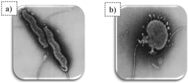
(34) Chapter I. Literature Review.. Campylobacter spp. are unable to grow at temperatures under 30°C and a sudden growth decline near the lower temperature limit is observed, it has been noted that they survive up to 15 times longer at 2°C than at 20°C (Hazeleger et al. 1998). At freezing temperatures the ability of Campylobacter ssp. to survive decreases rapidly. In pure cultures, Campylobacter spp. are normally inactivated by frozen storage at −15°C in as few as 3!days (Stern and Kotula, 1982). As result, freezing appears to be an efficient way to reduce the level of Campylobacter in chicken meat (Georgsson et al. 2006; Rosenquist et al. 2006; Meldrum and Wilson 2007; FSA 2009). However, freezing does not eliminate the pathogen from contaminated foods (Lee et al., 1998). Campylobacter spp. may still be isolated from frozen poultry (Lee et al. 1998; Sandberg et al. 2005). C. jejuni and C. coli, together with C. lari and C. upsaliensis, belong to the socalled thermophilic group of Campylobacter spp., which are able to grow at 42ºC (Penner, 1991). Under appropriate atmospheric and nutritional conditions, C. jejuni grows at temperatures between 32 and 45ºC, while the optimal growth temperature ranges between 42 and 45ºC (Doyle and Roman, 1981). Campylobacter appears to be highly sensitive to environmental factors such as drying conditions and osmotic stress. During exposure to unfavourable environmental conditions, such as high oxygen concentration, extreme temperatures, low nutrient availability or low osmolality environments, Campylobacter may form coccoid cells, which have been associated with loss of culturability using traditional culture methods (Klančnik et al., 2013). Rosenquist et al. (2006) related this coccoid form as a nonviable, degenerative form, or a dormant state that is non-culturable with metabolically active, and is recoverable in a suitable animal host (VBNC, Viable But Non-Culturable) (Figure 2). The bacteria can decrease its metabolic activities and undergo morphological transformation from the motile spiral form to a coccoid form. As for other pathogens, it is still not clear whether this coccoid form retains the potential to be revived to a colonisation/infectious form (Ziprin et al. 2003; Oliver 2005). However, Cappelier (1997) observed under laboratory conditions, that Campylobacter strains, isolated from the soil around the broiler house, may have been transformed into viable but noncultivable forms and might have become cultivable after passing through the intestinal tract of chickens.. 6.
(35) Chapter I. Literature review.. a) ). b) ). Figure 2. Campylobacter jejuni forms: a) spiral form. b) coccoid form. (Pead, 1979).. I.1.3. Nomenclature. The. genus. Campylobacter. currently. comprises. 34. species. (http://www.bacterio.net/Campylobacter.html, last accessed 6 September 2016) and 14 subspecies. The species C. jejuni comprises two subspecies (C. jejuni subsp. jejuni and C. jejuni subsp. doylei). Currently, the family Campylobacteraceae consists of 3 genera: Campylobacter, Sulfurospirillum, and Arcobacter (Vandamme, 2000; Vandamme et al., 2005; Vandamme and De Ley, 1991). Within the genus, three species (C. jejuni, C. coli, and C. lari) are known as thermophilic members of the genus and of clinical significance as they are the dominant 2 causative agents of human campylobacteriosis. C. jejuni accounts for the majority of food-borne Campylobacter enteritis in humans, followed by C. coli, and to a lesser extent, by C. lari (Pearson et al., 1996; Hazeleger et al., 1998). Within the past 20 years the genus Campylobacter has been subjected to a continuos evolution in nomenclature. In 1991, Vandamme et al. (1991) proposed a new family, Campylobacteraceae, consisting of the genus Campylobacter, with eleven species, and the new genus Arcobacter. Since C. jejuni and to a lesser extent C. coli are the most frequently isolated Campylobacter species both from patients with enteritis (Endtz, 1991, Skirrow and Blaser, 1992) and from poultry (Jacobs-Reitsma et al., 1994) the following paragraphs will focus on these two species.. 7.
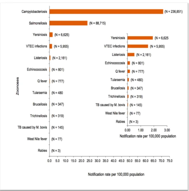
(36) Chapter I. Literature Review.. Although originally placed in the genus Vibrio, a new genus name of Campylobacter was proposed (Sebald and Véron, 1963) to reflect fundamental differences from the vibrios. It was not until the 1970s before they were isolated successfully from the stools of humans with acute enterocolitis (Butzler et al., 1973; Skirrow, 1977). Their presence in the gut had been suspected before this time (Levy, 1946; King, 1957), but the techniques traditionally used in clinical laboratorios were not suitable for the isolation of campylobacters. Although the species names of C. jejuni and C. coli were derived from an initial association with enteric disease in animals (Jones et al., 1931; Doyle, 1948), they are the most important human pathogens in this genus, with the former usually responsible for the majority of enteric Campylobacter infections (80-90%).. I.2. Campylobacter epidemiology in humans. I.2.1. Campylobacteriosis in humans Campylobacter is a zoonotic pathogen and is the main cause of human bacterial gastroenteritis in the world (Scallan et al., 2011; EFSA and ECDC, 2015b). In Europe, results from the European Summary Report on Trends and Sources of Zoonoses, Zoonotic Agents and Foodborne Outbreaks in 2014, revealed that Campylobacter has been the most commonly reported gastrointestinal bacterial pathogen in humans in the EU since 2005, with 236,851 reported confirmed cases (Figure 3). The notification rate was 71.0 cases per 100,000 of the population in European countries, with a case-fatality rate of 0.01%. In Spain, the reported incidence was higher than the average in UE accounting for 83.3 cases per 100,000 persons. However, it is well recognised that the actual numbers of human campylobacteriosis cases are underestimated as not all cases are reported in the laboratory due to the self-limiting nature of the disease and that it can be associated with mild symptoms (Allos, 2001; EFSA, 2011; Tam et al., 2012). In Europe, the true incidence of human campylobacteriosis is estimated to be approximately nine million cases per year (EFSA, 2011).. 8.
(37) Chapter I. Literature review.. Figure 3. Reported numbers and notification rates of confirmed human zoonoses cases in the EU in 2014. Total number of confirmed cases is indicated in parenthesis at the end of each bar. Exception is made for West Nile fever where the total number of cases was used (EFSA and ECDC, 2015b).. This infection has major economic repercussions on human health care. Indeed there are direct illness costs such as health consultations, laboratory diagnosis, medical treatment or hospitalisation and indirect costs such as loss of work productivity due to sickness, product recalls and legal costs (Roberts et al. 2003; Bogaardt et al., 2004). In the EU the cost of campylobacteriosis to public health systems is estimated to be about €2.4 billion per year and the disease burden was calculated at 35,000 disability-adjusted life years (DALYs) (EFSA, 2011).. 9.
(38) Chapter I. Literature Review.. As reported above, the most frequently identified Camplylobacter species associated with human disease have been identified as C. jejuni and C. coli (Nachamkin and Blaser, 2000; Allos, 2001; Friedman et al., 2004; Lin, 2009; Hermans et al., 2012). In fact, it was observed that C. jejuni accounted for 81.8% of human campylobacteriosis cases in the EU in 2014, followed by C. coli, C. lari and C. upsaliensis corresponding to 7.13%, 0.13%, and 0.07% of the isolates respectively. Most Campylobacter infections appear to be sporadic rather than outbreak associated, and in majority of cases, the original source of infection cannot be determined (EFSA and ECDC, 2015b).. I.2.2. Human clinical aspects. In susceptible humans, campylobacteriosis infection is associated with acute enteritis and abdominal pain lasting for up to seven days or longer. The infective dose is generally low, induced by 500-800 bacteria (Conlan et al., 2011). The incubation period is two to five days, but estimates have extended up to ten days. The infection results in an acute self-limiting gastrointestinal illness typically resolved in one week, characterised by mild to severe watery/bloody diarrhoea, fever, nausea, malaise and abdominal pain (Blaser, 1997). Common campylobacteriosis symptoms include acute gastroenteritis, cramping abdominal pain, fever, vomiting and headaches (WHO, 2011). Diarrhoea occurs shortly after onset of abdominal pain and varies from mild, noninflammatory, watery to severe and bloody. The incubation period of Campylobacter is 3 days and falls within a range of 18 h to 8 days (Horn and Lake, 2013). Disease outcome is likely to be influenced by both host (age, health status, preexisting immunity), and pathogen specific factors, such as the virulence of the infecting strain (Altekruse et al., 1999). The disease is self-limited in most cases in adults and non-immunecompromised individuals. However, complications may occur and include bacteraemia, irritable bowel syndrome, and reactive arthritis characterized by conjunctivitis, urethritis and/or arthritis (Havelaar et al., 2000; Helms et al., 2003; Mangen et al., 2005; Gradel et al., 2009). Guillain Barré Syndrome (GBS) is the most commonly reported chronic sequelae (Zautner et al., 2014). This complication is a demyelinating neuropathy (Rajabally et al., 2014) and is characterised by ascending paralysis (Zilbauer et al.,. 10.
(39) Chapter I. Literature review.. 2008). It is estimated that one in 1,000 Campylobacter infections leads to GBS, with 23% of fatal cases (Allos, 1997). The bacterium surface has lipooligosaccharides (LOS), which are important for GBS development. LOS stimulate peripheral nerve gangliosides to result in the generation of autoreactive antibodies inflammation and tissue damage (Nyati and Nyati, 2013). Miller Fisher syndrome is a non-paralytic variant of GBS and causes inability to move eyes with non-reactive pupils (Mori et al., 2012). Reactive arthritis is also associated with Campylobacter post-infection with 7 in every 100 cases. Reactive arthritis occurs mainly in joints, particularly knees and ankles (Ajene et al., 2013). In limited cases, C. jejuni has been associated with intestinal haemorrhaging (Chamovitz et al., 1983), toxic megacolon (McKinley et al., 1980), haemolytic uraemic syndrome (Shulman and Moel, 1983) and bowel syndrome (Gradel et al., 2009). Human campylobacteriosis may rarely result in long-term disabilities or even death (Helms et al., 2003). Some persons are at higher risk of suffering severe symptoms (deriving in hospitalization and/or death) such as immunocompromised individuals, very young and very old persons (Helms et al., 2003; Gradel et al., 2009). Furthermore, Campylobacter strains that are resistant to the most commonly used antibiotics represent a challenge for the treatment of human campylobacteriosis (Moore et al., 2006).. I.2.3. Main sources of human campylobacteriosis. The bacteria are widespread in the environment and have been detected in various animal reservoirs, including poultry, cattle, swine, and dogs (Man, 2011). Therefore, they can be a source for food or water contamination and subsequently a risk factor for human campylobacteriosis. Campylobacter may be transmitted from these reservoirs to humans by many different routes. Several risk factors for human campylobacteriosis have been reported in various studies conducted in developed countries, with the most common ones being: consumption and handling of chicken, and in particular undercooked chicken or commercially prepared chicken, unpasteurised milk and dairy products, consumption of untreated water, contact with domestic pets like dogs and cats, contact with farm animals, and travel abroad (Eberhart-Phillips et al., 1997; Studahl and Anderson, 2000; Rodrigues et al., 2001; Tenkate and Stafford, 2001;. 11.
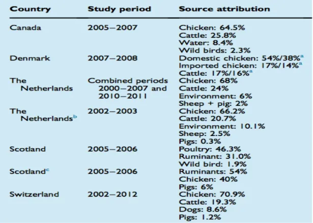
(40) Chapter I. Literature Review.. Potter et al., 2003; Friedman et al., 2004; Schonberg-Norio et al., 2004; Stafford et al., 2007, Figure 4).. Figure 4. Distribution of food vehicles in strong-evidence outbreaks caused by Campylobacter (excluding strong-evidence water-borne outbreaks) in the EU in 2013. Data from 32 outbreaks are included: Austria (5), Belgium (3), Germany (1), Netherlands (1), Spain (6), and United Kingdom (16). Number after the label refers to the number of outbreaks (EFSA and ECDC, 2015a).. Broiler meat is considered to be a major source of human campylobacteriosis, as a result of undercooking and cross-contamination, either directly onto other foods or via the kitchen environment from poultry meat during food preparation (Figure 5) (EFSA, 2010). Data from the EU summary report on zoonoses, zoonotic agents and food-borne outbreaks revealed that half of the Campylobacter outbreaks in the EU (16 out of 32) during 2014 were linked to broiler meat (EFSA and ECDC, 2015b). According to EFSA, handling, preparation and consumption of broiler meat is associated with 20% to 30% of human cases, while 50% to 80% could be attributable to the chicken reservoirs as a whole (EFSA, 2010). As it is well established that poultry meat is the most significant source of Campylobacter in the food chain (Wilson et al. 2008; Mullner et al. 2009; Sheppard et al. 2009), it can be predicted that a reduction of Campylobacter in chickens will reduce the number of cases in the human population. Therefore, implementation of Campylobacter control measures at the primary production level would not only reduce the contamination of broiler meat along the food chain, but also. 12.
(41) Chapter I. Literature review.. it would lower the human exposure to the bacteria through pathways other than meat consumption (EFSA, 2010). Thus, this fact is expected to have a bigger impact on the reduction of human disease. In addition, safe handling of raw meat, thorough cooking and strict kitchen hygiene should prevent or reduce the risk posed by Campylobactercontaminated broiler meat.. Figure 5. Comprehensive overview of Campylobacter source attribution studies published between 2010 and 2015. aThe first percentage indicates source attribution determined by asymmetric island model. The second percentage indicates source attribution by the Campylobacter source attribution model developed by authors. b Animal data supplemented with data from UK, Scotland, Switzerland, New Zealand, Curaçao, Finland and USA. cOnly C. coli included (Skarp et al., 2016).. Pigs seem to be a natural reservoir of Campylobacter spp. with prevalence between 50% and 100% and excretion levels ranging from 102 to 107 CFU/g (Munroe et al., 1983; Nielsen et al., 1997; Alter et al., 2005; Boes et al., 2005). In a national baseline survey of cattle and pigs conducted in Great Britain, the carriage rates were 54.6% and 69.3%, respectively (Milnes et al., 2008). Also, recent results on the investigation of Campylobacter in pork meat at the slaughterhouse among European countries revealed a mean prevalence of 9.75%, ranging from 5.91 to 50.0% (EFSA and ECDC, 2015b). Despite the medium-high carriage rates, there is little information concerning the contribution of pork meat to Campylobacter outbreaks. Several authors found that transmission of Campylobacter spp. from pigs appears to be non-evident for. 13.
(42) Chapter I. Literature Review.. C. jejuni and of very low risk for C. coli, where only two out of 4604 incidents of infectious intestinal disease, investigated and reported to the Public Health Laboratory Service in the UK, over an eight year period, were linked to pork meat and one of these was due to cross contamination (Kramer et al., 2000). In contrast to the number of foodborne outbreaks attributed to consumption of undercooked poultry contaminated with Campylobacter, only 1% of the reported outbreaks were associated with consumption of pork meat in the EU during 2013 (EFSA and ECDC, 2015b). ! Cattle are also common carriers of campylobacters (Humphrey and Beckett. 1987; Stanley et al., 1998; Inglis et al., 2004). Data for Campylobacter prevalence in cattle in the European Union range from 0 % to 16.5 % (EFSA and ECDC, 2015b). However, beef is not considered to be an important vehicle of transmission in human infections, because campylobacters are not commonly detected on carcasses or in beef. In surveys of retail beef only 0 to 5% of the samples have tested positive for campylobacters (Stern et al., 1985; Ono and Yamamoto, 1999; Whyte et al., 2004). Nevertheless, a recent study from the United States found that 5% (12/262) of campylobacteriosis outbreaks from 1997–2008 were due to consumption of contaminated pork, beef or game (Taylor et al., 2013). In addition, molecular typing studies of C. jejuni isolates from cattle have demonstrated a similarity with human strains (Fitzgerald et al., 2001; Schouls et al., 2003). Sporadic outbreaks of campylobacteriosis have been linked to contaminated red meat (Itoh et al., 1980; Inglis et al., 2004). Raw milk has also been identified as a vehicle of human gastroenteritis caused by Campylobacter spp. in several epidemiological studies (Studahl and Anderson, 2000; Michaud et al., 2004). Contamination of milk with Campylobacter can arise from a number of different sources involving intrinsic contamination from infection in the animal prior to milking, or extrinsic contamination arising from environmental contamination of the milk with faecal material either directly from the animal at the time of milking, or indirectly from the milking equipment, farm environment or at the point of use (Rapp et al., 2012; Moatsou and Moschopoulou, 2014). Data from the EU summary report on zoonoses, zoonotic agents and food-borne outbreaks in 2014 revealed that the Campylobacter was detected in up to 16.7% of the tested units (single. 14.
(43) Chapter I. Literature review.. or batch) of raw cow’s milk intended for direct human consumption or manufacture of raw or minimal heat-treated products (EFSA and ECDC, 2015). Raw-milk outbreaks involving Campylobacter have been consistently reported by several authors (Evans et al., 1996; Lehner et al., 2000; Studahl and Andersson, 2000; Schildt et al., 2006; CDC, 2013a; Mungai et al., 2015). Recently, the CDC’s Emerging Infectious Disease Journal reported an increase of the outbreaks associated with raw milk from 30 (2007-2009) to 51 (2010-2012) (Mungai et al., 2015). Also, the European Food Safety Authority’s Panel on Biological Hazards reported that 21 of the 27 raw milk-related outbreaks were attributed to Campylobacter spp., predominantly C. jejuni (EFSA and ECDC, 2015a). Consumption of untreated water (Schorr et al., 1994) has also been considered as a risk factor for campylobacteriosis. In an ecological study in Sweden, positive associations were found between the incidence of Campylobacter spp. and the average volume of water consumed per person and similar associations were found with ruminant density. These observations suggest that drinking water and contamination from livestock might also be important factors in explaining at least a proportion of human sporadic campylobacteriosis cases (Nygard et al, 2004). The faeces of livestock, domestic and wild animals, wild birds, poultry and also sewage effluents are usually the sources of Campylobacter in water environments (Jones, 2001). Campylobacter species have been described as common causative agents in waterborne gastrointestinal illness outbreaks all over the world and most of the illness cases have been associated with C. jejuni (Pitkänen et al., 2008). Untreated drinking water has been worldwide implicated in several Campylobacter outbreaks (Brieseman, 1987; Aho et al., 1989; Stehr-Green et al., 1991; Duke et al., 1996; Furtado et al., 1998; Miettinen et al., 2001; Jakopanec et al., 2008; Karagiannis et al., 2010; CDC, 2013b) and has been found as a risk factor in several case-control studies from other countries (Eberhart-Phillips et al., 1997; Friedman et al., 2004; Domingues et al., 2012). Specifically, Campylobacter was identified as the major pathogen in outbreaks traced to private water supplies in England and Wales (Said et al. 2003), and in Canada Campylobacter was the second most common pathogen in 24 waterborne disease outbreaks during 1974–2001 (Schuster et al. 2005). Also, in Finland, Campylobacter. 15.
(44) Chapter I. Literature Review.. has been the most common bacterial pathogen identified in waterborne disease outbreaks, being implicated in 11 incidents between 1998 and 2004 (Kuusi et al. 2005). The proportion of Campylobacter-positive cats and dogs is generally low, but in two clinical investigations from the Netherlands and Norway 40.4 % and 31.2 %, respectively, of the tested dogs were found to be Campylobacter-positive (EFSA and ECDC, 2015a). Species information was reported by Norway, where 101 of the 119 Campylobacter-positive dogs were infected with C. upsaliensis and the rest of the findings were due to species more commonly causing human disease (C. jejuni in 12 dogs and C. coli in one dog). Campylobacter can be transported directly from animals (i.e. skin contaminated with faeces) to humans (petting the animal and then subsequently using the hands to touch food or mouth directly) (EFSA, 2011).. I.3. Campylobacter epidemiology in broiler production. I.3.1. Avian campylobacteriosis. Campylobacter can colonize the intestinal mucosa of most a wide range of warm-blooded host species (Newell and Fearnley, 2003). The avian species are the most common hosts for Campylobacter spp. probably because of their higher body temperature (Skirrow, 1977). Although all commercial poultry species can carry Campylobacter spp., the risk is greater from chicken because of the large quantities consumed (Humphrey et al., 2007). In broiler chickens Campylobacter is a commensal organism that establishes persistent and benign infections with colonization level up to 1010 colony-forming units (CFU) per gram of faeces (Sahin et al., 2002; Newell and Fearnley, 2003; Dhillon et al., 2006). Campylobacter can be isolated from most intestinal sites of broiler chickens, but it is mainly found in the caecal and cloacal crypts, where it does not adhere to epithelial cells but is found in the mucous layer (Beery et al., 1988; Achen et al., 1998). In contrast to infection in humans, the bacteria does not induce any pathology in chickens and inhabits the lower intestine in a commensal relationship (Dhillon et al. 2006). Histopathological studies reveal no evidence of necrosis and no significant change in crypt architecture (Berry et al., 1988; Dhillon et al., 2006; Shaughnessy et al., 2011). Moreover, colonization is persistent suggesting that the immune response is ineffective. 16.
(45) Chapter I. Literature review.. in the elimination of infection, at least under these circumstances, although older birds, e.g. layers, may have reduced colonization with time (Newell and Fearnley, 2003). In summary, this situation has large benefits for the bacterium and no detrimental effects on the host.. I.3.2. Colonization and immunology against Campylobacter. Ingestion of Campylobacter numbers as few as 35 CFU can be sufficient for successful colonization of chicks (Stern et al., 1988). After ingestion, the bacterium reaches the cecum and multiplies, resulting in an established colonizing Campylobacter population within 24 hours after entrance (Coward et al., 2008). Campylobacter is not routinely detected in birds younger than 2-3 weeks old (Newell and Fearnley, 2003; van Gerwe et al., 2009). This early age-related resistance (commonly called lag phase), extended against different Campylobacter species, is not completely understood. Campylobacter specific maternal antibodies (MAB) are common in young chickens and could be involved in this protection: the high level of these antibodies observed during the first weeks, falls at 14 days, reaching minimal levels at 3-4 weeks (Sahin et al., 2002). Also the stage of intestinal development has been hypothesised to be involved in this age resistance, as avian intestinal niches go through physiological change during the first weeks of life (van Der Wielen et al., 2000). Changes in the microbial flora and competitive caecal microflora (Mead, 2002) are also considered in relation to the lag phase, together with management adjustments, such as changes in feed and medication that occur during the rearing period. Campylobacter colonization of chickens is rapid and widespread, so that once flock colonization is detected, the majority (>95%) of the birds of that flock is colonized within several days (Stern et al., 2001), and stay so until slaughter (Coward et al., 2008; Stern, 2008). However, it has been observed that, after 8 weeks, colonisation could decrease in terms of number of bacteria and number of birds colonised which is likely to be associated with the development of an adaptive immunity and changes in the intestinal microflora (Achen et al. 1998; Sahin et al. 2003a; Vandeplas et al. 2010). Although no pathology is associated with chicken colonization, an intestinal immune response to infection has been illustrated with increased cytokine expression. 17.
(46) Chapter I. Literature Review.. (Borrmann et al., 2007; Smith et al., 2005, 2008; Larson et al., 2008; Li et al., 2008) and toll-like receptor (TLR) activation (de Zoete et al., 2010). Campylobacter is able to stimulate both a systemic and mucosal immune response in chickens, as it has been shown by different studies that reported the induction of immune-associated gene and protein expression after Campylobacter colonization of chicken (de Zoete et al., 2007). However it is still largely unknown how Campylobacter interacts with the chicken immune system to trigger the immune response (Lin et al., 2009). Analysis of isolated chicken tissue displayed an increase in cytokine expression (Smith et al., 2008) and circulating monocytes/ macrophages (Meade et al., 2009), and several different types of chicken cells produce or upregulate cytokines during in vitro infection (Smith et al., 2005; Larson et al., 2008; Li et al., 2008). However, Campylobacter-specific antibody response is slow and moderate in chicken host because the infection does not cause a strong inflammatory response or tissue damage in intestine (Lin et al., 2009). In some studies, Campylobacter was also isolated from the bursa of Fabricius, thymus, reproductive tract, spleen, liver and blood in young chickens, suggesting that Campylobacter may invade intestinal epithelial cells and become systemic (Cox et al., 2005a, 2006, 2009; Knudsen et al., 2006; Lamb-Rosteski et al., 2008; Van Deun et al., 2008; Meade et al., 2009; Richardson et al., 2011). Recent studies further demonstrated that C. jejuni could adhere to and invade chicken intestinal epithelial cells in vitro and in vivo. However, the C. jejuni strains that invaded chicken epithelial cells were not able to proliferate intracellularly, but quickly evaded from the cells (Byrne et al., 2007; Van Deun et al., 2008). Therefore, Van Deun et al. (2008) proposed a novel colonization mechanism of C. jejuni by escaping rapid clearance through shortterm epithelial invasion and evasion, combined with fast replication in the mucus. Other studies have demonstrated that the chick immune system may be inefficiently activated upon Campylobacter colonization and expression of several antimicrobial peptide genes may be reduced (Meade et al., 2009; Hermans et al., 2012). All these observations may indicate that C. jejuni is well adapted to the poultry host, and bacteria may be seen as a normal enteric flora by the host. This fact may contribute to the persistent colonization of Campylobacter in the avian gut.. 18.
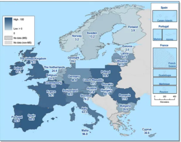
(47) Chapter I. Literature review.. I.3.3. Campylobacter in primary production. I.3.3.1. Prevalence in broiler batches. The prevalence in commercial broiler flocks varies greatly depending on the age of birds (Kazwala et al., 1990; Berndtson et al., 1996a, 1996b; Evans and Sayers, 2000). Campylobacter is rarely detected in broiler chickens less than 2–3 weeks old under commercial production conditions, although newly hatched chickens can be experimentally infected with C. jejuni (Shanker et al., 1986; Stern et al., 1988; Sahin et al., 2001). For the majority of commercial flocks, Campylobacter infection is usually detected after the third week of age. Once some birds become infected, C. jejuni spreads rapidly to most of the birds in the flock, which remain colonized up to slaughter, leading to carcass contamination at the processing plants (Jacobs-Reitsma et al., 1995; Berndtson et al., 1996a; Gregory et al., 1997; Evans and Sayers, 2000; Shreeve et al., 2000). Commercial poultry are the major natural reservoirs of C. jejuni, and up to 100% of broilers at slaughter age may harbor the organism (Jacobs-Reitsma et al., 1995; Jacobs-Reitsma, 1997). Results from a European Union survey in member states to estimate the prevalence of Campylobacter in broiler batches revealed a mean prevalence of 71.2% (95% CI: 68.5; 73.7), with results ranged from a minimum of 2.0% (Estonia) to a maximum of 100.0% (Luxembourg) (Figure 6). The median of Member States prevalence of Campylobacter-colonised broiler batches was 57.1% (EFSA, 2010). In addition, results from the European Summary Report on Trends and Sources of Zoonoses, Zoonotic Agents and Foodborne Outbreaks in 2014, reported an overall occurrence of Campylobacter in fresh broiler meat, sampled at slaughter, processing and retail of 38.4% of the 6,703 tested units (single or batch, aggregated data from all sampling stages) (EFSA and ECDC, 2015b). The proportion of Campylobacter-positive samples of broiler meat varied greatly between reporting Member States. Campylobacter was detected in 35.5% of single samples at retail; six of eleven MS reporting at retail level found ≥ 50.0% positive samples. At slaughterhouse level, 44.4% of the single samples tested positive for Campylobacter (EFSA and ECDC, 2015b).. 19.
(48) Chapter I. Literature Review.. Shedding of Campyloacter by chickens varies by season, being highest in the summer (Jacobs-Reitsma et al., 1994; Gregory et al., 1997; Evans and Sayers, 2000; Newell and Wagenaar, 2000; Wedderkopp et al., 2000, 2001). Even though C. jejuni is highly prevalent in broiler chickens, some flocks remain free of Campylobacter throughout their lifespan (Berndtson et al., 1996b; Wedderkopp et al., 2000; Stern et al., 2001). Campylobacter is also highly prevalent in chickens raised on organic or freerange farms (Rivoal et al., 1999; Heuer et al., 2001), indicating that different production systems are equally vulnerable to invasion by this organism.. Figure 6. Prevalence (%) of Campylobacter spp. colonised broiler batches in the EU, 2008, EFSA (EFSA, 2010).. I.3.3.2. Risk factors and sources of contamination at broiler farms. The possible sources and transmission routes of Campylobacter for poultry flocks have been investigated extensively, but no definitive factor(s) have been identified that explain the occurrence of the organism in commercial poultry flocks (Sahin et al., 2002; Cox et al., 2012). Circumstantial evidence has been accumulated in. 20.
(49) Chapter I. Literature review.. favour of horizontal transmission from the environment as the most probable source of poultry infection by C. jejuni (Sahin et al., 2002; Newell and Fearnley, 2003; O’Mahony et al., 2011). Potential sources include old litter (Thakur et al., 2013), untreated drinking water (Pearson et al., 1993; Stanley et al., 1998; Zimmer et al., 2003), other farm animals or domestic pets (van de Giessen et al., 1996, 1998; Bouwknegt et al. 2004; Lyngstad et al., 2008; Zweifel et al., 2008; Ellis-Iversen et al., 2009), insects (Shane et al., 1985; Jacobs-Reitsma, 1997; Hald et al., 2008; Hazeleger et al., 2008), rodents (Gregory et al., 1997; McDowell et al., 2008), equipment and transport vehicles and farm workers (Johnsen et al., 2006; Lyngstad et al., 2008; Ridley et al., 2008). Campylobacter is very sensitive to oxygen and drying, thus it is generally unable to grow in litter under normal ambient conditions (Kazwala et al., 1990). The organism is usually absent in fresh litter or feed samples before broilers are infected (Humphrey et al., 1993; Pearson et al., 1993; Jacobs-Reitsma et al., 1995; Gregory et al., 1997; van de Giessen et al., 1998; Thakur et al., 2013). Used litter may become contaminated by Campylobacter and may play a role in maintaining the bacteria in the farm environment (Montrose et al., 1985). In European countries, since broiler houses are usually cleaned and disinfected and the litter is changed between consecutive flocks, litter seems an unlikely source of infection in commercial broiler production (Evans, 1992). Also, a nationwide epidemiological study in the USA indicated that there were no marked differences in the prevalence and onset time of Campylobacter shedding among flocks on different grow-out farms having different practices of litter use (Stern et al., 2001). Farm animals such as cattle, pigs and other poultry have also been recognised as a potential reservoir of the Campylobacter (van de Giessen et al., 1996, 1998; Bouwknegt et al., 2004; Hald et al. 2004; Lynngstad et al. 2008; Zweifel et al. 2008). Molecular epidemiological investigations on farm livestocks showed that strains colonizing target poultry flocks can sometimes be found in adjacent livestock, including cattle and pigs (Jacobs-Reitsma et al. 1995; Johnsen et al., 2006; Ridley et al., 2011), and this occurrence can be detected prior to poultry flock colonization in longitudinal studies, indicating that the direction of transmission is from the livestock to the broilers. Moreover, models from the Netherlands (Katsma et al., 2007) indicate that removal of other livestock from a poultry farm would reduce infection only from 44% to 41%.. 21.
(50) Chapter I. Literature Review.. However, this seems to be a relatively rare event (Johnsen et al., 2006), and the majority of the strains in adjacent livestock are not recovered subsequently from broilers. In addition, domestic animals such as dogs and cats have been found to frequently carry and shed C. jejuni and C. coli, and their presence on farm facilities has also been recognised as a risk factor of Campylobacter infection of the broiler flocks (EllisIversen et al. 2011; Torralbo et al., 2014). Insects such as flies, beetles, etc., may act as mechanical vectors for Campylobacter transmission from various animals to chickens (Shane et al., 1985; Jacobs-Reitsma, 1997; Hald et al. 2008; Hazeleger et al., 2008). Campylobacter transmission between chicken flocks by flies under controlled laboratory conditions was first demonstrated by Shane et al. (1985). Thereafter, Hald et al. (2004) showed that C. jejuni most likely was carried by house flies from livestock (sheep) close to the farm and to the broiler flocks through ventilation inlets. In addition, a recent study successfully proved the effect of hygiene barriers and fly screens as a way to reduce prevalence of Campylobacter spp. among flocks of broiler chickens, suggesting that flies may have a linking role in Campylobacter epidemiology (Bahrndorff et al., 2013). Despite of these findings the contribution of flies as a significant source of Campylobacter infection of broiler flocks has been debated. Even though identical serotypes and genotypes of Campylobacter have been isolated from insects and broilers within broiler houses, the direction of spread was not determined (Jacobs-Reitsma et al., 1995; Berndtson et al., 1996a; Stern et al., 1997). In fact, the insects in a chicken house were usually not positive for Campylobacter until the broilers were determined to be positive, suggesting that insects might not be important as an original source of Campylobacter for a broiler house (Berndtson et al., 1996a; Nesbit et al., 2001). Also, some studies have shown the role of darkling beetles (Alphitobius diaperinus) and their larvae as potential vectors for the transfer of C. jejuni between successive rearing cycles (Refrégier-Petton et al., 2001; Hazeleger et al., 2008). The evidence for rodents as infection sources for Campylobacter is also circumstantial (Newell et al., 2011). The presence of rodents on farms can have a strong association with flock positivity (Gregory et al., 1997; McDowell et al., 2008), and the efficacy of vermin control is a risk factor (Arsenault et al., 2007; Huneau-Salaun et al., 2007). Although rodents are detected within the poultry houses of some modern farms. 22.
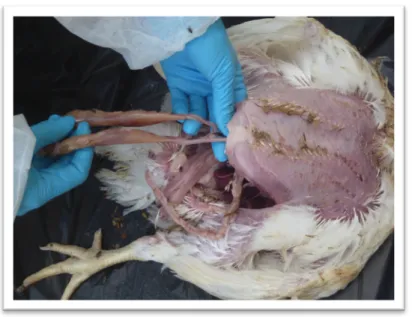
Figure


+7


Documento similar
In these and the rest of online media that reached a medium level of participation in relation to news production, the user acquires, to some extent, roles
In the “big picture” perspective of the recent years that we have described in Brazil, Spain, Portugal and Puerto Rico there are some similarities and important differences,
This article describes the current situation of desalinated seawater production and supply to agriculture in the southeast of Spain, and analyzes key questions such as its role
At the same time, however, it would also be misleading and simplistic to assume that such Aikido habituses were attained merely through abstracted thought
Influence of farm management on the dynamics of Salmonella enterica serovar Infantis shedding and antibiotic resistance during the growing period of
• How does the global consumption of rice relate to the use of different kinds of water at production regions?. • What is the role of the blue (surface and ground) and green water
Therefore, the aim of this research was to estimate the sensory shelf life of breast and leg quarter meat obtained from chickens fed on the HC diet (based on HC maize)
These searches are dubbed resonant, since they allow to directly probe B(Υ → γa) in a model-independent way, regardless of the non-resonant contribution from figure 1. Searches
