Heme oxygenase 1 contribution to modulating the severity of salmonella enterica serovar typhimurium infection in mice
75
0
0
Texto completo
(2) 2. 20 21 22 23. DOCTORAL THESIS:. 24. HEME OXYGENASE 1 CONTRIBUTION TO MODULATING THE SEVERITY OF. 25. SALMONELLA ENTERICA SEROVAR TYPHIMURIUM INFECTION IN MICE.. 26. Tesis entregada a la Pontificia Universidad Católica de Chile en cumplimiento parcial de. 27. los requisitos para optar al Grado de Doctora en Ciencias con Mención en Genética. 28. Molecular y Microbiología. 29 30. By. 31 32. VALENTINA PILAR SEBASTIAN QUIJADA. 33 34. Tutor: Dr. Susan Bueno Ramírez. 35. Thesis Committee: Dr. Alexis Kalergis. 36. Dr. Luis Larrondo. 37. Dr. Juan Pablo Mackern. 38 39. October 2019.
(3) 3. 40 41 42 43 44 45 46 47 48 49 50 51 52 53 54 55 56 57 58 59 60 61 62. ACTA APROBACIÓN TESIS.
(4) 4. 63. ACKNOWLEDGEMENTS. 64. First of all, I would like to thank the Vicerrectoría de Investigación from the Pontificia. 65. Universidad Católica de Chile for the financial support given to me during all of the PhD. 66. program. Also, the Facultad de Ciencias Biológicas from the Pontificia Universidad Católica de. 67. Chile was very important in this issue, because of the complementary grant given the first four. 68. years of my program. The Facultad de Ciencias Biológicas also helped me with financial support. 69. for attendance to international and national congresses. In addition, I would like to thank the. 70. Millenium Institute on Immunology and Immunotherapy for financial support granted to the. 71. development of this thesis.. 72. I would like to thank my lab team. Bárbara Schultz, Geraldyne Salazar, Catalina Pardo,. 73. Francisco Salazar and Omar Vallejos were fundamental for the development of this thesis, both. 74. experimental and intellectually. I also want to thank Loreani Noguera, Isidora Suazo, Liliana. 75. González, Irenice Coronado and Hernán Peñaloza for all the support. These professionals were. 76. not only my co-workers, but they became close friends and emotional support in the past years.. 77. I want to include in these acknowledgements the former members of Dr. Alexis Kalergis. 78. laboratory, especially Natalia Muñoz, Pablo Céspedes and Roberto Gómez. In addition, I deeply. 79. appreciate all the confidence my tutor Dr. Susan placed in me and my work. None of these. 80. would have been possible without her support and constant preoccupation on me. She believed. 81. in my abilities since the beginning and pushed me to stay strong and keep working hard.. 82. Finally, I would like to thank my mother Marcela, my father Jaime, my sisters Gabriela and. 83. Amparo, my grandmothers and all the members of my big family and friends that supported me. 84. from my decision to choosing science as my career, until my last years finishing this thesis,. 85. always feeling very proud and encouraging me to keep forward..
(5) 5. 86. INDEX. 87. ACKNOWLEDGEMENTS…………………………………………………………………….4. 88. FIGURES INDEX……………………………………………………………………………...7. 89. TABLES INDEX……………………………………………………………………………….8. 90. ABBREVIATIONS…………………………………………………………………………….9. 91. RESUMEN……………………………………………………………………………………11. 92. ABSTRACT…………………………………………………………………………………...13. 93. 1. GENERAL INTRODUCTION……………………………………………………………15. 94. 1.1. Acute and persistent Salmonella infection…………………………………………...15. 95. 1.2. Heme oxygenase 1……..……………………………………………………………..18. 96. 1.3. HMOX1 and microbial infections……………………………………………………21. 97. 1.4. HMOX1 and Salmonella infection…………………………………………………...23. 98. 2. SUBMITTED MANUSCRIPT: HEME OXYGENASE 1 CONTRIBUTION TO. 99. MODULATING THE SEVERITY OF Salmonella enterica SEROVAR TYPHIMURIUM. 100. INFECTION IN MICE……………………………………………………………………25. 101. 2.1. ABSTRACT………………………………………………………………………….25. 102. 2.2. INTRODUCTION……………………………………………………………………26. 103. 2.3. MATERIALS AND METHODS…………………………………………………….30. 104. 2.3.1. Mice…………………………………………………………………………...30. 105. 2.3.2. In vivo infection and monitoring……………………………………………....30. 106. 2.3.3. Bacteria and infective dose preparation……………………………………….32. 107. 2.3.4. CoPP and SnPP preparation…………………………………………………...32. 108. 2.3.5. Enrofloxacin administration…………………………………………………...32.
(6) 6. 109. 2.3.6. Bacterial loads in organs and tissues………………………………………….33. 110. 2.3.7. RNA extraction………………………………………………………………..33. 111. 2.3.8. Quantitative real time PCR……………………………………………………34. 112. 2.3.9. Western blot…………………………………………………………………...36. 113 114. 2.4 RESULTS……………………………………………………………………………37 2.4.1. 115 116. infection…………………………………………………………………...…..37 2.4.2. 117 118. HMOX1 induction by CoPP treatment reduces bacteria load in tissues of S. Typhimurium-infected mice…………………………………………………..41. 2.4.3. 119 120. CoPP-mediated HMOX1 induction does not influence an acute S. Typhimurium. Prophylactic HMOX1 induction by CoPP reduces persistence of S. Typhimurium in mice…………………………………………………………43. 2.4.4. HMOX1 induction by CoPP treatment reduces persistence of S. Typhimurium. 121. when administrated post-infection…………………………………………….47. 122. 2.5 DISCUSSION………………………………………………………………………..49. 123. 2.6 ACKNOWLEDGEMENTS…………………………………………………………...52. 124. 3. GENERAL DISCUSSION AND CONCLUSIONS………………………………………53. 125. 4. REFERENCES……………………………………………………………………………59. 126. 5. APPENDIX………………………………………………………………………………..72. 127. 5.1. Scientific meetings attended during this thesis……………………………………….72. 128. 5.2. Scientific publications generated in this thesis……………………………………….73. 129 130 131.
(7) 7. 132. FIGURE INDEX. 133. FIGURE 1: HMOX1 induction has no major effect on the clinical score and survival rates in. 134. the acute model of S. Typhimurium infection in mice………………………………………...38. 135. FIGURE 2: HMOX1 induction has a mild anti-inflammatory effect in the acute model of S.. 136. Typhimurium infection in mice……………………………………………………………….40. 137. FIGURE 3: HMOX1 induction with CoPP treatment reduces bacterial loads in an acute S.. 138. Typhimurium infection model in mice………………………………………………………..42. 139. FIGURE 4: HMOX1 induction by CoPP treatment reduces persistent S. Typhimurium infection. 140. model in mice………………………………………………………………………………….44. 141. FIGURE 5: Evaluation of hmox-1 transcription in tissues of S. Typhimurium-infected mice46. 142. FIGURE 6: Model proposed for HMOX1 modulation of S. Typhimurium infection in murine. 143. model…………………………………………………………………………………………..58. 144 145 146 147 148 149 150 151 152 153 154.
(8) 8. 155. TABLE INDEX. 156. TABLE 1: Daily supervision chart for Salmonella enterica serovar Typhimurium infected. 157. C57BL6 female mice………………………………………………………………………….31. 158. TABLE 2: Probes for gene targets and its ID used in qRT-PCR……………………………..35. 159. TABLE 3: HMOX1 induction by prophylactic treatment with CoPP reduces S. Typhimurium. 160. persistence in mice…………………………………………………………………………….45. 161. TABLE 4: HMOX1 induction by treatment with CoPP reduces persistence of S. Typhimurium. 162. in mice when applied after infection…………………………………………………………..48. 163 164 165 166 167 168 169 170 171 172 173 174 175 176 177.
(9) 9. 178. ABBREVIATIONS. 179. TTSS: Type Three Secretion System. 180. SPI-1: Salmonella-pathogenicity island 1. 181. SPI-2: Salmonella-pathogenicity island 1. 182. IL-10: interleukin 10. 183. HMOX1: Heme oxygenase 1. 184. CO: carbon monoxide. 185. CoPP: cobalt protoporphyrin. 186. SnPP: tin protoporphyrin. 187. DC: dendritic cells. 188. SLE: systemic lupus erythematosus. 189. EAE: experimental autoimmune encephalomyelitis. 190. IRF3: interferon regulatory factor 3. 191. TLR: Toll-like receptor. 192. IFN-: interferon . 193. RSV: respiratory syncytial virus. 194. HBV: hepatitis B virus. 195. NALP3: NACHT, LRR and PYD domains-containing protein 3. 196. shRNA: short hairpin RNA. 197. IBD: inflammatory bowel disease. 198. ATCC: American Type Culture Collection. 199. ON: over night. 200. OD: optical density.
(10) 10. 201. LB: Luria broth. 202. NF: nuclease free. 203. qRT-PCR: quantitative real time PCR. 204 205 206 207 208 209 210 211 212 213 214 215 216 217 218 219 220 221 222 223.
(11) 11. 224. RESUMEN. 225. Salmonella enterica es un bacilo Gramnegativo perteneciente a la clase de las. 226. Gammaproteobacterias, cuyos serovares son capaces de causar enfermedades gastrointestinales. 227. y sistémicas en animales y humanos. En particular Salmonella enterica serovar Typhimurium. 228. (S. Typhimurium) es la causa más común de intoxicación por alimentos contaminados y en. 229. ratones es capaz de causar una enfermedad sistémica, muy parecida a la fiebre tifoidea causada. 230. por Salmonella enterica serovar Typhi en humanos. Su reservorio natural comprende aves de. 231. corral y sus huevos, reptiles y otros mamíferos, y se transmite a través del consumo de alimentos. 232. o agua contaminada. Una de las principales características que hacen de Salmonella enterica. 233. serovar Typhimurium una bacteria virulenta es su habilidad de evadir el sistema inmune del. 234. hospedero, generando infecciones sistémicas y persistentes. Una de las moléculas. 235. inmunomoduladoras expresadas por las células del hospedero que juega un rol en la eliminación. 236. de bacterias es Hemoxigenasa 1. Hemoxigenasa 1 es una enzima que cataliza la degradación del. 237. grupo hemo en Fe3+, biliverdina y monóxido de carbono. El rol de la actividad de Hemoxigenasa. 238. 1 durante una infección por S. Typhimurium no está claro y estudios previos muestran resultados. 239. contradictorios. En este estudio, evaluamos el efecto de la inducción farmacológica de HMOX1. 240. en un modelo de infección aguda y persistente por S. Typhimurium. Para abordar esto, indujimos. 241. la expresión de Hemoxigenasa 1 e inhibimos su actividad enzimática en ratones mediante el. 242. tratamiento con protoporfirina de cobalto o protoporfirina de estaño, respectivamente, antes de. 243. una infección con S. Typhimurium. Observamos que la inducción de Hemoxigenasa 1 con. 244. protoporfirina de cobalto no tiene mayor efecto en el score clínico y en la supervivencia de los. 245. ratones infectados con S. Typhimurium. Sin embargo, el tratamiento con protoporfirina de. 246. cobalto redujo la carga bacteriana en órganos 5 días post-infección, mientras que los ratones.
(12) 12. 247. tratados con protoporfirina de estaño mostraron cargas similares a las de los ratones tratados. 248. con vehículo. Además, la inducción de Hemoxigenasa 1 elimina la carga bacteriana cuando el. 249. tratamiento con protoporfirina de cobalto se realiza después de la infección en un modelo de. 250. infección persistente por S. Typhimurium, mientras que el tratamiento con protoporfirina de. 251. estaño resultó en valores de bacterias persistentes similares a los observados en ratones tratados. 252. con vehículo. Nuestros resultados sugieren que la actividad de Hemoxigenasa 1 puede promover. 253. la eliminación de S. Typhimurium, reduciendo la diseminación y la persistencia de la bacteria. 254. en ratones.. 255 256 257 258 259 260 261 262 263 264 265 266 267 268 269.
(13) 13. 270. ABSTRACT. 271. Salmonella enterica is a Gram-negative bacillus that belongs to Gammaproteobacteria class,. 272. which serovars are capable of causing gastrointestinal and systemic diseases in animals and. 273. humans. Particularly Salmonella enterica serovar Typhimurium (S. Typhimurium) is the most. 274. common cause of food poisoning and can cause a typhoid-like fever in mice. Its natural reservoir. 275. comprises poultry, eggs, reptiles and other mammals, and can be transmitted by consumption of. 276. contaminated food and water. An important virulence trait of Salmonella enterica serovar. 277. Typhimurium is the ability to avoid the host immune response, generating systemic and. 278. persistent infections. One of the immunomodulatory molecules expressed by host cells that. 279. plays a role in bacterial clearance is Heme oxygenase 1. Heme oxygenase 1 is an enzyme that. 280. catalyzes the degradation of heme groups into Fe3+, biliverdin and carbon monoxide (CO). The. 281. role of Heme oxygenase 1activity during S. Typhimurium infection is not clear and previous. 282. studies have shown contradictory results. In this study we evaluated the effect of pharmacologic. 283. modulation of Heme oxygenase 1 in a mouse model of acute and persistent S. Typhimurium. 284. infection. To approach this question, we induced Heme oxygenase 1 expression and inhibited. 285. Heme oxygenase 1 enzymatic activity in mice by treating with cobalt protoporphyrin or tin. 286. protoporphyrin, respectively, prior to infection with S. Typhimurium. We observed that Heme. 287. oxygenase 1 induction with cobalt protoporphyrin has no major effect on the clinical score and. 288. survival of S. Typhimurium-infected mice. However, cobalt protoporphyrin reduced the. 289. bacterial burden in organs 5 days post-infection and tin protoporphyrin-treated mice show. 290. bacterial loads similar to vehicle-treated mice. Furthermore, Heme oxygenase 1 induction. 291. eliminated bacterial loads when cobalt protoporphyrin was administrated after infection in a. 292. persistent infection model of S. Typhimurium, while tin protoporphyrin treatment resulted.
(14) 14. 293. persistent bacterial burden in similar of vehicle-treated mice. Our results suggest that Heme. 294. oxygenase 1 activity can promote S. Typhimurium clearance, reducing bacterial dissemination. 295. and persistence in mice.. 296 297 298 299 300 301 302 303 304 305 306 307 308 309 310 311 312 313 314 315.
(15) 15. 316. 1. GENERAL INTRODUCTION. 317 318. 1.1 Acute and persistent Salmonella infection. 319. Salmonella enterica serovar Typhimurium (S. Typhimurium) is a Gram-negative. 320. facultative anaerobic bacterium that causes foodborne illnesses in humans, with more than 1.2. 321. million cases only in the United States (Scallan et al., 2011). The World Health Organization. 322. estimates that non-typhoidal Salmonella are one of the most important causes of dead due to. 323. foodborne diseases, together with norovirus and Campylobacter spp. (Havelaar et al., 2015). In. 324. Chile, salmonellosis is a mandatory reportable disease and the Public Health Institute report. 325. more than 11,000 cases between 2012 and 2016 (MINSAL, 2016), being children between 0-4. 326. years old the most affected. The natural host of S. Typhimurium can be poultry, swine, horses,. 327. cattle, wild rodents and human. In humans, the period of incubation vary between 4 and 72 hours. 328. after the consumption of contaminated food or water, and the symptoms include high fever,. 329. nausea and diarrhea (Chen et al., 2013; Lampel, 2012).. 330. The ability of Salmonella to cause systemic diseases is due to its capacity of survive and. 331. replicate inside phagocytic cells, evading the capacity of the immune system of the host to. 332. respond to bacterial infections (Bueno et al., 2012). Once Salmonella is ingested and accesses. 333. to the intestinal lumen, it invades epithelial cells and then is engulfed by macrophages and. 334. dendritic cells. Inside those cells, the bacteria can travel and spread to deeper organs such as. 335. spleen, liver and lymph nodes (de Jong et al., 2012; Salazar et al., 2017). The capacity of. 336. Salmonella to invade eukaryotic cells relays on a secretion system named Type III Secretion. 337. System (TTSS), that consist in a complex syringe-like structure used by the bacterium to inject. 338. effector proteins into host cell (Moest and Méresse, 2013; Troisfontaines and Cornelis, 2005)..
(16) 16. 339. TTSS-1 and TTSS-2 are encoded in Salmonella pathogenicity islands 1 and 2 (SPI-1 and SPI-. 340. 2), respectively, and allows the bacteria to invade epithelial and immune cells (Bueno et al.,. 341. 2010; Tobar et al., 2006). SPI-1 and SPI-2 also encodes other virulence factors required not only. 342. to enter host cells, but also to survive and replicate inside them (Bierschenk et al., 2019; Nieto. 343. et al., 2015).. 344. Multidrug and antibacterial resistance of pathogen bacteria have increased in last years,. 345. representing a major public health issue (Parisi et al., 2018; Prestinaci et al., 2015). This. 346. phenomenon can be driven by several mechanisms such as efflux pumps (Sun et al., 2014) and. 347. plasmids (Oliva et al., 2017). In the case of Salmonella spp. many cases of multidrug resistant. 348. have been reported not only in strains isolated from contaminated food but also in strains. 349. recovered from infected people (Mandomando et al., 2009; Nghiem et al., 2017; Sanchez-. 350. Maldonado et al., 2017; Ziech et al., 2016). This evidence represents a high risk, due to the. 351. persistent infections that this bacteria can cause (Michael and Schwarz, 2016).. 352. Several studies have found evidence of persistent Salmonella infections in mice, many. 353. days post infection and after an antibiotic treatment (Helms et al., 2005; Monack et al., 2004;. 354. Somedutta Barat, Benjamin Steeb, Alain Maze, 2012). Furthermore, it has been demonstrated. 355. that Salmonella can form biofilms and persist on gallstones (Adcox et al., 2016; Nath et al.,. 356. 2010), as well as in liver, spleen and mesenteric lymph nodes, where the bacteria persist inside. 357. macrophages (Monack et al., 2004; Søndberg and Jelsbak, 2016). Moreover, several recent. 358. investigations have shown that antibiotic treatment promotes the selection of “persisters”, which. 359. are clones of Salmonella that undergo genetic modifications during infection of mice treated. 360. with antibiotics. These persisters become resistant to antibiotics, are not able to replicate and. 361. reside intracellularly (Claudi et al., 2014; Diard et al., 2014; Helaine et al., 2014; Søndberg and.
(17) 17. 362. Jelsbak, 2016). Supporting this idea, a recent study has shown that an acetyltransferase toxin. 363. TacT, which blocks protein synthesis by affecting the function of tRNA molecules, is required. 364. by S. Typhimurium to become antibiotic resistant and persist in the infected tissues of mice. 365. (Cheverton et al., 2016). Importantly, it has been demonstrated that with the withdraw of. 366. antibiotics, persistent bacteria can resume active growth from infected tissues and produce. 367. relapsing infections (Cheverton et al., 2016; Diard et al., 2014). This data support the idea that. 368. the persisters can use reservoir cell to protect themselves from the action of the antibiotics,. 369. resulting in cell permanently infected. This is an important feature of the disease caused by. 370. Salmonella, because it has been reported that persistent bacterial infection leads to severe. 371. consequences, such as pancreatitis (DelGiorno et al., 2014).. 372. Our previous data showed that after an enrofloxacin treatment, resident bacteria are. 373. found in spleen and liver at day 42 post-infection in mice that do not produce the anti-. 374. inflammatory cytokine interleukin 10 (IL-10-/- mice), which is a mouse model of Inflammatory. 375. Bowel Disease (IBD). We have described that these mice are less susceptible to the acute disease. 376. caused by Salmonella than WT mice that produce IL-10 (Salazar et al., 2017), but are more. 377. susceptible to develop chronic intestinal inflammation than uninfected mice (Schultz et al.,. 378. 2018). Importantly, in both WT and IL-10-/- mice, persistent infections with Salmonella are. 379. observed after antibiotic treatment, which may have an important role in this disease,. 380. considering that antibiotics are used as part of the treatment in several patients (Schultz et al.,. 381. 2018). Moreover, another study described the persistence of S. Typhimurium in the cecum. 382. lymph node of infected mice, even after 10 days of ciprofloxacin treatment, which is a widely. 383. used antibiotic in humans to treat Salmonellosis (Kaiser et al., 2014). Furthermore, presence of. 384. S. Typhimurium has been observed after almost 3 months of infection in Gr1+ cells of mesenteric.
(18) 18. 385. lymph nodes of infected mice (Monack et al., 2004). Given this evidence, it is important to. 386. elucidate the state of the cells containing S. Typhimurium in a persistent manner. Preliminarily,. 387. a recent publication that tracks the presence of S. Typhimurium by using fluorescent bacteria in. 388. mice concludes that this bacterium resides inside iNOS-producing macrophages, associated with. 389. a granuloma in spleen, that keeps it isolated from Th1 T-cells (Goldberg et al., 2018).. 390. Because persistent infections with intracellular bacterial pathogens, which cannot be. 391. eliminated with the available antibiotics, could lead to several disorders such as chronic. 392. inflammation and re-activation of the infectious disease in the future, it is relevant to identify. 393. natural bactericidal mechanisms of the host that could be activated to eliminate intracellular. 394. bacteria and prevent persistent infections. In this way, it would be possible to achieve. 395. sterilization of the affected cells and tissues through the mechanisms of the host.. 396 397. 1.2 Heme oxygenase 1. 398. Among the molecules that are involved in the clearance of intracellular pathogens is. 399. Heme oxygenase 1 (HMOX1). HMOX1 is an enzyme that catalyzes the first step of the. 400. oxidative degradation of the heme group, which is a rate-limiting reaction that releases as by-. 401. products the following molecules: carbon monoxide (CO), free iron, and biliverdine (which is. 402. lately reduced to bilirubin)(Ryter et al., 2006). HMOX1 is composed of 288 residues and its. 403. active site is located between the first two alpha-helixes (Schuller et al., 1999). It is expressed. 404. in all mammalian tissues at basal undetectable levels, but in tissues where red blood cells or. 405. hemoglobin are degraded, such as spleen, liver, bone marrow and kidney, HMOX1 can be. 406. induced either by its substrate (heme), cobalt protoporphyrin (CoPP), and other physical and. 407. chemical stimuli (Vareille et al., 2008) (Espinoza et al., 2017) (Sebastian et al., 2018)..
(19) 19. 408. The exact mechanisms and the precise site where the reaction occurs that involves the. 409. conversion of hemoglobin, hematin and myoglobin into bilirubin, were not known until 1969,. 410. when Tenhunen, Marver and Schmid described that this reaction was catalyzed by a Heme. 411. oxygenase, based on two observations. First, the reaction required a metal chelate and the. 412. formation of only one isomer of bile pigment, suggesting the participation of an enzyme in this. 413. reaction. Secondly, the insertion of two hydroxyl groups indicates that the cleavage of the. 414. porphyrin ring is an oxidation. With this criteria, this group described that Heme oxygenase. 415. stoichiometrically require NADPH and molecular oxygen to generate the same amount of. 416. carbon monoxide and bilirubin, and that this enzyme is localized in the microsomal fraction of. 417. spleen and liver of Sprague-Dawley rats (Tenhunen et al., 1968),(Tenhunen et al., 1969).. 418. Through the following decades, Heme oxygenase was fully described in terms of its. 419. interactions with other proteins and the conditions that induces its expression and activity.. 420. However, it was not until late 90s that this enzyme was associated with the immune response. 421. against several stimuli. The immune mechanisms involved in the rejection to transplanted. 422. organs is an issue of major importance. In 1998, Soares and collaborators found that the. 423. mechanism that protects xenografts from being rejected involves the rapid increase of HMOX1. 424. expression by the endothelial and smooth muscle cells from mouse cardiac xenografts. 425. transplanted to rats, probably because of its anti-inflammatory properties (Soares et al., 1998).. 426. Two years later, the same group used mouse-to-rat cardiac transplant model, observing that. 427. inhibition of HMOX1 activity by tin-protoporphyrin resulted in earlier rejection of the organ,. 428. and that when these same rats were treated with exogenous CO, the long-term survival was. 429. restored (Sato et al., 2001). These studies demonstrate the important role of HMOX1 in immune. 430. modulation and that this effect is driven by CO..
(20) 20. 431. In a recent study, Dendritic Cells (DCs) treated with LPS and CO showed a diminished. 432. capacity of presenting antigens to T cells, and this effect was due to a reduced fusion of. 433. endosomes and lysosomes, that is necessary for antigen presentation (Tardif et al., 2013). This. 434. reduced fusion of endosomes and lysosomes is probably caused by the impaired mitochondrial. 435. function produced by CO (Riquelme et al., 2015). The process of inflammation requires antigen. 436. presentation and the consequent cytokine secretion that finally derive in cell infiltration. These. 437. results show a variety of targets of CO acting as an anti-inflammatory and cytoprotective. 438. molecule. Another of the effects of HMOX1 is the induction of anti-inflammatory cytokines,. 439. such as IL-10 (Zhang et al., 2017). However, it has been demonstrated that in murine. 440. macrophages, treatment with IL-10 also induce HMOX1 protein production in a dose-dependent. 441. manner; and this induction was through a MAPK called p38 (Lee and Chau, 2002) that is. 442. modulated by CO (Sheikh et al., 2011). In addition, HMOX1 induction has been reported to. 443. enhance the polarization of IL-10-producing anti-inflammatory macrophages (M2 phenotype). 444. (Sierra-Filardi et al., 2010). Moreover, HMOX1 can have protective effects mediated by the. 445. anti-inflammatory effect, in different tissues. For instance, hypoxia can induce the secretion of. 446. several pro-inflammatory cytokines in lungs, producing severe inflammation and structural. 447. changes of vessels. Transgenic mice that constitutively express HMOX1 that were submitted to. 448. hypoxia, had less inflammation and less vessel hypertrophy as compared with wild type mice. 449. (Minamino et al., 2001). Furthermore, the pro-inflammatory cytokines and chemokines. 450. expressed in normal conditions, were suppressed in HMOX1 transgenic mice (Minamino et al.,. 451. 2001). Similar anti-inflammatory effects were observed in a mouse model of lupus. 452. erythematosus (SLE). These FcRIIb-/- mice develop proteinuria and renal inflammation that is. 453. improved with HMOX1 induction or carbon monoxide treatment. Indeed, carbon monoxide.
(21) 21. 454. administration decreased the expansion of CD11b+ cells, the proportion of CD4+ FoxP3+ Treg. 455. cells and the anti-histone antibodies (Mackern-Oberti et al., 2013). It has been reported that. 456. patients with SLE show reduced expression of HMOX1 in circulating monocytes, suggesting. 457. that myeloid cell HMOX1 expression could contribute to modulate the inflammatory response. 458. in the host (Herrada et al., 2012). Moreover, several studies have shown association between. 459. HMOX1 overexpression in the brain and spinal cord of multiple sclerosis patients and in. 460. experimental autoimmune encephalomyelitis (EAE) (Stahnke et al., 2007). In multiple sclerosis. 461. patients, HMOX1 expression was decreased in peripheral blood mononuclear cells during. 462. disease exacerbation, similar to patients with SLE (Fagone et al., 2013). A recent study suggests. 463. an association between HMOX1 and HO-2 polymorphisms and the risk of developing multiple. 464. sclerosis in Spanish Caucasian men (Agúndez et al., 2016). Besides, possible associations. 465. between variants in HMOX-1 genes and the risk of Parkinson's disease have been reported. 466. (Ayuso et al., 2014), (Tian et al., 2017). In addition, in essential tremor and restless legs. 467. syndrome HMOX1 was up-regulated, suggesting a possible link between these diseases and the. 468. enzyme (García-Martín et al., 2015).. 469 470. 1.3 HMOX1 and microbial infections. 471. Other conditions where HMOX1 is required for immune responses are infections. In a. 472. mouse model which myeloid cells do not express HMOX1 (HMOX1M/KO mice), it was. 473. demonstrated that HMOX1 is required for the activation of Interferon Regulatory Factor 3. 474. (IRF3), a transcription factor of IFN, after viral infection or activation of Toll-Like Receptors. 475. (TLRs) (Tzima et al., 2009). It is well described that interferon β (IFN-β) protects against EAE.
(22) 22. 476. through the activation of TLR3 (Touil et al., 2006). Induction of EAE in HMOX1M/KO mice. 477. revealed higher incidence and exacerbated clinical disease (Tzima et al., 2009), effects that were. 478. reversed by administration of IFN-β, indicating an important role of HMOX1 in the innate. 479. immune response. Moreover, recent studies have demonstrated that HMOX1 regulated. 480. neutrophil infiltration and activation, reducing oxidative tissue damage in lungs (Konrad et al.,. 481. 2015). In this study, HMOX1 induction by hemin has a protective role against pulmonary. 482. inflammation due to decreased polymorphonuclear cells influx, the release of chemokines, and. 483. enhanced endothelial integrity. Also, HMOX1 has a role in lung protection from viral-induced. 484. inflammation. HMOX1-deficient mice showed decreased survival and antibody production after. 485. influenza virus vaccination and infection, as compared to wild type mice (Cummins et al., 2012).. 486. A recent study showed that treatment with hemin could regulate the immune response to. 487. influenza A virus infection (Wang et al., 2017). In this study, mice treated with hemin showed. 488. an improved survival rate and a reduced lung tissue injury, suggesting that HMOX1 chemical. 489. inducer could ameliorate disease severity in influenza infection. Similar results have been. 490. reported for lung disease caused by the human respiratory syncytial virus (RSV). Espinoza et. 491. al. showed that viral replication in mice treated with CoPP is reduced and lung inflammation. 492. after RSV challenge is decreased. Interestingly, transgenic mice that overexpress human. 493. HMOX1 in myeloid cells showed a protected phenotype against RSV, which favored disease. 494. resolution (Espinoza et al., 2017). These data suggest an important role of HMOX1 in protecting. 495. lungs with different inflammatory stimuli.. 496. Another example of the wide effect of HMOX1 is observed in liver infection. In two. 497. murine models of acute and chronic hepatitis B induced by adenoviral transfer of Hepatitis B. 498. Virus (HBV) genome to mice and HBV transgenic mice, respectively, induction of HMOX1 by.
(23) 23. 499. cobalt-protoporphyrin result in a significant reduction of liver injury with antiviral effect. 500. (Protzer et al., 2007). Also, after the treatment for malaria infection, liver damage is common.. 501. In mice, liver function after Plasmodium yoelii infection and antimalarial treatment, is recovered. 502. in 25 days. In a study, mice infected with Plasmodium yoelli were treated with an antimalarial. 503. drug (α/β-arteether), which is effective against chloroquine-resistant parasites and then treated. 504. with zinc protoporphyrin, another HMOX1 inductor. The results showed that treated mice had. 505. less liver inflammation, injury and apoptosis(Dey et al., 2014). In conclusion, as well as in lungs,. 506. HMOX1 has an anti-inflammatory effect in liver, and represents a potential therapeutic focus.. 507 508. 1.4 HMOX1 and Salmonella infection. 509. Although little is known about the exact effect of HMOX1 on general bacterial. 510. infections, it has been described that HMOX1 plays a role in the clearance of Salmonella by the. 511. immune system, and that this effect is exerted by CO (Rana et al., 2014a). For instance, in an. 512. acute inflammation model, where C57BL/6 mice were treated with streptomycin prior to S.. 513. Typhimurium infection, treatment with the HMOX1 substrate CoPP reduced the presence of. 514. Salmonella DNA in the mesenteric lymph nodes, lamina propria, liver and spleen after 3 days. 515. post infection (Onyiah et al., 2013). This effect can be explained by the release of CO, as a result. 516. of HMOX1 activity, which may regulate macrophage function against S. Typhimurium. Thus,. 517. in vitro depletion of HMOX1 in murine macrophages reduced its bactericidal capacity against. 518. S. Typhimurium, demonstrating that the effect of CO observed is due to its capacity to promote. 519. bacterial clearance in phagocytic cells associated to the intestine (Onyiah et al., 2013). An in. 520. vitro study supports these findings, in which the bacterial clearance capacity of murine and. 521. human macrophages was tested. Specifically, it was observed that in HMOX1-deficient mice,.
(24) 24. 522. macrophages have bacterial killing defects that were restored with the administration of CO.. 523. Also, in mouse and human macrophages, CO treatment results in an enhanced bacterial. 524. clearance capacity. In contrast, CO has no effect on bactericidal activity of macrophages isolated. 525. from NALP3-deficient and caspase 3-deficient mice. Consequently, the capacity of. 526. macrophages of killing bacteria depends on CO-mediated inflammasome activation (Wegiel et. 527. al., 2014). All these results suggest that administration of CO, a product of HMOX1 activity,. 528. has an antibacterial effect in macrophages. However, contradictory results were observed in. 529. murine macrophages transfected with a hmox1 shRNA. In these macrophages, intracellular S.. 530. Typhimurium survival was reduced upon hmox1 knockdown, effect attributed to iron. 531. availability inside the cell (Mitterstiller et al., 2016). Therefore, reduced levels of HMOX1. 532. expression can also lead to increased clearance of intracellular S. Typhimurium.. 533. Based on these evidences linking HMOX1 expression and intracellular S. Typhimurium. 534. clearance, we explored the role of HMOX1 activation or inhibition in vivo during acute and. 535. persistent infection by this bacterium.. 536 537 538 539 540 541 542 543 544.
(25) 25. 545 546. 2. SUBMITTED MANUSCRIPT: Heme oxygenase 1 Contribution to Modulating the Severity of Salmonella enterica serovar Typhimurium Infection in Mice. 547 548. 2.1 Abstract. 549. An important virulence trait of Salmonella enterica serovar Typhimurium (S. Typhimurium) is. 550. the ability to avoid the host immune response, generating systemic and persistent infections.. 551. One of the immunomodulatory molecules expressed by host cells that plays a role in bacterial. 552. clearance is Heme oxygenase 1 (HMOX1). HMOX1 is an enzyme that catalyzes the degradation. 553. of heme groups into Fe3+, biliverdin and carbon monoxide (CO). The role of HMOX1 activity. 554. during S. Typhimurium infection is not clear and previous studies have shown contradictory. 555. results. In this study we evaluated the effect of pharmacologic modulation of HMOX1 in a. 556. mouse model of acute and persistent S. Typhimurium infection. To approach this question, we. 557. induced HMOX1 expression and inhibited HMOX1 enzymatic activity in mice by treating with. 558. cobalt protoporphyrin (CoPP) or tin protoporphyrin (SnPP), respectively, prior to infection with. 559. S. Typhimurium. We observed that HMOX1 induction with CoPP has no major effect on the. 560. clinical score and survival of S. Typhimurium-infected mice. However, CoPP reduced the. 561. bacterial burden in organs 5 days post-infection and SnPP-treated mice show bacterial loads. 562. similar to vehicle-treated mice. Furthermore, HMOX1 induction eliminated bacterial loads. 563. when CoPP was administrated after infection in a persistent infection model of S. Typhimurium,. 564. while SnPP treatment resulted bacterial burden in similar of vehicle-treated mice. Our results. 565. suggest that HMOX1 activity can promote S. Typhimurium clearance, reducing bacterial. 566. dissemination and persistence in mice..
(26) 26. 567. 2.2 Introduction. 568. S. Typhimurium is a Gram-negative facultative anaerobic bacterium that causes. 569. foodborne illnesses in humans, with more than 1.2 million cases only in the United States. 570. (Scallan et al., 2011). The natural host for these bacteria can be poultry, swine, horses, cattle,. 571. wild rodents and humans, in which this pathogen causes gastroenteritis, fever, septicemia and. 572. systemic disease (Chen et al., 2013). S. Typhimurium is a major world health problem due to. 573. the emergence of many cases of multidrug resistance that have increased over the last 30 years. 574. (Helms et al., 2005).. 575. The ability of Salmonella to cause systemic diseases is due to the capacity to survive and. 576. replicate within phagocytic cells, evading the host immune response directed to clearing. 577. bacterial infection (Bueno et al., 2012). Once Salmonella is ingested and accesses to the. 578. intestinal lumen, it invades epithelial cells and then is engulfed by macrophages and dendritic. 579. cells (DCs). Inside those cells, the bacteria can travel and spread to deeper organs, such as the. 580. spleen, liver and lymph nodes (de Jong et al., 2012; Salazar et al., 2017). Several studies have. 581. provided evidence of persistent Salmonella infections in mice various days post-infection and. 582. after antibiotic treatment (Helms et al. 2005). This is an important feature of the Salmonella-. 583. caused disease, because it has been reported that persistent bacterial infection can lead to severe. 584. consequences, such as pancreatitis and other chronic inflammatory diseases (DelGiorno et al.,. 585. 2014). Our previous data showed that after enrofloxacin treatment, resident bacteria were found. 586. in the spleen and liver at day 42 post-infection in mice that did not produce the anti-. 587. inflammatory cytokine interleukin 10 (IL-10-/- mice), which is a mouse model of Inflammatory. 588. Bowel Disease (IBD). We described that these mice were less susceptible to the acute disease.
(27) 27. 589. caused by Salmonella than did WT IL-10-producing mice (Salazar et al., 2017), but were more. 590. susceptible to developing chronic intestinal inflammation than uninfected control mice (Schultz. 591. et al., 2018). Importantly, in both WT and IL-10-/- mice, persistent infections with Salmonella. 592. were observed after antibiotic treatment. This observation might have important clinical. 593. implications for disease progression, considering that antibiotics are used as part of the treatment. 594. in most IBD patients (Schultz et al., 2018). Moreover, another study described the persistence. 595. of S. Typhimurium in the cecum lymph node of infected mice, even after 10 days of. 596. ciprofloxacin treatment, which is a widely used antibiotic in humans to treat salmonellosis. 597. (Kaiser et al., 2014). Furthermore, the presence of S. Typhimurium has been observed after. 598. almost 3 months of infection in Gr1+ cells of the mesenteric lymph nodes of infected mice. 599. (Monack et al., 2004). A recent publication that tracks the presence of S. Typhimurium by using. 600. fluorescent bacteria in mice concluded that this bacterium would reside inside iNOS-producing. 601. macrophages, associated with a granuloma in spleen, that keeps bacteria away from T cells. 602. (Goldberg et al., 2018), that are known to help resolve infection (Salazar et al., 2017).. 603. Because persistent infections with intracellular bacterial pathogens that cannot be. 604. cleared with antibiotics could lead to inflammatory disorders, such as chronic inflammation and. 605. re-activation of the infectious disease in the future, it is relevant to identify natural bactericidal. 606. mechanisms of the host that could be boosted to eliminate persistent intracellular bacteria. Along. 607. these lines, it might be possible to achieve sterilization of the affected cells and tissues through. 608. the defense mechanisms of the host.. 609. Among the molecules that are involved in the clearance of intracellular pathogens is. 610. Heme oxygenase 1 (HMOX1). HMOX1 is an enzyme that catalyzes the first step of the.
(28) 28. 611. oxidative degradation of the heme group, which is the rate-limiting reaction that releases carbon. 612. monoxide (CO), free iron (Fe2+), and biliverdin, which is rapidly reduced to bilirubin (Ryter et. 613. al., 2006). HMOX1 is composed of 288 residues and its active site is located between the first. 614. two alpha-helixes of the protein (Schuller et al., 1999). This enzyme is expressed in all. 615. mammalian tissues at basal undetectable levels, but in tissues where red blood cells or. 616. hemoglobin are degraded, such as the spleen, liver, bone marrow and kidney, HMOX1 can be. 617. induced either by its substrate (heme), or drugs such as cobalt protoporphyrin (CoPP) and other. 618. physical and chemical stimuli (Vareille et al., 2008) (Espinoza et al., 2017) (Sebastian et al.,. 619. 2018).. 620. Although little is known about the exact effect of HMOX1 on general bacterial. 621. infections, it has been shown that HMOX1 plays a role in the clearance of Salmonella by the. 622. immune system and that this effect is exerted by CO (Rana et al., 2014a). For instance, in an. 623. acute inflammation model, where C57BL/6 mice were treated with streptomycin prior to S.. 624. Typhimurium infection, CoPP treatment reduced the presence of Salmonella DNA in mesenteric. 625. lymph nodes, lamina propria, liver and spleen three days post-infection (Onyiah et al., 2013).. 626. This effect was explained by the release of CO, as a result of HMOX1 activity, which may. 627. regulate the macrophage response S. Typhimurium. Thus, in vitro depletion of HMOX1 in. 628. murine macrophages reduced their bactericidal capacity against S. Typhimurium, suggesting. 629. that the effect observed for CO was due to the capacity of this gas to promote bacterial clearance. 630. in phagocytic cells associated to the intestine (Onyiah et al., 2013). An in vitro study supports. 631. these findings, in which the bacterial clearance capacity of murine and human macrophages was. 632. tested. Specifically, it was observed that macrophages from HMOX1-deficient mice have. 633. bacterial killing defects that were restored with the administration of CO. Also, in mouse and.
(29) 29. 634. human macrophages, CO treatment results in an enhanced bacterial clearance capacity. In. 635. contrast, CO has no effect on bactericidal activity of macrophages isolated from NALP3-. 636. deficient and caspase 3-deficient mice. Consequently, the capacity of macrophages of killing. 637. bacteria depends on CO-mediated inflammasome activation (Wegiel et al., 2014). These results. 638. suggest that CO produced by HMOX1 activity has an antibacterial effect in macrophages.. 639. However, contradictory results were observed in murine macrophages transfected with a hmox1. 640. shRNA. In these macrophages, intracellular S. Typhimurium survival was reduced upon hmox1. 641. knockdown, an effect that was attributed to decreased iron availability inside the cell. 642. (Mitterstiller et al., 2016). Therefore, reduced levels of HMOX1 expression can also lead to. 643. increased clearance of intracellular S. Typhimurium.. 644. Based on these reports linking HMOX1 expression with intracellular S. Typhimurium. 645. clearance, we explored the role of HMOX1 activation in vivo during acute and persistent. 646. infections with this bacterium. We further evaluated whether pharmacological modulation of. 647. HMOX-1 can facilitate persistent bacterial clearance by the host. CoPP treatment resulted in. 648. reduced bacterial load in acute S. Typhimurium infection by and no bacterial load in the. 649. persistent infection model. Although further work is needed to evaluate the molecular. 650. mechanism behind this phenomenon, the results observed here suggest the potential of the. 651. modulation of endogenous targets in the treatment of infectious diseases such as Salmonella. 652. Typhimurium..
(30) 30. 653. 2.3 Materials and Methods. 654. 2.3.1 Mice. 655. C57BL6/J female mice (6 to 8 weeks of age) were originally obtained from Jackson Laboratories. 656. and maintained at the specific pathogen free central facility of the Pontificia Universidad. 657. Católica de Chile in ventilated racks. All animal work was reviewed and approved by the. 658. Scientific Ethical Committee for Animal and Environment Care of the Pontificia Universidad. 659. Católica de Chile and the Scientific Committee for Research Biosafety (Protocol number. 660. 170721004). Experiments were preformed and conducted in agreement to institutional and. 661. international Guidelines for Animal Care.. 662. 2.3.2 In vivo infection and monitoring. 663. Mice were anesthetized by inhalation of isoflurane at 2% (1L/min) and inoculated with. 664. 1x10⁶CFU of wild type S. Typhimurium by intragastric gavage, using an intravenous catheter. 665. (20GA x 1.88’’). In the case of acute infections, mice were monitored to measure weight loss. 666. and clinical score daily for 5 days until euthanasia. For persistent infection, mice were monitored. 667. to measure weight loss and clinical score daily for 6 days, and then every other day until day 42. 668. post-infection, when mice were euthanized (Table 1).. 669 670 671 672.
(31) 31. 673 674. TABLE 1: Daily supervision chart for Salmonella enterica serovar Typhimurium infected C57BL6 female mice. Physiological parameters: -Weight loss Appearance parameters: -Posture. Spontaneous behavior: -Response to stimuli. 675 676 677 678 679 680 681 682 683 684 685 686 687. Observations Normal (no weight loss). Weight loss under 10%. Weight loss between 10%-20%. Weight loss greater than 20%. Normal (erect, intense activity, clean fur). Lower activity, hirsute fur, presence or absence of porphyric secretion in eyes and nose. Bowed position, very low activity, stays at the bottom of the cage, hirsute fur, porphyric secretion in eyes and nose. Prostrate, lateral decubitus, obvious dehydration, hollow eyes. Normal (watchful, interacts with mates, groom). Little changes: less groom and motion, accelerated breathing, tends to stay at the bottom of the cage. Wobbly motion, inactive or stays at the bottom of the cage, abdominal breathing. Motionless, prostrate, lateral decubitus open its mouth to breath.. Score 0 1 2 3 0 1. 2. 3 0 1. 2 3. Total score: 12. Score from 0 to 3: normal*; from 4 to 6: supervise carefully; from 7 to 9: intense suffering. Consider and evaluate euthanasia**; from 10 to 12: practice euthanasia immediately. *When one mouse has reached score 3 in more than one parameter, all “3” must be considered as “4”. **Score from 8 to 12 are indicators of significant deterioration and intense suffering. Therefore, euthanasia must be considered. If this happens, anesthetic overdose will be applied followed by cervical dislocation..
(32) 32. 688. 2.3.3 Bacteria and infective dose preparation. 689. Wild type S. Typhimurium 14028 was obtained from the American Type Culture Collection. 690. (ATCC) and kindly provided by Dr. Carlos Santiviago (Universidad de Chile, Santiago, Chile).. 691. Stocks of frozen bacteria were stored at -80°C in glycerol 20% in Cryobank system. For each. 692. infection experiment, one bead of the tube with S. Typhimurium stock was grown in LB broth. 693. in agitation at 37°C overnight (ON). Then, a subculture was done in LB broth 1:1,000 until. 694. OD600 0.6. Depending on the exact OD that was reached, the volume with the CFU needed for. 695. infection and for the number of mice to infect was calculated. This volume was centrifuged at. 696. 6,200g for 10 min at 4°C and resuspended in 200 µl of PBS per dose. To verify the dose of. 697. infection, the inoculum was seeded in LB Agar plates in serial dilutions.. 698. 2.3.4 CoPP and SnPP preparation. 699. Cobalt protoporphyrin and tin protoporphyrin (Sigma-Aldrich) were diluted in NaOH 1 N at 10. 700. mM. This stock was stored at 4°C in amber tubes. The volume needed for each animal was. 701. calculated based on the weight of the mouse (5 mg/kg for both drugs) and diluted in 150 µl of. 702. sterile PBS per dose. The control vehicle was prepared by replacing the volume of the drug for. 703. NaOH 1 N diluted in 150 µl of sterile PBS per dose. The administration of the drugs was. 704. performed by an intraperitoneal injection using an insulin syringe (BD Ultra Fine).. 705. 2.3.5 Enrofloxacin administration. 706. Enrofloxacin 10% (Enromic® 10%, Centrovet) was administrated to mice in the drinking water. 707. at 2 mg/ml, from day 3 post-infection for 28 days. The bottle of water with Enrofloxacin was. 708. replaced every other day during the time of the treatment..
(33) 33. 709. 2.3.6 Bacterial loads in organs and tissues. 710. Organs and tissues were weighed once extracted from the mouse and placed in sterile PBS. Then. 711. the organs were disrupted with a 70 µm cell strainer. In the case of feces samples, those were. 712. disaggregated in 1.5ml tubes with autoclaved plastic pistils. The samples were diluted serially. 713. six times in sterile PBS and one drop of 10 µl of each dilution were seeded in triplicates in LB. 714. Agar (Difco, BD) for liver and spleen, and one drop of 5 µl of each dilution was seeded in. 715. triplicates in MacConkey Agar (Difco, BD) for mesenteric lymph nodes and blood, or. 716. Salmonella-Shigella Agar (Difco, BD) for feces. Agar plates were incubated at 37ºC for 16 h. 717. approximately.. 718. 2.3.7 RNA extraction. 719. RNA extraction from tissues was performed using Trizol Reagent (Invitrogen) following the. 720. protocol recommended by the manufacturer. Organs were homogenized in 1 to 3 ml depending. 721. on the size of the organ, 200 µl of chloroform (Merck) were added, and then the sample was. 722. mixed by agitation for 15 seg and incubated at room temperature (RT) for 2-3min. Then, the. 723. samples were centrifuged at 13,000 g for 15min at 4°C. The aqueous phase was transferred to a. 724. new tube and 500 µl of Isopropanol were added and the samples to be incubated for 30min at -. 725. 20°C. Then the tubes were centrifuged at 12,000 g for 10min at 4°C and the supernatant was. 726. eliminated. 1ml of 75% of Ethanol was added to the pellet and the tubes were centrifuged at. 727. 21,000 g for 5min at 4°C. The supernatant was eliminated again and the pellet was air-dried for. 728. 5min. The pellet was diluted in nuclease free (NF) water, and the samples were measured in. 729. Nanodrop to obtain RNA concentration and purity..
(34) 34. 730. 2.3.8 Quantitative Real time PCR. 731. Quantitative real time PCR (qPCR) was performed to detect transcription of genes coding for. 732. HMOX1, IL-10, IFN-γ and IL-1β, using Taqman RNA-to-Ct 1-Step kit (Applied Biosystems),. 733. following the manufacturer instructions for a 10 l reaction. HMOX1 and cytokines. 734. transcription was normalized with β-2-microglobulin (B2M) transcription. Probes and primers. 735. for HMOX1 and cytokines were purchased from Thermo Fisher (Table 2).. 736 737 738 739 740 741 742 743 744 745 746.
(35) 35. 747 748. TABLE 2: Probes for gene targets and its ID (Thermo Fisher) Probe Gene Target Hmox-1 Il10 Il1β Ifnγ B2m. 749 750 751 752 753 754 755 756 757 758 759 760 761 762 763. ID Mm00516005_m1 Mm00439614_m1 Mm00434228_m1 Mm01168134_m1 Mm00437762_m1. Probe gene targets and its ID. Sequences for probes are not provided by the manufacturer (Thermo Fisher).
(36) 36. 764. 2.3.9 Western blot. 765. Western blot analyses were performed to assess the expression of HMOX-1. Protein. 766. preparations from liver and spleen were extracted using RIPA protein extraction RIPA buffer. 767. after been frozen in liquid nitrogen immediately after extraction from mice. Proteins in the. 768. soluble fraction were then quantified using Pierce BCA Protein Assay Kit (Thermo Fisher. 769. Scientific). 50 g of protein was loaded onto SDS-PAGE polyacrylamide 15% gels. 770. (Miniprotean II, BIO-RAD Laboratories) and transferred onto 0.45µm nitrocellulose. 771. membranes (BIO-RAD). After transfer, membranes were blocked with milk 5% in TBS-Tween. 772. and incubated, either with HMOX1 monoclonal antibody (HO-1-1, Invitrogen, MA1-112) at a. 773. dilution of 1:1000 at 4ºC overnight or an anti-β-actin antibody (Biolegend, clone 2F1-1) at a. 774. dilution of 1:1000 at 4ºC overnight in 5% BSA TBS-Tween. After incubation, membranes were. 775. washed thrice with TBS-Tween 0.01% (ChemCruz) and incubated with Goat Anti-Mouse IgG. 776. (H +L)-HRP Conjugate, (BIO-RAD, Cat 1706516) for 45 minutes at room temperature at a. 777. dilution of 1:5000. After incubation with the secondary antibody, membranes were washed. 778. thrice with TBS-Tween 0.01% and incubated with a luminol:coumaric acid solution to detect. 779. membrane-bounded antibodies. Quimioluminiscence derived from this reaction was visualized. 780. using a ChemiDoc Imaging System (BIO-RAD).. 781 782 783 784.
(37) 37. 785. 2.4 Results. 786. 2.4.1 CoPP-mediated HMOX1 induction does not influence an acute S. Typhimurium. 787. infection.. 788. Previous studies have shown that HMOX1 has a role in S. Typhimurium clearance after 72 h of. 789. infection (Onyiah et al., 2013). To evaluate whether this phenomenon is also observed during. 790. an acute systemic model of infection, we treated C57BL6/J female mice with 5 mg/kg CoPP. 791. and the HMOX1 competitive inhibitor tin protoporphyrin-IX (SnPP) and 24 h later mice were. 792. infected with 1x10⁶ CFU of S. Typhimurium. For fourteen days post-infection, clinical scores. 793. and survival rates of animals was monitored.. 794. As shown in Figure 1, we observed that CoPP treatment tends to reduce clinical score as. 795. compared with SnPP-treated and vehicle-treated mice, although no significant differences were. 796. found between treatments (Fig. 1A). Survival assessment also showed that both, CoPP and SnPP. 797. treatments slightly increased survival of the infected mice, although no statistically significant. 798. differences were observed (Fig. 1B). These results suggest that modulation of HMOX1 through. 799. a single pharmacological induction during an acute S. Typhimurium infection has no significant. 800. effects on the disease caused by this bacterium in mice.. 801 802 803 804.
(38) 38. 805 806 807 808 809 810 811 812 813 814 815. FIGURE 1. HMOX1 induction has no major effect on the clinical score and survival rates in the acute model of S. Typhimurium infection in mice. Female C57BL/6 mice were treated with CoPP, SnPP or vehicle 24 h before intragastric infection with 1x10⁶ CFU of S. Typhimurium. Clinical score (A) and survival rates (B) were evaluated daily until day 14 postinfection (STm-Veh n=11; STm-CoPP n=10; STm-SnPP n=8; 3 independent experiments). No significant differences were observed between treatments. 2-way ANOVA was used to compare differences between days in the same treatment. Statistical significance of days 4 and 5 postinfection compared with day 1 post-infection is indicated by letters: a: p<0.0001 of STm-Veh; b: p=0.02 of STm-CoPP; c: p=0.001 and d: p<0.0001 of STm-SnPP..
(39) 39. 816. Because pro-inflammatory cytokine production is a parameter used to measure the inflammation. 817. caused by infections in tissues, here we measured transcription of the pro-inflammatory. 818. cytokines ifn-γ and il-1β mRNA by qRT-PCR in liver and spleen of infected mice that were. 819. treated either with CoPP, SnPP or vehicle. Due to the dispersion of the data obtained for each. 820. group, no significant differences were found. Nevertheless, we observed a tendency in CoPP-. 821. treated mice to reduce pro-inflammatory cytokines in the liver (Fig 2A and B) and spleen (Fig.. 822. 2D and E), as compared to vehicle or SnPP-treated mice. These results may indicate that, in this. 823. model, CoPP could have either a mild anti-inflammatory effect or a preventive effect in S.. 824. Typhimurium dissemination.. 825. Previous studies from our laboratory have shown that acute S. Typhimurium infection induces. 826. the production of high levels of IL-10 5 days post-infection in the liver and spleen, which. 827. associates positively to infection severity (Salazar et al., 2017). A similar result was observed. 828. in vehicle-treated and SnPP-treated mice, but less transcription of IL-10 was observed in the. 829. liver (Fig. 2C) and spleen (Fig. 2F) of CoPP-treated mice. This result suggests that CoPP. 830. treatment may limit the systemic spread of S. Typhimurium, which results in reduced IL-10. 831. production.. 832 833 834 835.
(40) 40. 836 837 838 839 840 841 842 843 844 845 846 847 848. FIGURE 2. HMOX1 induction has a mild anti-inflammatory effect in the acute model of S. Typhimurium infection in mice. Female C57BL/6 mice were treated with CoPP, SnPP or vehicle 24 h before intragastric infection with 1x10⁶ CFU of S. Typhimurium. Mice were euthanized at day 5 post-infection and quantitative real time PCR (qPCR) was performed to detect transcription of genes coding for IFN-γ, IL-1β and IL-10 in the liver and spleen. (n=4, 1 independent experiment). t test was used to evaluate differences and results with statistically significant differences are shown..
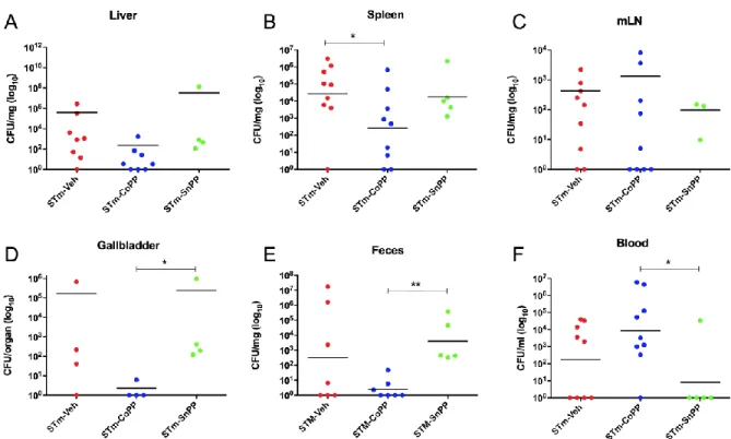
(41) 41. 849. 2.4.2 HMOX1 induction by CoPP treatment reduces bacteria load in tissues of S.. 850. Typhimurium-infected mice.. 851. C57BL6/J female mice were treated as described above with 5 mg/kg CoPP and 5 mg/kg SnPP.. 852. Twenty-four hours later, mice were infected with 1x10⁶ CFU of S. Typhimurium, and after 5. 853. days of infection mice were euthanized to evaluate bacterial burden in organs. As shown in. 854. Figure 3, reduced bacterial loads were observed in spleen, gallbladder and feces derived from. 855. CoPP-treated mice, as compared with vehicle- or SnPP-treated mice, suggesting a role of. 856. HMOX1 in bacterial clearance. However, the opposite result was observed in blood, where. 857. CoPP-treated mice displayed higher bacterial loads as compared to the other experimental. 858. groups.. 859 860 861 862 863 864 865 866 867.
(42) 42. 868 869 870 871 872 873 874 875 876 877 878 879 880 881. FIGURE 3. HMOX1 induction with CoPP treatment reduces bacterial loads in an acute S. Typhimurium infection model in mice. Female C57BL/6 mice were treated with CoPP, SnPP or vehicle 24h before intragastric infection with 1x10⁶ CFU of S. Typhimurium. Mice were euthanized at day 5 post-infection and bacterial loads in organs, blood and feces were measured. Geometric means are shown. (STm-Veh n=11; STm-CoPP n=10; STm-SnPP n=8; 3 independent experiments). Student’s t test was used to evaluate differences and results with statistically significant differences are shown..
(43) 43. 882. 2.4.3 Prophylactic HMOX1 induction by CoPP reduces persistence of S. Typhimurium in. 883. mice. 884. To evaluate whether HMOX1 induction through CoPP treatment can modulate S. Typhimurium. 885. persistent infection, mice were treated with CoPP 5 mg/kg, SnPP 5 mg/kg or vehicle ip 24 h. 886. before infection with 1x10⁶CFU of S. Typhimurium. The treatment was then repeated once a. 887. week. Because of the severity of this infection, mice were treated after 3 days of infection with. 888. the antibiotic Enrofloxacin in the drinking water for 3 weeks, to allow mice to survive until the. 889. end of the experiment. Clinical parameters and survival proportions were measured until the end. 890. of the experiment at day 38 post-infection. Weight loss of three groups of mice were not. 891. significantly different between each other (Fig. 4A). Bacterial loads from spleen, liver, lymph. 892. nodes and blood were measured at day 38 pi by plating homogenized tissues in agar plates. 893. (Table 3). With this information, a prospective analysis was performed, revealing a significant. 894. difference (p=0.0055) in persistence in at least one organ/blood between CoPP treated mice and. 895. the other two groups (Fig. 4C). These results indicate that a consistent state of high expression. 896. of HMOX1 modulates persistence infection of S. Typhimurium in mice. It is important to. 897. mention that HMOX1 protein expression was corroborated by western blot (Fig. 5).. 898 899 900 901.
(44) 44. 902 903 904 905 906 907 908 909 910 911 912 913 914 915. FIGURE 4. HMOX1 induction by CoPP treatment reduces persistent S. Typhimurium infection model in mice. Female C57BL/6 mice were treated with CoPP, SnPP or vehicle 24h before (A, B and C) or 72h after (D, E and F) intragastric infection with 1x10⁶ CFU of S. Typhimurium. In both cases, Enrofloxacin was administrated in the drinking water from day 3 to day 24, to allow mice to survive to infection and promote bacterial persistence until the end of the experiment. Weight change is represented as percentage of initial weight. (A and D). Survival proportions are represented as percentage of total initial number of mice (B and E). At day 38 post-infection mice were euthanized and bacterial loads were measured in organs and blood (Tables 3 and 4). Percentage of mice with persistent infection at 38 post-infection in at least one organ/blood. (STm-preVeh n=8; STm-preCoPP n=10; STm-preSnPP n=14; STmpostVeh=5; STm-postCoPP=6; STm-postSnPP n=12; 3 independent experiments) No significant differences were observed in weight change of mice. Prospective analysis was performed: for C: chi-square=10.30 and p=0.0055; for F: chi-square=84.34 and p<0.0001..
(45) 45. 916 917. TABLE 3. HMOX1 induction by prophylactic treatment with CoPP reduces S. Typhimurium persistence in mice S. Typhimurium burden STm pre-Veh. STm pre-CoPP. STm pre-SnPP. 918 919 920 921 922 923 924. Mouse (n=8) a b c d e f g h Mouse (n=10) a b c d e f g h i j Mouse (n=14) a b c d e f g h i j k l m n. Liver (CFU/mg). Spleen (CFU/mg). mLN (CFU/mg). Blood (CFU/ml). >5000 >5000 -. 1.44 >5000 -. 4.4 -. 333.3 -. Liver (CFU/mg). Spleen (CFU/mg). mLN (CFU/mg). Blood (CFU/ml). 41.7 -. -. -. -. Liver (CFU/mg). Spleen (CFU/mg). mLN (CFU/mg). Blood (CFU/ml). 181.8 6.41. >5000 >5000 12.9. -. 466.7 -. Female C57BL/6 mice were treated with CoPP, SnPP or its vehicle 24 h before intragastric infection with 1x10⁶ CFU of S. Typhimurium. Bacterial loads were quantified at day 38 postinfection in blood spleen, liver, mLN and feces. Bacterial loads of feces are not shown because no CFUs were observed..
(46) 46. 925 926 927 928 929 930 931 932 933 934 935 936 937 938 939 940 941. FIGURE 5: Evaluation of hmox-1 transcription in tissues of S. Typhimurium-infected mice. Female C57BL/6 mice were treated with CoPP, SnPP or vehicle as uninfected controls. Twenty-four hours later, mice were intragastrically infected with 1x10^6 CFUs of S. Typhimurium 14028 and at day 6 post-treatment (day 5 post-infection of infected groups) the liver and spleen were recovered to purify RNA or proteins. A and C. Quantitative real time PCR (qPCR) was performed to detect transcription of genes coding for HMOX-1, using the indicated probes in Supp Table S2. As a housekeeping gene, quantification of beta-2microglobulin mRNA was performed. Data is expressed as fold-increase of Ct. Student’s t test was used to evaluate differences. Results with statistically significant differences are shown (*: p<0.05). B and D. Representative images of Western blot analyses performed to assess the production of HMOX-1. Protein preparations from the liver and spleen were performed using RIPA protein extraction buffer and a Western blot assay was performed using anti-HMOX1 and anti-β-actin antibodies. SH-SY5Y cells were treated for 14 h with 10 M SnPP (negative control) or 10 M CoPP (positive control)..
(47) 47. 942. 2.4.4 HMOX1 induction by CoPP treatment reduces persistence of S. Typhimurium when. 943. administrated post-infection.. 944. To further evaluate the beneficial effect of HMOX1 in bacterial persistence, we treated mice. 945. after the beginning of the infection using the drug as a post-disease treatment. Then, mice were. 946. infected with 1x10⁶ CFU of S. Typhimurium and 3 days later were injected ip with CoPP 5. 947. mg/kg, SnPP 5 mg/kg or vehicle. The same day enrofloxacin treatment was started. Clinical. 948. parameters and survival proportions were measured until the end of the experiment at day 38. 949. post-infection. As well as observed in the prophylactic treatment, weight loss was not. 950. statistically different (Fig. 4D). Bacterial loads from spleen, liver, lymph nodes and blood were. 951. measured at day 38 p.i. by plating homogenized tissues in agar plates (Table 4). A prospective. 952. analysis was used as described before, and significant differences (p<0.0001) was observed in. 953. persistence between the groups (Fig. 4F), indicating that CoPP effectively can be used as a. 954. treatment to reduce persistence of the bacteria when the treatment is started after the onset of. 955. the infection.. 956.
(48) 48. 957 958. TABLE 4. HMOX1 induction by treatment with CoPP reduces persistence of S. Typhimurium in mice when applied after infection. S. Typhimurium burden STm post-Veh. STm post-CoPP. STm post-SnPP. 959 960 961 962 963 964 965 966 967. Mouse (n=5) a b c d e Mouse (n=6) a b c d e f Mouse (n=12) a b c d e f g h i j k l. Liver (CFU/mg). Spleen (CFU/mg). mLN (CFU/mg). Blood (CFU/ml). -. 171.8 61.7 61.7 -. -. -. Liver (CFU/mg). Spleen (CFU/mg). mLN (CFU/mg). Blood (CFU/ml). -. -. -. -. Liver (CFU/mg). Spleen (CFU/mg). mLN (CFU/mg). Blood (CFU/ml). -. 13.6 95.5 >5000 11 -. >5000 40.9 -. >5000 -. Female C57BL/6 mice were treated with CoPP, SnPP or its vehicle 72 h after intragastric infection with 1x10⁶ CFU of S. Typhimurium. Bacterial loads were quantified at day 38 postinfection in blood spleen, liver, mLN and feces. Bacterial loads of feces are not shown because no CFUs were observed..
(49) 49. 968. 2.5 Discussion. 969. Salmonella infections are an important public health problem due to the capacity of this. 970. bacterium to cause persistent infections (Griffin et al., 2011). It is well described that HMOX1. 971. has an immunomodulatory function in bacterial infections both, in vitro and in vivo (Jamal. 972. Uddin et al., 2015). Likewise, HMOX1 or its product CO have been proposed as a target for. 973. treatments for many diseases (Takagi et al., 2015). In this study, we explored the effects of. 974. HMOX1 induction or inhibition in the outcome of an acute and persistent infection caused by. 975. S. Typhimurium in mice.. 976. In the initial phase of S. Typhimurium infection in mice, the bacterium reaches deep organs and. 977. blood, and induces death before day 10 post-infection (Salazar et al., 2017). While non-. 978. significant, CoPP treatment was found to induce a tendency to reduce clinical score, allowing. 979. the survival of 20% of mice (Fig 1). Similar results were observed in SnPP-treated mice.. 980. Although SnPP is frequently used as a control with an opposite effect to CoPP, this drug acts. 981. by inhibiting the activity of HMOX1 through competition with its substrates, but actually may. 982. increase HMOX1 expression (Sardana and Kappas, 1987). Because of this, we thought that the. 983. inhibition of HMOX1 activity with SnPP in this model of acute infection may result in the. 984. induction of hmox1 gene expression and increase HMOX1 protein production, as we observed. 985. in the spleen of uninfected mice (Figure 5D). This could lead to the activation of additional. 986. genes involved in host defense, which are not necessarily related to the enzymatic activity of. 987. HMOX1 and its products.. 988. IL-10 is a crucial mediator of the immune response to S. Typhimurium (Neves et al., 2010) and. 989. previous data from our laboratory have described that IL-10 has a peak of expression at day 5.
Figure


+5


Documento similar
Exploring the immune response of porcine mesenteric lymph nodes to Salmonella enterica serovar Typhimurium: an analysis of transcriptional changes, morphological alterations and
As an example, Figure 19 shows at the top the neu- tral meta-model obtained by the induction process when the fragments in Figures 3, 5, 8 and 16 are consid- ered, and the
Probability of being alive according to the different treatments used, in comparison with no treatment adjusted by the inverse probability of treatment weight. Corticosteroidsm
The decrease in CAT activity (Figure 6) in shoots and roots after treatment with low concentrations of Cd observed in our study can be explained either by inhibition
107 Figure 19: Beta3-adrenergic receptor overexpression in cardiomyocytes reduces myocytes hypertrophy, cardiac fibrosis and protects against heart failure .... 108
Figure 5. Haeme oxygenase-1 is localized in the red pulp of the spleen and is induced by activation of nAChRs.. that HO-1 overexpression has an anti-inflammatory
In diabetic mice treated with SOCS1 peptidomimetic the number of Iba-1 positive cells was significantly reduced in comparison with diabetic mice treated with vehicle (Figure
Treatment of AngII-infused mice with anti-TLR4 antibody improved these structural parameters (Figure 2A-D); the treatment also improved the reduced number of smooth
