Development and validation of a capillary electrophoresis method for short chain organic acids measurement in natural rubber latex / V Galli, N Olmo and C Barbas
11
0
0
Texto completo
(2) Journal of Chromatography A, 894 (2000) 135–144 www.elsevier.com / locate / chroma. Development and validation of a capillary electrophoresis method for the measurement of short-chain organic acids in natural rubber latex V. Galli, N. Olmo, C. Barbas* ´ ´ Monteprıncipe ´ Facultad de CC Experimentales y Tecnicas , Universidad San Pablo-CEU, Urbanizacion , Ctra. Boadilla del Monte km 5,3, 28668 Madrid, Spain. Abstract Short-chain organic acid contents in serum of natural latex are interesting to measure and capillary electrophoresis (CE) has proved to be a good tool for their study. In the present work a method has been developed to identify the short-chain organic acids present in sera of natural rubber latex (oxalic, formic, fumaric, aconitic, succinic, malic, glutaric, citric, acetic, glycollic, propionic and quinic acids), the separation was optimised and the quantification method validated. The separation was performed on a CE system with UV detection at 200 nm. The separation was carried out with an uncoated fused-silica capillary (57 cm350 mm I.D.) and was operated at 210 kV potential. The separation buffers were prepared with 0.5 M H 3 PO 4 , 0.5 mM cetyltrimethylammonium bromide and pH adjusted by adding NaOH to 6.25 except for propionic acid which was better measured at pH 7.00. Validation parameters are adequate and limits of detection range from 0.005 mM to 1.6 mM. Short-chain organic acids were measured with this method in sera of three different types of latex. 2000 Elsevier Science B.V. All rights reserved. Keywords: Validation; Rubber; Latex; Organic acids; Fatty acids. 1. Introduction The natural latex concentrates used for the manufacture of dipped goods, such as gloves, are products both of nature and technology. Nature provides the raw material, field latex, from the tree species Hevea brasiliensis and it is then modified, preserved, concentrated and tested to produce the commercial concentrates [1]. Nowadays, the increasing use of latex products in healthcare areas has also increased new scientific *Corresponding author. Fax: 134-91-3510-475. E-mail address: [email protected] (C. Barbas).. controls of raw material and manufactured products in order to maintain higher standards in quality control. The aqueous phase of fresh natural latex contains many different chemical species, of which the principal types are carbohydrates, electrolytes, proteins and amino acids. In the absence of adequate preservation, the carbohydrates become microbiologically oxidised to the so-called volatile fatty acids (VFAs), which comprise mainly formic, acetic and propionic acids [2]. For this reason their measurement gives an indication of the extent of microorganic activity which has occurred during the time since the latex exuded from the tree. These matters are of consider-. 0021-9673 / 00 / $ – see front matter 2000 Elsevier Science B.V. All rights reserved. PII: S0021-9673( 00 )00525-2.
(3) 136. V. Galli et al. / J. Chromatogr. A 894 (2000) 135 – 144. able industrial importance, because several months may elapse between the production of preserved and concentrated natural rubber latex and its eventual use. VFA number measured in the steam of a quantity of latex is a classical test in quality control. The ionic content of latex is of continuing interest since it is believed to contribute to the properties and especially the processing of the latex, at least for natural rubber latices of low VFA number, it is thought that the salts of non-volatile acids (NVAs) significantly influence latex mechanical stability [3]. According to diverse sources [1,4–6], the principal non-volatile organic acids found in natural rubber latex are citric and malic acids. Minor amounts of trans-aconitic, fumaric, glycollic, succinic, quinic and glutaric acids may also be present. Furthermore, some of these acids mediate the biogenesis of polyisoprenes and their measurement can contribute to the study of the metabolism of the plants which produce them, since their biological function remains obscure. Previously reported works on the measurement of short-chain organic acids in natural rubber latex were carried out using ion chromatography [1,5]. Capillary electrophoresis (CE) has proved to be a good choice for investigation of samples in aqueous media, since usually no more than a simple dilution of samples is needed. Supporting this statement are the studies of Kenney [7] who reported the separation of some organic acids of interest in a variety of food matrices with indirect detection employing a commercial pack. Huang et al. [8] reported the separation of a six-component mixture of monocarboxylic acids from formate to hexanoate with conductivity deˆ tection. Devevre et al. [9] presented the separation of 14 organic acids with indirect UV detection using a commercial modifier; however, fumaric acid is not detected in the working conditions with this method. Turcat et al. [10] reported the monitoring of six organic acids in snow and rain water and Shirao et al. [11] reported the determination of organic acids in urine. Our research group has developed several methods of separation of organic acids in diverse matrices [12–14] with direct UV detection at 200 nm where the absorbance has a maxim. This low wavelength can be employed when working with non UV-absorbing aqueous buffers. The aim of the present work was the development of a CE method to identify the short-chain organic. acids present in sera of natural rubber latex, optimise the separation and validate the quantification method.. 2. Materials and methods. 2.1. Instrumentation The separation was performed on a CE P/ACE 5500 system (Beckman) with UV detection at 200 nm. The injection was by pressure (3.3 bar) for 5 s. The separation was carried out with an uncoated fused-silica capillary (57 cm350 mm I.D.) and was operated at 210 kV potential. Temperature was maintained at 258C. For optimising the pH of the separation buffers were prepared with 0.5 M H 3 PO 4 , 0.5 mM cetyltrimethylammonium bromide (CTAB), as cationic surfactant to avoid electroosmotic flow, and pH adjusted by adding NaOH in the range from 5.5 to 7.5. The best pH found for separation was 6.25 except for propionic acid which was better measured at pH 7.00.. 2.2. Chemicals Standards were obtained from Sigma (St. Louis, MO, USA). Phosphoric acid (85%) was from Merck (Darmstadt, Germany), sodium hydroxide from Panreac (Madrid, Spain) and organic solvents from Scharlau (Barcelona, Spain).. 2.3. Samples Samples of Thailand natural latex double centrifuged were directly imported and were representative of commercial material. For valuation, samples of natural latex coming from Guatemala taken directly from the trees or single centrifuged were also used. To obtain serum from latex ultracentrifugation has been utilised, but it needs 30 000 rpm for 60 min, so different chemical procedures for easier colloidal destabilisation were assayed, bearing in mind that many additives could alter or interfere with the electrophoretic process. Therefore, acidic substances such as H 3 PO 4 , water-soluble electrolytes as Ca(NO 3 ) 2 , (NH 4 ) 2 SO 4 or (NH 4 ) 3 PO 4 and hydrophobic organic solvents as acetone or chloroform were assayed. The best conditions found were to mix 10% (v / v).
(4) V. Galli et al. / J. Chromatogr. A 894 (2000) 135 – 144. H 3 PO 4 with latex 1:2 (v / w), to stir vigorously and keep at 2208C overnight. The next day, after tempering, serum was filtered through 0.45-mm nylon filters and diluted 1:3 (v / v) with purified water prior to analysis.. 2.4. Validation Individual stock solutions of each organic acid, 400 mM in water, were prepared and stored at 2208C at this concentration. On the day of the analysis they were adequately diluted. Linearity of response for standards was tested assaying by triplicate six levels of concentrations, ranging from 0.05 to 0.5 mM for nitrate; oxalic, fumaric and aconitic acids; from 0.5 to 10 mM for formic, glycollic and quinic acids; from 0.1 to 5.0 mM for succinic and glutaric acids; from 0.25 to 2.5 mM for malic and citric acids; from 0.5 to 5 mM for acetic acid and 0.49 to 7.81 mM for propionic acid. Linearity of response for samples was tested in the same way but replacing water with latex serum. Recovery was estimated comparing the values obtained in the linearity of the calibration serum, with the standards linearity, taking into account the endogenous serum concentrations, which had been previously quantified. Within-day precision was tested both to check the constancy of instrumental response to a given analyte and the repetitiveness of concentrations and migration times, since the latter is a key parameter for peak assignment. For this purpose, the assay was performed with 10 solutions of standards and 10 of samples, in the medium concentration of the calibration curve for all the compounds. Limits of detection (LODs) were calculated following IUPAC recommendations [(a13SB ) /b] for chromatographic methods [15] by extrapolating to zero concentration the standards deviation of the last three points of linearity and interpolating this value in the corresponding equation.. 3. Results and discussion. 3.1. Coagulation The addition of acidic substances to latices reduces the colloid stability and is commonly attribu-. 137. ted to chemical interaction between the added hydrogen ions and the surface-bound carboxylate ions which confer colloid stability upon the latex [3]. Phosphoric acid was chosen because it is the same acid included in the electrolyte buffer and it is not UV absorbing. The concentration used was varied from 30% to 10% to get the least ionic strength possible in the sample as lower ionic strength in samples than in buffers have been recommended to reduce electrodispersion broadening and extremely high ionic strength in buffers causes high Joule heating. Different water-soluble electrolytes were also assayed. Monovalent salts need the addition of large amounts to bring about flocculation and this is not useful in the electrophoresis process. Ammonium sulphate and ammonium phosphate were highly effective as destabilising agents, but two big system peaks appeared in the electropherogram overlapping. Fig. 1. Electropherogram of standards at different pH. Peaks: (1) oxalic, (2) formic, (3) fumaric, (4) aconitic, (5) succinic, (6) malic, (7) glutaric, (8) citric, (9) acetic, (10) glycollic, (11) propionic, (12) furanoic and (13) quinic acids. Buffer, 0.5 M H 3 PO 4 –0.5 mM CTAB. UV detection at 200 nm. Injection by pressure (3.3 bar) for 5 s. Uncoated fused-silica capillary (57 cm350 mm I.D.), 210 kV potential. Temperature 258C..
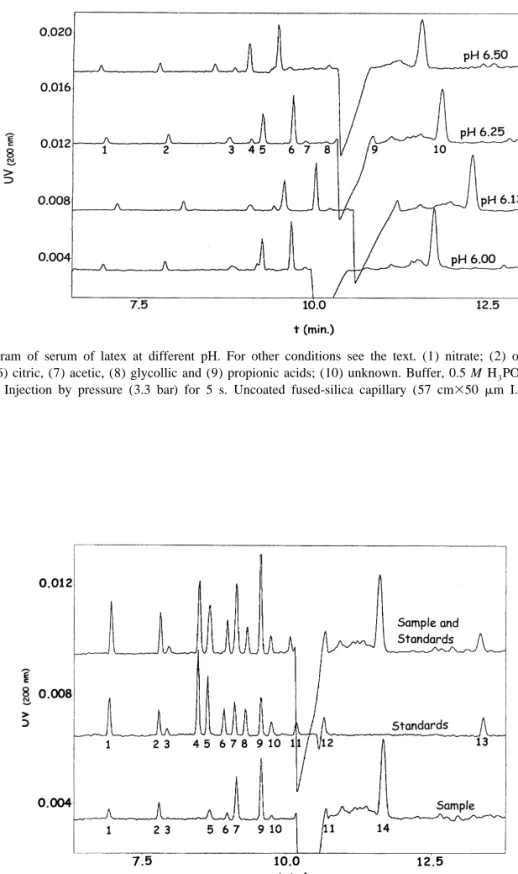
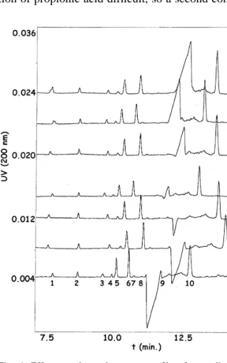
(5) 138. V. Galli et al. / J. Chromatogr. A 894 (2000) 135 – 144. Fig. 2. Electropherogram of serum of latex at different pH. For other conditions see the text. (1) nitrate; (2) oxalic, (3) aconitic, (4) succinic, (5) malic, (6) citric, (7) acetic, (8) glycollic and (9) propionic acids; (10) unknown. Buffer, 0.5 M H 3 PO 4 –0.5 mM CTAB. UV detection at 200 nm. Injection by pressure (3.3 bar) for 5 s. Uncoated fused-silica capillary (57 cm350 mm I.D.), 210 kV potential. Temperature 258C.. Fig. 3. Identification of peaks by spiking samples with standards. Standards: (1) nitrate; (2) oxalic, (3) formic, (4) fumaric, (5) aconitic, (6) succinic, (7) malic, (8) glutaric, (9) citric, (10) acetic, (11) glycollic, (12) propionic and (13) quinic acids. Sample: (1) nitrate; (2) oxalic, (3) formic, (5) aconitic, (6) succinic, (7) malic, (9) citric, (10) acetic and (12) propionic acids; (14) unknown. Buffer, 0.5 M H 3 PO 4 –0.5 mM CTAB pH 6.25 with NaOH. UV detection at 200 nm. Injection by pressure (3.3 bar) for 5 s. Uncoated fused-silica capillary (57 cm350 mm I.D.), 210 kV potential. Temperature 258C..
(6) V. Galli et al. / J. Chromatogr. A 894 (2000) 135 – 144. with analytes. It is well known that salts of divalent cations such as calcium cause rapid particle agglomeration of the latex and separation of the polymer but, after separation, a white precipitate in aqueous phase was observed, probably due to the calcic salts of the organic acids, and so these salts were avoided. Finally, none of the assayed organic solvents in the studied range (10–30%, v / w) gave adequate results. Therefore, the conditions established for the method were those explained above with phosphoric acid and freeze–thaw destabilisation to force the process.. 139. double centrifuged commercial latex alone and with the working standards added. The ghost negative peak in the electropherogram is a system peak due to the high electric field created by the elevated phosphoric acid concentration in samples, necessary to coagulate latex. When samples were neutralised with NaOH, previous to injection, at increasing pH, the peak diminished or disappeared but, as could be seen in Fig. 4, measurement is not possible in this area anyway. As this treatment increases the operations without any further improvement, it was avoided. During identification it could be seen that the system peak appearing in samples made quantification of propionic acid difficult, so a second condition. 3.2. Optimisation Starting from the experience in our laboratory separating short-chain organic acids in different matrices, described above, we aimed at optimising sensitivity and selectivity of the electrophoretic method by achieving good peak shapes and separation of peaks. For this purpose, the ionic strength was raised by increasing the concentration of the buffer, to get the biggest possible stacking effect. As samples were coagulated in highly concentrated H 3 PO 4 the buffer was made 0.5 M in phosphate. It poses a problem with high current intensities and high heat generation by Joule effect, but with the cooling system in the equipment it had no deleterious effects on separation. As some of the short-chain organic acids existing in latex serum partially overlapped under previously developed conditions, pH in the buffer was optimised varying between 5.50 and 7.50 with 0.25 increments. Fig. 1 shows the separation obtained for standards at the different pH values. The resolution for standards is complete in the range 6.00 to 6.50. Under pH 6.00 some peaks disappeared overlapping with others and over pH 6.50 resolution is not complete in some cases. Samples were then analysed in the same range (6.00–6.50) and, as shown in Fig. 2, the best results ocurred at 6.13 and 6.25 so finally the latter value was established. The identification of peaks was performed by comparing with the migration times of pure standards and by spiking the sample with each pure standard separately. Fig. 3 shows the electropherograms under final conditions for standards, a. Fig. 4. Effect on electropherogram profile of neutralisation in samples: (1) nitrate; (2) oxalic, (3) formic, (4) aconitic, (5) succinic, (6) malic, (7) citric, (8) acetic, (9) glycollic and (10) propionic acids; (11) unknown. Buffer, 0.5 M H 3 PO 4 –0.5 mM CTAB. UV detection at 200 nm. Injection by pressure (3.3 bar) for 5 s. Uncoated fused-silica capillary (57 cm350 mm I.D.), 210 kV potential. Temperature 258C. A: Sample–water (1:1); B: sample– 1% NaOH (1:1); C: sample–2% NaOH (1:1); D: sample–2.5% NaOH (1:1); E: sample–3% NaOH (1:1); F: sample–4% NaOH (1:1); G: sample–5% NaOH (1:1)..
(7) V. Galli et al. / J. Chromatogr. A 894 (2000) 135 – 144. 140. was developed just for this acid to displace it out of the negative peak, which was pH 7.00. So for measuring propionic acid a second injection must be done in a different buffer, but everything is done automatically. An unknown peak appears near 12.5 min. Great efforts have been made in order to identify it. A long list of compounds, described as possible latex constituents and with adequate size for the migration time, has been assayed: aspartic acid, glutaric acid, arginine, alanine, hystidine, glycine, lysine, glycerophosphate, levulinic acid, mevalonic acid, caprylic acid, shikimic acid, isovaleric acid, ketoisovaleric acid, a-ketoglutaric acid, benzoic acid,. valproic acid, ftalic acid, lactic acid, maleic acid, methionine, butyric acid, gluthation and ascorbic acid, but currently it remains unknown.. 3.3. Validation As shown in Table 1, standards and samples fit the linear model (r.0.99) for all the organic acids. Although a small bias was found in some of them because the intercept (a)6its limits of confidence (LCs) does not include the zero value, it has statistical significance, but it has no practical consequences as can be seen in the recoveries that are near 100%. Table 1 Main parameters of validation for linearity and accuracy Linearity a6LC 39679 64694. Range (mM). Accuracy: Recovery (%)6RSD (%). LOD (mM). 0.999 0.999. 0.050–0.500 0.016–0.166. 10064 10967. 0.022 0.005. 0.997 0.992. 0.050–0.500 0.086–0.236. 100612 104610. 0.020 0.071. 414635 448629. 0.991 0.995. 0.500–10.00 0.375–7.500. 93622 10869. 1.612 1.037. b6LC. r. 35 8396889 38 94261053 98296615 12 1136933. Nitrate. Standard Sample. Oxalic acid. Standard Sample. 1116124 21226141. Formic acid. Standard Sample. 1556216 1556139. Fumaric acid. Standard Sample. 2816352 22996388. 64 89861735 67 95562095. 0.999 0.999. 0.037–0.375 0.037–0.375. 10063 10064. 0.003 0.003. Aconitic acid. Standard Sample. 2726504 2816141. 52 20362494 56 85661595. 0.998 0.999. 0.050–0.500 0.015–0.165. 9966 11065. 0.002 0.005. Succinic acid. Standard Sample. 23886332 24556172. 39776135 4165695. 0.998 0.999. 0.100–5.000 0.193–3.943. 107612 10469. 0.049 0.117. Malic acid. Standard Sample. 3236132 24056246. 41226131 50816235. 0.999 0.997. 0.250–2.500 0.740–1.490. 9966 10566. 0.204 0.224. Glutaric acid. Standard Sample. 24426370 23736338. 44646150 48086164. 0.998 0.999. 0.100–5.00 0.375–3.750. 10268 113610. 0.076 0.046. Citric acid. Standard Sample. 2966193 22056403. 62346191 72946427. 0.999 0.995. 0.250–2.500 0.635–1.385. 10065 10765. 0.159 0.140. Acetic acid. Standard Sample. 22866196 2354696. 1853671 1807651. 0.998 0.998. 0.500–5.000 0.294–4.044. Glycollic acid. Standard Sample. 4386437 21876113. 1184670 1412624. 0.995 0.999. 0.500–10.00 0.375–7.500. 9967 10268. 1.414 0.185. Propionic acid. Standard Sample. 21356173 3206137. 889636 844638. 0.998 0.998. 0.488–7.805 0.366–5.854. 10062 11168. 0.650 1.600. Quinic acid. Standard Sample. 30076182 3593645. 0.995 0.999. 0.500–10.00 0.375–7.500. 96612 101615. 1.188 0.413. 101161138 1136213. 9966 95610. 0.177 0.234.
(8) V. Galli et al. / J. Chromatogr. A 894 (2000) 135 – 144. in the whole range and it can be due to the good fit of the points to the regression line. During the calculation of recoveries it could be detected that for fumaric acid a recovery of 140% was obtained if pH of standards was not the same as in samples, so standards were prepared with the same quantity of 10% (v / v) H 3 PO 4 as in samples and all the recoveries obtained were near 100%. Mathematical LODs range from 0.005 mM to 1.6 mM. It is a problem, due to the statistical approach, that some of them are over the validated range.. 141. Obviously, the method is applicable for quantification in the range experimentally proved, which many times is under the calculated detection limit. It is interesting to see the doubtful meaning of a mathematical approach. In the same line, samples have lower detection limits than calculated for standards in some acids. That is justified because when the sample contents one of the acids in high concentration the lowest points in the regression line are not so low, and the corresponding standard deviation are smaller, giving better detection limits.. Table 2 Main validation parameters for precision Precision Intra-assay. Inter-assay. Mean (mM). RSD (%). tm a (min). RSD (%). Mean (mM). RSD (%). tm a (min). RSD (%). Nitrate. Standard Sample. 0.244 0.178. 2 2. 7.162 7.192. 0.04 0.20. 0.244 0.188. 6 8. 7.080 7.094. 1 1. Oxalic acid. Standard Sample. 0.244 0.341. 5 4. 7.824 7.857. 0.45 0.38. 0.244 0.319. 9 9. 7.874 7.914. 1 1. Formic acid. Standard Sample. 4.879 2.771. 5 6. 8.213 8.262. 0.37 0.21. 4.879 2.820. 7 8. 8.108 8.130. 2 1. Fumaric acid. Standard Sample. 0.244 0.131. 2 1. 8.772 8.830. 0.31 0.23. 0.244 0.136. 6 6. 8.634 8.681. 2 1. Aconitic acid. Standard Sample. 0.244 0.162. 2 1. 8.927 9.027. 0.83 0.21. 0.244 0.170. 8 7. 8.814 8.869. 2 1. Succinic acid. Standard Sample. 2.444 1.510. 1 2. 9.247 9.352. 0.37 0.24. 2.444 1.572. 4 5. 9.115 9.281. 2 1. Malic acid. Standard Sample. 1.220 2.276. 3 2. 9.442 9.527. 0.37 0.26. 1.220 2.319. 4 4. 9.304 9.355. 2 1. Glutaric acid. Standard Sample. 2.444 1.264. 2 2. 9.892 9.727. 0.83 0.26. 2.444 1.306. 6 7. 9.582 9.547. 3 1. Citric acid. Standard Sample. 1.220 2.383. 1 2. 9.918 9.990. 0.38 0.27. 1.220 2.473. 5 5. 9.771 9.799. 2 1. Acetic acid. Standard Sample. 2.444 1.620. 4 4. 10.110 10.175. 0.38 0.26. 2.444 1.664. 7 5. 9.953 9.978. 2 2. Glycollic acid. Standard Sample. 4.878 2.251. 2 2. 10.550 10.558. 0.39 0.25. 4.878 2.418. 4 6. 10.382 10.345. 2 2. Propionic acid. Standard Sample. 3.902 2.394. 2 6. 10.553 10.687. 0.36 0.40. 3.902 2.591. 3 8. 10.485 10.541. 1 2. Quinic acid. Standard Sample. 4.878 2.598. 3 3. 13.963 14.150. 0.51 0.41. 4.878 2.772. 4 5. 13.677 13.780. 3 2. a. t m 5Migration time..
(9) V. Galli et al. / J. Chromatogr. A 894 (2000) 135 – 144. 142. Table 3 Short-chain organic acid content in sera of different types of latices a Anion concentration (mmol l 21 serum). Nitrate Oxalic acid Formic acid Fumaric acid Aconitic acid Succinic acid Malic acid Glutaric acid Citric acid Acetic acid Glycollic acid Propionic acid Quinic acid. A. B. C. D. E. 0.14 0.82 3.13 n.d 0.14 1.51 6.27 n.d 7.36 2.95 n.d 2.83 n.d. n.d 0.52 9.50 n.d 0.23 23.10 1.80 n.d 8.08 24.50 n.d n.d n.d. n.d 0.37 6.92 n.d 0.16 16.68 1.90 n.d 5.74 17.04 n.d n.d n.d. – 0.97–1.99 2.00–6.66 – – n.d–5.00 6.39–11.66 – 2.00–7.73 6.39–16.77 – – –. – 0.12–1.37 n.d–8.22 – – n.d–10.25 4.21–10.14 – 3.73–6.89 n.d–18.15 – – –. a. Sample A: latex double centrifuged from Thailand (DRC 60.27%); sample B: natural latex from Guatenmala (DRC 29 / 85%); sample C: natural latex single centrifuged from Guatemala (DRC 58.46%); sample D: high ammoniated concentrated latex from Malaysia (DRC 59.73%) [5]; sample E: low ammoniated concentrated latex from Malaysia (DRC 59.78%) [5].. When running 10 runs per day of both standards and samples, daily relative standard deviation (RSDs) in concentrations are low enough to consider the method acceptable (1 to 6%) and variations when they exist are mainly due to difficulties in integration. of small peaks. RSDs for migration times, Table 2, are very low when they are measured in the same assay for both standards and samples (0.04 to 0.8%). The intermediate precision evaluated on different days with a total of 25 runs provided RSD values. Fig. 5. Electropherogram of serum obtained by freezing. Peaks: (1) nitrate; (2) thiosulfate; (3) oxalic, (4) formic, (5) aconitic, (6) succinic, (7) malic, (8) citric and (9) acetic acids; (10) unknown. Buffer, 0.5 M H 3 PO 4 –0.5 mM CTAB. UV detection at 200 nm. Injection by pressure (3.3 bar) for 5 s. Uncoated fused-silica capillary (57 cm350 mm I.D.), 210 kV potential. Temperature 258C..
(10) V. Galli et al. / J. Chromatogr. A 894 (2000) 135 – 144. slightly superior to intra-assay precision (3 to 9% for concentrations and 1 to 3% for migration times), as could be expected.. Table 4 Results obtained from 10 samples coagulated with phosphoric acid and 10 samples coagulated by freezing a. 3.4. Quantification of samples After development, the method was applied to the quantification of these acids in serum of three different types of latices: the one double centrifuged coming from Thailand used during validation (sample A), one just obtained from the tree without any treatment (sample B) and one centrifuged once (sample C), both coming from Guatemala. Results are shown in Table 3. As it could be expected, natural latex without any treatment has higher organic acids content in the same weigh, mainly because during the centrifugation process the ratio of rubber increases while the serum is partially eliminated. In these acids DRC (dry rubber content) is 60.27% for A, 29.85% for B and 58.46% for C. But, variations in this parameter do not fully explain differences in short-chain organic acids content. So further studies must be done to establish the correlation between these acids and different processes in latex. When comparing the values obtained and those existing in the literature [1,5,6] it can be found that the published ranges are wide and so we are in agreement with them in most cases, but it is not very clarifying.. 3.5. Coagulation by freezing While assaying the best way to receive samples from different countries to apply the method in phytochemical studies, the possibility of destabilising the latex by freezing at 2208C overnight, without any other addition could be seen. This is very interesting because, just the acids naturally free in serum are measured, avoiding the liberation of acids bound to solid matter in some way or those lost due to insolubilisation of some acids in high acidic media. The method consisted of filling 50-ml Falcon plastic tubes with around 37 g of latex and placing them in the freezer at 2208C overnight, after tempering, the tubes were centrifuged at 4000 g for 20 min and serum was filtered and injected. As the ionic strength is lower than in the previous method, favouring stacking effect, and no dilution is needed,. 143. Nitrate Oxalic acid Aconitic acid Succinic acid Malic acid Citric acid Acetic acid a. 10% H 3 PO 4 (mmol l 21 serum). Frozen (mmol l 21 serum). 0.1860.02 0.9160.06 0.1460.01 1.2460.12 8.0060.41 6.2560.29 2.6560.31. 0.7660.02 1.0560.02 0.1660.01 1.4160.03 8.8560.20 7.0160.23 3.0460.19. Mean6limits of confidence (P.95%).. the electropherogram profile improved considerably, as can be observed in Fig. 5. Only a new peak appeared in the electropherogram due to an unfolding of the nitrate peak previously missed. It was identified as tiosulfate by retention time and by spiking the sample with it. In order to compare the results obtained coagulating with phosphoric acid and just freezing, two series of 10 samples of the same latex were processed individually coagulating by both methods. Results can be found in Table 4. Only nitrate poses a great difference, which was previously justified. Concentration of organic acids are slightly superior and limits of confidence slightly inferior in the frozen way than when coagulating with phosphoric acid. The value could be justified because losses by volatisation are minimum and the smaller deviations because the peaks are better measured.. 4. Conclusions A CE method has been developed and validated for measuring short-chain organic acids in sera of natural rubber latex directly without any sample pre-treatment more than coagulation. It has been applied to different types of samples but further studies must be done to establish the correlation between these acids and different processes in latex.. Acknowledgements The present work has been developed with the collaboration of Tecnilatex S.A..
(11) 144. V. Galli et al. / J. Chromatogr. A 894 (2000) 135 – 144. References [1] T.D. Pendle, Rubber Chem. Technol. 63 (3) (1990) 234. [2] D.C. Blackley, Polymer Latices – Science and Technology, 2nd ed., Types of Latices, Vol. 2, Chapman and Hall, London, 1997. [3] D.C. Blackley, Polymer Latices – Science and Technology, 2nd ed., Fundamental Principles, Vol. 1, Chapman and Hall, London, 1997. [4] K.C. Calvert, Plastics Rubber: Mater. Appl. 2 (1977) 59. [5] R.C. Crafts, A.D.T. Gorton, T.D. Pendle, NR Technol. 16 (1985) 12. [6] R.T. Davies, T.D. Pendle, Rubber Dev. 44 (4) (1991) 94. [7] B.F. Kenney, J. Chromatogr. 546 (1991) 423.. [8] X. Huang, J.A. Luckey, M.J. Gordon, R.N. Zare, Anal. Chem. 61 (1989) 766. ` [9] O. Devevre, D.P. Putra, B. Botton, J. Garbaye, J. Chromatogr. A 679 (1994) 349. [10] S. Turcat, P. Masclet, T. Lissol, Sci. Total Eviron. 158 (1994) 21. [11] M. Shirao, R. Furuta, S. Suzuki, H. Nakazawa, S. Fujita, T. Maruyama, J. Chromatogr. A 680 (1994) 247. [12] C. Barbas, N. Adeva, R. Aguilar, M. Rosillo, T. Rubio, M. Castro, Clin. Chem. 44 (1998) 1340. ´ C. Barbas, R. Aguilar, M. Castro, Clin. Chem. 44 [13] A. Garcıa, (1998) 1905. ˜ ´ [14] C. Barbas, J.A. Lucas, F.J. Gutierrez-Manero, Phytochem. Anal. 10 (1999) 55. [15] G.L. Long, J.D. Winefordner, Anal. Chem. 55 (1983) 712..
(12)
Figure
Documento similar
- Competition for water and land for non-food supply - Very high energy input agriculture is not replicable - High rates of losses and waste of food. - Environmental implications
50 The goal is to help people to reach an optimum level in the dimensions of psychological well- being: environmental mastery, personal growth, purpose in life,
Validation is an important feature in any method of measurement because it is closely related to the quality of the results. A method of analysis is characterised by its
It was possible to identify the main compounds produced upon Fenton oxidation in the final aqueous effluents (acetic, oxalic and formic acids) and the results seem to suggest that
The characterization in amino acids, organic acids, sugars, trigonelline, volatiles compounds, fatty acids, total phenolic, carotenoids, vitamin C content, and antioxidant capacity
In the present work, the development of an electronic platform for remote real-time monitoring of long-term cold chain transport operations is detailed where a comparative analysis
High-performance liquid chromatography- ultraviolet detection method for the simultaneous determination of typical biogenic amines and precursor amino acids. applications in
The expansionary monetary policy measures have had a negative impact on net interest margins both via the reduction in interest rates and –less powerfully- the flattening of the
