Evaluation of hemochemical changes in the blood of piglets supplemented with two microbial preparations
9
0
0
Texto completo
(2) 58. Cuban Journal of Agricultural Science, Volume 52, Number 1, 2018.. as growth promoters in animals (Dadvar et al. 2015). Therefore, taking into account the economic losses caused by diarrheal disorders, it was necessary to look for alternatives to mitigate these physiological disorders in animals (Lye et al. 2012). Biological media, such as probiotics, are microbial additives obtained from monocultures or mixed cultures, which inhibit the growth of pathogens in the gastrointestinal tract (Colina et al. 2011). Likewise, they play a role in the maintenance of the function of intestinal barrier and local and systemic modulation of the immune system, which contributes to improve the health of the host (Londoño and Parra 2015). Hence, the objective of this study is to evaluate the effect of two microbial preparations on hematological values and blood biochemicals of piglets, at 14 and 42 d of age. Materials and Methods The experimental study was carried out in the pig production unit “Gahuijón Alto”, Canton Colta, Ecuador. The unit is located at 1° 53' 12.248" SL longitude 78° 43' 22.454" LW, 3 510 m.o.s.l. (meters over sea level), with annual precipitation between 500 and 1,000 mm. Minimum, maximum and average temperature is 3, 14 and 10 ºC, with annual relative humidity of 80 % and annual evapotranspiration of 69.03. Experimental treatments and design. A completely randomized design with four repetitions per treatment was used, where each experimental unit was composed by twelve piglets. Evaluated treatments were: T1) control; T2) microbial preparation A and T3) microbial preparation B. Animals. An amount of 120 Duroc x Landrace/Yorshire crossbreed piglets were used, which descendants of 12 first farrowing sows (Landrace/ Yorkshire), with 135.6 ± 2 kg of liveweight (LW) and 235 ± 3 d of age. Four pregnant sows were used per treatment, housed in collective pens of 6 x 6.5 m wide and cement floor, with a density of 1.6 m2 per animal, from gestation to 110 d of gestation. After parturition, piglets were distributed at random to form the experimental groups with 40 animals per treatment (20 females and 20 males). From this moment until weaning, they were settled in maternity area. Piglets after weaning were regrouped (without altering the groups of origin) with 20 piglet, in pens of 6 x 6.5 m in cement floors. The food used was Bioalimentar® (Ambato, Ecuador), which meets the nutritional requirements for pigs, recommended by NRC (2012). Bioalimentar was offered to sows twice a day, at 7:00 a.m. and 4:00 p.m., and the piglets received it ad libitum from 7 d of age up to weaning. From there until 42 d of age, they were fed at the same time as their mothers. Water was also provided ad libitum in nipple troughs. Animal management system. Maternity was. los antibióticos provocó que la Unión Europea desde 2006 prohibiera su utilización como promotores del crecimiento en los animales (Dadvar et al.2015). Por ello, al tener en cuenta las pérdidas económicas causadas por los trastornos diarreicos, fue necesario buscar alternativas atenuantes para mitigar estos trastornos fisiológicos en los animales (Lye et al.2012). Los medios biológicos, como los probióticos, son aditivos microbianos obtenidos a partir de los monocultivos o cultivos mixtos, que inhiben el crecimiento de agentes patógenos en el tracto gastrointestinal (Colina et al.2011). Asimismo, intervienen en el mantenimiento de la función de la barrera intestinal y la modulación local y sistémica del sistema inmune, lo que contribuye a mejorar la salud del huésped (Londoño y Parra 2015). De ahí que el objetivo de este estudio sea evaluar el efecto de dos preparados microbianos en los valores hematológicos y bioquímicos sanguíneos de lechones, a los 14 y 42 d de edad. Materiales y Métodos El trabajo experimental se realizó en la unidad de producción porcina Gahuijón Alto, Cantón Colta, Ecuador. La unidad está localizada en 1° 53' 12.248 "LS longitud 78° 43' 22.454 "LW, 3 510 msnm (metros sobre nivel del mar), con precipitación anual entre 500– 1 000 mm. La temperatura mínima, máxima y media es de 3, 14 y 10 ºC, con humedad relativa anual de 80 % y evapotranspiración anual de 69.03. Diseño y tratamientos experimentales. Se utilizó un diseño completamente aleatorizado con cuatro repeticiones por tratamiento, donde cada unidad experimental estuvo compuesta por doce lechones. Los tratamientos evaluados fueron: T1) control; T2) preparado microbiano A y T3) preparado microbiano B. Animales. Se emplearon 120 cerditos, cruce Duroc x Landrace/Yorshire, descendientes de 12 cerdas primerizas (Landrace/Yorkshire), con 135.6 ± 2 kg de peso vivo (PV) y 235 ± 3 d de edad. Se emplearon cuatro cerdas gestantes por tratamiento, alojadas en corrales colectivos de 6 x 6.5 m y piso de cemento, con densidad de 1.6 m2 por animal, desde la gestación hasta 110 d de gestación. Después del parto, los cerditos se distribuyeron al azar para conformar los grupos experimentales con 40 animales por tratamiento (20 hembras y 20 machos). Desde este momento hasta el destete se ubicaron en la maternidad. Los lechones a partir del destete se reagruparon (sin alterar los grupos de procedencia) con 20 cerditos, en corrales de 6 x 6.5 m en pisos de cemento. El alimento utilizado fue Bioalimentar® (Ambato, Ecuador), que cumple con los requerimientos nutricionales para cerdos, recomendados por NRC (2012). El Bioalimentar se ofreció a las reproductoras dos veces por día, a las 7:00 a.m. y 4:00 p.m., y a los cerditos se les ofreció ad libitum a partir de los 7 d de edad hasta el destete. De ahí hasta los 42 d de edad, se alimentaron en el mismo horario que a sus madres. El agua también se suministró ad libitum en bebederos tipo tetinas. Sistema de manejo de los animales. La maternidad se.
(3) 59. Cuban Journal of Agricultural Science, Volume 52, Number 1, 2018.. maintained at 28 °C during the first two weeks after parturition. Later, this temperature was reduced in 1.5 °C every week until weaning. Photoperiod was controlled with 12 h of light and 12 h of darkness. The litters of each treatment were located distant from each other (with an intermediate pen on both sides of the corridor) to avoid self-inoculation. Piglets were weaned at 33 d of age. Piglets from each group under study received the proper veterinary attention, according to the Manual de Manejo de Hembras y Primerizas (Coates et al. 2013). Microbial preparations. The strains used in the preparations were Kluyveromyces fragilis (L-4 UCLV), from the Bank of Microorganisms of the Universidad Central “Marta Abreu” of Las Villas, and four ATCC (American Type Cultures Collection, USA) strains, which were Lactobacillus acidophilus, L. bulgaricus, Streptococcus thermophilus and Saccharomyces cerevisiae. These strains were activated in skim milk at 37 °C for 24 h. To obtain the preparations, the mixture of sugarcane molasses and orange vinasse was used as a substrate and fermented at 37ºC for 24 h, according to the methodology described by Miranda et al.(2017). In preparation A, L. acidophilus, L. bulgaricus and S. thermophilus were used and in B, the previous bacteria plus S. cerevisiae and K. fragilis yeasts (L-4 UCLV) were also used. Chemical composition and microbial concentration of each preparation are shown in table 1.. mantuvo a 28 °C de temperatura durante las dos primeras semanas postparto, posteriormente se redujo en 1.5 °C cada semana hasta el destete. El fotoperíodo se controló con 12 h de luz y 12 h de oscuridad. Las camadas de cada tratamiento se ubicaron distantes unas de otras (con un cuartón intermedio a ambos lados del pasillo) para evitar la auto inoculación. Los lechones se destetaron a los 33 d de edad. Los cerditos provenientes de cada grupo en estudio recibieron las atenciones veterinarias pertinentes, según el Manual de Manejo de Hembras y Primerizas (Coates et al.2013). Preparados microbianos. Las cepas utilizadas en los preparados fueron Kluyveromyces fragilis (L-4 UCLV), proveniente del Banco de Microorganismos de la Universidad Central “Marta Abreu” de Las Villas, y cuatro cepas ATCC (American Type Cultures Collection, EEUU): Lactobacillus acidophilus, L. bulgaricus, Streptococcus thermophilus y Saccharomyces cerevisiae. Estas cepas se activaron en leche descremada a 37 ºC durante 24 h. Para la obtención de los preparados se utilizó como sustrato la mezcla de melaza de caña de azúcar y vinaza de naranja y se fermentó a 37 ºC durante 24 h, según la metodología descrita por Miranda et al.(2017). En el preparado A se emplearon las bacterias L. acidophilus, L. bulgaricus y S. thermophilus y en el B las bacterias anteriores, además de las levaduras S. cerevisiae y K. fragilis (L-4 UCLV). La composición química y la concentración microbiana de cada preparado se presentan en la tabla 1.. Table 1. Composition of microbial preparations to be included in the base diet of mother sows and their offspring Indicators Dry Matter, % Crude Protein, % True Protein, % Ether Extract, % Ashes, % pH Lactic acid, mmol/L Microbial concentration, CFU/mL. Microbial preparations Preparation A (T2) Preparation B (T3) 16.5 17.5 15.2 16.1 10.3 11.1 2.86 2.53 2.82 2.76 3.88 3.85 0.68 0.72 7 9.2 x 10 9.5 x 10⁸. Use of additives in pigs. Microbial preparations were provided to animals of groups T2 and T3 at 7:00 a.m. every three days. The breeding sows received 15 mL of additive, by mixing 0.3 kg of balanced food plus 0.5 L of water, from the day after the confirmation of pregnancy until weaning, according to the assigned treatment. The offspring continued to receive the same additive. The first dose applied to piglets was a single dose before having colostrum. Oral dosage varied according to age: 1 mL in the first week; 1.5 mL in weeks 2 and 3; 2 mL in weeks 4 and 5, and 2.5 mL in the following up to 42 d of age. Control group received normal saline. Empleo de los aditivos en los cerdos. Los preparados microbianos se suministraron a los animales de los grupos T2 y T3 a las 7:00 a.m. cada tres días. A las cerdas reproductoras se les proporcionó 15 mL de aditivo, al mezclar 0.3 kg de balanceado más 0.5 L de agua, a partir del día posterior de la confirmación de la gestación hasta el destete, según el tratamiento asignado. La descendencia continuó recibiendo el mismo aditivo. La primera dosis aplicada a los cerditos fue en mono dosis antes de tomar el calostro. La dosificación, en forma oral, varió de acuerdo con la edad: 1 mL en la primera semana; 1.5 mL en las semanas 2 y 3; 2 mL en.
(4) 60. Cuban Journal of Agricultural Science, Volume 52, Number 1, 2018.. solution in the same amount as the treated groups. Experimental procedure for taking and analyzing samples. At 14 and 42 d of age of the animals, 12 pigs of each treatment were randomly selected. They were immobilized and extracted 8 mL of blood from the jugular vein. These samples were taken in vacutainer tubes, with and without ethylenediaminetetraacetic acid (EDTA), using a California-type needle. Subsequently, they were transferred to the laboratory within the first three hours for further processing. The evaluation of the blood profile consisted of the determination of hemoglobin (Hb), hematocrit, erythrocyte, mean corpuscular volume (MCV), mean corpuscular hemoglobin (MCH), mean corpuscular hemoglobin concentration (MCHC), leukocytes, basophils, eosinophils, lymphocytes and monocytes, through the methodology described by Kraft (1998) and Corredor (2012). Indicators of blood biochemistry were total proteins, albumin, glucose, triglycerides, total cholesterol, high-density lipoprotein-cholesterol (HDL-C) and low-density lipoprotein cholesterol (LDL-C), determined according to Mejía et al.(2012) and Londoño and Parra (2015). Statistical analysis. The experimental data were processed with the statistical package Statgraphic plus 15.1 for Windows. Analysis of variance was carried out according to a completely randomized design. In the necessary cases, Duncan (1955) comparison test was applied to discriminate differences between means at P <0.05.. las semanas 4 y 5, y 2.5 mL en las siguientes hasta los 42 d de edad. El grupo control recibió suero fisiológico en igual cantidad que los grupos tratados. Procedimiento experimental para la toma y análisis de las muestras. A los 14 y 42 d de edad de los animales, se seleccionaron al azar 12 cerdos de cada tratamiento. Se inmovilizaron y se les extrajo 8 mL de sangre de la vena yugular. Estas muestras se tomaron en tubos vacutainer, con etilendiaminotetraacético (EDTA) y sin él, mediante una aguja tipo California. Posteriormente, se trasladaron al laboratorio dentro de las tres primeras horas para su posterior procesamiento. La evaluación del perfil hemático consistió en la determinación de hemoglobina (Hb), hematocrito, eritrocito, volumen corpuscular media (VCM), hemoglobina corpuscular media (HCM), concentración de hemoglobina corpuscular media (CHCM), leucocitos, basófilos, eosinófilos, linfocitos y monocitos, mediante la metodología descrita por Kraft (1998) y Corredor (2012). Los indicadores de la bioquímica sanguínea fueron proteínas totales, albúmina, glucosa, triglicéridos, colesterol total, colesterol-lipoproteína de alta densidad (C-HDL) y colesterol-lipoproteína de baja densidad (C-LDL), determinados según la metodología de Mejía et al.(2012) y Londoño y Parra (2015). Análisis estadístico. Los datos experimentales se procesaron con el paquete estadístico Statgraphic plus 15.1 para Windows. Se realizó análisis de varianza según diseño completamente aleatorizado. En los casos necesarios, se aplicó la dócima de comparación de Duncan (1955) para discriminar diferencias entre medias a P < 0.05.. Results. Resultados. Table 2 shows the blood profile of the piglets, evaluated at 14 and 42 d of age. In both ages, differences were found with respect to control for all the evaluated indicators, except lymphocytes at 14 d of age. With the inclusion of microbial preparations, in both measurements, hemoglobin, hematocrit and erythrocytes increased (P <0.05). Hemoglobin was higher in T3, while hematocrit and erythrocytes did not differ between T2 and T3. At 14 and 42 d, MCV and MCHC were higher (P <0.05) in T1 with respect to T2 and T3, without differences between these groups. This same effect was found for MCH at 42 d of age, not being so at 14 d, when differences were detected between T2 and T3, with a lower value in T3. Leukocytes and basophils also decreased with the inclusion of microbial preparations, without differences between T2 and T3 (table 2). With respect to monocytes, in the evaluation performed at 14 d, the values were higher (P <0.05) for the animals treated with additives, being superior in T3. However, at 42 d old, this indicator was higher (P <0.05) in the control treatment, without differences between T2 and T3. There were no differences (P <0.05) among treatments for lymphocytes at 14 d.. En la tabla 2 se muestra el perfil hemático de los lechones, evaluados a los 14 y 42 d de edad. En ambas edades, se encontraron diferencias con respecto al control para todos los indicadores evaluados, excepto los linfocitos a los 14 d de edad. Con la inclusión de los preparados microbianos, en las dos mediciones, aumentó la hemoglobina, hematocrito y eritrocitos (P < 0.05). La hemoglobina fue mayor en el T3, mientras que el hematocrito y los eritrocitos no difirieron entre T2 y T3. A los 14 y 42 d edad, el VCM y CHCM fueron mayores (P < 0.05) en el T1 con respecto a T2 y T3, sin diferencias entre estos grupos. Este mismo efecto se encontró para HCM a los 42 d de edad, no siendo así a los 14 d, cuando se detectaron diferencias entre T2 y T3, con menor valor en T3. Los leucocitos y basófilos también disminuyeron con la inclusión de los preparados microbianos, sin diferencias entre T2 y T3 (tabla 2). Con respecto a los monocitos, en la evaluación realizada a los 14 d, los valores fueron mayores (P < 0.05) para los animales tratados con los aditivos, siendo superior en el T3. Sin embargo, a los 42 d de edad, este indicador fue mayor (P < 0.05) en el tratamiento control, sin diferencias entre T2 y T3. No hubo diferencias (P < 0.05) entre tratamientos para los.
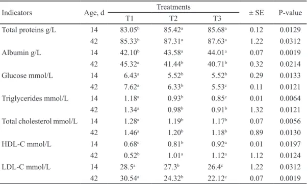
(5) 61. Cuban Journal of Agricultural Science, Volume 52, Number 1, 2018.. Table 2. Full blood count (FBC) of piglets at 14 and 42 d of age, when supplemented with two microbial preárations Treatments Parameters Age, d ± SE P Value T1 T2 T3 Hemoglobin, g/L 14 104.5c 105.4b 108.8a 0.89 0.0130 c b a 42 110.7 112.6 114.4 0.02 0.0070 b a a Hematocrit, L/L 14 0.21 0.23 0.24 0.01 0.0064 b a a 42 0.28 0.30 0.31 1.32 0.0121 Erythrocytes, x1012/L 14 3.45b 4.33a 4.72a 0.07 0.0056 b a a 42 5.32 6.21 6.65 0.01 0.0241 a b b MCV, fL 14 49 45 45 0.12 0.0129 a b b 42 39 37 36 1.22 0.0312 MCH, pg 14 29a 27b 25c 0.07 0.0019 a b b 42 20 18 17 0.32 0.0214 a b b MCHC, g/L 14 421 408 406 0.29 0.0133 a b b 42 345 335 330 0.11 0.0121 Leucocytes, x109/L 14 6.08a 5.05b 5.06b 0.17 0.0129 42 13.25a 12.85b 12.42b 1.03 0.0034 9 a b b Basophils, x10 /L 14 0.12 0.11 0.10 0.02 0.0132 42 0.38a 0.25b 0.23b 0.11 0.0125 9 a b b Eosinophils, x10 /L 14 0.23 0.21 0.20 0.03 0.0017 a b c 42 0.87 0.54 0.47 0.02 0.0012 9 c b a Monocytes, x10 /L 14 0.35 0.42 0.50 0.01 0.0197 42 2.45a 1.85b 1.74b 1.12 0.0124 9 Lymphocytes, x10 /L 14 1.12 1.01 1.05 0.04 0.6495 a b c 42 3.2 2.1 1.88 0.12 0.0021 a,b,c Means with different superscripts in the same row differ at P<0.05 (Duncan 1955) T1, Base diet without additive. T2, L. acidophilus + L. bulgaricus and S. thermophilus. T3, L. acidophilus + L. bulgaricus + S. thermophilus + S. cerevisiae and K. fragilis (L-4 UCLV). MCV, mean corpuscular volume. MCH, mean corpuscular hemoglobin. MCHC, mean corpuscular hemoglobin concentration. Blood values used as reference of Kraft (1998) and Corredor (2012). While at 42 d, it was higher (P <0.05) in the animals that did not consume the microbial preparations (T1). Table 3 shows the performance of blood biochemistry of piglets at 14 and 42 d old. In both ages, differences were found with respect to the control for all the evaluated indicators. Total protein was lower (P <0.05) in the control with respect to the piglets that consumed the microbial preparations. Albumin was also lower (P <0.05) in control animals with 14 d of age, without variations among treatments (T2 and T3). However, the opposite effect was observed for this indicator in the measurement at 42 d of age. The glucose values decreased in piglets treated with the microbial preparations. Out of these, the lowest was (P <0.05) in T3, at 42 d of age. Triglyceride, total cholesterol and LDL-C levels were lower in T2 and T3 with respect to animals in control group T1, while HDL-C values were increased in piglets treated with microbial cultures (T2 and T3).. linfocitos a los 14 d. Mientras que a los 42 d fue mayor (P < 0.05) en los animales que no consumieron los preparados microbianos (T1). En la tabla 3 se muestra el comportamiento de la bioquímica sanguínea de los lechones a los 14 y 42 d de edad. En ambas edades, se encontraron diferencias con respecto al control para todos los indicadores evaluados. La proteína total fue menor (P < 0.05) en el control con respecto a los lechones que consumieron los preparados microbianos. La albúmina también fue menor (P < 0.05) en los animales del control con 14 d de edad, sin variaciones entre tratamientos (T2 y T3). Sin embargo, se observó efecto contrario para este indicador en la medición a los 42 d de edad. Los valores de glucosa disminuyeron en los cerditos tratados con los preparados microbianos. De estos, el menor fue (P < 0.05) en el T3, a los 42 d de edad. Los índices de los triglicéridos, colesterol total y C-LDL fueron menores en T2 y T3 con respecto a los animales del grupo control T1), mientras que los valores de C-HDL se incrementaron en lechones tratados con los cultivos microbianos (T2 y T3)..
(6) 62. Cuban Journal of Agricultural Science, Volume 52, Number 1, 2018.. Table 3. Performance of blood biochemistry of pigs at 14 and 42 d of age, supplemented with two microbial preparations Treatments ± SE P-value T1 T2 T3 Total proteins g/L 14 83.05b 85.42a 85.68a 0.12 0.0129 b a a 42 85.33 87.31 87.63 1.22 0.0312 Albumin g/L 14 42.10b 43.58a 44.01a 0.07 0.0019 a b b 42 45.32 41.44 40.71 0.32 0.0214 a b b Glucose mmol/L 14 6.43 5.52 5.52 0.29 0.0133 42 7.62a 6.33b 5.53c 0.11 0.0121 Triglycerides mmol/L 14 1.18a 0.93b 0.85c 0.01 0.0064 a b b 42 1.34 0.98 0.91 1.32 0.0121 a b b Total cholesterol mmol/L 14 1.28 1.19 1.17 0.07 0.0056 42 1.46a 1.20b 1.18b 0.89 0.0130 c b a HDL-C mmol/L 14 0.68 0.81 0.92 0.01 0.0197 b a a 42 0.52 1.01 1.12 1.12 0.0124 a b c LDL-C mmol/L 14 28.5 27.3 26.4 1.22 0.0312 42 30.54a 24.32b 22.12c 0.07 0.0019 a,b,c Means with different superscripts in the same row differ at P<0.05 (Duncan 1955) T1, Base diet without additive. T2, L. acidophilus + L. bulgaricus and S. thermophilus. T3, L. acidophilus + L. bulgaricus + S. thermophilus + S. cerevisiae and K. fragilis (L-4 UCLV). MCV, mean corpuscular volume. MCH, mean corpuscular hemoglobin. MCHC, mean corpuscular hemoglobin concentration. Blood values used as reference of Kraft (1998) and Corredor (2012) Indicators. Age, d. Discussion. Discusión. Blood profile results of this study (Table 2) could be related to the substrate to ferment the previously mentioned microorganisms and with the microorganisms used and introduced with the diet to the mother sows and their offspring. This could favor the physiological changes in the piglets, possibly by improving the imbalance of the autochthonous microbiota of the gastrointestinal tract, which affected the values of blood indicators. In this sense, the results of this research agree in part with those reported by Ayala et al.(2008), who, when supplementing Bacillus subtilis with sows in the last gestation stage, observed higher (P<0.05) concentration of Hb and Hto in the animals that consumed this bioproduct, which indicates a probiotic effect on health of pigs. Muñoz et al.(2010) reported a slight decrease in physiological changes, as well as in the stabilization of blood values in pigs, by supplementing a mixed culture of lactic acid bacteria, based on the assumption that microbial preparations in piglets could improve passive immunity and reduce blood disorders, such as hemolysis or destruction of red blood cells (Sun et al. 2015). Regarding other blood indicators, values obtained in this study were among the reference limits for this species (Muñoz et al. 2010). However, some blood indicators obtained were favorable for the animals that consumed the microbial preparations, which can be attributed to the fact that probiotics had a favorable influence with a constant contribution. Los resultados de perfil hemático de este estudio (tabla 2) podrían estar relacionados con el sustrato para fermentar los microorganismos antes mencionados y con los microorganismos utilizados e introducidos con la dieta a las cerdas madres y su descendencia. Esto pudo favorecer los cambios fisiológicos en los cerdos jóvenes, posiblemente al mejorar el desequilibrio de la microbiota autóctona del tracto gastrointestinal, lo que repercutió en los valores de los indicadores hemáticos. En este sentido, los resultados de esta investigación concuerdan en parte con los informados por Ayala et al.(2008), quienes al suplementar Bacillus subtilis a cerdas madres en la última etapa de la gestación, observaron mayor (P < 0.05) concentración de Hb y Hto en los animales que consumieron dicho bioproducto, lo que indica el efecto probiótico en la salud de los cerdos. Muñoz et al.(2010) informaron una ligera disminución de las alteraciones fisiológicas, así como de la estabilización en los valores hemáticos en cerdos, al suplementar un cultivo mixto de bacterias lácticas, a partir del supuesto de que los preparados microbianos en los cerdos jóvenes podrían mejorar la inmunidad pasiva y reducir trastornos hemáticos, como la hemólisis o la destrucción de los glóbulos rojos (Sun et al.2015). En lo que se refiere a los otros indicadores hemáticos, los valores obtenidos en este estudio se encontraron entre los límites de referencia para esta especie (Muñoz et al.2010). Sin embargo, algunos indicadores hemáticos obtenidos fueron favorables para los animales que consumieron los preparados microbianos, lo que se puede atribuir a que los probióticos influyeron.
(7) Cuban Journal of Agricultural Science, Volume 52, Number 1, 2018.. of blood-forming elements (Ayala et al. 2008). González et al.(1993) reported similar responses by supplementing microbial additives in pigs with six weeks old. Regarding blood biochemistry of piglets at 14 and 42 d of age (table 3), it is confirmed that the use of the additives did not cause a negative effect on the animals, since the values of blood biochemistry of piglets were among the values considered normal for pigs by González et al.(1993) and Espinosa et al.(2008). Likewise, uniformity and superior degree of immunity is observed in the treated animals, attributed to the response that microbial preparations with probiotic capacity may cause, associated with eubiosis of intestinal biota that stimulates the response of lymphoid tissue and promotes better health state in piglets (Ahmed et al. 2014 and Dadvar et al. 2015). Similar results were reported by Ponnampalam et al.(2011) and Londoño and Parra (2015), by supplementing microbial additives in pigs. Colina et al.(2011) observed a reduction (P <0.05) in serum levels by supplementing lactic acid bacteria. Muñoz et al.(2010) also reported similar results, which could affirm the beneficial effect of microbial additives on animal health and its favorable repercussion for final consumer of pork (Lye et al. 2012). Pajarillo et al.(2014) and Sun et al.(2015) argued that the beneficial effect of probiotics occurs when they are ingested in adequate amounts and that the intestinal ecosystem of the host is modified. This, in turn, generates a beneficial microbial balance, which means a better animal health. Another beneficial effect of microbial preparations is to act against hypercholesterolemia, reducing total cholesterol and LDL-C by balancing the natural microbiota of the gastrointestinal tract of the host (Ahmed et al. 2014). This study showed an increase of HDL-C and a reduction of LDL-C at 42 d of age, which may indicate that the animals consuming microbial preparations may have had better functioning of metabolic organs, despite that levels found in all evaluated treatments were within the normal reference range (Colina et al. 2011). There is evidence of a decrease of postprandial curves of glycemia and insulin after providing S. cerevisiae in diet for goats (Dadvar et al. 2015). It is known for some time that ingestion of non-digestible carbohydrates modifies the kinetics of glucose absorption, reducing postprandial peaks of glycemia and insulinemia (Ros 2003). Changes in intestinal expression of released insulinotropic peptides in response to ingestion of microbial preparations may also have an important function in the lipid effects observed in this study. This could be, in turn, closely related to metabolic diseases and the composition of bacterial populations in the intestine of the host (Lye et al. 2012). In this study, glucose reduction was observed. 63 favorablemente con un aporte constante de elementos hemoformadores (Ayala et al.2008). Similares respuestas informaron González et al.(1993), al suplementar aditivos microbianos en cerdos con seis semanas de edad. En lo que se refiere a la bioquímica sanguínea de los lechones a los 14 y 42 d de edad (tabla 3), se reafirma que la utilización de los aditivos no provocó un efecto negativo en los animales, ya que los valores de la bioquímica sanguínea de los lechones estuvieron entre los valores considerados normales para los cerdos por González et al.(1993) y Espinosa et al.(2008). A su vez, se constata uniformidad y grado de inmunidad superior en los animales tratados, atribuible a la respuesta que pueden provocar los preparados microbianos con capacidad probiótica, asociados a la eubiosis de la biota intestinal que estimula la respuesta del tejido linfoide y propicia mejor estado de salud en cerdos jóvenes (Ahmed et al.2014 y Dadvar et al.2015). Similares resultados informaron Ponnampalam et al.(2011) y Londoño y Parra (2015), al suplementar aditivos microbianos en cerdos. Colina et al.(2011) observaron reducción (P < 0.05) en los niveles séricos al suplementar bacterias lácticas. Muñoz et al.(2010) refieren también resultados parecidos, lo que pudiera afirmar el efecto benéfico de los aditivos microbianos en la salud del animal y su repercusión favorable para el consumidor final de la carne de cerdo (Lye et al.2012). Pajarillo et al.(2014) y Sun et al.(2015) argumentaron que el efecto benéfico de los probióticos se produce cuando se ingieren en cantidades adecuadas y se logra modificar el ecosistema intestinal del huésped. Esto, a su vez, genera un balance microbiano beneficioso, que se traduce en una mejor salud del animal. Otro de los efectos benéficos de los preparados microbianos es actuar contra la hipercolesterolemia, reduciendo colesteroles totales y C-LDL mediante el equilibrio de la microbiota natural del tracto gastrointestinal del huésped (Ahmed et al. 2014). En esta investigación se observó aumento de C-HDL y reducción de C-LDL a los 42 d de edad, lo que puede indicar que los animales que consumieron preparados microbianos pudieron haber tenido mejor funcionamiento de órganos metabólicos, a pesar de que los niveles encontrados en todos los tratamientos evaluados estuvieron en el rango de referencia normal (Colina et al. 2011). Existen evidencias de una disminución de las curvas posprandiales de glucemia e insulina tras la administración de S. cerevisiae en la dieta de las cabras (Dadvar et al.2015). Es conocido desde hace tiempo que la ingestión de hidratos de carbono no digeribles modifica la cinética de absorción de glucosa, reduciendo los picos posprandiales de glucemia e insulinemia (Ros 2003). Los cambios en la expresión intestinal de péptidos insulinotrópicos liberados en respuesta a la ingestión de preparados microbianos puede también representar una función importante en los efectos lipídicos observados en este estudio. Esto podría estar a su vez estrechamente relacionado con las enfermedades metabólicas y la composición de las poblaciones.
(8) 64. Cuban Journal of Agricultural Science, Volume 52, Number 1, 2018.. at 14 and 42 d of age in the animals that consumed the microbial preparations, which may indicate that animals had better functioning of metabolic organs, even though the levels found in all treatments were in the normal reference range (Colina et al. 2011 and Mejía et al. 2012). It is concluded that the addition of the evaluated microbial preparations with probiotic capacity in diets for piglets favorably improves the values of the blood and biochemical profile of the blood and with it, the physiological state and the health of the animals. Acknowledgements The main author thanks the Secretary of Higher Education, Science, Technology and Innovation for the Doctoral training scholarship. Likewise, gratitude is expressed to the Faculty of Sciences, Escuela Superior Politécnica de Chimborazo, Riobamba (Ecuador) for facilitating the use of equipment and microbiology, clinical and bromatology laboratories to develop this study and the Ibero-American Agro-Bigdata Network and "Decision Support Systems" (DSS) for a sustainable agricultural sector (BIGDSSAGRO) for their support to this research.. bacterianas en el intestino del huésped (Lye et al.2012). En este estudio se observó disminución de glucosa a los 14 y 42 d de edad en los animales que consumieron los preparados microbianos, lo que puede indicar que los animales tuvieron mejor funcionamiento de los órganos metabólicos, a pesar de que los niveles encontrados en todos los tratamientos estuvieron en el rango de referencia normal (Colina et al.2011 y Mejía et al.2012). Se concluye que la adición de los preparados microbianos evaluados con capacidad probiótica en las dietas de cerdos jóvenes mejora favorablemente los valores del perfil hemático y bioquímico de la sangre y con ello, el estado fisiológico y la salud de los animales. Agradecimientos El autor principal agradece a Secretaria de Educación Superior, Ciencia, Tecnología e Innovación por la beca de formación de Doctoral. Asimismo, se expresa gratitud a la Facultad de Ciencias, Escuela Superior Politécnica de Chimborazo, Riobamba (Ecuador) por facilitar el uso de los equipos y los laboratorios de microbiología, clínica y bromatología para desarrollar el presente estudio y a la Red Iberoamericana de Agro-Bigdata y "Decision Support Systems" (DSS) para un sector agropecuario sostenible (BIGDSSAGRO) por su apoyo a esta investigación.. References Ahmed, S.T., Hoon, J., Hong, M. & Chul, Y. 2014. Evaluation of Lactobacillus and Bacillus-based probiotics as alternatives to antibiotics in enteric microbial challenged weaned piglets. African J Microbiol Res 8(1): 96-101. Ayala, L., Bocourt, R., Martínez, M., Castro, M. & Hernández, L. 2008. Respuesta productiva, hematológica y morfométrica de un probiótico comercial en cerdos jóvenes. Rev Cubana de Cienc Agríc 42(2):181-184. Coates, J., Corns, P.J., Juarez, A., MacDonald, R., McCulley, N., Melody, B., Minton, A., Molinari, R., Montes De Oca, H., Mosqueira, P., Neill, C., Pinilla, J.C., Piva & J., Teuber, R. 2013. Manual PIC de Manejo de Hembras y Primerizas. 100 Bluegrass Commons Blvd: Suite 2200: Hendersonville, TN 37075: EEUU. Colina, J., Méndez, A., Araque, H., Rueda, E., León, M. & Rossini, M. 2011. Lípidos sanguíneos en cerdos alimentados con pijiguao (Bactris gasipaes Kunth) y lisina sintética Rev Mvz Córdoba 16(3): 2668-2677 Corredor, R. 2012. Perfil hemático de cerdos alimentados con follaje de Morera Morus alba. Rev CITECSA 2(3): 27-35. Dadvar, P., Dayani, O., Mehdipour, M. & Morovat, M. 2015. Determination of physical characteristics, chemical composition and digestion coefficients of treated lemon pulp with Saccharomyces cerevisiae in goat diet. J Anim Physiol and Anim Nutrit 99(1):107–113. Duncan, D.B. 1955. Multiple range and multiple F tests. Biometrics 11:1. Espinosa, V., García, A., Herrera, J., Álvarez, A., Estrada, S. & Meza, M. 2008. Efecto del extracto de Yucca schidigera en el perfil bioquímico y hemático de cerdos en crecimiento y engorde. Rev Científica (Maracaibo) 18(1):51 – 58. González, D., Cisneros, M., Martínez, A., Zendejasm, V. & Morilla, A. 1993. Perfil inmunológico de los cerdos durante las primeras diez semanas de edad. Rev Tec Pecuaria México 24(3): 217-221. Kraft, H. 1998. Hematología. Análisis químico clínico de sangre. En: Métodos de laboratorio clínico en medicina veterinaria de mamíferos domésticos. Ed. Acribia 1998. España pp 23-82. Londoño, S. & Parra, J. 2015. Efecto de la adición de cepas probióticas sobre metabolitos sanguíneos en cerdos en crecimiento. Biotecnología en el Sector Agropecuario y Agroindustrial 13(2): 49-56. Lye, H., Khoo, Y., Karim, A., Rusul, G. & Liong, T. 2012. Growth properties and cholesterol removal ability of electroporated Lactobacillus acidophilus BT 1088. J Microbiol and Biotechnol 22(7): 981-989. Mejía, K., Lemus, C., Huerta, R., Almague, R. & Ly, J. 2012. Niveles séricos de triglicéridos y colesterol en cerdos cuino mexicano. Rev Comput de Prod Porc (RCPP) 19(1): 37-41. Miranda, J.E., Marin, A. & Baño, D. 2017. Elaboration of a bioprepared with probiotic effect from a mixed culture of lactic bacteria and yeasts. Bionatura 2(1): 245-247. Muñoz, D., Ruiz, A, González, M., Islas, A., Díaz, N. & Quezada, M. 2010. Estudios hematológicos y patológicos comparativos de cerdos inoculados con un aislado de campo y el serotipo 5 ATCC de Actinobacillus pleuropneumoniae. Arch Med Vet 42(1): 57-65 National Research Council (NRC) 2012. Nutrient Requirements of Swine: Eleventh Revised Edition. Washington, DC: The.
(9) 65. Cuban Journal of Agricultural Science, Volume 52, Number 1, 2018.. National Academies. Pajarillo, E., Chae, J., Balolong, M., Kim, H. & Kang, D. 2014. Assessment of fecal bacterial diversity among healthy piglets during the weaning transition. J Gener and Appl Microbiol 60(4): 140‒146. Ponnampalam, E., Lewandowski, P., Nesarstnam, K., Dunshea, R. & Gill, H. 2011. Differential effects of natural palm oil, chemically-and enzymatically-modified palm oil on weight gain, blood lipid metabolites and fat deposition in a pediatric pig model. Nutrition Journal 10(53): 1-7. Ros, E. 2003. Prebióticos y probióticos en la regulación del metabolismo de los lípidos. Gastroenterol Hepatol 26(1): 31-36 Sun, Y., Park, I., Guo, J., Weaver, A. & Sung, K. 2015. Impacts of low level aflatoxin in feed and the use of modified yeast cell wall extract on growth and health of nursery pigs. Animal Nutrition 1(3):177–183 Received: September 19, 2017.
(10)
Figure


Documento similar
The aim of the this study is to evaluate the hemodynamic changes of blood pressure and heart rate on hypertensive patients receiving three different types of local anesthe-
monocytogenes in two fish products and describe inter-species competition was based on (i) the determination of the kinetic parameters of the two microbial pop- ulations
Astrometric and photometric star cata- logues derived from the ESA HIPPARCOS Space Astrometry Mission.
The photometry of the 236 238 objects detected in the reference images was grouped into the reference catalog (Table 3) 5 , which contains the object identifier, the right
performance of the primiparous sow and the adopted litter. Absorption of orally supplied immunoglobulins in neonatal piglets. Appearance of immunoglobulin G in the plasma of
Two series of coatings (using two different peak powers) were deposited with increasing V content, in order to evaluate the effect of V addition on the surface and cross
In addition, precise distance determinations to Local Group galaxies enable the calibration of cosmological distance determination methods, such as supernovae,
Abstract: Transepidermal water-loss (TEWL), stratum-corneum hydration (SCH), erythema, elas- ticity, pH and melanin, are parameters of the epidermal barrier function and
