TítuloContemporary management of acute right ventricular failure: a statement from the Heart Failure Association and the Working Group on Pulmonary Circulation and Right Ventricular Function of the European Society of Cardiology
25
0
0
Texto completo
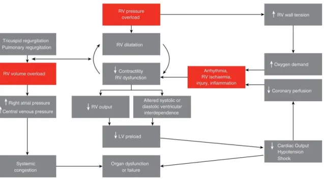
(2) Abstract Acute right ventricular (RV) failure is a complex clinical syndrome that results from many causes. Research efforts have disproportionately focused on the failing left ventricle, but recently the need has been recognized to achieve a more comprehensive understanding of RV anatomy, physiology, and pathophysiology, and of management approaches. Right ventricular mechanics and function are altered in the setting of either pressure overload or volume overload. Failure may also result from a primary reduction of myocardial contractility owing to ischaemia, cardiomyopathy, or arrhythmia. Dysfunction leads to impaired RV filling and increased right atrial pressures. As dysfunction progresses to overt RV failure, the RV chamber becomes more spherical and tricuspid regurgitation is aggravated, a cascade leading to increasing venous congestion. Ventricular interdependence results in impaired left ventricular filling, a decrease in left ventricular stroke volume, and ultimately low cardiac output and cardiogenic shock. Identification and treatment of the underlying cause of RV failure, such as acute pulmonary embolism, acute respiratory distress syndrome, acute decompensation of chronic pulmonary hypertension, RV infarction, or arrhythmia, is the primary management strategy. Judicious fluid management, use of inotropes and vasopressors, assist devices, and a strategy focusing on RV protection for mechanical ventilation if required all play a role in the clinical care of these patients. Future research should aim to address the remaining areas of uncertainty which result from the complexity of RV haemodynamics and lack of conclusive evidence regarding RV‐specific treatment approaches. Keywords: Right ventricular dysfunction; Right ventricular function; Heart failure; Intensive care; Cardiogenic shock. Introduction Acute right ventricular (RV) failure can be defined as a rapidly progressive syndrome with systemic congestion resulting from impaired RV filling and/or reduced RV flow output. Most often it is associated with increased RV afterload or preload and consequent RV chamber dilatation and tricuspid regurgitation (Figure 1).1, 2 The prevalence of acute RV failure is difficult to estimate, but its predominant causes [i.e. left‐sided heart failure, acute pulmonary embolism (PE), acute myocardial ischaemia] are common. 3-5 It is observed in 3–9% of acute heart failure admissions, and the in‐hospital mortality of patients with acute RV failure ranges from 5 to 17%.6-10 Research efforts have disproportionately focused on the failing left ventricle, but recently more attention has been placed on understanding RV anatomy, physiology, dysfunction, and management. 11 Right ventricular failure is a heterogeneous syndrome, and its varied aetiologies require individualized treatment. Recognizing the numerous aspects of RV failure, the Heart Failure Association (HFA) and the Working Group on Pulmonary Circulation and Right Ventricular Function of the European Society of Cardiology (ESC) convened a multidisciplinary group of experts to discuss state‐of‐the‐art principles pertaining to RV failure and its aetiology, clinical presentation, assessment, treatment, and areas where focused research is needed. This paper summarizes the dialogue and suggests priorities for research in this field..
(3) Figure 1. Pathophysiology of acute right ventricular failure. LV, left ventricular; RV, right ventricular.. Pathophysiology Anatomy and mechanics of right ventricular function The thin‐walled right ventricle pumps the entire systemic venous return into the pulmonary circulation for gas exchange. Right ventricular function integrates preload, afterload, contractility, pericardial constraint, interaction with the left ventricle, and cardiac rhythm.12-14 Venous return depends on the pressure gradient between the peripheral vasculature where the mean systemic filling pressure (although not well characterized in humans) is approximately 7–10 mmHg, and the right atrial (or central venous) pressure, which is usually 0 mmHg at rest.15 In contrast to the left ventricle, twisting and rotational movements do not contribute significantly to RV contraction. Instead, the most important mechanisms are the bellows‐like inward movement of the free wall, the contraction of the longitudinal fibres drawing the tricuspid annulus toward the apex, and the traction on the free wall as a result of left ventricular contraction. The contraction of the right ventricle is sequential, starting with the trabeculated myocardium and ending with the contraction of the infundibulum (25–50 ms delay).16 Because RV afterload is very low under normal conditions, blood flows from the RV into the pulmonary circulation both during systole and during the early part of diastole, leading to the absence of isovolumetric relaxation. 15 Aetiology and pathogenesis of right ventricular failure Right ventricular mechanics and function are altered in the setting of either pressure overload or volume overload (Figure 1, Table 1). Failure may also result from a primary reduction of myocardial contractility owing to ischaemia, cardiomyopathy, or arrhythmia..
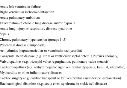
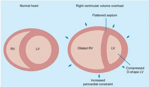
(4) Table 1. Causes and differential diagnosis of acute right ventricular failure. Acute left ventricular failure Right ventricular ischaemia/infarction Acute pulmonary embolism Exacerbation of chronic lung disease and/or hypoxia Acute lung injury or respiratory distress syndrome Sepsis Chronic pulmonary hypertension (groups 1–5) Pericardial disease (tamponade) Arrhythmias (supraventricular or ventricular tachycardia) Congenital heart disease (e.g. atrial or ventricular septal defect, Ebstein's anomaly) Valvulopathies (e.g. tricuspid valve regurgitation, pulmonary valve stenosis) Cardiomyopathies (e.g. arrhythmogenic right ventricular dysplasia, familial, idiopathic) Myocarditis or other inflammatory diseases Cardiac surgery (e.g. cardiac transplant or left ventricular assist device implantation) Haematological disorders (e.g. acute chest syndrome in sickle cell disease). The right ventricle is not built to handle large or rapid increases in pulmonary artery pressure. However, it possesses, like the left ventricle, the capacity to adapt its systolic function to preserve ventriculo‐arterial coupling. During the acute response, the right ventricle uses a homeometric or systolic functional adaptation (Anrep's law of the heart) within minutes of a rise in pulmonary artery pressure; chronically it implements a heterometric or dimensional adaptation (Starling's law of the heart) to preserve flow output. Insufficient systolic functional adaptation will limit cardiac output and ultimately result in systemic hypotension and cardiogenic shock; dilatation with eventual diastolic dysfunction causes systemic congestion. Dyssynchrony, or inhomogeneous regional contraction, occurs early in the adaptation of the right ventricle to increased systolic function demands. Asynchrony, or delayed RV systole (i.e. the right ventricle is still ejecting blood while the left ventricle is already filling), appears when the right ventricle dilates and the septum shifts. At this stage, the left ventricle becomes underfilled, with resultant hypotension and altered systolic ventricular interactions. Systolic or diastolic ventricular interdependence relates to the concept that the functioning of the left ventricle affects the functioning of the right ventricle and vice versa (Figure 2). The main anatomical determinants of ventricular interdependence include the interventricular septum, the pericardium, and the continuity between myocardial fibres of the left and right ventricle. 16 Some (up to 40%) of the RV contractile force originates from left ventricular contraction. 17 Moreover, when the right ventricle dilates acutely, the interventricular septum is shifted leftward, both in systole and diastole as both ventricles ‘compete’ for space within the pericardium. Notably, this shift occurs only in diastole when ventricular pressures are low. Septal shift compresses the left ventricle, impairs its filling, and leads to reduced left ventricular contractility (see the Supplementary material online, Cases S1, S2).12, 13 By the same mechanism, acute RV dilatation also leads to increased pericardial constraint, which in turn decreases the distensibility of the left ventricle and reduces left ventricular filling (preload), ultimately leading to a drop in stroke volume.1, 13.
(5) Figure 2. Ventricular interdependence in right ventricular failure. Dilatation of the right ventricle shifts the interventricular septum toward the left, changing left ventricular geometry. Acute right ventricular distension may also lead to an increase in pericardial constraint (arrows). These changes may contribute to low cardiac output state by decreasing left ventricular distensibility, preload, and ventricular elastance. Reprinted with permission from Haddad F, Doyle R, Murphy DJ, Hunt SA. Right ventricular function in cardiovascular disease, part II: pathophysiology, clinical importance, and management of right ventricular failure. Circulation 2008;117:1717–1731.. Consequences of right ventricular failure The typical dilatation of the right ventricle in response to volume overload is shown in the Supplementary material online, Case S3. In the setting of acutely increased afterload, the right ventricle responds in a similar manner by increasing its end‐diastolic volume and contractility, but RV failure may occur rapidly if these mechanisms prove unable to generate sufficient pressure to maintain flow (e.g. past the thromboembolic obstruction in the case of acute PE) (see the Supplementary material online, Cases S4, S5).1 In chronically evolving pulmonary hypertension from precapillary or postcapillary causes, the right ventricle responds to increasing afterload with progressive hypertrophy, which allows it to maintain cardiac output at rest over long periods of time. The right ventricle finally dilates in end‐stage disease, leading to tricuspid regurgitation and, ultimately, decreased cardiac output (see the Supplementary material online, Cases S1, S2). Acute decompensations of chronic pulmonary hypertension may lead to a clinical presentation barely distinguishable from that of ‘truly’ acute RV failure as, for example, in acute pulmonary embolism.1 Venous congestion commonly occurs in acute RV failure. Dysfunction leads to impaired RV filling and increased right atrial pressures.1 As RV dysfunction progresses to overt RV failure, the RV chamber becomes more spherical and tricuspid regurgitation is aggravated, leading to progressive venous congestion.15, 18 Venous congestion and increased central venous pressure also lead to impairment of renal, intestinal, and hepatic function,19-26 which are important predictors of poor prognosis in patients with acute RV failure.22, 23, 26-28.
(6) Clinical presentation and assessment Initial triage: clinical assessment and biochemical markers The clinical presentation of acute RV failure varies depending on the underlying cause and presence of comorbidities (Table 2).12, 17. Table 2. Clinical signs and biochemical markers of acute right ventricular (RV) failure Clinical signs. Biochemical markers. Hypoxaemia. Increased lactate levels. Signs of systemic congestion. Elevated natriuretic peptides (BNP or NT‐proBNP). Jugular venous distension, hepatojugular reflux. Elevated cardiac troponin I or T. Peripheral oedema, pericardial effusion, congestive hepato/splenomegaly, ascites, anasarca*. Abnormal liver biochemistry (e.g. elevated transaminases, bilirubin, prolonged prothrombin time). Signs of right ventricular dysfunction. Abnormal renal function (blood urea nitrogen, creatinine). Third heart sound, systolic murmur of tricuspid regurgitation, hepatic D‐dimer levels† pulse, signs of concomitant left ventricular dysfunction Paradoxical pulse Signs of low cardiac output state Hypotension, tachycardia, cool extremities, central nervous system abnormalities, oliguria. BNP, brain natriuretic peptide; NT‐proBNP, N‐terminal pro brain natriuretic peptide. * Particularly in acute decompensation of chronic RV failure. † D‐dimer testing is used in the diagnostic workup of suspected pulmonary embolism.. The primary goal of pre‐hospital and emergency department triage is to assess the acuity and urgency of the clinical situation. The aetiology of RV failure should be sought (Table 1), and the diagnosis or exclusion of causes requiring specific treatment (such as PE) should be prioritized. The initial triage is based on clinical history and physical examination. The electrocardiogram, arterial blood gases, and blood lactate should also be assessed.29 Examples of typical electrocardiograms in different clinical settings are shown in the Supplementary ECG material online. On hospital admission, focused bedside echocardiography provides rapid information on cardiac structure and function (see section on Echocardiography). Chest X‐ray is routinely obtained and occasionally yields specific findings. There are currently no biomarkers specific for RV failure.30 Consequently, the clinical utility of B‐ type natriuretic peptides and cardiac troponin testing depends on the clinical context in which acute RV failure presents. These markers possess high sensitivity for the early detection of RV failure and myocardial injury, respectively, in patients with confirmed acute PE; 31-34 they were also associated with poor prognosis in RV failure related to pulmonary arterial hypertension (PAH). 22, 35 Novel biomarkers for detecting acute RV failure are under evaluation.36-39.
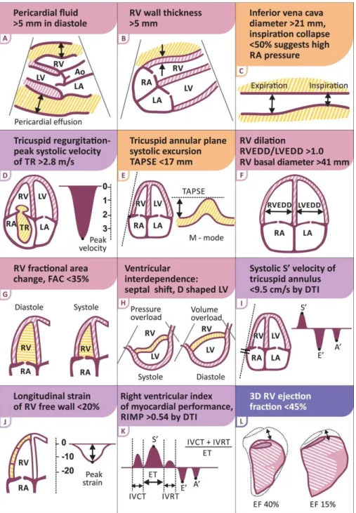
(7) Echocardiographic assessment Bedside focused cardiac ultrasound in the emergency department or intensive/coronary care unit is a first‐line test in the assessment of RV size, function and load. 40, 41 It can be used to exclude extrinsic causes of acute RV failure, especially those needing immediate treatment (such as pericardial tamponade which mimics acute RV failure), and to estimate right atrial pressure by assessing the diameter and the respiratory collapse of the inferior vena cava. The quantification of pulmonary artery systolic pressure by the trans‐tricuspid pressure gradient is a reliable method compared with invasive measurements. 42 All available views of the right heart, including apical four‐chamber RV‐focused (see the Supplementary material online, Cases S1–S3) and subcostal views, should be used to estimate accurately RV enlargement, sphericity, and degree of RV dilation compared with left ventricular dilation. 42 The latest advances in ultrasound techniques are shifting from mostly qualitative assessment of global and segmental RV function in the past to quantitative evaluation. 43 Recently updated recommendations from the American Society of Echocardiography and the European Association of Cardiovascular Imaging44 advocate quantitative assessment of global RV function by at least one of the following parameters: fractional area change (FAC; Supplementary material online, Case S2); tricuspid annular plane systolic excursion (TAPSE; see the Supplementary material online, Cases S2, S3, S6); Doppler tissue imaging‐derived systolic S′ velocity of the tricuspid annulus (see the Supplementary material online, Cases S2–S4); or RV index of myocardial performance (RIMP; see the Supplementary material online, Case S2). In addition to TAPSE and S′ velocity, RV global and regional longitudinal shortening may be estimated by strain echocardiography. Assessment of RV ejection fraction based on two‐dimensional echocardiography is not recommended; experienced and appropriately equipped centres employ three‐dimensional RV volumes and ejection faction with considerable accuracy. Frequently utilized parameters and reference values of RV dilation, global and segmental function, haemodynamic overload, and ventricular interdependence are graphically represented in Figure 3.42, 44 Typical ultrasound features of the most common clinical scenarios are shown in Table 3 and in the Supplementary material online, Cases S1–S8.42, 44 A comprehensive, more simple evaluation of RV function, which takes into account the effect of tidal inflation on RV ejection flow of mechanically ventilated patients, has been proposed in the intensive care unit and is part of advanced critical care echocardiography.40.
(8) Figure 3. Graphic representation of echocardiographic parameters in the assessment of right ventricular failure. Ao, aorta; DTI, Doppler tissue imaging; EF, ejection fraction; ET, ejection time; FAC, fractional area change; IVC, inferior vena cava; IVCT, isovolumic contraction time; IVRT, isovolumic relaxation time; LA, left atrium; LV, left ventricle; LVEDD, left ventricular end‐ diastolic diameter; RA, right atrium; RIMP, right ventricular index of myocardial performance; RV, right ventricle/ventricular; RVEDD, right ventricular end‐diastolic diameter; TAPSE, tricuspid annular plane systolic excursion; TR, tricuspid regurgitation..
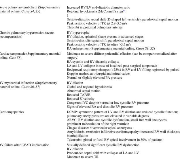
(9) Table 3. Echocardiographic findings in specific clinical settings. Acute pulmonary embolism (Supplementary material online, Cases S4, S5). Increased RV/LV end‐diastolic diameter ratio Regional hypokinesia (McConnell's sign* ) Systole‐diastolic septal shift (D‐shaped left ventricle), paradoxical septal motion Peak systolic velocity of TR jet 2.8–3.5 m/s Thrombi in proximal pulmonary arteries. Chronic pulmonary hypertension (acute decompensation). RV hypertrophy RV dilation, spherical shape present in advanced stages Systolic/diastolic septal shift, paradoxical septal motion Peak systolic velocity of TR jet often >3.5 m/s RA enlargement (Supplementary material online, Cases S1, S2). Cardiac tamponade (Supplementary material online, Case S8). Moderate to severe diffuse pericardial effusion (can be compartmentalized after surgery) RA systolic and RV diastolic collapse LA and LV collapse in case of localized post‐surgical tamponade Reciprocal respiratory changes (>25%) in RV and LV filling registered by pulsed Doppler method at tricuspid and mitral valves Normal or slightly elevated PA pressure. RV myocardial infarction (Supplementary material online, Cases S6, S7). RV dilation Global and regional hypokinesia Abnormal septal motion Reduced TAPSE Reduced S′ velocity Congested IVC despite normal or low systolic RV pressure Signs of elevated RA and diastolic RV pressure. Cardiomyopathies. DCMP: symmetric pattern of LV and RV dilation and reduced systolic function; pulmonary artery pressures are elevated in variable degrees ARVC: RV dilation and systolic dysfunction, small free wall aneurysms, prominent trabeculation of the right ventricle Chagas disease: biventricular apical aneurysms Amyloidosis, restrictive infiltrative cardiomyopathy: increased RV wall thickness, biatrial dilation Takotsubo: global or focal RV apical involvement in 50% of patients. RV failure after LVAD implantation. Visually defined significant systolic RV dysfunction RV dilation Pronounced septal shift with collapse of LA and LV Moderate to severe TR. ARVC, arrhythmogenic right ventricular cardiomyopathy; DCMP, dilated cardiomyopathy; IVC, inferior vena cava; LA, left atrium; LV, left ventricle; PA, pulmonary artery; RA, right atrial; RV, right ventricular; RVEF, right ventricular ejection fraction; TAPSE, tricuspid annular plane systolic excursion; TR, tricuspid regurgitation. * Akinetic mid RV free wall segment with spared apical contractility.. Invasive haemodynamic assessment with pulmonary artery catheter Invasive haemodynamic assessment is recommended in unexplained diagnostic or therapy‐resistant cases; it provides continuous, accurate, and valuable information about right and left atrial pressure, cardiac output, and pulmonary vascular resistance. In general, invasive monitoring should be used for the shortest possible period..
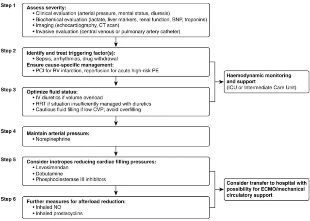
(10) Clinical management General overview of managing right ventricular failure Effective treatment of RV failure requires a skilled multidisciplinary team 45-47 to rapidly assess and triage the patient to the appropriate environment (Figure 4). The ongoing monitoring varies according to the clinical scenario, but its focus is on supporting the RV, managing the consequences of failure, and alleviating distressing physical (e.g. breathlessness, pain) and emotional (e.g. anxiety) symptoms (Figure 4, Table 4).. Figure 4 Comprehensive care of patients affected by right ventricular failure. Modified from Sztrymf B et al. Acute right heart failure in pulmonary hypertension patients. In Gaine S, Naeije R, Peacock A, eds. The Right Heart. Springer Verlag, London, 2013, pages 261–275. BNP, B‐type natriuretic peptide; CT, computed tomography; CVP, central venous pressure; ECMO, extracorporeal membrane oxygenation; ICU, intensive care unit; IV, intravenous; NO, nitric oxide; PCI, percutaneous coronary intervention; PE, pulmonary embolism; RRT, renal replacement therapy; RV, right ventricular..
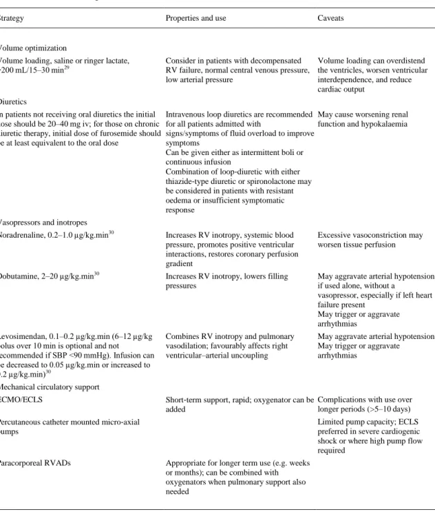
(11) Table 4. Overview of acute right ventricular (RV) failure treatment Strategy. Properties and use. Caveats. Volume optimization Volume loading, saline or ringer lactate, >200 mL/15–30 min29. Consider in patients with decompensated Volume loading can overdistend RV failure, normal central venous pressure, the ventricles, worsen ventricular low arterial pressure interdependence, and reduce cardiac output. Diuretics In patients not receiving oral diuretics the initial dose should be 20–40 mg iv; for those on chronic diuretic therapy, initial dose of furosemide should be at least equivalent to the oral dose. Intravenous loop diuretics are recommended May cause worsening renal for all patients admitted with function and hypokalaemia signs/symptoms of fluid overload to improve symptoms Can be given either as intermittent boli or continuous infusion Combination of loop‐diuretic with either thiazide‐type diuretic or spironolactone may be considered in patients with resistant oedema or insufficient symptomatic response. Vasopressors and inotropes Noradrenaline, 0.2–1.0 µg/kg.min30. Increases RV inotropy, systemic blood pressure, promotes positive ventricular interactions, restores coronary perfusion gradient. Excessive vasoconstriction may worsen tissue perfusion. Dobutamine, 2–20 µg/kg.min30. Increases RV inotropy, lowers filling pressures. May aggravate arterial hypotension if used alone, without a vasopressor, especially if left heart failure present May trigger or aggravate arrhythmias. Levosimendan, 0.1–0.2 µg/kg.min (6–12 µg/kg bolus over 10 min is optional and not recommended if SBP <90 mmHg). Infusion can be decreased to 0.05 µg/kg.min or increased to 0.2 µg/kg.min)30. Combines RV inotropy and pulmonary vasodilation; favourably affects right ventricular–arterial uncoupling. May aggravate arterial hypotension May trigger or aggravate arrhythmias. Mechanical circulatory support ECMO/ECLS. Short‐term support, rapid; oxygenator can be Complications with use over longer periods (>5–10 days) added Limited pump capacity; ECLS preferred in severe cardiogenic shock or where high pump flow required. Percutaneous catheter mounted micro‐axial pumps. Paracorporeal RVADs. Appropriate for longer term use (e.g. weeks or months); can be combined with oxygenators when pulmonary support also needed. ECLS, extracorporeal life support; ECMO, extracorporeal membrane oxygenation; RVAD, right ventricular assist device; SBP, systolic blood pressure..
(12) Volume optimization Patients with RV failure may be preload‐dependent, but volume loading has the potential to overdistend the RV and thereby increase wall tension, decrease contractility, aggravate tricuspid regurgitation, increase ventricular interdependence, impair left ventricular filling, and ultimately reduce systemic cardiac output.48 Cautious volume loading guided by central venous pressure monitoring may be appropriate if low arterial pressure is combined with the absence of elevated filling pressures. As RV failure is often caused, associated with, or aggravated by RV volume overload, as mentioned above, diuretics are often the first option for most patients with RV failure who present with signs of venous congestion along with maintained arterial blood pressure. Volume redistribution in the venous system under diuretic treatment can contribute to rapid clinical improvement (Table 4). Vasopressor and inotrope treatment Vasopressors and/or inotropes are indicated in acute RV failure with haemodynamic instability (Table 4). Vasopressors such as noradrenaline are primarily indicated to restore blood pressure and improve cerebral, coronary, and other organ perfusion. Noradrenaline can improve systemic haemodynamics by improvement of ventricular systolic interaction and coronary perfusion without change in pulmonary vascular resistance.49 Data for vasopressin are lacking in acute RV failure. Dobutamine, levosimendan and phosphodiesterase III inhibitors improve contractility and increase cardiac output. Dobutamine may reduce blood pressure; in that case, a vasopressor, such as noradrenaline, is recommended. Levosimendan may favourably affect RV–arterial uncoupling13, 48, 50 by combining RV inotropy and pulmonary vasodilation. Phosphodiesterase III receptors are absent in the pulmonary vasculature. Thus, phosphodiesterase III inhibitors exert a positive inotrope effect on the right ventricle without the deleterious effects on pulmonary vascular resistance that occur with catecholamines. Similar to dobutamine, these drugs may aggravate arterial hypotension and should be combined with noradrenaline if needed. Levosimendan or phosphodiesterase III inhibitors may be preferentially indicated over dobutamine in cardiac patients with pulmonary hypertension caused by left heart disease. Mechanical circulatory support Acute mechanical circulatory support of the right ventricle may be required in certain clinical situations such as RV myocardial infarction (MI), acute PE, following left ventricular assist device implantation, or primary graft failure after heart transplantation (Table 4). The most important determinant of success is the correct timing of implantation to avoid significant, potentially irreversible end‐organ injury. Multiorgan failure is the leading cause of death in unsuccessful cases. Early transfer of the patient to an appropriate centre is essential for success. Device selection depends on the anticipated duration of mechanical support. Extracorporeal membrane oxygenation (ECMO) or life support (ECLS) carries the potential for increasing use as short‐term mechanical support in the future. It is less expensive than other assist devices and can be inserted quickly, even percutaneously.51 After 5–10 days, a decision should be made to wean the patient and explant the ECMO, or switch to an intermediate or long‐term device, to avoid typical ECMO complications (e.g. infections or formation of thrombus around the cannulae, limb hypoperfusion, or local infection). Alternatively, percutaneously inserted catheter‐mounted microaxial pumps can be used, but these devices may have limited maximum pump capacity. 51.
(13) Right ventricular assist devices (RVADs) can be implanted either surgically52 or percutaneously.53 Paracorporeal RVADs can be used for weeks or even months,54 but they are only approved for up to 4 weeks. These devices can easily be combined with oxygenators when needed. In rare cases, if RV function is not restored, the insertion of implantable continuous‐flow ventricular assist devices has been effective in some reports.55 Retrospective reports have shown good results in terms of haemodynamic status and functional recovery enabling RVAD explantation in 42–75% of patients.56, 57 Bleeding or thrombus formation are the most common complication related to RVADs.58 Poor left ventricular function usually predicts worse outcomes as isolated RVAD support under these circumstances is insufficient to improve systemic perfusion.56 Despite case reports of prolonged RV support with an assist device intended for destination therapy, 59 options for long‐term mechanical circulatory support have been lacking. Thus, cardiac transplant remains the ultimate treatment for refractory RV failure. Clinical management in specific clinical scenarios Pulmonary embolism Acute PE is one of the most frequent causes of acute RV failure (see the Supplementary material online, Cases 4, 5). Conversely, RV failure is the principal determinant of early mortality in the acute phase of PE. Accordingly, and as emphasized in the recently updated (2014) ESC Guidelines on the Diagnosis and Management of Acute Pulmonary Embolism, 60 early detection of clinical, imaging (echocardiographic, computed tomographic), and/or laboratory (e.g. natriuretic peptides, cardiac troponins) indicators of RV dysfunction and myocardial injury is the cornerstone of a successful risk‐ adjusted therapeutic approach to PE. Based on the contemporary risk classification, ‘high risk’ (formerly ‘massive’) PE is defined by persistent arterial hypotension or shock caused by overt RV failure, while ‘intermediate risk’ identifies normotensive patients with a high clinical prognostic score (such as the Pulmonary Embolism Severity Index) accompanied by imaging or biochemical markers of RV dysfunction, alone (i.e. ‘intermediate‐low risk’) or in combination (i.e. ‘intermediate‐high risk’).60 Reperfusion treatment, mainly systemic (intravenous) thrombolysis, is recommended for patients who present with high‐risk PE; in contrast, the haemorrhagic risks of thrombolysis appear to outweigh its clinical benefits in patients who are haemodynamically stable at presentation. 61 Among these latter normotensive patients, those with ‘intermediate–high‐risk’ PE should be clinically and haemodynamically monitored over the first 2–3 days, as rescue thrombolysis may be necessary if RV dysfunction leads to haemodynamic decompensation. Surgical pulmonary embolectomy is an alternative option for haemodynamically unstable patients with high‐risk PE, particularly if thrombolysis is contraindicated or has failed. It may also be considered as a rescue procedure in intermediate high‐risk patients in whom haemodynamic decompensation appears imminent and the anticipated bleeding risk under systemic fibrinolysis is high. 60 Catheter‐directed techniques for the removal of obstructing thrombi from the main pulmonary arteries have been available for several years. ‘Purely interventional’ options such as thrombus fragmentation with pigtail or balloon catheter, rheolytic thrombectomy with hydrodynamic catheter devices, suction thrombectomy with aspiration catheters and rotational thrombectomy have been reserved for patients with absolute contraindications for fibrinolysis. When ‘high‐risk’ patients, or occasionally ‘intermediate‐high‐risk’ patients with signs of developing haemodynamic decompensation, have relative contraindications for fibrinolysis and an increased bleeding risk, conventional catheter‐directed fibrinolysis through a multi‐ sidehole catheter placed into the thrombus, or pharmaco‐mechanical fibrinolysis, are preferred approaches.62.
(14) Pulmonary hypertension Right ventricular function is the major determinant of morbidity and mortality in the pulmonary hypertension population, and signs of RV failure frequently dominate the clinical presentation of patients with pulmonary arterial hypertension (Group 1) or chronic thromboembolic pulmonary hypertension (Group 4; Supplementary material online, Cases S1, S2). Pulmonary hypertension secondary to chronic (left) heart (Group 2; Supplementary material online, Case S6) or lung (Group 3) disease may also exhibit signs and symptoms of RV failure, but they usually present with clinical features of the underlying cardiac or pulmonary disease. Patients with previously unknown PAH are occasionally seen for the first time in the emergency department. If a patient with PAH is seen on an emergency basis, echocardiography is extremely important to assess the current status of RV function. 63, 64 A central venous catheter allows for monitoring of SvO2 and central venous pressure in response to therapy. Continuous and complete haemodynamic assessment might require the placement of a pulmonary artery catheter, but the expected benefits for patient management should be weighed against the risk of complications, including complications with vein access, life‐threatening arrhythmias as well as infections and thrombosis. 65 Possible triggers for acute RV failure in patients with PAH should be identified. 48, 66-68 The most frequent cause is an infection, and sepsis increases mortality significantly. 67 Supraventricular arrhythmias are common,69 and may require electrical cardioversion. Anaemia is also common and may need correction. In particular, iron deficiency should trigger a search for potential reasons, and iron replacement should be considered; oral iron absorption may be impaired in patients with PAH, therefore, intravenous administration may be preferable.70 However, the threshold may differ according to the clinical situation (e.g., right to left shunting, Eisenmenger syndrome, cyanosis) and there are no studies to determine the optimal value for haemoglobin or haematocrit in patients with RV failure. Finally, non‐ adherence to or withdrawal from pulmonary hypertension treatment is another major cause of decompensation. Hypoxia and hypercapnia, as well as acidosis and hypothermia, promote pulmonary vasoconstriction and further increase the afterload of the right ventricle. Oxygen therapy should be used to keep an arterial oxygen saturation >90%. Non‐invasive ventilation may be indicated in patients with respiratory failure and hypercapnia who do not respond to general measures; this may particularly apply to those with pulmonary hypertension caused by left heart disease. Positive pressure ventilation should generally be avoided because it increases RV afterload and further decreases LV preload. In addition, intubation requires sedation that may lead to systemic hypotension.71 Diuretics should be the first option for most patients with PAH who present with signs of venous and systemic congestion. Renal replacement therapy may be needed in patients with diuretic resistance, but it is associated with a dismal prognosis.72 Fluid status should be closely monitored by cardiac ultrasound and, if comprehensive haemodynamic monitoring is necessary, by pulmonary artery catheter. Intravenous prostacyclin analogues effectively reduce RV afterload, although care must be taken to avoid systemic hypotension.48, 66-68 Epoprostenol is the only PAH‐specific drug that has been shown to improve survival in World Health Organization (WHO) functional class IV PAH patients.73 Inhaled nitric oxide60 or prostacyclin70 may be used in patients who do not tolerate parenteral prostanoids because of hypotension. Nitric oxide is frequently used in post cardiac surgery patients. It seems especially promising in patients having right ventricular failure after orthotopic heart transplant. 74-76 Balloon atrio‐septostomy decompresses the right ventricle and, in very specific cases, improves left ventricular filling and cardiac output. It may also improve systemic O 2 transport despite arterial O2 desaturation. This is a high‐risk technique and it is not recommended as an emergency procedure because the risk of fatal complications is high in unstable patients with high RV filling pressures (right atrial pressure >20 mmHg) or low arterial oxygen saturation (<85% at rest on room air).70, 77 In unresponsive patients ECMO or RVADs may be necessary as a bridge to recovery or to lung transplant..
(15) Right ventricular infarction Acute RV infarction usually occurs in relation to acute inferior wall MI caused by occlusion of the proximal right coronary artery (see the Supplementary material online, Cases S6, S7).78 It is an independent, strongly age‐dependent predictor of short‐term mortality in patients with inferior MI.79, 80 The anatomic occlusion of the infarct‐related artery and functional impairment of the right ventricle are poorly correlated. The right ventricle tolerates ischaemic injury better than the left ventricle because it has a lower oxygen demand, greater oxygen extraction reserve capability during stress, dual anatomical supply from the right and left coronary arteries, relatively homogeneous transmural perfusion across the cardiac cycle, and increased propensity to acute collateral development. 81, 82 Although between 30% and 50% of patients with acute inferior wall MI may have some degree of RV involvement, severe hypotension and low cardiac output are uncommon complications in the reperfusion era.80 Complications such as bradycardia, high‐grade atrioventricular (AV) block, ventricular tachycardia, and rupture of the interventricular septum are more frequent in patients with RV infarction. 80, 83, 84 Treatment of RV infarction includes early myocardial reperfusion, preferably with primary percutaneous coronary intervention or, alternatively, with thrombolysis. 79, 85-88 Complete reperfusion of the proximal right coronary artery and the major RV branches may lead to immediate improvement and later complete recovery of RV function and to a better clinical outcome. Unlike the left ventricle, the right ventricle may remain viable for days after an infarct. 89 Therefore, late reperfusion is an option that may be considered in patients with inferior MI complicated by RV dysfunction. 90-92 Treatments compromising RV preload (i.e. nitrates, diuretics) may be deleterious and volume optimization must be done carefully in order to prevent haemodynamic compromise. In non‐responsive patients, inotropic support should be used. In these cases, maintenance of the contribution of the atrial kick to maintain cardiac output is also essential. Avoidance or correction of bradycardia by atropine or aminophylline may become necessary. 93 High‐ grade AV block usually responds immediately to reperfusion. If it does not, pacing, ideally with AV sequential pacing, may be needed.94, 95 Reversion of acute atrial fibrillation also improves haemodynamics. Cardiogenic shock is the most severe form of acute MI and acute RV failure as the primary cause of cardiogenic shock has been reported in 16% of cases in a recent registry. 96 Despite younger age, less multivessel disease, and better left ventricular ejection fraction, prognosis is no different in cardiogenic shock patients with or without acute RV failure.97. Tamponade Cardiac tamponade is a clinical syndrome that mimics acute RV failure, and it should be included in the differential diagnosis. It is caused by rapid accumulation of fluid (transudate), pus, blood, clots, or gas within the pericardial space that compresses the heart, resulting in impaired diastolic filling and reduced cardiac output.98 The classical clinical picture includes distended jugular veins, an exaggerated inspiratory decrease in arterial systolic pressure (paradoxical pulse), arterial hypotension, and diminished heart sounds on cardiac auscultation. These signs may be lacking in patients with slowly accumulating pericardial fluid. Moreover, typical clinical manifestations maybe absent in the case of localized tamponade after cardiac surgery. Acute cardiac tamponade is usually associated with low blood pressure (<90 mmHg), but blood pressure is only slightly reduced in subacute, chronic tamponade. Hypertensive patients may have normal to mildly elevated blood pressure concomitant with cardiac tamponade. Fever is a non‐specific sign that may be associated with infectious or immune‐mediated pericarditis..
(16) Cardiac tamponade is primarily a clinical diagnosis, and it should be confirmed non‐invasively by bedside echocardiography (see the Supplementary material online, Case S8).99 Differential diagnosis may include acute MI (especially with RV involvement), PE, and aortic dissection in acute‐onset cases. In patients with a subacute onset of symptoms, the differential diagnosis should exclude constrictive pericarditis (see the Supplementary material online, Case S8), congestive heart failure, and advanced liver disease with cirrhosis. Volume resuscitation can be deleterious, except in case of associated hypovolaemia. A vasopressor may be needed to correct blood pressure before urgent percutaneous or surgical pericardial drainage.100. Acute right ventricular failure in the intensive care setting Acute RV failure is frequently encountered in the intensive care unit.101, 102 The main cause is ARDS, with a reported incidence of RV failure ranging from 25% to 50% depending on severity and mechanical ventilation settings.101, 103, 104 Most ARDS patients are under positive pressure ventilation. This can contribute to an uncoupling between the pulmonary circulation and the RV, predisposing to an inability to sustain sufficient blood flow, especially if myocardial contraction is also impaired. Infection as the cause of lung injury, plateau pressure, driving pressure, and significant hypercapnia are reported predictors of RV failure in ARDS.102, 105 Acute RV failure is easily detected by echocardiography, 106 and transoesophageal imaging can be used for monitoring in intubated patients. 101 A protective ventilation strategy with focus on maintaining plateau pressure <27 cmH2O, partial pressure of arterial carbon dioxide (PaCO2) <8 kPa (60 mmHg), adapting positive end‐expiratory pressure to RV function, and considering prone positioning for PaO2/fraction of inspired oxygen (FiO2) <20 kPa (150 mmHg) has been recommended to prevent acute RV failure or ameliorate its complications. 101 How this strategy may alter prognosis remains to be evaluated.. Valvular disease Acute RV dysfunction may be seen in both left‐sided and right‐sided valvular heart disease. Central venous pressure may be insufficient to maintain pulmonary arterial flow in the setting of elevated left atrial filling pressure that occurs with left‐sided valvular diseases, resulting in RV failure. Right‐sided valvular diseases have a significant and independent impact on morbidity and mortality. Tricuspid regurgitation is the most frequent right‐sided valvular disease involved in pathogenesis of acute RV dysfunction. With functional tricuspid regurgitation being the most common cause, this valvular disorder produces adverse haemodynamic consequences on RV function as result of volume overload and RV enlargement. An enlarged right ventricle is associated with increased mortality, 107 and the severity of tricuspid regurgitation correlates with worse survival. 108 Furthermore, moderate or greater tricuspid regurgitation is associated with a worse prognosis, even in the absence of ventricular dysfunction or pulmonary hypertension.108 Right‐sided infective endocarditis accounts for 5–10% of all cases of infective endocarditis.109, 110 It is the most prevalent in intravenous drug abusers. Right‐sided endocarditis may occur on native valves, prosthetic valves, congenital heart defects, and on implanted devices. 111 Acute RV dysfunction may arise as a consequence of tricuspid valve destruction and severe tricuspid regurgitation with RV volume overload and dilatation. Surgery is recommended for patients with right heart failure, severe tricuspid regurgitation and a poor response to diuretics, difficult to eradicate infective endocarditis, large vegetations, and recurrent emboli.112. Surgery In non‐cardiac surgery, perioperative RV failure is most often, although not exclusively, secondary to acute pulmonary hypertension (increased afterload). In cardiac surgery, RV failure is also frequently caused by volume overload, myocardial ischaemia, pre‐existing RV dysfunction, or arrhythmias.16, 113 Right ventricular failure in the setting of adult congenital heart disease can be observed in either cardiac or non‐cardiac surgery..
(17) Cardiac surgery should be planned anticipating RV failure, and RV function management should focus on prevention. Delayed coronary artery bypass graft surgery may be considered for stable patients with predominant RV involvement because poor RV function worsens prognosis, but the integrity of blood supply to the RV should be restored in patients with RV dysfunction and concurrent ischaemia.114, 115 Patients at risk of acute postoperative RV failure such as those with previous pulmonary hypertension, RV dysfunction, severe left ventricular dysfunction, and those with planned long cardiopulmonary bypass periods, should be managed by specialized multidisciplinary teams ensuring optimized continued medical and surgical care. Right ventricular function can be jeopardized by several intraoperative factors, such as suboptimal myocardial protection, myocardial stunning after long durations on cardiopulmonary bypass, air or thromboembolism to the right coronary artery, and mechanical occlusion or kinking of the right coronary button or bypass graft. Combined anterograde and retrograde cardioplegia provide superior RV myocardial protection.116 Avoiding cardiopulmonary bypass may theoretically improve RV myocardial protection, although no differences were observed in long‐term follow‐up.117 Liberal transfusion strategies should be avoided to prevent increased RV load and negative outcomes. 118 Maintenance of right atrial contraction and AV synchrony was experimentally shown to be crucial preventing deterioration in RV failure.119 Although LVAD ultimately improves RV function, transient disturbances in loading and geometry lead to RV failure that may compromise late outcomes. In addition to demographic and clinical parameters (female sex, low body surface area, renal dysfunction), preoperative echocardiographic findings (two‐dimensional RV global longitudinal strain <9.6%; three‐dimensional RV ejection fraction <20%) have been proposed to assess the risk of RV failure and the potential benefit of planned use of biventricular assist devices, but accurate prediction remains challenging. 120-122 Occasionally, patients may not tolerate sternal closure after cardiac surgery; delayed sternal closure for 24 h or more may reduce oedema and extrinsic RV compression. 123. Conclusions Acute RV failure is a complex clinical scenario and its appropriate management requires an understanding of RV anatomy and mechanics, rapid identification and treatment of underlying causes, and knowledge of supportive treatment measures. Many uncertainties remain, and there is a need for randomized trials to investigate the efficacy and safety of pharmacological and mechanical interventions for the treatment of acute RV failure.. Acknowledgements The authors acknowledge the Heart Failure Association of the European Society of Cardiology for organizing the meeting during which this topic was discussed. The work of S.K. was supported by the German Federal Ministry of Education and Research (BMBF 01EO1003). The author is responsible for the contents of this publication. The work of J.Č. was supported by the grant MIP‐049/2015 from the Research Council of Lithuania. The authors acknowledge Wendy Gattis Stough (Campbell University College of Pharmacy and Health Sciences), supported by the Heart Failure Association of the European Society of Cardiology for contributions to content development, writing, and editing the manuscript.. Funding No funding was received for this paper..
(18) Conflict of interest: V‐P.H. has received speaker fees and has research contracts with Novartis, Orion Pharma, and Servier. A.M. has received personal fees from Novartis, Orion, Roche, Servier, ZS Pharma, Cardiorentis (Steering Committee), Adrenomed (Steering Committee), a research grant from Adrenomed, MyCartis (Steering Committee), and Critical Diagnostics. J.Č. has received speaker and investigator fees from Servier, Amgen Inc., Bayer, and Berlin‐Chemie AG. H.B. has received personal fees from Novartis, BMS‐Pfizer, Servier, AstraZeneca, Eli Lilly, Daichii‐Sankyo, Menarini, Bayer, and Sanofi, Ferrer, and a research grant from AstraZeneca. O.C. is a steering committee member for Novartis clinical trial. V.F. has received institutional research grants from HeartWare and Berlin Heart, speaker fees to institution from Edwards and St Jude, and is a principal investigator of Respond Trial (Boston Scientific). C.M. has received research grants and personal fees from several diagnostic and therapeutic companies. S.G. has received personal fees from Actelion, Bayer, Gilead, GSK, Novartis, and Pfizer. A.V.N. has received personal fees from GlaxoSmithKline and speaker fees from Actelion. J.P. has received honoraria from Orion Pharma, Novartis, and Servier. A.R. has received personal fees from Roche Diagnostics (advisory board), Merck Serono (advisory board), Astra Zeneca, Servier, Krka, Berlin Chemie‐Menarini, Boehringer Ingelheim (advisory board), Genzyme, Sanofi Aventis, Hemofarm AD, and Pfizer and a research grant from Ministry of Science of the Republic of Serbia. A.R. has received personal fees from Novartis, AOP Pharma AG, and Baxter (honoraria for lectures and expert meetings). M.B.Y. has received investigator fees from Cardiorentis, Novartis, Amgen, and Bayer. S.K has received has received speaker fees and advisory board honoraria by Bayer HealthCare, Boehringer Ingelheim, Pfizer – Bristol‐Myers Squibb, and Daiichi Sankyo. D.B. A.L‐M., J.L., J.M.,W.M., R.N., J.R., G.R., P.S., B.S., A.V‐B., G.F. and F.R. have nothing to disclose.. Supplementary Information Additional Supporting Information may be found in the online version of this article: Appendix S1. Examples of ECG and echocardiography findings in clinical cases. Supplementary ECGs for cases 1–5 and 7. Case S1. Chronic pulmonary hypertension. Case S2. Chronic pulmonary thromboembolic disease. Case S3. Volume overload of right chambers in a case of ASD. Case S4. Acute pulmonary embolism. Case S5. Acute pulmonary embolism in patient after ASD repair. Case S6. Ischaemic cardiomyopathy, old RV MI. Case S7. First 72 h of acute RV MI with cardiogenic shock. Case S8. Cardiac tamponade. References 1.. 2.. 3.. 4.. Haddad, F, Doyle, R, Murphy, DJ, Hunt, SA. Right ventricular function in cardiovascular disease, part II: pathophysiology, clinical importance, and management of right ventricular failure. Circulation 2008; 117: 1717– 1731. Vonk‐Noordegraaf, A, Haddad, F, Chin, KM, Forfia, PR, Kawut, SM, Lumens, J, Naeije, R, Newman, J, Oudiz, RJ, Provencher, S, Torbicki, A, Voelkel, NF, Hassoun, PM. Right heart adaptation to pulmonary arterial hypertension: physiology and pathobiology. J Am Coll Cardiol 2013; 62: D22– D33. Moran, AE, Forouzanfar, MH, Roth, GA, Mensah, GA, Ezzati, M, Flaxman, A, Murray, CJ, Naghavi, M. The global burden of ischemic heart disease in 1990 and 2010: the Global Burden of Disease 2010 study. Circulation 2014; 129: 1493– 1501. Belohlavek, J, Dytrych, V, Linhart, A. Pulmonary embolism, part I: Epidemiology, risk factors and risk stratification, pathophysiology, clinical presentation, diagnosis and nonthrombotic pulmonary embolism. Exp Clin Cardiol 2013; 18: 129– 138..
(19) 5. 6.. 7.. 8.. 9.. 10.. 11.. 12.. 13. 14. 15. 16. 17.. 18. 19.. 20.. 21.. 22.. 23.. 24. 25.. Kinch, JW, Ryan, TJ. Right ventricular infarction. N Engl J Med 1994; 330: 1211– 1217. Chioncel, O, Vinereanu, D, Datcu, M, Ionescu, D, Capalneanu, R, Brukner, I, Dorobantu, M, Ambrosy, A, Macarie, C, Gheorghiade, M. The Romanian Acute Heart Failure Syndromes (RO‐AHFS) registry. Am Heart J 2011; 162: 142– 153. Logeart, D, Isnard, R, Resche‐Rigon, M, Seronde, M, Groote, P, Jondeau, G, Galinier, M, Mulak, G, Donal, E, Delahaye, F, Juilliere, Y, Damy, T, Jourdain, P, Bauer, F, Eicher, J, Neuder, Y, Trochu, J. Current aspects of the spectrum of acute heart failure syndromes in a real‐life setting: the OFICA study. Eur J Heart Fail 2013; 15: 465– 476. Maggioni, AP, Dahlstrom, U, Filippatos, G, Chioncel, O, Leiro, MC, Drozdz, J, Fruhwald, F, Gullestad, L, Logeart, D, Metra, M, Parissis, J, Persson, H, Ponikowski, P, Rauchhaus, M, Voors, A, Nielsen, OW, Zannad, F, Tavazzi, L. EURObservational Research Programme: the Heart Failure Pilot Survey (ESC‐ HF Pilot). Eur J Heart Fail 2010; 12: 1076– 1084. Nieminen, MS, Brutsaert, D, Dickstein, K, Drexler, H, Follath, F, Harjola, VP, Hochadel, M, Komajda, M, Lassus, J, Lopez‐Sendon, JL, Ponikowski, P, Tavazzi, L. EuroHeart Failure Survey II (EHFS II): a survey on hospitalized acute heart failure patients: description of population. Eur Heart J 2006; 27: 2725– 2736. Spinar, J, Parenica, J, Vitovec, J, Widimsky, P, Linhart, A, Fedorco, M, Malek, F, Cihalik, C, Spinarova, L, Miklik, R, Felsoci, M, Bambuch, M, Dusek, L, Jarkovsky, J. Baseline characteristics and hospital mortality in the Acute Heart Failure Database (AHEAD) Main registry. Crit Care 2011; 15: R291. Voelkel, NF, Quaife, RA, Leinwand, LA, Barst, RJ, McGoon, MD, Meldrum, DR, Dupuis, J, Long, CS, Rubin, LJ, Smart, FW, Suzuki, YJ, Gladwin, M, Denholm, EM, Gail, DB. Right ventricular function and failure: report of a National Heart, Lung, and Blood Institute working group on cellular and molecular mechanisms of right heart failure. Circulation 2006; 114: 1883– 1891. Haddad, F, Hunt, SA, Rosenthal, DN, Murphy, DJ. Right ventricular function in cardiovascular disease, part I: anatomy, physiology, aging, and functional assessment of the right ventricle. Circulation 2008; 117: 1436– 1448. Naeije, R, Brimioulle, S, Dewachter, L. Biomechanics of the right ventricle in health and disease (2013 Grover Conference series). Pulm Circ 2014; 4: 395– 406. Voelkel, NF, Gomez‐Arroyo, J, Abbate, A, Bogaard, HJ. Mechanisms of right heart failure—a work in progress and a plea for failure prevention. Pulm Circ 2013; 3: 137– 143. Berlin, DA, Bakker, J. Understanding venous return. Intensive Care Med 2014; 40: 1564– 1566. Haddad, F, Couture, P, Tousignant, C, Denault, AY. The right ventricle in cardiac surgery, a perioperative perspective: I. Anatomy, physiology, and assessment. Anesth Analg 2009; 108: 407– 421. Markel, TA, Wairiuko, GM, Lahm, T, Crisostomo, PR, Wang, M, Herring, CM, Meldrum, DR. The right heart and its distinct mechanisms of development, function, and failure. J Surg Res 2008; 146: 304– 313. Walker, LA, Buttrick, PM. The right ventricle: biologic insights and response to disease: updated. Curr Cardiol Rev 2013; 9: 73– 81. Damman, K, Deursen, VM, Navis, G, Voors, AA, Veldhuisen, DJ, Hillege, HL. Increased central venous pressure is associated with impaired renal function and mortality in a broad spectrum of patients with cardiovascular disease. J Am Coll Cardiol 2009; 53: 582– 588. Mullens, W, Abrahams, Z, Francis, GS, Sokos, G, Taylor, DO, Starling, RC, Young, JB, Tang, WH. Importance of venous congestion for worsening of renal function in advanced decompensated heart failure. J Am Coll Cardiol 2009; 53: 589– 596. Damman, K, Navis, G, Smilde, TD, Voors, AA, Bij, W, Veldhuisen, DJ, Hillege, HL. Decreased cardiac output, venous congestion and the association with renal impairment in patients with cardiac dysfunction. Eur J Heart Fail 2007; 9: 872– 878. Sztrymf, B, Souza, R, Bertoletti, L, Jais, X, Sitbon, O, Price, LC, Simonneau, G, Humbert, M. Prognostic factors of acute heart failure in patients with pulmonary arterial hypertension. Eur Respir J 2010; 35: 1286– 1293. Abe, S, Yoshihisa, A, Takiguchi, M, Shimizu, T, Nakamura, Y, Yamauchi, H, Iwaya, S, Owada, T, Miyata, M, Sato, T, Suzuki, S, Oikawa, M, Kobayashi, A, Yamaki, T, Sugimoto, K, Kunii, H, Nakazato, K, Suzuki, H, Saitoh, S, Takeishi, Y. Liver dysfunction assessed by model for end‐stage liver disease excluding INR (MELD‐XI) scoring system predicts adverse prognosis in heart failure. PLoS One 2014; 9:e100618. Lau, GT, Tan, HC, Kritharides, L. Type of liver dysfunction in heart failure and its relation to the severity of tricuspid regurgitation. Am J Cardiol 2002; 90: 1405– 1409. Megalla, S, Holtzman, D, Aronow, WS, Nazari, R, Korenfeld, S, Schwarcz, A, Goldberg, Y, Spevack, DM. Predictors of cardiac hepatopathy in patients with right heart failure. Med Sci Monit 2011; 17: CR537– CR541..
(20) 26. 27.. 28.. 29.. 30.. 31.. 32.. 33.. 34.. 35.. 36.. 37.. 38.. 39.. Deursen, VM, Damman, K, Hillege, HL, Beek, AP, Veldhuisen, DJ, Voors, AA. Abnormal liver function in relation to hemodynamic profile in heart failure patients. J Card Fail 2010; 16: 84– 90. Haddad, F, Peterson, T, Fuh, E, Kudelko, KT, JP, V, Skhiri, M, Vagelos, R, Schnittger, I, Denault, AY, Rosenthal, DN, Doyle, RL, Zamanian, RT. Characteristics and outcome after hospitalization for acute right heart failure in patients with pulmonary arterial hypertension. Circ Heart Fail 2011; 4: 692– 699. Verbrugge, FH, Dupont, M, Steels, P, Grieten, L, Malbrain, M, Tang, WH, Mullens, W. Abdominal contributions to cardiorenal dysfunction in congestive heart failure. J Am Coll Cardiol 2013; 62: 485– 495. Mebazaa, A, Yilmaz, MB, Levy, P, Ponikowski, P, Peacock, WF, Laribi, S, Ristic, AD, Lambrinou, E, Masip, J, Riley, JP, McDonagh, T, Mueller, C, deFilippi, C, Harjola, VP, Thiele, H, Piepoli, MF, Metra, M, Maggioni, A, McMurray, J, Dickstein, K, Damman, K, Seferovic, PM, Ruschitzka, F, Leite‐ Moreira, AF, Bellou, A, Anker, SD, Filippatos, G. Recommendations on pre‐hospital & early hospital management of acute heart failure: a consensus paper from the Heart Failure Association of the European Society of Cardiology, the European Society of Emergency Medicine and the Society of Academic Emergency Medicine. Eur J Heart Fail 2015; 17: 544– 558. McMurray, JJ, Adamopoulos, S, Anker, SD, Auricchio, A, Bohm, M, Dickstein, K, Falk, V, Filippatos, G, Fonseca, C, Gomez‐Sanchez, MA, Jaarsma, T, Kober, L, Lip, GY, Maggioni, AP, Parkhomenko, A, Pieske, BM, Popescu, BA, Ronnevik, PK, Rutten, FH, Schwitter, J, Seferovic, P, Stepinska, J, Trindade, PT, Voors, AA, Zannad, F, Zeiher, A, Bax, JJ, Baumgartner, H, Ceconi, C, Dean, V, Deaton, C, Fagard, R, Funck‐Brentano, C, Hasdai, D, Hoes, A, Kirchhof, P, Knuuti, J, Kolh, P, McDonagh, T, Moulin, C, Popescu, BA, Reiner, Z, Sechtem, U, Sirnes, PA, Tendera, M, Torbicki, A, Vahanian, A, Windecker, S, McDonagh, T, Sechtem, U, Bonet, LA, Avraamides, P, Ben Lamin, HA, Brignole, M, Coca, A, Cowburn, P, Dargie, H, Elliott, P, Flachskampf, FA, Guida, GF, Hardman, S, Iung, B, Merkely, B, Mueller, C, Nanas, JN, Nielsen, OW, Orn, S, Parissis, JT, Ponikowski, P. ESC guidelines for the diagnosis and treatment of acute and chronic heart failure 2012: The Task Force for the Diagnosis and Treatment of Acute and Chronic Heart Failure 2012 of the European Society of Cardiology. Developed in collaboration with the Heart Failure Association (HFA) of the ESC. Eur J Heart Fail 2012; 14: 803– 869. Maisel, A, Mueller, C, Adams, K, Anker, SD, Aspromonte, N, Cleland, JG, Cohen‐Solal, A, Dahlstrom, U, DeMaria, A, Di, SS, Filippatos, GS, Fonarow, GC, Jourdain, P, Komajda, M, Liu, PP, McDonagh, T, McDonald, K, Mebazaa, A, Nieminen, MS, Peacock, WF, Tubaro, M, Valle, R, Vanderhyden, M, Yancy, CW, Zannad, F, Braunwald, E. State of the art: using natriuretic peptide levels in clinical practice. Eur J Heart Fail 2008; 10: 824– 839. Januzzi, JL, Filippatos, G, Nieminen, M, Gheorghiade, M. Troponin elevation in patients with heart failure: on behalf of the third Universal Definition of Myocardial Infarction Global Task Force: Heart Failure Section. Eur Heart J 2012; 33: 2265– 2271. Lankeit, M, Jimenez, D, Kostrubiec, M, Dellas, C, Hasenfuss, G, Pruszczyk, P, Konstantinides, S. Predictive value of the high‐sensitivity troponin T assay and the simplified Pulmonary Embolism Severity Index in hemodynamically stable patients with acute pulmonary embolism: a prospective validation study. Circulation 2011; 124: 2716– 2724. Lankeit, M, Friesen, D, Aschoff, J, Dellas, C, Hasenfuss, G, Katus, H, Konstantinides, S, Giannitsis, E. Highly sensitive troponin T assay in normotensive patients with acute pulmonary embolism. Eur Heart J 2010; 31: 1836– 1844. Kurzyna, M, Zylkowska, J, Fijalkowska, A, Florczyk, M, Wieteska, M, Kacprzak, A, Burakowski, J, Szturmowicz, M, Wawrzynska, L, Torbicki, A. Characteristics and prognosis of patients with decompensated right ventricular failure during the course of pulmonary hypertension. Kardiol Pol 2008; 66: 1033– 1039. Puls, M, Dellas, C, Lankeit, M, Olschewski, M, Binder, L, Geibel, A, Reiner, C, Schafer, K, Hasenfuss, G, Konstantinides, S. Heart‐type fatty acid‐binding protein permits early risk stratification of pulmonary embolism. Eur Heart J 2007; 28: 224– 229. Naito, A, Tanabe, N, Jujo, T, Shigeta, A, Sugiura, T, Sakao, S, Ishida, K, Tatsumi, K. Pentraxin3 in chronic thromboembolic pulmonary hypertension: a new biomarker for screening from remitted pulmonary thromboembolism. PLoS One 2014; 9:e113086. Breidthardt, T, Vanpoucke, G, Potocki, M, Mosimann, T, Ziller, R, Thomas, G, Laroy, W, Moerman, P, Socrates, T, Drexler, B, Mebazaa, A, Kas, K, Mueller, C. The novel marker LTBP2 predicts all‐cause and pulmonary death in patients with acute dyspnoea. Clin Sci (Lond) 2012; 123: 557– 566. Lankeit, M, Kempf, T, Dellas, C, Cuny, M, Tapken, H, Peter, T, Olschewski, M, Konstantinides, S, Wollert, KC. Growth differentiation factor‐15 for prognostic assessment of patients with acute pulmonary embolism. Am J Respir Crit Care Med 2008; 177: 1018– 1025..
(21) 40.. 41.. 42.. 43. 44.. 45.. 46. 47. 48. 49.. 50.. 51. 52.. 53. 54. 55.. 56.. 57.. 58.. Mayo, PH, Beaulieu, Y, Doelken, P, Feller‐Kopman, D, Harrod, C, Kaplan, A, Oropello, J, Vieillard‐ Baron, A, Axler, O, Lichtenstein, D, Maury, E, Slama, M, Vignon, P. American College of Chest Physicians/La Societe de Reanimation de Langue Francaise statement on competence in critical care ultrasonography. Chest 2009; 135: 1050– 1060. Lancellotti, P, Price, S, Edvardsen, T, Cosyns, B, Neskovic, AN, Dulgheru, R, Flachskampf, FA, Hassager, C, Pasquet, A, Gargani, L, Galderisi, M, Cardim, N, Haugaa, KH, Ancion, A, Zamorano, JL, Donal, E, Bueno, H, Habib, G. The use of echocardiography in acute cardiovascular care: recommendations of the European Association of Cardiovascular Imaging and the Acute Cardiovascular Care Association. Eur Heart J Acute Cardiovasc Care 2015; 16: 119– 146. Rudski, LG, Lai, WW, Afilalo, J, Hua, L, Handschumacher, MD, Chandrasekaran, K, Solomon, SD, Louie, EK, Schiller, NB. Guidelines for the echocardiographic assessment of the right heart in adults: a report from the American Society of Echocardiography endorsed by the European Association of Echocardiography, a registered branch of the European Society of Cardiology, and the Canadian Society of Echocardiography. J Am Soc Echocardiogr 2010; 23: 685– 713. Jurcut, R, Giusca, S, La, GA, Vasile, S, Ginghina, C, Voigt, JU. The echocardiographic assessment of the right ventricle: what to do in 2010? Eur J Echocardiogr 2010; 11: 81– 96. Lang, RM, Badano, LP, Mor‐Avi, V, Afilalo, J, Armstrong, A, Ernande, L, Flachskampf, FA, Foster, E, Goldstein, SA, Kuznetsova, T, Lancellotti, P, Muraru, D, Picard, MH, Rietzschel, ER, Rudski, L, Spencer, KT, Tsang, W, Voigt, JU. Recommendations for cardiac chamber quantification by echocardiography in adults: an update from the American Society of Echocardiography and the European Association of Cardiovascular Imaging. J Am Soc Echocardiogr 2015; 28: 1– 39. Aiken, LH, Sloane, DM, Bruyneel, L, Van den Heede, K, Griffiths, P, Busse, R, Diomidous, M, Kinnunen, J, Kozka, M, Lesaffre, E, McHugh, MD, Moreno‐Casbas, MT, Rafferty, AM, Schwendimann, R, Scott, PA, Tishelman, C, van AT, Sermeus, W. Nurse staffing and education and hospital mortality in nine European countries: a retrospective observational study. Lancet 2014; 383: 1824– 1830. Kelly, DM, Kutney‐Lee, A, McHugh, MD, Sloane, DM, Aiken, LH. Impact of critical care nursing on 30‐day mortality of mechanically ventilated older adults. Crit Care Med 2014; 42: 1089– 1095. West, E, Barron, DN, Harrison, D, Rafferty, AM, Rowan, K, Sanderson, C. Nurse staffing, medical staffing and mortality in intensive care: an observational study. Int J Nurs Stud 2014; 51: 781– 794. Green, EM, Givertz, MM. Management of acute right ventricular failure in the intensive care unit. Curr Heart Fail Rep 2012; 9: 228– 235. Ghignone, M, Girling, L, Prewitt, RM. Volume expansion versus norepinephrine in treatment of a low cardiac output complicating an acute increase in right ventricular afterload in dogs. Anesthesiology 1984; 60: 132– 135. Morelli, A, Teboul, JL, Maggiore, SM, Vieillard‐Baron, A, Rocco, M, Conti, G, De, GA, Picchini, U, Orecchioni, A, Carbone, I, Tritapepe, L, Pietropaoli, P, Westphal, M. Effects of levosimendan on right ventricular afterload in patients with acute respiratory distress syndrome: a pilot study. Crit Care Med 2006; 34: 2287– 2293. Werdan, K, Gielen, S, Ebelt, H, Hochman, JS. Mechanical circulatory support in cardiogenic shock. Eur Heart J 2014; 35: 156– 167. Chen, JM, Levin, HR, Rose, EA, Addonizio, LJ, Landry, DW, Sistino, JJ, Michler, RE, Oz, MC. Experience with right ventricular assist devices for perioperative right‐sided circulatory failure. Ann Thorac Surg 1996; 61: 305– 310. Atiemo, AD, Conte, JV, Heldman, AW. Resuscitation and recovery from acute right ventricular failure using a percutaneous right ventricular assist device. Catheter Cardiovasc Interv 2006; 68: 78– 82. Griffith, KE, Jenkins, E, Stulak, J, Paugh, T, Pagani, FD. Long‐term use of the CentriMag(R) Ventricular Assist System as a right ventricular assist device: a case report. Perfusion 2012; 27: 65– 70. Krabatsch, T, Potapov, E, Stepanenko, A, Schweiger, M, Kukucka, M, Huebler, M, Hennig, E, Hetzer, R. Biventricular circulatory support with two miniaturized implantable assist devices. Circulation 2011; 124: S179– S186. Kapur, NK, Paruchuri, V, Jagannathan, A, Steinberg, D, Chakrabarti, AK, Pinto, D, Aghili, N, Najjar, S, Finley, J, Orr, NM, Tempelhof, M, Mudd, JO, Kiernan, MS, Pham, DT, DeNofrio, D. Mechanical circulatory support for right ventricular failure. JACC Heart Fail 2013; 1: 127– 134. Cheung, AW, White, CW, Davis, MK, Freed, DH. Short‐term mechanical circulatory support for recovery from acute right ventricular failure: clinical outcomes. J Heart Lung Transplant 2014; 33: 794– 799. Schürner, A, Wilhelm, MJ, Falk, V, Ruschitzka, F, Bettex, D, Rudiger, A. Recurrent clotting of a continuous‐flow right ventricular assist device: repeated thrombolysis with two different protocols. J Cardiothorac Vasc Anesth 2015; 29: 1614– 1617..
(22) 59. 60.. 61.. 62.. 63.. 64. 65.. 66. 67. 68. 69. 70.. 71. 72.. 73. 74.. 75. 76.. 77. 78. 79.. Deuse, T, Schirmer, J, Kubik, M, Reichenspurner, H. Isolated permanent right ventricular assistance using the HVAD continuous‐flow pump. Ann Thorac Surg 2013; 95: 1434– 1436. Konstantinides, SV, Torbicki, A, Agnelli, G, Danchin, N, Fitzmaurice, D, Galie, N, Gibbs, JS, Huisman, MV, Humbert, M, Kucher, N, Lang, I, Lankeit, M, Lekakis, J, Maack, C, Mayer, E, Meneveau, N, Perrier, A, Pruszczyk, P, Rasmussen, LH, Schindler, TH, Svitil, P, Vonk, NA, Zamorano, JL, Zompatori, M. 2014 ESC guidelines on the diagnosis and management of acute pulmonary embolism. Eur Heart J 2014; 35: 3033– 3069. Meyer, G, Vicaut, E, Danays, T, Agnelli, G, Becattini, C, Beyer‐Westendorf, J, Bluhmki, E, Bouvaist, H, Brenner, B, Couturaud, F, Dellas, C, Empen, K, Franca, A, Galie, N, Geibel, A, Goldhaber, SZ, Jimenez, D, Kozak, M, Kupatt, C, Kucher, N, Lang, IM, Lankeit, M, Meneveau, N, Pacouret, G, Palazzini, M, Petris, A, Pruszczyk, P, Rugolotto, M, Salvi, A, Schellong, S, Sebbane, M, Sobkowicz, B, Stefanovic, BS, Thiele, H, Torbicki, A, Verschuren, F, Konstantinides, SV. Fibrinolysis for patients with intermediate‐risk pulmonary embolism. N Engl J Med 2014; 370: 1402– 1411. Kucher, N, Boekstegers, P, Muller, OJ, Kupatt, C, Beyer‐Westendorf, J, Heitzer, T, Tebbe, U, Horstkotte, J, Muller, R, Blessing, E, Greif, M, Lange, P, Hoffmann, RT, Werth, S, Barmeyer, A, Hartel, D, Grunwald, H, Empen, K, Baumgartner, I. Randomized, controlled trial of ultrasound‐assisted catheter‐directed thrombolysis for acute intermediate‐risk pulmonary embolism. Circulation 2014; 129: 479– 486. Ghio, S, Klersy, C, Magrini, G, D'Armini, AM, Scelsi, L, Raineri, C, Pasotti, M, Serio, A, Campana, C, Vigano, M. Prognostic relevance of the echocardiographic assessment of right ventricular function in patients with idiopathic pulmonary arterial hypertension. Int J Cardiol 2010; 140: 272– 278. Ayuela Azcarate, JM, Clau, TF, Ochagavia, A, Vicho, PR. [ Role of echocardiography in the hemodynamic monitorization of critical patients]. Med Intensiva 2012; 36: 220– 232. Rajaram, SS, Desai, NK, Kalra, A, Gajera, M, Cavanaugh, SK, Brampton, W, Young, D, Harvey, S, Rowan, K. Pulmonary artery catheters for adult patients in intensive care. Cochrane Database Syst Rev 2013; 2:CD003408. Gayat, E, Mebazaa, A. Pulmonary hypertension in critical care. Curr Opin Crit Care 2011; 17: 439– 448. Hoeper, MM, Granton, J. Intensive care unit management of patients with severe pulmonary hypertension and right heart failure. Am J Respir Crit Care Med 2011; 184: 1114– 1124. Ventetuolo, CE, Klinger, JR. Management of acute right ventricular failure in the intensive care unit. Ann Am Thorac Soc 2014; 11: 811– 822. Rajdev, A, Garan, H, Biviano, A. Arrhythmias in pulmonary arterial hypertension. Prog Cardiovasc Dis 2012; 55: 180– 186. Galie, N, Humbert, M, Vachiery, JL, Gibbs, S, Lang, I, Torbicki, A, Simonneau, G, Peacock, A, Vonk Noordegraaf, A, Beghetti, M, Ghofrani, A, Gomez Sanchez, M, Hansmann, G, Klepetko, W, Lancellotti, P, Matucci, M, McDonagh, T, Pierard, LA, Trindade, PT, Zompatori, M, Hoeper, M. 2015 ESC/ERS Guidelines for the diagnosis and treatment of pulmonary hypertension. Eur Heart J 2016; 37: 67– 119. Dalabih, M, Rischard, F, Mosier, J. What's new: the management of acute right ventricular decompensation of chronic pulmonary hypertension. Intensive Care Med 2014; 40: 1930– 1933. Sztrymf, B, Prat, D, Jacobs, FM, Brivet, FG, O'Callaghan, DS, Price, LC, Jais, X, Sitbon, O, Simonneau, G, Humbert, M. Renal replacement therapy in patients with severe precapillary pulmonary hypertension with acute right heart failure. Respiration 2013; 85: 464– 470. McLaughlin, VV, Shillington, A, Rich, S. Survival in primary pulmonary hypertension: the impact of epoprostenol therapy. Circulation 2002; 106: 1477– 1482. George, I, Xydas, S, Topkara, VK, Ferdinando, C, Barnwell, EC, Gableman, L, Sladen, RN, Naka, Y, Oz, MC. Clinical indication for use and outcomes after inhaled nitric oxide therapy. Ann Thorac Surg 2006; 82: 2161– 2169. Hoeper, MM, Granton, J. Intensive care unit management of patients with severe pulmonary hypertension and right heart failure. Am J Respir Crit Care Med 2011; 184: 1114– 1124. Price, LC, Wort, SJ, Finney, SJ, Marino, PS, Brett, SJ. Pulmonary vascular and right ventricular dysfunction in adult critical care: current and emerging options for management: a systematic literature review. Crit Care 2010; 14: R169. Sandoval, J, Torbicki, A. Atrial Septostomy. In N Voelkel, D Schranz, eds. The Right Ventricle in Health and Disease. New York: Humana Press, Springer; 2015; p. 419– 437. Bowers, TR, O'Neill, WW, Pica, M, Goldstein, JA. Patterns of coronary compromise resulting in acute right ventricular ischemic dysfunction. Circulation 2002; 106: 1104– 1109. Zehender, M, Kasper, W, Kauder, E, Schonthaler, M, Geibel, A, Olschewski, M, Just, H. Right ventricular infarction as an independent predictor of prognosis after acute inferior myocardial infarction. N Engl J Med 1993; 328: 981– 988..
Figure

+5
Documento similar
