Facultad de Odontología Vol. 16, No. 1 January-March 2012
pp 18-30
Revista Odontológica Mexicana
ORIGINAL RESEARCH
www.medigraphic.org.mx
Nasoalveolar bone graft integration range in patients
with cleft lip and palate sequels
Grado de integración de injertos óseos nasoalveolares,
en pacientes con secuelas de labio y paladar fi surados
Alejandro Montaño López,* Héctor Rincón Rodríguez,§ Carlos Landa SolísII
* Maxillofacial and Oral Surgery Service HMG Hospital, Coyoa-can, Mexico, General Pathology professor, Centro de Estudios Tecnológicos Industrial y de Servicios (Industrial Technology and Service Center). Mexico.
§ Head of Pediatric Maxillofacial Surgery, Stomatology
Depart-ment, Mexico´s Children Federico Gomez.
II «B» Research Assistant, Yissue Engineering, Tissue Therapy
and Regenerative Unit, Instituto Nacional de Rehabilitación (Unit, Rehabilitation National Institute), Mexico.
Este artículo puede ser consultado en versión completa en http://www.medigraphic.com/facultadodontologiaunam
ABSTRACT
Introduction: Cleft lip and palate are growth and development
condi-tions accounting for 15% of congenital malformacondi-tions, and are associ-ated to external and internal factors. One of the most linked sequels to this condition is the nasoalveolar cleft (NAC). Treatment for this condi-tion is generally based on a periosteal surgery or a bone graft. These can be classifi ed according to the age or time when they are adminis-tered, as well as to the type or nature of the material used. Objective: To determine bone integration extent in nasoalveolar secondary bone grafts, obtained from the iliac crest in patients with unilateral cleft palate and lip sequels (CPLS) surgically treated following Dr Phillipe Boyne’s technique, published in 1972. These patients were treated at the Max-illofacial Surgery Service of the Mexico’s Children Hospital, Federico Gomez. Methods: 104 clinical and radiographic fi les of unilateral CPLS patients were examined. Patients age range was 7 to 14 years. Patients were classifi ed according to the following criteria : gender, age, side of the cleft and development of complications. Grafts were radiographically evaluated, height with respect to the nasal fl oor was determined, and grafts were classifi ed according to publications by Brusati and Garattin in 2,000. Results: A dependence was established between the extent of bone integration and the development of complications. No other depen-dence was observed in any of the variables. Conclusions: In the sample of studied population, Dr Philippe Boyne s technique for reconstruction of NAC and CPLS with extirpation and application of the iliac crest was very effective since adequate amounts of bone integration were achieved.
RESUMEN
Introducción: El labio y paladar fi surados son patologías del
creci-miento y desarrollo que constituyen el 15% de las malformaciones con-génitas, asociadas tanto a factores intrínsecos como extrínsecos. Una de las secuelas más frecuentes vinculadas a este padecimiento es la fi sura nasoalveolar (FNA), cuyo tratamiento es a partir de una perios-teoplastia o con un injerto óseo. Éstas pueden clasifi carse con base en el tiempo o edad en la cual se apliquen así como el tipo o naturale-za del material utilinaturale-zado. Objetivo: Determinar el grado de integración ósea en injertos óseos secundarios nasoalveolares (INA), obtenidos de la cresta iliaca en pacientes con secuelas de labio y paladar fi sura-do (SLPF) unilateral, tratasura-dos quirúrgicamente bajo la técnica del Dr. Phillipe Boyne, publicada en 1972; realizados en el Servicio de Cirugía Maxilofacial del Departamento de Estomatología del Hospital Infantil de México Federico Gómez. Métodos: Se estudiaron 104 expedientes clí-nicos y radiográfi cos de pacientes con SLPF unilateral de entre 7 y 14 años de edad que fueron clasifi cados bajo los criterios de edad, sexo, lado de la fi sura y desarrollo de complicaciones, así mismo se evalua-ron radiográfi camente los injertos determinando su altura con respecto al piso nasal y se clasifi caron sobre la base de lo publicado por Bru-sati y Garattini en el año 2000. Resultados: Se determinó que existe una dependencia entre el grado de integración ósea y el desarrollo de complicaciones; sin que exista otra dependencia con alguna de las variables. Conclusiones: La técnica descrita por el Dr. Phillipe Boyne, para la reconstrucción de la FNA como SLPF, con toma y aplicación de injerto de la cresta iliaca, mostró su efi cacia al obtenerse grados sufi cientes de integración ósea en la población estudiada.
Key words: Nasoalveolar bone graft, nasoalveolar cleft, bone integration. Palabras clave: Injerto nasoalveolar, fi sura nasoalveolar, integración ósea.
INTRODUCTION
Growth and development disorders associated to the craniofacial region are of a very diverse nature. Within their scope we include oral and facial clefts or fi ssures. The most frequent oral and facial fi ssures are nasolabial and nasopalatine, these are also known as cleft lip (CL) and cleft palate (CP). In due time, general classifi cation of these fi ssures will be mentioned.
From the embryological point of view, labiopala-tine clefts are defi ned as alterations of the
develop-www.medigraphic.org.mx
ment and union of the facial embryological processes.Specifi cally, CL develops due to a lack of thickening of the mesoderm and of the lateral and medial nasal processes. These are initially joined by a thin epithelial wall in their most lower or caudal region. At this point, around the fourth week of gestation, the mesoderm must migrate towards the cephalic region and the mid-line, invading thus this epithelial attachment (union) and allowing the thickening of the same, which now is also composed of mesenchymal tissue. A continu-ity is thus created between both processes; enabling thus the formation of the upper lip architecture, and the consequent formation of the nasal fl oor.
Defi ciency of this phenomenon generates an evi-dent lack in the mesodermal tissue migration. And this culminates in the resorption of the primitive epithelial union bridge and producing a more inferior border of the medial and lateral nasal processes up to the base of the nasal cavity.
The nasopalatine fi ssure is formed by the horizon-tal processes pertaining to the maxillary process. At around the seventh week of gestation, the borders of mesenchymal tissue covered with epithelial tissue must develop a migration towards the midline and must later bond with the contralateral portion originat-ing thus the fl oor of the nasal fossae as well as the palatine vault or ceiling. This develops due to the apoptosis phenomenon which must be expressed by the epithelial cells from the covering of the aforemen-tioned medial borders in both processes. This allows the exposition of mesenchymal tissue, later migration, as well as contact and union of these processes, and, fi nally, their re-epithelialization. Apparently, in the case of CP, this apoptotic process of the palatine processes of the epithelial border does not take place; the pres-ence of the tongue in the fi ssure sometimes contrib-utes to this situation, since, in a normal situation, the tongue should descend to allow for this union.1,2
Nasal and palatine fissures, from the etiological point of view are considered two pathologically dif-ferent entities from the pathological or embryological standpoint. Nevertheless they are considered to be in-timately related in their genetics and function, for these reasons they are considered a congenital disorder with a genetic component.1
We can specifically classify etiological agents of cleft lip and palate into:
Intrinsic or genetic factors.
Extrinsic or environmental factors.
In the genetic factor group we include cases of a single disorder, as those associated to syndromic complexes. Less than 40% of all LPC have genetic etiology. If a child is born with LPC, and neither of the
parents is so affected, there is a 4.4% possibility of the next child being affl icted with CLP. In cases when one of the parents is affected by CLP, there is a 3.2% chance that the fi rst born child develops CLP. If this is the case, there is a 17% probability of CLP develop-ment in the second child.2
With respect to syndromic disorders associated to CLP, we can mention the following:
a) 21 Trisomy or Downs syndrome. In this section the following anomalies are included: mental retar-dation, auricular dysplasia, macroglossia, mandibu-lar prognatism, heart disease in 12 % of cases, and CLP in 6% of cases.
b) 17-18 trisomy or Edwards syndrome. Here we can include mental retardation, congenital heart malformations, low insertion of auricular pavilion, hyperlaxity of limbs, micrognatia, renal function al-terations, bone malformations, as well as CLP in 15-17% of cases.
c) 13-14 trisomy or Patau syndrome. In this disor-der, the following factors are observed: mental re-tardation, congenital heart defects, deafness, eye development alterations, micrognatia, auricular dys-plasia, urogenital disorders and CLP in 70 to 80% of cases.
d) Van der Woude syndrome. This is a dominant au-tosomal disorder. Non linked to gender, associated to the deletion of the 32 chromosome. It is char-acterized by cleft lip and/or palate, fossae in the lower lip (vermillion border) linked to defects in the minor salivary glands, partial maxillary anondon-tia, ankyloglossia , as well as temporomandibular joint disorders. Other manifestations have been re-ported such as: heart and leg malformations. This syndrome occurs in approximately one out of every 100,000 live births. 2% of CLP patients are linked world wide to this syndrome.
e) Tracher Collins syndrome. This constitutes a dominant autosomal disorder, not linked to gender and affecting the structures of the fi rst and second branchial arches. It is characterized by hypoplasia which in the lower jaw can be moderate or severe, cygomaticapofi sis of the temporal bone, maxillary jaw, malar bone and middle to external ear (micro-tia). Linear coloboma of the lower lid middle third can be observed (50% of reported cases) as well as antimongoloid obliquity. 30% of cases are associ-ated to CLP and 15% to macrostomia.2,3
f) Cardiofacial Veil syndrome (Shprintzen
syn-drome). This constitutes a dominant autosomal
dis-order associated to the deletion of the 22 chromo-some. It is characterized by CLP, heart anomalies,
www.medigraphic.org.mx
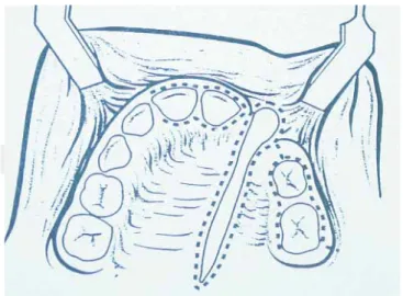
Figure 1. Schematic depiction of modifi cation of approach
and surgical technique described by Phillipe J. Boyne (1972).
slow learning, as well as several endocrine, ortho-pedic, ophthalmologic an immunological non con-stant alterations.4
g) Robins sequence. A set of face and mouth altera-tions characterized by micrognathia, glossoptosis, and distinctive «u» shaped cleft palate. The severity of micrognathia can give rise to obstructions of the upper airway.5
In the case of environmental factors, which are con-sidered as directly linked to hereditary disorders, we beg to mention the following:
a) Infectious agents. In this section we can men-tion the most frequent: congenital rubella (togavi-rus) which, besides CLP can originate ophthalmic malformations (cataracts, chorioretinitis, glaucoma and microphtalmia; ear malformations, mainly as-sociated to deafness through lesions of the Corti organ; heart malformations which manifest through the persistence of the ductus arteriosus, septal de-fects as well as auricular-ventricular malformations and pulmonary stenosis. Also included in this sec-tion are other infecsec-tions due to cytomegalovirus, congenital toxoplasmosis (Toxoplasma Gondii) and congenital syphilis (Treponema Pallidum).6
b) Pharmacological agents. Pharmacological groups associated to this condition are: Corticosteroids (cor-tisone), benzodiazepines, anticonvulsants, thalido-mide, and antimetabolites such as aminopterin and methrotexate which antagonize folic acid metabo-lism. In this group we are also including hormonal defi ciencies, where growth hormone defi ciencies is a factor associated to CLP development. Retinoids, as well as tobacco and alcohol consumption have also been associated as etiological agents.6
c) Physical agents. In this group we mainly find Roentgen or x radiation which besides being linked to CLP cases, is associated to microcephaly, spina bifi da and alterations in limb development. The de-velopment of pathological (fever) or induced (sau-na) hyperthermia, has been in addition related to SNC alterations and neural tube defects.4
d) Other congenital agents. In this group we can mention severe oligohydramnios, which can gener-ate anterior-posterior compression on the lower jaw in the placental membrane walls. This will prevent the descent of the tongue and preclude the union of the horizontal portions of the maxillary processes. This is one of the causes for Robin s Sequence.1,4
When speaking from the epidemiological stand-point, lip and palate clefts are two different entities
which can occur either isolated or together, as has been already mentioned.7 It is considered a relatively frequent condition, and constitutes 15% of hereditary malformations, being second only to club foot and con-genital heart defects.8,9 In Subjects affl icted with CLP, 45% of cleft lip cases are associated with cleft palate, 30% of cases present only cleft lip and the remain-ing 25% present cleft palate only. Unilateral cleft lip presents a 2:1 ratio. More cases are observed in the left side, this 2:1 ratio also applies to the male: female ratio. Cleft lip proportion is 1.5:1 in males with respect to females.10
Cleft lip and palate, presented as isolated patho-logical entities, present an incidence of 30 % and 20 % respectively, the remaining 50% is accounted for in cases where both conditions appear together. We must mention that, in cases of isolated cleft palate, 40 to 50% of reported cases are associated to some sys-temic condition or congenital malformation. In cases with isolated cleft lip only 7 to 13% of cases bear this relationship and in cases of combined cleft lip and pal-ate, 2 to 11% of cases are thus related.9,10 Mean world ethnic incidence of live births is expressed as follows: a) In Mexico: 1:700
b) In Latin America: 1:650 c) Caucasia race: 1:1,000 d) Asian races: 1:500 e) African races: 1:2,000
The classifi cation of cleft lip and palate is subject to great controversy due to the multiple criteria that can be used to reach that goal. The most accepted
www.medigraphic.org.mx
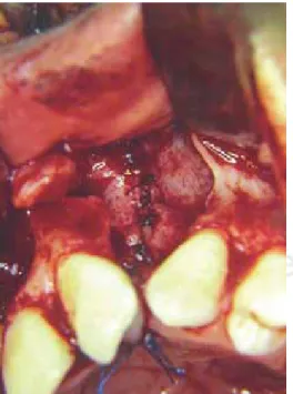
Figure 2. Technique described by Philippe J. Boyne allows
adequate visualization of the nasal floor fissure, creating thus appropriate posterior seal t.
Figure 3. A suffi cient amount of bone tissue to be grafted
in the nasoalveolar fi ssure is one of the characteristics pro-vided by this technique, and optimizes the objectives which the NAG strives for.
and established lip and palate cleft classifi cation in the course of the last 70 years are:
a) Davies and Ritchie. Clefts are grouped in three cat-egories:
I. Anterior (nasolabial) clefts. II. Posterior (palatine) clefts. III. Nasolabial and palatine clefts.
b) Veau. Four groups are formed (either numerical or alphabetical). Group 1 or A includes fi ssures of the soft palate, group 2 includes fi ssures of both soft and hard palate, group 3 or C includes defects of palate and alveolar ridge, which generally unilater-ally involve the lip, and group 4 or D which includes bilateral fi ssures.
c) Kernahan and Stark. The incisive foramen acts as division point between the two embryological enti-ties which involve CLP; they are named primary and secondary. Labiopalatine clefts are divided into four groups: A) primary labial clefts, B) soft palate clefts, C) hard palate clefts, D) palatine and lip clefts. d) The National Confederation of Plastic And
Recon-structive Surgery mentions three groups: a) alveolar and labial defects, b) primary and secondary pal-ate clefts, and c) any combination of clefts involving both primary and secondary palate.4,10
Other well accepted classifi cation and nomencla-ture is based upon the anatomical description of clefts.
Cleft lip with or without cleft palate:
a) Unilateral left clip
b) Unilateral cleft lip and palate c) Bilateral cleft lip
d) Bilateral cleft lip and palate
Cleft Palate:
a) Cleft palate
b) Submucosal palate
c) Velopharyngeal insuffi ciency d) Robin’s sequence
We must also incorporate into the classifi cations the following adjectives: 1) «incomplete» when ana-tomical extension in the labiopalatine cleft is so ex-pressed, 2) «primary» when the lip and palate cleft involves the most forward region of the premaxilla and 3) «secondary» when it affects the posterior re-gion of the apical foramen regardless of whether it affects the soft palate, the hard palate, or both.
Treatment of palate and lip clefts involves many dis-ciplines. We can divide treatment into two broad
cat-www.medigraphic.org.mx
egories: surgical and non surgical. The scope of thepresent paper will only describe the first mentioned category.
Surgical treatment for cleft lip and palate can be classifi ed into two stages: primary and secondary.
Primary surgical treatment encompasses the clo-sure of cleft lip and palate (primary cloclo-sure) as well as a nasal surgical review and the correction of naso-pharyngeal insuffi ciencies. These events are normally carried out during the patients fi rst fi ve years of life. Nevertheless, some authors mention that primary pro-cedures must be limited to the closure of cleft lip and palate. Specifi cally, with respect to primary closure of cleft lip, there is the trend to perform an early closure, at the postnatal stage. This due to the fact that the phoetal collagen levels are still high, and this guaran-tees a better connective tissue regenerations, avoiding thus the development of conspicuous scarring. Like-wise, the presence of considerable amounts of mater-nal immunoglobulin and natural plasma corticosteroids reduce possible postsurgical complications. Another advantage of cleft lip early primary closure is that it al-lows for better patient feeding, and parents and family experience psychological acceptance.4 Nevertheless, not all reports on primary closure have been positive. Some authors have initiated controversy pointing out to the possibility of complications occurring during an-aesthesia due to the relatively larger amount of drugs required, greater diffi culty when intubating as well as the smaller anatomical dimensions of the cleft lip. This admits then to consider a criterion of a longer waiting time for this primary closure. Among other elements, Wilhelmsen and Musgrave 196615 «rule of ten» can be observed (a minimum of 10 mg hemoglobin, at least 10 pounds of weight, up to ten thousand leukocytes and 10 weeks of life). In an almost parallel manner, Veau suggested that the optimum time for the proce-dure was when the patient reached 18 months of age, supporting his claim by stating that this is the period in which the patient initiates the process of language. Nevertheless, other studies showed that an early clo-sure can be performed from 6 months onwards, based on the development of an improved muscular activity of the soft palate muscles, a better velopharyngeal competence which results from a smaller scar fl ange applied to the soft tissues of the cleft palate. The early closure of the hard palate considerably compromises maxillary anteroposterior growth; for these reasons it has been suggested to postpone closure until the patient reaches 18 months of age. The main objec-tives to strive for in a primary closure process are: a) optimize feeding, b) improve airway, especially in syn-dromic associations or cases which might imply
man-dibular retrognathia, c) treatment of middle ear condi-tions where palatine fi ssure condicondi-tions a serous otitis due to the sphincter muscle insuffi ciency of the mouth of the auditory tube d) decrease of velopharyngeal in-suffi ciency, which promotes hypernasal voice. Age is considered the main criterion when programming the closure, since speech develops fi xation in the cerebral cortex at around six years of age.
The second surgical stage, also known as second-ary, begins at around the patient´s 7 years of age. It in-volves treatment of nasoalveolar fi ssures and fi stulas (NAF), orthodontic and orthopedic correction of maxil-lary-mandibular skeletal disharmonies or defi ciencies. In these, common denominators are jaw anterior pos-terior and transversal hypoplasia, as well as the ver-tical growth of the mandible. The aforementioned is known as cleft lip and palate sequels (CLPS).12
Within this state of secondary surgical treatment, we specifically refer to NAF treatment procedures, where the bone defect elicits an important sequence of functional and aesthetic sequels, which involve soft and hard tissues and is frequently associated to the presence of nasoalveolar fi stulas.
One of the fundamental principles of NAF treatment is the closure of fi stulas whenever present, and their reconstruction with a bone graft.
Through the years, several materials have been used to reconstruct human bone defects. In 1901, Dr Von Eiselberg, considered one of the pioneers of NAF treatment, applied a pedicle bone graft to correct an alveolar defect.14 At a later date, in 1907, Dr Axhausen performed one of the greater contribution to the area of bone transplant and osteogenesis: He formulated the biological principle that the periosteum stimulated osteogenesis cellular index from certain autologous grafts. This ocurrence is considerably low in the event of heterografts and practically nil in xenografts. His research provided the bases upon which bone graft biology, as we nowadays know it, was developed.3,14 In 1914, Dr Drachter performed the fi rst attempt of a nasoalveolar bone graft for the reconstruction of NAF.
NAF reconstructions through autologous primary bone graft application were very popular during the 50’s, in 1955 specifi cally with Dr Nodin, who is con-sidered pioneer of NAF reconstruction with autologous graft. He performed this procedure in an attempt to prevent maxillary collapse, achieve transversal sta-bility, and at the same time allow optimal craniofacial growth, thus facilitating eruption of primary teeth at the graft site. In spite of these expectations, Dr Jol-ley, in 1968, determined that, in the long run, patients showed considerably compromised facial growth and high incidence of malocclusion phenomena.11
www.medigraphic.org.mx
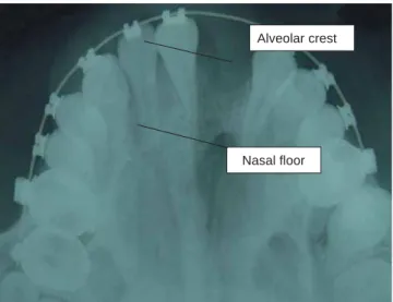
Alveolar crest
Nasal fl oor
Figure 4. The space found between the two lines indicated
in x-ray image determine classifi cation through visual crite-rion in degrees I, II, III or IV. This image represents an ex-ample of grade II.
After this trend, came the technique of primary surgical periosteal surgery. In it, only closure of soft tissues was created, with the aim of promoting bone growth at the fi ssure site through the biological prin-ciple of stimulation or periosteal induction. This aim was partially met when obtaining narrow bone bridges which reconstruct the fi ssure with the advantage of not disturbing facial growth. Nevertheless, the amount of newly formed bone was not suffi cient to cover neces-sary objectives or expectations, and neither was it suf-fi cient to meet requirements or objectives for a nasoal-veolar reconstruction graft.8
In the decade of the seventies, secondary recon-struction of nasoalveolar fissures gains new impor-tance with the 1972 publication of surgeons Phillipe J. Boyne and N.R. Sands: «Secondary Bone Grafts in Palatine and Alveolar Clefts». In this essay, NAF secondary reconstruction concepts are reinforced and a new technique is presented (Figure 1). This tech-nique allows, among other advantages, an appropri-ate closure of the nasal fl oor through the dissection of the mouth´s nasal mucosa. It also affords application of a more considerable volume of bone tissue, which grants better expectations of the NAG. This subject will be dealt with at a later moment. Due to all the afore-mentioned reasons, this technique is then considered part of the protocol for treatment of NAF with NAGin the present study.
As mentioned before, establishing biological bases for a bone graft allowed the determination of ideal cri-teria and properties for bone reconstruction macri-terials. These are: 1) biocompatibility, 2) viability, 3) osteo-genic potential, 4) bone matrix neoformation and 5) mechanical stability.
Through biological development and clinical evo-lution, these criteria have indicated that autologous bone is a primary choice for bone reconstruction.
We also deem important to mention the nasoalveo-lar graft classifi cation. NAG classifi cation based on ap-plication chronology can be as follows:
Early secondary bone grafts: Early secondary
bone grafts are performed during the primary dentition phase, which varies from 2 to 5 years of age. Pres-ence of bone tissue to allow eruption of primary denti-tion as well as provide appropriate periodontal health is one of the fundamental considerations for this early graft. Notwithstanding, in patients subject to this type of grafts, some alterations and defi ciencies in hemifa-cial growth and development have been observed.
Secondary bone grafts: Secondary bone grafts
are applied during the mixed dentition stage, which varies between 6 and 12 years of age. Most authors
consider this the optimal moment to place a graft, since the bone provides support for the eruption of the permanent cuspid at the site of the fi ssure, it also pro-vides suffi cient bone tissue to give appropiate height to the alveolar process. In these cases there is expec-tation of minimal interference with facial growth.12
Late secondary bone grafts: Bone graft
require-ments in a skeletally mature patient are much lesser than those of a young one. Te need for bone tissue to allow dental eruption is made unnecessary. No al-teration whatsoever in facial growth is observed in this type of grafts.12
It was previously mentioned that NAF reconstruc-tion is part of LPF sequels treatment. An important factor among these sequels will depend directly on the development of an hypoplasia, a hypofunction, or a combination of both, in hard and soft tissues of the anatomical regions involved in the fi ssures. Therefore, the main objective of the surgical correction of these deformities or sequels must focus on the re-establish-ment of a proper physiology which will later allow opti-mal facial growth.8
For all the aforementioned reasons, nasoalveo-lar bone grafts must ideally comply with the following characteristics:
1) Allow the closure of the nasoalveolar anterior fi s-tula.
2) Provide suffi cient bone tissue to the fi ssure site, not only to allow the proper eruption of the permanent
www.medigraphic.org.mx
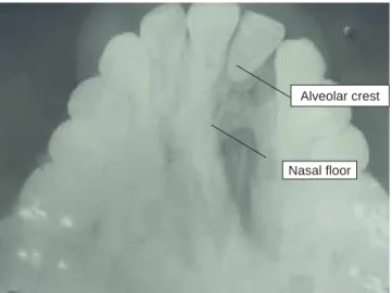
Alveolar crest
Nasal fl oor
Figure 5. Radiographic image shows an example of
Nasoal-veolar graft with grade III bone integration.
cuspid but also provide maxillary stability (premax-illa) and provide appropriate root support to teeth involved in the fi ssure.
3) Provide a proper bone architecture (continuity) to the premaxilla region, providing at the same time optimal lip support.
4) Establish a functional airway in the nasal cavity in-volved with the lip and palate fi ssure.
5) Provide appropriate bone volume to allow the best possible dental rehabilitation.1,12
Among controversial points related to nasoalveolar bone grafts the following can be mentioned: a) surgical time required for NAF reconstruction, b) type of bone and donor site to be used for the graft, c) orthodontic and orthopedic aspects involving the fi ssure site such as maxillary expansion for the correction of anterior-posterior discrepancies.11
In this study we are going to stress on donor bone types and sites.
We have already mentioned there is an ample ar-ray of materials taken from the human body for the reconstruction of bone defects. Two main groups can be observed:
• Bone grafts or transplants which are divided ac-cording to their origin into autologous grafts (those harvested from the same individual where the graft is going to be applied), homologous grafts (those obtained from different subjects belonging to the same species) and heterologous grafts (obtained from subjects of a species different from that of the receptor). These biological materials are subject to
varied processes like freezing, deminerilization or lyophilization, which allow, among other properties, to modify the graft’s antigenic capacity. The second group encompasses aloplastic materials, these are all of synthetic or semi-synthetic materials which al-low to achieve bone reconstruction. Among these we can mention hydroxyapatite of even synthetic bone.
Research and experience of several authors have demonstrated that autologous bone grafts cover the best part of expectations for bone reconstruction grafts. This quality is mainly due to the ability of the bone to integrate to the recipient bed through the bio-logical processes of osteogenesis, osteoconduction and osteoinduction.12
Within the category of autologous bone grafts we can fi nd three variants: a) bloc non vascularized corti-cal cancellous bone in where the transplants consists of greater amount of bone mineral matrix than bone cellular components; b) vascularized cortical cancel-lous bone depending for its application on anastomo-sis of vascular tissues. This has to bear the disadvan-tage of the size and anatomical shape of the donor site, such the fi bula or rib, c) cortical particulate bone or bone marrow which provides adequate cellular den-sity to the graft, mineral components, as well as the bone morphogenetic protein needed for the graft’s os-teoinduction.
We hereby provide a list of autologous donor sites for maxillofacial region bone reconstruction deemed adequate due to their histological and ossification characteristics as well as to their approach site and convenient amount of bone to harvest:
1) Iliac crest upper border and lateral, anterior and posterior sides.
2) Cranial parietal eminence 3) Mid-lateral side of the tibia
4) Rib (cortical cancellous or costochondral) 5) Chin region
The iliac crest has been systematically used for maxillofacial region bone reconstruction. It presents advantages, since it is possible to obtain from it the three types of bone we have mentioned (cortical, cancellous and corticalcancancellous) and it provides suffi -cient amount of material. The posterior region of this bone is indicated to obtain larger volumes. The two main disadvantages of using this donor site are based on the fact that this a growth center, which could result affected with the process of donation and the morbid-ity associated to approach a harvest of the graft. In
www.medigraphic.org.mx
Este documento es elaborado por Medigraphic
the fi rst case, the harvesting of the graft in a growing and developing patient, the surgical technique is modi-fi ed so as to preserve the growth cartilage located at the upper and anterior portion of the iliac crest. In the second case, main morbidity is associated to the le-sion of the iliac or gluteus maximus muscles, which, in the patient, elicits pain when walking. Nevertheless, a meticulous tissue dissection and a conservative su-ture technique will allow adequate reposition of these structures and decrease risk of complications.2
For the aforementioned reasons autologous na-soalveolar bone grafts harvested from the iliac crest are the best alternative for the reconstruction of bone defects of NAF in CLPS patients. Finally concerning the degree or determination of amount of bone present in the grafted NAF, or the degree of bone integration, the evaluation of NAG is based on Bergland O. and Sembs 1986 classifi cation3 as well as on the modifi -cation and re-proposal developed by Brusati and Ga-rattini, published in 2000.5 In this latter, classifi cation is determined by the radiographic appreciation of the NAF occlusal projection and its perspective of bone volume based upon the observable radiopacity in the NAF space, as well as its relationship with nostrils and alveolar ridge. All the aforementioned facts allowed the authors to establish the following classifi cation:
Type 1: Bone graft with complete fi ssure ossifi
ca-tion and reconstrucca-tion from the fl oor of the nostrils up to the entirety of the alveolar ridge (100%).
Type 2: Ossification is observed from the nasal
fl oor, reaching a height of three quarters of the alveo-lar ridge (75%).
Type 3: Graft ossifi cation encompasses from the
nasal fl oor up to a height lesser than two thirds of the height of the alveolar ridge (50%).
Type 4: Ossification is limited to a scant bone
bridge amounting to a third of the required bone vol-ume (25% or less).3,5
See fi gure 2.
Within the important aspects of surgical treatment of NPF we must include handling of possible post surgical complications or sequels. Within the scope of nasoalveolar grafts, the most common complica-tions are soft tissue dehiscence and the resulting bone tissue exposition in the oral cavity; the development of a sinus infectious process which can compromise bone integration process. In the fi rst case, it is more frequent in large NAF (over 10 mm) where oral mu-cosa fl aps can be insuffi cient for NAF closure and re-covering. On the other hand, factors like eating habits and hygiene among others, are elements which can
infl uence in the development of soft tissues as well as in the process of localized infectious process. Devel-opment of these complications negatively modifi es the objectives sought when closing a nasoalveolar fi stula and NAF bone reconstruction. At the beginning of this paper, it was mentioned that the objectives sought with this reconstruction, nevertheless it must be real-ized that these aims are rarely met at 100%. Within NAF closure and reconstruction protocols the following characteristics are considered the minimum required to consider the nasoalveolar graft as a success: 1) Ap-propriate closure of nasoalveolar fi stula, 2) Suffi cient bone tissue integration to allow maxillary stabilization in the premaxilla and 3) eruption of permanent cuspid and/or its later prosthetic rehabilitation. For clarity of expression purposes, «suffi cient degree» is consid-ered for those NAG which can hold degree I, II or III in the Brusatti and Garatini classifi cation. Those deemed «not suffi cient» are those determined as grade IV.
PROBLEM APPROACH
Surgeons of the maxillofacial surgery service (MSS) of Mexico’s Children’s Hospital (Hospital Infantil de México) MCH have been performing an average of 160 surgeries in the last ten years. These surgical events are related to the closure and reconstruction of nasoalveolar fi ssures (NAF) taken from iliac crest autologous secondary bone grafts, as part of the reha-bilitation protocol and treatment of patients with CLPS. These interventions represent over 40% of all surgical procedures performed by staff of this Center. The re-quirements for scientifi c and statistical documentation as well as results to back up this therapeutic conduct show the need for a study that will sustain and pres-ent important elempres-ents that might corroborate, or, in a given case, modify treatment protocol for NAF recon-struction and closure procedures. This study aspires to contribute statistically signifi cant information on the degree of bone integration developed in patients sub-ject of NAF closure and reconstruction processes as well as factors that might modify them.
The aforementioned can be expressed in the follow-ing fashion:
a) How many patients have developed bone integra-tion with secondary nasoalveolar grafts of the iliac crest in surgical interventions performed at the Mex-ico’s Children’s Hospital’s maxillofacial service dur-ing 1991 to 2001? And what if any is the degree of bone integration in operated subjects.
b) Which factors such as age (within the secondary graft rank), site ( side) of the fi ssure and gender of
www.medigraphic.org.mx
Count Complication No 87 15 Sí 100 80 60 40 20 0 Degree Suffi cient degree Insuffi cient degreeIn this table we determine that 87 % of patients did not develop com-plications with appropriate level or degree of bone integration, and 15% of patients developed some complication with suffi cient de-gree of integration. It was then determined that there is dependency among variables corresponding to patients with suffi cient degree of NAG either with or without complications.
Figure 6. Complications vs nasoalveolar grafts
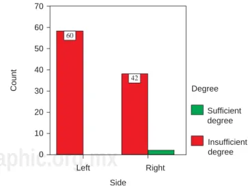
Count 60 42 Side Left Right 70 60 50 40 30 20 10 0 Degree Suffi cient degree Insuffi cient degree
This table shows that there was no dependance among variables found. The side of the nasoalveolar fi ssure and the degree of na-soalveolar graft bone integration, where 100% (60) of left NAG pre-sented suffi cient degree of integration when compared to 95.5% (42) of right NAG which presented similar amount of integration.
Figure 7. Side vs nasoalveolar graft degree of bone
integra-tion.
patient could be associated to the NAG bone inte-gration degree?
OBJECTIVES
A) General Objective:
To ascertain the degree of bone integration in pa-tients with CLPS subject of secondary nasoalveolar grafts of the iliac crest at the maxillofacial surgery service of the children s hospital in Mexico City dur-ing the period 1991 to 2001.
B) Specifi c objectives:
1) To obtain frequency of iliac crest secondary na-soalveolar bone grafts performed ond on pa-tients of the Maxillofacial Service of the Mexi-co s Children s Hospital in the period between 1991 and 2001, and the number of these which developed any degree of bone integration 2) To ascertain whether there is any relationship
among factors like age and gender of patient, location of the fi ssure (side) and the degree of bone integration.
HYPOTHESIS
1) 80% of all NAF reconstruction performed with iliac crest secondary graft at the maxillofacial service of the Mexico, s Children’s Hospital developed some degree of bone integration.
2) 70% of patients who experienced some degree of bone integration are within the rank of functional grafts corresponding to degree I, II and III.
RESEARCH DESIGN
a) Type of Research: The present study has been deemed documental since it is based on written and radiographic records of clinical fi les of CLPS, operated for NAF reconstruction at the Maxillofacial clinic of the Mexicos Children hospital. The study is also retrospective since already performed surgical procedures are subject of research. It is as well lon-gitudinal because the study encompasses patients subject of surgical intervention during the period from 1991 to 2001.
b) Consideration of variables: Variables to be applied to this study are qualitative and quantitative.
1) The independent variable is the degree of bone integration developed in patients subject of a graft.
2) Dependent variables are the following: i. Side of the cleft (right or left)
ii. Age of patients subject of the surgical proce-dure
iii. Sex of patients subject of the surgical proce-dure
www.medigraphic.org.mx
Count ID I 56 33 13 2 II III IV 60 50 40 30 20 10 0 IV 2.00/1.9% III 13.00/12.5% II 33.00/31.7% I 56.00/53.8%This table shows the existence of a high success rate in nasoalveolar grafts with an comprehensive total of 98.1% of grafts with suffi cient amount of bone integration. 53.8% correspond to grade I, 31.7% to grade II, 12.5% to grade III and only 1.9% to grade IV. Grade IV is consid-ered insuffi cient degree of bone integration.
Figure 10. Subject number and percentage according to developed degree of bone integration
Count Gender 50 52 Male Female 60 50 40 30 20 10 0 Degree Suffi cient degree Insuffi cient degree
No dependency was found in correlation of gender and bone inte-gration degree. A 100% of male patients with suffi cient degree of bone integration was found as compared to 96.3% of female pa-tients with similar bone integration degree.
Figure 9. Gender vs nasoalveolar graft degree of bone
in-tegration. Count 38 64 Age 7 a 10 11 a 14 70 60 50 40 30 20 10 0 Degree Suffi cient degree Insuffi cient degree
Comparative bone integration degree with age does not present sta-tistical signifi cance between both groups (7 to 10 years of age and 11 to 14 years of age) since 95% of patients belonging to the fi rst group (7 to 10 years of age) presented satisfactory degree of bone integration, whereas 100% of the second group (11 to 14 years of age) presented the same satisfactory degree of bone integration.
Figure 8.Age vs nasoalveolar graft degree of bone
integra-tion.
iv. Development of complications 1. Postoperative re-fi stulization of the NAF.
2. Development of infectious rino-sinusite process. c) Material: Clinical fi les and offi ce studies (occlusal
radiographs) of unilateral CLPS patients operated at the MFC of the MCH.
d) Method: An initial selection of all fi les was carried out. These fi les encompassed all patients operated for reconstruction and closure of NAF from 1991 to 2001. 176 fi les were obtained. A second selection was later performed in which to admit only patients with diagnose of unilateral CLPS, with reconstruc-tion based solely on autologous bone harvested from the iliac crest, and whose age corresponded to a secondary graft. Finally all cases which lacked
www.medigraphic.org.mx
suffi cient radiographic fi les to allow evaluation withor without the characteristics just mentioned were precluded, leaving thus a an encompassing sample of 104 fi les. Clinical fi le evaluation was initiated, in which the age of patient at the time of the graft was determined. Those found in the lower section of the age rank (7 to 10 years) and those in the higher section (11 to 14 years) were sub-grouped. Other factors considered were gender of patient, side of the NAF and postoperative evolution during the six months following the nasoalveolar reconstruction. Results were included in a resume table. Radio-graphic characteristics of the graft will allow the to be initially classifi ed as:
1) Integrated 2) Non integrated.
The fi rst classifi cation is based upon radiographic evaluation, which establishes presence or absence of calcifi ed integrated bone tissue (radio opacity) in the NAF zone, regardlessly of the amount, density or in re-lation to other anatomical sites. The second classifi ca-tion is considered when the radiographic image does not show any trace of calcifi ed bone material (radiolu-cency) present in the NAF. In the array of NAF, once selected those with presence of bone integration, the amount or degree of bone present must be assessed. This can be determined with a second radiographic assessment, to determine the amount of bone present with the total NAF area, and can be assessed follow-ing criteria mentioned by Brusati and Garattini.13
The assessment was undertaken by three members of the MFS of the MCH. Their assessments were averaged.
Finally, results were analyzed applying Chi Square statistical tests, to assess the hypothesis with respect to variables. Fisher’s Exact Test was used to deter-mine, at every step, dependence among analyzed variables. Phi test was used to determine whether the cases are suffi cient for the analysis. These three tests were applied with STATS v.2. software.
e) Inclusion criteria:
1) Patients who have received a graft at the cor-responding age to secondary grafts, with autol-ogous bone harvested from the iliac crest, with procedure performed at the Maxillofacial Service of the Mexico s Childrens Hospital during the pe-riod encompassed between 1991 and 2001. 2) Patients with unilateral CLPS.
3) Patients with full (complete) radiographic and clinical fi le.
f) Exclusion Criteria:
1) Patients with bilateral CLPS.
2) Patients not operated at the MFS of the MCH. 3) Patients nasoalveolar grafts are considered
pri-mare or late secondary.
4) Patients whose grafts are not considered autol-ogous in type and harvested from the iliac crest. 5) Patients with insuffi cient radiographic fi le.
RESULTS
One hundred and four clinical and radiographic fi les corresponding to the 1991 to 2001 period were examined. 44.26% (46) corresponded to female pa-tients and 55.76% (58) to male papa-tients. Average age of patients was 11.52 years, with maximum 15 years of age, minimum 8.
With respect to anatomical location of fissures, 59.61% (62) were on the left side and the remaining 40.48% (42) on the right side.
Total number of grafts presenting sufficient or functional bone integration grades I, II, and III) cor-respond to 98.07% (102) of cases and the remain-ing 1.98 % (2) to grafts which did not develop suffi-cient amount of bone integration (grade IV). These two cases where the graft was considered insuf-ficient were a consequence of rino-sinusitic condi-tion sitis).
Quantitative correlation of the independent variable with dependent variables is as follows:
1) Total number of grafts with grade I integration: (53.84%) (56) a) Gender a. Female : 57.14 (32) b. Male: 42.85% (24) b) Side: a. Left: 57.14% (32) b. Right: 42.85% (24)
c) Age average 9.3 years (max 15, min. 8) d) Complications: None.
2) Total number of grafts with grade II integration: a) Gender: a. Female: 43.75 (14) b. Male: 56.25% (18) b) Side: a. Left: 68.75% (22) b. Right: 31.25 (10)
c) Age average: 10.8 years (max: 14, min: 8). d) Complications: 7 (21.8%)
3) Total number of grafts with grade III integration: 13.46 % (14)
www.medigraphic.org.mx
a) Gender: a. Female: 42.85% (6) b. Male: 57.14% (8) b) Side: a. Left: 14.2% (4) b. Right: 71.42% (10)c) Age average: 12 years (max: 14, min: 11) d) Complications: 8 (57.14%)
4) Total number of grafts with grade IV integration: 1.9% (2) a) Gender: a. Female: 0% b. Male: 100% (2) b) Side: a. Left: 100% (2) b. Right: 0%
c) Age average: 9 years (10 and 8) d) Complications: 2 (100%).
See fi gures 6 to 9.
DISCUSSION
Within the scope of the studied sample, 83.3% (87) cases did no present local postoperative complica-tions. All of them experienced suffi cient degree of bone integration. The remaining 1.9% which did present complications did not attain suffi cient degree of bone integration. When establishing a comparison with the 2000 study performed by Brusati and Garattini which mentions the degree of bone integration developed in a 70 subject sample, and where 100% of cases devel-oped a suffi cient degree of bone integration. In those cases presenting right NAF only 95.5% of cases expe-rienced suffi cient amount of bone integration.
With respect to this study, the development of NAG complications were classifi ed into two groups: those that, complications notwithstanding, developed suffi -cient or satisfactory degree of bone integration, and those which did not achieve this goal. The difference is statistically signifi cant.
Subject of the study, were also divided into two groups according to age. The first group encom-passed patients ages 7-11, second group comprised patients ages 11-14. In the fi rst group (37.3% of the sample) it was determined that 95% of cases psented suffi cient degree of bone integration, the re-maining 5% did not achieve this goal. In the second group, (62.7% of the sample) 100% presented suf-ficient integration degree. The gender of the sam-ple population was determinant, since it revealed that 100% of female patients (48.1% of the sample) showed suffi cient degree of bone integration. 96.3%
of male patients developed suffi cient degree of bone integration, and the remaining 3.7% did not.
See fi gures 10 and 11.
The application of the technique described in 1972 by Dr Phillipe J Boyne for the reconstruction of NAF such as CLPS resulted effi cient in patients with uni-lateral LPF, since it permits to obtain bone integration degrees deemed suffi cient for the purposes of naso-alveolar grafts. On the other hand, the use of bone harvested from the iliac crest as reconstruction mate-rial resulted equally successful. It has been demon-strated that there is a signifi cant degree of depen-dence among patients with suffi cient degree of bone integration and the development of complications, especially those related to infection. The question of comparative studies among different techniques for NAF closure in comparison to the one here presented also remains on the table. Also of interest would be the attainment of comparative studies of different re-construction materials.
REFERENCES
1. Johnson DC. Cleft lip and palate. In: Behrman RE, Kliegman RM, Arvin AM, and editors. Textbook of pediatrics. 15th edition.
Madrid (ES) McGraw-Hill. 1997; I: 1312.
2. Bloomquist DS, Turvey TA. Bone grafting in dentofacial deformi-ties. In: Bell WH. Modern practice in orthognatic and
reconstruc-tive surgery. 2nd ed. Philadelphia: W.B. Saunders; 1992: 834-35,
839-841.
3. Bergland O, Semb G, Abyholm FE. Elimination of the residual alveolar cleft by secondary bone grafting and subsequent orth-odontic treatment. Cleft Palate Craniofacial J 1986; 23: 175. 4. Kernahan DA, Rosenstein SW. Cleft lip and palate. Baltimore,
Maryland Williams & Wilkins; 1990: 13-18, 115-118.
5. Brusati R, Garattini G. The early secondary gingivoperiostoplas-ty. Oral and Maxillofacial Surgery Clinics of North America 2000; 3: 443-453.
6. Cohen MM. Etiology and pathogenesis of orofacial clefting.
Oral and Maxillofacial Surgery Clinics of North America 2000;
3: 361-376.
7. Bauer BS, Vicari FA. Cleft palate. In: Georgiade GS. Gregory NG, Riefkhol R, Barwick WJ. Textbook of plastic, maxillofacial
and reconstructive surgery. 2nd ed. New York: Wiliams & Wilkins
1992: 299-306.
8. Boyne JP. The evolution of guided tissue regeneration, in: al-veolar ridge reconstruction/guided tissue regeneration and bone grafting. Oral and Maxillofacial Surgery Clinics of North America 2001; 13: 3.
9. Marx RE. Philosophy and particulars of autogenous bone graft-ing. Oral and Maxillofacial Surgery Clinics of North America 1993; 4: 599-612.
10. Optiz, Meier, Stoll. Subklew radiographic evaluation of the trans-plant bone height in patients with clefts of the lip, alveolus palate, after secondary bone grafting. Journal of Orofacial Orthopedics 1999; 60 (6): 383-91.
11. Bardach J, Kenneth ES. Técnicas quirúrgicas en labio y paladar
hendidos. Medilibros 1989, Madrid, España.
12. Turvey TA, Fonseca RJ. Facial clefts and craniosinostosis,
www.medigraphic.org.mx
13. Amin K, Jefrey WS, Raymod JF. Secondary grafting in the alveo-lar cleft patient. Oral And Maxillofacial Surgery Clinics of North
America 2002; 4: 477-489.
14. Regezi JA, Sciubba JJ. Oral pathology, clinical pathologic
cor-relations. 3ª ed. México, D.F: McGraw-Hill; 2000.
15. Vig WL, Turvey TA, Fonseca RJ. Orthodontic and surgical con-siderations in bone grafting in the cleft maxilla and palate. In:
Surgical management of craniofacial deformities. New York: WB
Saunders 1998; 2.
Correspondence address:
Alejandro Montaño López