TECHNOLOGICAL INSTITUTE OF COSTA RICA
SCHOOL OF BIOLOGY
The University of Texas at Austin
Molecular Cell and Developmental Biology
The Gross Lab
THE ROLE OF CUGBP1 IN THE DEVELOPMENT OF
ZEBRAFISH LENS
Report of Final Graduation Work
In Partial fulfillment of the Requirements for the Degree of
Bachelor of Biotechnology Engineering
Linette Pérez-Campos
THE ROLE OF CUGBP1 IN THE DEVELOPMENT OF ZEBRAFISH
LENS
Abstract
The lens is a transparent tissue in the anterior of the eye and its main role is to refract light on the retina. The lens consists of two types of cells: epithelial cells and fibers. Epithelial cells surround the anterior and lateral limits of the lens, remain proliferative and at the equator of the lens they differentiate into lens fibers. In this process newly generated lens fibers elongate and gradually lose their organelles, enabling transparency. Cataracts are any opacification of the lens that compromises its ability to refract light onto the retina, and can be genetic or environmentally induced.
Since it has been demonstrated that the zebrafish (Danio rerio) is an ideal organism to study human ocular disorders, this model system was utilized to study gene products that regulate normal lens development and that in pathological states contribute to cataracts. CUGBP1 is an mRNA binding protein that has been implicated in the multisystemic disease Myotonic Dystrophy 1 (DM1). DM1 is caused by a (CTG)n repeat expansion within the 3'UTR region of the DMPK gene. Its mechanism implies a toxic gain of function where
expanded CUG mRNA repeats increase steady state levels of CUGBP1 protein among other effects. Patients with DM1 develop cataracts. So, it can be hypothesized that CUGBP1 disrupted expression in lenses from DM1 patients can be, at least, one of the causes that leads to cataracts in this disease.
In situ hybridization results show that cugbp1 is expressed in the zebrafish lens at early embryonic
development in newly formed lens fibers. Transgenic embryos expressing nuclear or membrane localized-EGFP under the control of a 1.2kb cugbp1 promoter further demonstrate its expression in the lens. Knocking down
expression of cugbp1 with a splice-altering morpholino results in cataracts as early as 3dpf. Hence, the latter reveals that cugbp1 expression is a requirement for normal lens early development. In morphant embryos, lens fiber compaction is disturbed. In addition, these cells retain nuclei. Lens overall shape and size is also affected. Furthermore, the defective phenotype includes a general developmental delay, little mobility and dilated cardiomyopathy, symptoms that are also observed in DM1 patients.
EL ROL DE CUGBP1 EN EL DESARROLLO DEL CRISTALINO DEL
PEZ CEBRA
Resumen
El cristalino (lente del ojo) es un tejido transparente en la región anterior del ojo y su rol principal es refractar la luz sobre la retina. El cristalino está constituido por dos tipos de células: células epiteliales y fibras.
Las células epiteliales rodean la parte anterior y los límites laterales de la lente del ojo, mantienen su capacidad de proliferación y en el ecuador del cristalino se diferencian para generar fibras. En este proceso, las fibras recién generadas se elongan y gradualmente pierden sus organelas, permitiendo así la transparencia. Las cataratas se refieren a cualquier opacificación del cristalino que comprometa su habilidad de refractar la luz hacia la retina y pueden ser inducidas por la genética o el ambiente.
Debido a que se ha demostrado que el pez cebra (Danio rerio) es un organismo ideal para estudiar desordenes oculares humanos, este sistema modelo se utilizó para estudiar productos génicos que regulan el desarrollo normal de la lente y que en estados patológicos contribuyen a la aparición de cataratas. CUGBP1 es una proteína de unión a ARNm que ha sido implicada en la enfermedad multisistémica Distrofia Miotónica 1 conllevan a la formación de cataratas en esta enfermedad.
Resultados de ensayos de hibridación in situ muestran que cugbp1 se expresa en la lente del ojo del pez
cebra durante el desarrollo embrionario temprano en fibras recién formadas. Embriones transgénicos que expresan la proteína verde fluorescente co-localizada en el núcleo o la membrana celular bajo el control de una región promotora de 1.2kb del gen cugbp1 evidencian aún más su expresión en el cristalino. El disminuir la expresión (knock down) de cugbp1 con un morfolino que altera el proceso de corte y empalme (splicing) resulta en la formación de cataratas a partir de los 3 días después de la fertilización. Por lo tanto, lo anterior revela que la
expresión de cugbp1 es un requerimiento para el desarrollo temprano normal de la lente del ojo. En embriones inyectados con el morfolino, la compactación de las fibras del cristalino se ve perturbada. Además, estas células retienen el núcleo. La forma y el tamaño generales de la lente del ojo también se ven afectados. Asimismo, el fenotipo defectuoso incluye un retraso general en el desarrollo, poca movilidad y miocardiopatía dilatada, síntomas que también se observan en pacientes con DM1.
THE ROLE OF CUGBP1 IN THE DEVELOPMENT OF ZEBRAFISH
LENS
Accreditation
Report of final graduation work presented to the School of Biology
Costa Rica Institute of Technology
In partial fulfillment of the requirements for the Degree of
Bachelor of Biotechnology Engineering
Tribunal Members
____________________________
M.Sc. María Clara Soto-Bernardini Professor Costa Rica Institute of Technology
____________________________
Ph.D. Jeffrey Gross
Professor University of Texas at Austin
____________________________
Dedication
To my mother, that even though we think very differently,
Acknowledgements
I would like to thank Dr. Jeffrey Gross for allowing me to perform this amazing and
interesting project in his laboratory.
Thank you to Julie Hayes for always guiding me in the experimental procedures of this
project, teaching me how to work in a laboratory and always having a good and helpful
attitude.
Thank you to Professor María Clara Soto-Bernardini for guiding me through the steps of the writing process of this project.
To Richard Nuckels, Julie Hayes and Jeffrey Gross thank you for performing previous
valuable work in this project.
To Rosa Uribe thank you for always helping me and teaching me experimental
procedures.
Thank you to all The Gross Lab members for always answering my questions and helping me
along the way.
Thank you to Professor Olga Rivas-Solano for being the lector of this project.
Thank you to all my academic professors at the Biology School for teaching me
theoretical and basic scientific knowledge.
Thank you to my mother and father for always supporting me and being helpful
General Index
Abstract 2
Resumen 3
Accreditation 4
Dedication 5
Acknowledgements 6
General Index 7
Index of Figures 10
Index of Annexes 11
Chapter 1. Introduction 12
Chapter 2. Research Aims 14
2.1 General objective 14
2.2 Specific objectives 14
Chapter 3. Theoretical Framework 15
3.1 Zebrafish lens early development 15
3.2 Differences between mammalian and zebrafish lens development and early morphology
19
3.3 Lens fiber cells differentiation 21
3.3.1 Aquaporin0 (AQP0) expression and function 22
3.3.2 Lens actin cytoskeleton 24
3.3.3 Organelle degradation in lens fibers: emphasis in nuclei 29 3.4 CUG binding protein 1 (CUGBP1): an mRNA binding protein 32
3.5 Myotonic dystrophy 1 (DM1) 33
3.5.1 CDM phenotype 33
3.5.2 DM1 phenotypes 34
3.5.3 Molecular mechanisms of Myotonic dystrophy 1 36 3.5.4 CUGBP1 and its involvement in DM1 models and tissues 40
3.6 CUGBP1 and early development 41
Chapter 4. Materials and Methods 55
4.1 RNA in situ hybridization with digoxigenin-labeled probe 55
4.1.1 cugbp1 cDNA cloning 55
4.1.2 cDNA sequencing and protein sequence alignment 55
4.1.3 Digoxigenin-labeled probe synthesis 56
4.1.4 In situ hybridization 56
4.2 cugbp1 promoter activity in microinjected and transgenic zebrafish embryos
4.3 cugbp1 down regulation by splice-altering morpholino injections 61 4.3.1 Splice-altering morpholino injections 61 transverse cryosections from embryonic eyes of previously injected cugbp1-MO and cugbp1-MM embryos and statistical analysis
63
4.3.5 Aquaporin0 (Aq0) immunohistochemistry on transverse cryosections from embryonic eyes of previously injected cugbp1-MO and cugbp1-MM embryos
64
4.3.6 F-actin staining on transverse cryosections from embryonic eyes of previously injected cugbp1-MO and cugbp1-MM embryos
64
4.3.7 Nuclei staining on transverse cryosections from embryonic eyes of previously injected cugbp1-MO and cugbp1-MM embryos
65
Chapter 5. Results 66
5.1 RNA in situ hybridization with digoxigenin-labeled probe 66 5.1.1 cugbp1 cDNA sequencing and protein sequence alignment 66 5.1.2 In situ hybridization to detect mRNA expression 66 5.2 cugbp1 promoter activity in microinjected and transgenic zebrafish
zebrafish embryos marker of early fiber differentiation after cugbp1 down regulation
75
6.1 cugbp1 expression on zebrafish early lens development 84 6.1.1 cugbp1 cDNA sequencing and protein sequence alignment 84 6.1.2 cugbp1 mRNA expression and promoter activity 84 6.2 cugbp1 down regulation by splice-altering morpholino injections in
zebrafish embryos
Chapter 7. Recommendations and Future Directions 109
7.1 Identification of Cugbp1 RNA targets at the developing zebrafish lens 109 7.2 Reversal of cugbp1 morpholino phenotype by RNA rescue 112
7.3 Other experiments 113
References 114
Index of Figures
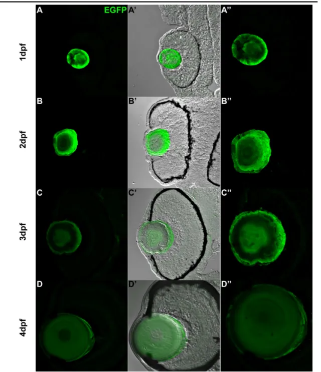
Diagram representing a transverse section of the zebrafish lens. 68
Figure 5.1.2b
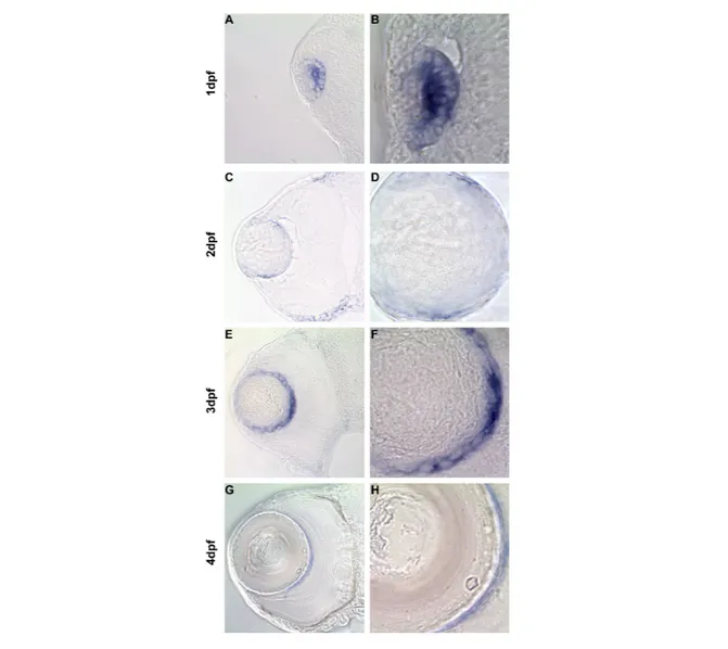
Transverse sections of zebrafish eyes show expression of cugbp1 mRNA in the lens by in situ hybridization assay.
69
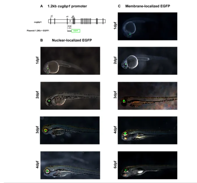
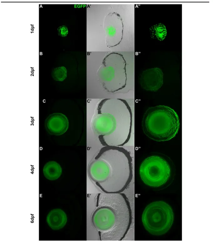
Figure 5.2.1
EGFP expression driven by a 1.2kb cugbp1 promoter in F0 zebrafish embryos.
71
Figure 5.2.2a
Transverse sections at the eye region from zebrafish F1 stable transgenic line embryos carrying the 1.2kb cugbp1:pME-nlsEGFP-polyA transgene.
72
Figure 5.2.2b
Transverse sections at the eye region from zebrafish F1 stable transgenic line embryos carrying the 1.2kb cugbp1:pME-EGFPCAAX-polyA transgene.
73
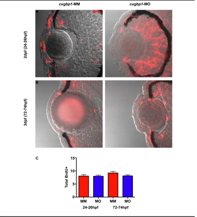
Figure 5.3.1
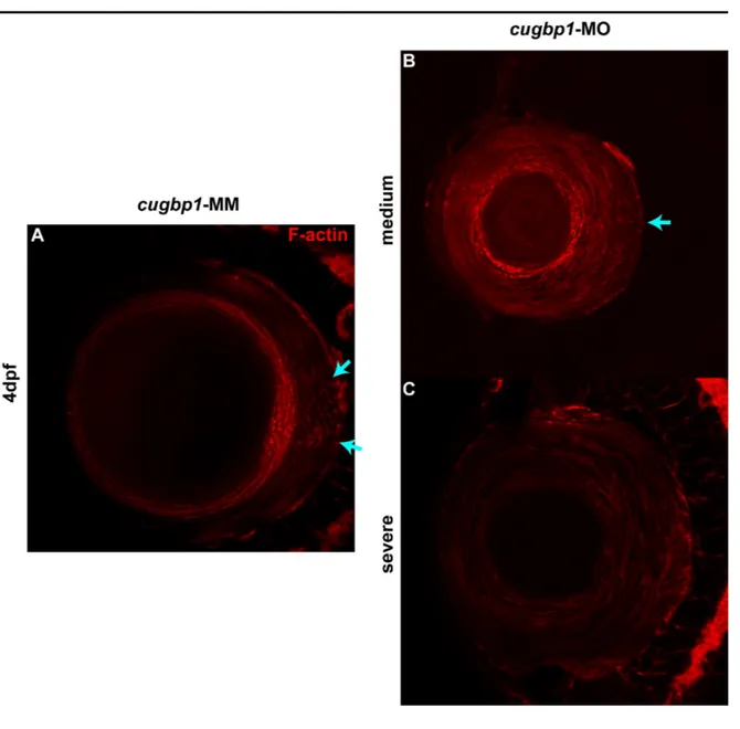
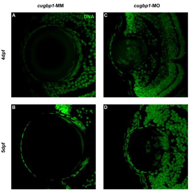
Knock down of cugbp1 function in zebrafish embryonic development by a splice-altering morpholino results in a cataract phenotype and
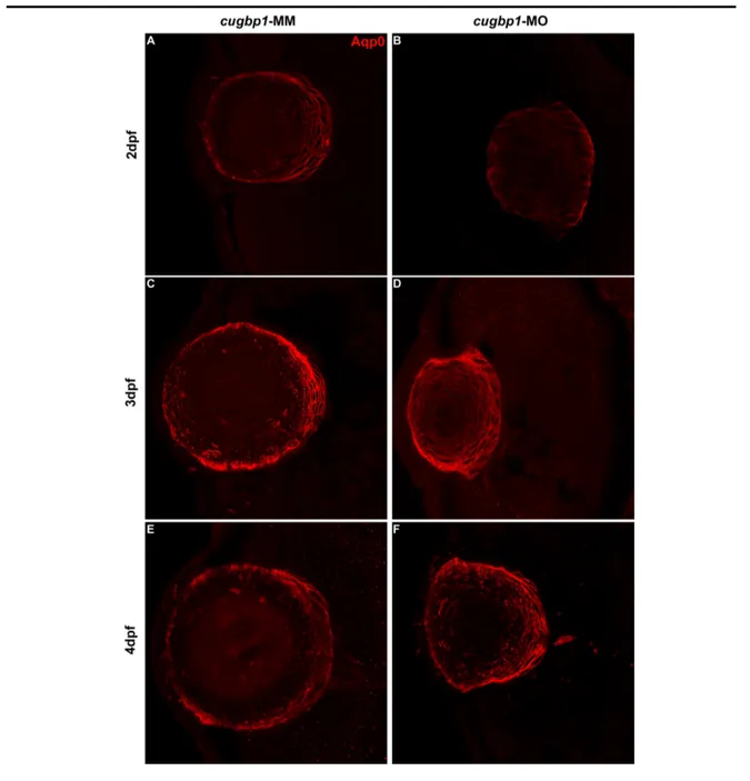
Lens fibers express differentiation marker Aqp0 in cugbp1-MO embryos.
79
Figure 5.3.5
F-actin organization in the lens mass shows differences between cugbp1-MO and cugbp1-MM embryos.
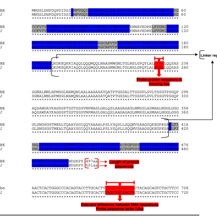
Splice-altering cugbp1 morpholino activity generates a frameshift and a premature stop codon by removing exon5.
Index of Annexes
Annex 1. Signed accreditation 128
Annex 2. CS10R plasmid 129
Annex 3. Sequence alignment of cugbp1 cDNA used for probe synthesis 130
Annex 4. Amino acid sequence from cugbp1 cDNA used for probe synthesis 132
Annex 5. Protocol for cugbp1 probe synthesis 133
Annex 6. Whole mount RNA in situ hybridization protocol 134
Annex 7. Immunohistochemistry protocol 137
Annex 8. cugbp1 promoter sequence and location 140
Annex 9. RT-PCR Morpholino Activity
A. Morpholino disrupted splicing by removing exon4
B. Mismatch morpholino did not disrupt splicing
141
142
Chapter 1. Introduction
The lens is a transparent and avascular tissue in the anterior of the eye and its main role
is to refract light onto the retina where light is transduced into neural signals. Afterwards,
these signals are transmitted to the brain. The vertebrate lens consists of two types of cells:
epithelial cells and fibers. Epithelial cells surround the anterior and lateral limits of the lens,
remain proliferative and at the equator of the lens, or near to it, they differentiate into lens
fibers (Chow and Lang 2001; Tsang and Gouras 2006; Greiling and Clark 2009). In this
process newly formed lens fibers elongate and gradually lose their organelles, enabling
transparency (Bassnett 2009).
Cataracts are defined as any opacification of the lens that compromises its ability to
refract light onto the retina (Graw 1999). According to the World Health Organization, the
latest estimates (Oct., 2011) say there are 285 million people visually impaired worldwide and
about 90% of them live in developing countries. Cataracts account for 33% of global visual
impairment (VI = VA ˂ 0.3) and are still the main cause of blindness in third world nations
(WHO 2011). Visual acuity (VA) is the ability to distinguish details and shapes of objects.
Any visual deprivation, such as lens opacities, will result in a decrease of VA. In humans, VA
develops from birth to adolescence and a VA of 1.0 is reached by 5-6 years of age (Ekström
2009). Cataracts can be genetic and/or environmentally induced and can happen via many
different cellular and molecular mechanisms. Therefore, it is necessary to investigate the
cellular and molecular basis of lens development and physiology to be able to understand the
reasons that cause cataracts. This will lead to aim for new and better therapeutic treatments
(Gross and Perkins 2007; Wormstone and Wride 2011).
The zebrafish (Danio rerio) has emerged as an ideal model to study early development
and disease of the visual system, including the lens of the eye. Some of the zebrafish
advantages to study overall embryonic development are: external fertilization, their rapid
development compared to other vertebrate model organisms, embryonic development occurs
ex utero. The embryo is transparent which facilitates visual identification of morphogenetic
movements and organogenesis with a standard dissection microscope. Zebrafish are easily
other vertebrate model systems. These freshwater fish reach sexual maturity in just 3-4 months
and a single pair of fish can produce >200 fertilized eggs per mating. These characteristics
have made zebrafish embryos ideal for the discovery of the function of genes implicated in
regulating embryonic development, including lens morphogenesis (Glass and Dahm 2004;
Fadool and Dowling 2008; Greiling and Clark 2009).
Zebrafish are very visually oriented and their lenses show much the same morphology
as other vertebrates, including humans (Glass and Dahm 2004). Their visual system is first
identified as a functional structure between the third and fourth days post fertilization (dpf;
Easter and Nicola 1996). Moreover, the lens shape and overall structure suggests it is a
functional optical element in the visual pathway as early as 3dpf (Greiling and Clark 2009).
All the aspects mentioned above make zebrafish well suited for examining lens development,
Chapter 2. Research aims
2.1 General objective
• Identify and investigate the role of Cugbp1 protein in early zebrafish (Danio rerio) lens development.
2.2 Specific objectives
• Detect if there is cugbp1 mRNA expression in zebrafish early lens development, and if so, where inside the lens and at what developmental stages is it expressed.
• Identify a promoter at the cugbp1 gene that directs expression at the zebrafish lens to estimate Cugbp1 protein expression.
• Identify if a specific cugbp1 morpholino alters splicing of cugbp1 mRNA.
• Identify phenotypical defects in whole embryos, at first sight, due to down regulation of cugbp1 and compare them with defects previously reported at DM1 disease where cugbp1 expression is also disrupted.
• Observe if lens development is affected when cugbp1 expression is down regulated and recognize a role for Cugbp1 protein in zebrafish early lens development.
• Detect if Cugbp1 is necessary for zebrafish lens cell proliferation.
• Identify if Cugbp1 is required for the expression of Aqp0 protein to detect if Cugbp1 is necessary for zebrafish lens fibers early differentiation.
• Recognize if cugbp1 has a role in F-actin distribution and/or arrangement in zebrafish lens fibers.
Chapter 3. Theoretical Framework
3.1 Zebrafish lens early development
Zebrafish lens establishment starts at the 14-15 somite stage (~16hpf; hours post
fertilization) with a contact between the surface cranial ectoderm and an evaginating solid
mass of cells that comes from the diencephalon (more posterior and ventral part of the
forebrain) and constitutes the optic primordium. The optic primordium is a solid mass of cells
that emerges from the anterior portion of the neural tube and will eventually give rise to the
retina. The forebrain or prosencephalon refers to the most anterior region of the brain and it
includes the diencephalon (Schmitt and Dowling 1994; Kimmel et al. 1995; Soules and Link
2005; Dahm et al. 2007; Greiling and Clark 2008).
The surface ectoderm-optic primordium interaction results in the thickening of the lens
placode and in zebrafish, this occurs at 16hpf. This thickening starts as a columnar epithelium
by doubling of the basal to apical height of cells from simple cuboidal epithelium of the
cranial surface ectoderm (Schmitt and Dowling 1994; Greiling and Clark 2008; Greiling and
Clark 2009). The lens placode is defined as the ectodermal primordium of the lens and it
overlies the center of the developing retina (Kimmel et al. 1995; Dahm et al. 2007; Greiling
and Clark 2009).
When observed from the surface the lens placode, from 16hpf zebrafish embryos,
looks circular, of approximately 8 cells in diameter, and it is composed of columnar cells
relatively uniform in size and shape. At this point, the lens placode of the zebrafish resembles
the mammalian or avian placode (Greiling and Clark 2009).
By 18hpf, many of the cells in the lens placode have more than double in height as
compared to 16hpf, and look as a solid mass of cells ordered as a flattened spheroid (plate-like
thickening organization). The lens mass is two or three cell-layers thick at the center with a
single layer remaining laterally. The change in morphology has made elongated cells of the
lens placode clearly distinguishable from cuboidal cells of the surface ectoderm. In the
anteromedial region of the lens mass, cells are shorter and more rounded than the elongated
cells present at the posterior and lateral lens borders (Greiling and Clark 2009). At the
start to invaginate instead of undergoing a delamination growth process as in zebrafish
embryos (Fadool and Dowling 2008; Greiling and Clark 2008).
At 20hpf the thickness of the lens mass increases bulging towards the retina and
narrows in the equatorial dimension. At this moment, it is quite obvious that the lens placode
has changed from a plate-like structure to a lentoid solid mass of cells that will eventually
acquire a more spherical shape (Fadool and Dowling 2008; Greiling and Clark 2008; Greiling
and Clark 2009). Elongated fiber-like cells appeared along the deep and lateral boundaries of
the lens mass surrounding a central core of rounded, undifferentiated cells. Three distinct cell
morphologies can be seen at this stage: (1) cells at the posterior and lateral surfaces are tall
and similar in height to the elongated cells at 18hpf. These cells formed a single layer that
establishes a posterior and lateral border of the lens and have a basal cell surface 2-3 times
wider than the apical surface. (2) Cells in the center of the elongated lens mass with round or
ovoid shape and irregularly clustered in the center of the lens core and stalk (region located in
the anterior-middle). (3) Cells at the anterior lens border in contact with the surface epithelium
are elongated and with a more parallel orientation to the surface ectoderm (Greiling and Clark
2009).
At 22hpf cells attached to the surface ectoderm narrow to form a stalk 2 to 3 cells wide
connecting the developing cornea with the developing lens. At this moment of development
the shape of the lens mass is rounded. Three morphologically distinct cell types are clearly
present: (1) the lateral and posterior borders of the lens are formed by a single-layer of tall
columnar cells with wider basal than apical cell surfaces. These cells radiated out from the
central core. (2) The central core is a cluster of cells in the middle of the lens; these cells
appear to have a tear-drop shape with their narrow edges facing towards the center. (3) Cells at
the anterior-middle of the lens and the surface ectoderm in contact with the lens are rounded or
cuboidal in shape with an irregular arrangement. These 3 cell types correspond to the primary
fiber cells, the embryonic nucleus, and undifferentiated cells of the original lens mass,
respectively (Greiling and Clark 2009). The analogous lens developmental stage of mammals
and birds shows a different formation pattern. In these superior vertebrates the lens placode
as a fluid-filled lens vesicle surrounded by a single layer of epithelial cells (Reza and Yasuda
2004; Soules and Link 2005; Greiling and Clark 2008).
The lens mass remains connected to the surface ectoderm at 23hpf by a stalk that is
only one cell wide. The surface ectoderm is a single-layer of flat, cuboidal epithelium above
the lens except where it is still attached to the lens in the very anterior-middle border.
Fiber-like cells begin to curve and appear to wrap around the central core. Cells in the central core of
the lens mass remain large and tear-drop shaped with their narrow edges facing the lens center.
The cells at the anterior-middle of the lens remained rounded or cuboidal in shape and in an
irregular arrangement (Greiling and Clark 2009).
At 24hpf the lens mass separates completely from the surface ectoderm which remains
a continuous single layer of epithelial cells at the surface of the head of the fish. Cells in the
posterior-middle region continue to enlarge and take on a rounded shape forming an
organizing center around which primary fiber cells elongate and migrate. Lateral columnar
cells elongate further to form arcs or layers of primary lens fibers surrounding the nucleus.
Cells at the anterior border organize into a single-layer of epithelium and cells deep to the
developing anterior epithelium are still disorganized and undifferentiated (Schmitt and
Dowling 1994; Greiling and Clark 2009).
The morphology of the differentiated lens cells at 28hpf represents the cell types
expected in the adult lens: (1) A single-layer of tall, cuboidal epithelium that covers the entire
anterior hemisphere of the lens and that in zebrafish, but not mammals, extends posteriorly
beyond the lens equator, but not in the posterior-most surface of the lens. (2) Primary fiber
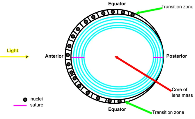
cells that wrap around the large round cells in the core of the lens nucleus. (3) Secondary fiber
cells that elongate and migrate from a developing transition region (Dahm et al. 2007;
Greiling and Clark 2009). In the zebrafish lens, this transition region is located more
posteriorly as compared to the mammalian and avian lenses that possess this region at their
equator. This type of region in the lens is common among vertebrates and it is where epithelial
cells exit the cell cycle and start differentiating into secondary lens fibers (Soules and Link
2005; Griep 2006; Weber and Menko 2006b; Greiling and Clark 2008).
By 36hpf the lens seems spherical in the equatorial dimension and lentoid in the
28hpf and the height of the anterior epithelial cells is decreased by half. Cell membranes
between the central cells look jagged like establishing shallow interdigitations between them
(Greiling and Clark 2009).
By 2dpf zebrafish embryos start their hatching period (Kimmel et al. 1995; Easter and
Nicola 1996; Easter and Nicola 1997). At 48hpf, the lens still appears spherical at the equator
and lentoid in the anterior-posterior direction. By 2days the lens has increased in size and cells
of the anterior epithelium continue to decrease in height and form a flat cuboidal epithelium.
Fiber cells in the cortex are narrow and elongated. The posterior tips of newly added
elongating fiber cells meet at the midline establishing a posterior suture. Borders between cells
in the lens mass are increasingly jagged (Greiling and Clark 2009).
At 72hpf (3dpf; first day post hatch; Kimmel et al. 1995), the width of the lens
increases at the equator making the lens shape spherical in all dimensions. An umbilical suture
(point-like) can be seen at both the anterior and posterior poles (Greiling and Clark 2009). A
thin extracellular capsule is apparent, which is an uninterrupted basement membrane
completely enclosing and protecting the lens. Newly generated lens fibers possess
interdigitations that have not yet achieve an eventual ball and socket organization (Soules and
Link 2005; Danysh and Duncan 2009). Between the third and fourth dpf the still growing
zebrafish visual system is first identified as a functional structure (Easter and Nicola 1996). By
4dpf the lens is a larger spherical version of the 3dpf lens, that increases in size by additional
layers of secondary fiber cells (Greiling and Clark 2009).
As lens development and growth proceeds through embryogenesis into postnatal
(mice) or larval (zebrafish) and subsequently throughout adult life a distinguished organization
of regions with high or low proliferative index arises at the lens epithelium. This epithelial
tissue has 4 distinct subpopulations: (1) a central zone (CZ) that comprises the biggest portion
of lens epithelial tissue covering most of the anterior surface of the lens. This region has a low
proliferative index with most of its cells in a quiescent state (G0); although they retain their
proliferative potential. (2) A pregerminative zone (PGZ) that constitutes cells comprising a
narrow, latitudinal band or ring peripheral to and limiting the CZ. A small portion of these
cells undergo mitosis to add to the lens epithelial mono-layer as the lens increases in size
germinative zone (GZ) are located at a narrow, latitudinal band peripheral and posterior to the
PGZ. In the GZ cells have a high proliferative index. Daughter cells from the GZ migrate into
the transition region of the lens. (4) A transition zone (TZ) is found posterior to the GZ at the
equatorial (mice) or posterior to the equatorial (zebrafish) arc or bow region of the lens. This
zone is a narrow ring of cells where proliferation does not happen. Cells in this place exit the
cell cycle, elongate and differentiate into secondary lens fibers as they form concentric layers
around previously formed lens fibers. This summarizes how cells are continuously added to
the differentiated fiber cell mass that originates from the epithelium (Graw 1999; Soules and
Link 2005; Griep 2006; Kuszak and Costello 2006; Mathias et al. 2010).
In addition, it is important to mention that as the lens matures, its fiber cells become
flattened and band-like shaped with a width to thickness ratio between 10:1 and 15:1. Lens
fibers develop interdigitating lateral membrane protrusions at their narrow edges and ball and
socket-like joints on their broad surfaces (Dahm et al. 2007).
3.2 Differences between mammalian and zebrafish lens development and early morphology
Although the mammalian, bird and zebrafish lens are all derived from surface
ectoderm, zebrafish early lens development possess noteworthy differences compared to
mammals and birds. During embryonic development, the mammalian and avian lens placode
invaginates to form a hollow lens vesicle bordered by a monolayer of ectoderm that constitutes
epithelial cells. Instead, in zebrafish the lens placode delaminates as a solid cluster of cells
from the surface ectoderm (Schmitt and Dowling 1994; Easter and Nicola 1996; Soules and
Link 2005; Dahm et al. 2007).
In birds and mammals, primary lens fibers are formed from epithelial cells located in
the posterior half of the lens vesicle. These posterior epithelial cells elongate in a posterior to
anterior direction and a parallel-like way. They differentiate to fill the lens vesicle cavity as
primary fibers. In contrast, in zebrafish primary lens fibers differentiate from cells in the
center of the delaminated solid cluster of cells by elongating in a circular fashion. Thus, giving
rise to concentrically arranged primary lens fibers. In both cases, primary lens fibers give rise
Cells in the anterior half of the still fluid-filled lens vesicle in birds and mammals as
well as cells in the anterior-most border of the lens solid mass in zebrafish become the lens
anterior epithelium. These epithelial cells exhibit a high mitotic activity to form a mono-layer
of epithelial cells that extends along the anterior and equatorial surface of the lens. However,
in contrast to birds and mammals, in zebrafish the epithelial single layer extends farther
towards the posterior-lateral surface of the lens. And, like birds and mammals, the epithelial
cell layer does not extend to the most-posterior border area in zebrafish lenses. At this region,
lens fibers are in direct contact with the lens capsule (Dahm et al. 2007; Greiling and Clark
2008).
As mentioned before, another difference corresponds to the location of the transition
region where epithelial cells differentiate to give rise to secondary lens fibers. In mammals
and birds this region is located at the lens equator; whereas in zebrafish, this transition zone is
at a more posterior region compared with the lens equator (Soules and Link 2005; Griep 2006;
Weber and Menko 2006b; Greiling and Clark 2008).
Lens suture formation as fibers elongate and meet at their narrow edges with another
fiber is another variant. Zebrafish, as well as avian lenses exhibit two umbilical sutures
(point-like), one at the center of the anterior pole and the other one at the center of the posterior pole.
In these types of sutures fiber cells are meridians and taper at the ends as they extend from
pole to pole. All fibers are sequentially overlaid onto existing growth shells of fibers, resulting
in radial cell columns that extend from the center (at the transitional zone) to both the anterior
and posterior poles of the lens. These fibers are seen as straight meridians (circular arcs) that
extend from pole to pole (Al-Ghoul et al. 2003). Dahm et al. (2007) have shown that when the
lens capsule, the monolayer of epithelial cells and some of the outer-most fiber cells are
removed the umbilical anterior lens suture, from zebrafish whole lens samples becomes
visible. Scanning electron microscopy micrograph pictures show that the secondary lens fibers
converge in a single point at each lens pole.
In contrast, the fibers of other superior vertebrates are not meridians. These lens fibers
possess ends that flare (spread gradually outward) and curve away from the poles in opposite
directions. As a result the end-to-end arrangement, where opposing fibers meet produces lens
(e.g., rabbit and frog lenses), feature two anterior branches oriented at 180º to each other to
form a vertical line-like suture. Opposite end curvature results in two posterior branches
forming a horizontal line-like suture (Al-Ghoul et al. 2003).
Other vertebrates (e.g., mice, rat, pig, cat, dog, bovine and primates at birth) have
lenses with Y-like sutures. In this type of lenses three anterior branches orient at 120º to each
other to form a Y-like suture. Opposite end curvature results in three posterior branches that
form an inverted Y-like suture. In primates, the overall type of suture changes over time.
During fetal development the previously described Y-like sutures form. During infancy an
anterior and posterior six branch suture, referred as simple star develops. At adolescence, both
sutures evolve to become nine branch sutures, known as star. Later on, at the adult stage the
sutures are gradually transformed and become 12 branch sutures, referred as complex star
(Al-Ghoul et al. 2003; Kuszak et al. 2004).
These differences must be taken into account when interpreting results of molecular
biology studies (Dahm et al. 2007). The present work is an example of this type of studies in
which the purpose is to try to identify the function of a gene (cugbp1) in early zebrafish lens
development. The results obtained have the ultimate goal to try to understand what can happen
if the pathway of the human ortholog CUGBP1 is interrupted in embryonic development and
unravel its early function in lens formation.
3.3 Lens fiber cells differentiation
At the TZ of the lens, epithelial cells initiate their differentiation to become fibers, a
process that comprises dramatic changes in gene expression as well as in cell shape. It has
been observed that actin filament reorganization is necessary for both types of changes to
happen (Weber and Menko 2006a). In the cortical region (outer layers, which are comprised of
differentiating lens fibers that still have not lost their organelles) of the embryonic lens, the
fiber cells elongation process occurs in parallel with the accumulation of lens
differentiation-specific proteins (e.g., AQP0; Varadaraj et al. 2007).
In fact, lens fibers can elongate more than 1,000 fold to reach the lens sutures. The
stretched fiber cells are arranged as a series of concentric layers in which they appear as
narrow lateral faces. The broad lateral faces are oriented parallel to the lens surface (Kuszak
and Costello 2006). After the morphogenetic changes associated with elongation have
happened, the maturing fiber cells lose their organelles, including nuclei to enable
transparency (Weber and Menko 2006a).
3.3.1 Aquaporin0 (AQP0) expression and function
Epithelial cells at the transition bow of the lens initiate a change in the pattern of gene
expression as they start to differentiate into lens fibers. The new pattern includes the
expression of structural proteins that can be soluble (e.g. crystallins) and also membrane
proteins (e.g. water channels) in the lens fibers. Crystalline proteins contribute to the
transparency and appropriate refractive index of the lens. This happens due to the crystallins
elevated concentration and short-range interaction. Membrane proteins maintain the
architecture needed for lens appropriate function, also contributing to lens transparency
(Chepelinsky 2009). Aquaporin proteins (AQPs) are transmembrane water channels that
mediate the permeation of water across cell membranes (Agre et al. 1998).
The lens lacks blood vessels since they would scatter the incident light (deviate light
from its original trajectory). Hence, this avascular region of the eye has evolved a standing and
efficient circulatory current known as microcirculatory system. This current enters at both the
anterior and posterior poles of the lens; then passes into and through the lens fibers. Finally,
the current exists at the equatorial region of the lens. This circulatory system depends on water
channels (Mathias et al. 2010).
In mice, it has been observed that Aquaporin0 (Aqp0; previously known as Mip)
mRNA as well as protein expression is observed in the lens. It begins at embryonic day 11.25
(E11.25) when the posterior lens epithelial cells simultaneously start to differentiate into
primary lens fibers. This expression continues throughout lens development and in the adult
lens. AQP0 protein is exclusively expressed at the cell membranes of primary and secondary
lens fibers since their early cell differentiation. This membrane location remains permanently
(Varadaraj et al. 2007).
It has also been shown that a second aquaporin gene, Aquaporin1 (Aqp1) is expressed
lens fibers. AQP1 expression at the lens starts at E17.5, 7.25 hours later than APQ0 expression
begins at lens fibers. This happens even though lens anterior epithelium develops earlier than
primary and secondary lens fibers. Secondary lens fibers that differentiate from epithelial cells
that have already expressed AQP1 show progressive decrease of AQP1 protein expression as
they differentiate (Varadaraj et al. 2007).
The temporal pattern of expression between AQP0 (E11.5) and AQP1 (E17.5) suggests
that as the lens body increases in size by the addition of new lens fibers, there is a growing
demand for higher epithelial membrane water permeability to establish the microcirculatory
system. In addition, the lens switch from AQP1 expression in epithelial cells, located at the
transitional ring of the lens, to AQP0 in the differentiating secondary lens fibers. This
indicates that AQP0 may have other important membrane functions. AQP0 might also
function as an adhesion protein to join adjacent fiber cells. AQP0 probably contributes to
reduce the extracellular space between lens fibers and to diminish light scattering (Varadaraj
et al. 2007).
It is thought that after the two principal evolutionary radiations of jawed vertebrate life
that separated the ray-finned fish (class Actinopterygia; includes the zebrafish) and the
sarcopterygian lineage (from where the land vertebrates evolved), a genome-wide duplication
event happened in a zebrafish early ancestor (Meyer and Schartl 1999). This possible incident
might explain why many single copy genes in mammals can be observed as duplicates in
zebrafish where the function and temporal-spatial expression of the single-copy mammalian
gene can be split up between both duplicates (Postlethwait et al. 2004).
Indeed, it has been shown that the zebrafish genome has two aqp0 genes referred as
aqp0a and aqp0b. Both genes are expressed during lens development in fiber membranes and
persist in the adult lens (Froger et al. 2010; Tingaud-Sequeira et al. 2010). Moreover,
knocking down aqp0a and/or aqp0b in zebrafish embryos by translation altering morpholino
results in an obvious cataract phenotype as early as 3dpf (Froger et al. 2010). Morpholinos are
chemically modified oligonucleotide analogous with a morpholino moiety instead of a ribose.
They also possess a non-ionic phosphorodiamidate linkage instead of an anionic
phosphodiester bond resulting in a neutrally charged backbone. These mentioned variations
and little cellular toxicity. Moreover, morpholinos are resistant to digestion by nucleases
(Ekker 2000; Corey and Abrams 2001).
Froger et al. (2010) experiments have indicated that both aqp0a and aqp0b are needed
for lens transparency. Nevertheless, water permeability assays suggest that Aqp0a protein
functions as a water channel, whereas Aqp0b does not. Aqp0b might supply adhesion and/or
interactions with other lens components. Mammalian Aqp0 functions might be distributed
between aqp0a and Aqp0b in zebrafish. However, additional work is needed to figure out
aqp0b function on the zebrafish lens.
The unique eye expression of Aqp0 protein in lens fibers as they start their
differentiation process from initial epithelial cells makes this protein an excellent marker to
asses for early fiber cell differentiation in the lens.
3.3.2 Lens actin cytoskeleton
During fibergenesis, epithelial cells undergo elongation, with the anterior and posterior
tips of the elongating lens fibers sliding along the epithelium and capsule at the anterior and
posterior direction, respectively and as these cells migrate inward. Lens fibers finally detach
from the epithelium and capsule when they reach the anterior and posterior sutures. At the
sutures, fiber cells form contacts with their counterparts from the opposite side of the lens. All
these cellular movements are greatly coordinated through actin cytoskeleton remodeling
events (Rao and Maddala 2006). Thus, the actin cytoskeleton plays an important role in
regulating fiber cell elongation, migration, lens capsule-cell and intercellular interactions, cell
packing, overall geometry and in the maintenance of fiber cell symmetry. Therefore, the
activities of the actin cytoskeleton are critical for the establishment of lens overall shape,
symmetry and ultimately, for lens optical properties (Rao and Maddala 2006).
The actin cytoskeleton is composed of F-actin (actin filaments or microfilaments) and
other accessory proteins that vary depending on the type of structure formed. F-actin
represents a helical protein filament formed by polymerization of globular actin molecules
(G-actin). During remodeling of the actin cytoskeleton, F-actin undergoes disassembly and
Studies performed in Quails (Coturnix japonica; class Aves) during embryonic lens
development by Weber and Menko (2006a) have shown that a disassembly of actin stress
fibers (contractile large bundle/parallel arrays of F-actin crosslinked by α-actinin) happens as
lens fiber differentiation is initiated. In central epithelial cells at the anterior region of the lens,
actin stress fibers are organized along these cells basal surfaces (face linked to the lens
capsule). Indeed, actin stress fibers are the primary actin filament structures of the
undifferentiated lens epithelium and are most possibly linked to extracellular matrix
components of the lens capsule through integrin receptors. In general, integrins are
transmembrane adhesion proteins that play part in cell-matrix junctions. The extracellular
domains of integrins bind to components of the extracellular matrix (in these case: lens
capsule), while the cytoplasmic tail binds indirectly to F-actin. This type of cell-extracellular
matrix junction in which there is an intracellular coupling to F-actin is called focal adhesion
(Cooper 2000; Alberts et al. 2008). Lamellipodial-like extensions are broad membrane
protrusions that contain a three-dimensional network of F-actin and can contribute in focal
adhesion formation (Cooper 2000; Lodish et al. 2000). These extensions were observed at the
central epithelial cells basal edges. At the apicolateral (apical: faces the center of the lens)
aspects of these cells in the region of tight junctions (physical attachments that seal the gaps
between cells in the apical side of epithelia making the sheet an impermeable or selectively
permeable barrier), F-actin had a cortical arrangement (Weber and Menko 2006a).
However, at the equatorial epithelium where differentiation has started, a different
actin organization was observed. In the anterior-most region of the equatorial epithelium,
F-actin staining in the cells basal and basolateral aspects was amorphous and diffused indicating
that actin stress fibers were no longer present. This loss of actin stress fibers was concomitant
with lens cell differentiation. At the cells lateral borders (region of cell to cell interfaces) few
F-actin was detected. F-actin at the apical domain remains cortical. At the very center of the
equator, F-actin staining was disorganized and diffuse in the basal surfaces. The latter is
indicative of a transition due to actin filament reorganization as cells moved through the
transition region. As cells moved to the posterior-most aspects of the equatorial epithelium,
F-actin became localized at the cell to cell interfaces at their basal surfaces as well as along the
the cell borders were evident at the basal surfaces of these cells. Cortical F-actin at the cells
lateral borders is consistent with their function in the assembly of stable N-cadherin cell-cell
junctions (Weber and Menko 2006a). Cadherins are transmembrane proteins that mediate
contacts between cells to form adherence junctions. Inside the cell, cadherins bind indirectly to
F-actin (Cooper 2000; Alberts et al. 2008).
At the most cortical fiber cell region in the posterior pole of the lens, F-actin was
present at the basal tips of lens fibers and organized in a dense meshwork pattern. The basal
tips of these newly formed cortical lens fibers correspond to the surface in contact with the
lens basement membrane/capsule at the posterior region of the lens. At the basal and
basolateral aspects, F-actin is present along all sides of the already hexagonal cells, but is
missing from the vertices (region where three cells meet). At the lateral surfaces of these lens
fibers, cortical F-actin extended around the entire perimeter of the cells and along the length of
these elongating lens fibers. Cortical F-actin became much more organized than at earlier
moments of differentiation probably helping to stabilize lens elongated and hexagonally
packed morphology (Weber and Menko 2006a).
In summary, Weber and Menko (2006a) have demonstrated that the initiation of lens
cells differentiation is coincident with the disassembly of the cellular projections
(lamellipodia) and actin stress fibers that provide cell attachment between the extracellular
matrix (capsule) and the undifferentiated lens epithelia. F-actin is reorganized as cortical actin
in the differentiating lens fibers. Indeed, stress fibers disassembly is sufficient to induce lens
fibers differentiation. Actin filaments organized as stress fibers interact with integrin receptors
at focal adhesion complexes where they mediate integrin/matrix adhesion. The lens epithelium
is the only region of the embryonic lens that expresses high levels of α5β1 integrin and
fibronectin (extracellular matrix ligand of α5β1 integrin). The interaction of both molecules
promotes actin stress fibers organization and their loss could signal stress fibers disassembly
that activates lens cell differentiation.
Moreover, lens cell culture studies have evidenced that actin stress fiber disassembly
as well as cortical F-actin organization are dependent on the assembly of N-cadherin cell-cell
adhesions. However, in the undifferentiated epithelium adhesion corresponds primarily to
(integrin/matrix adhesions) as they differentiate and cadherin based cell-cell junctions become
the principal form of adhesion as lens fibers develop. This switch may promote the
disassembly of actin stress fibers and induces lens fibers differentiation (Weber and Menko
2006a).
In addition, Fischer et al. (2000) experiments realized in chick lens cell cultures
determined that in undifferentiated epithelial cells F-actin was organized in polygonal arrays
of actin stress fibers that intersect with an adhesion belt (a type of cadherin cell-cell junction in
epithelial cells located just below the tight junctions and forming a continuous belt-like
structure around each cell in which an underlying contractile bundle of actin filaments is
linked to the plasma membrane; Cooper 2000; Alberts et al. 2008). As cells elongated to form
lentoid bodies (lens fiber-like cells) the arrays of stress fibers were lost. Actin filaments in
differentiated lentoid cells were predominantly associated with membranes in a reticular
pattern. Moreover, in late-stage differentiated lentoid cells, F-actin colocalized with
N-cadherin molecules in complex curvilinear patterns outlining membranes (Fischer et al. 2000).
Cortical actin filaments are also part of a complex structure in lens fibers, besides
N-cadherin cell-cell adhesions, referred as membrane skeleton. The membrane skeleton is a
highly cross-linked network of spectrin (a long thin flexible rod protein) tetramers linked to
short F-actin, and together they are associated with membrane attachment and actin regulatory
proteins. The membrane skeleton is associated with the inner surface of the lens fiber plasma
membrane. In this complex structure, F-actin stability depends on capping proteins at filament
ends that prevent assembly as well as disassembly, and on tropomyosins (TMs). TMs bind
along the sides of microfilaments blocking severing and reducing subunit dissociation.
Tropomodulins (Tmods) are actin pointed end-capping proteins that bind to TMs and cap
TM-coated F-actin preventing polymerization and depolymerization in post-mitotic cells like lens
fibers. Thus, Tmods and TMs regulate actin filament lengths and provide stability to the
membrane skeleton (Alberts et al. 2008; Nowak et al. 2009).
A Tmod1 viable knock out mice line with no detectable TMOD1 protein in the lens
indicated that the membrane skeleton is necessary for maintenance of fiber cell hexagonal
shape, packing geometry during maturation of lens fibers and radial column organization in
lenses in comparison with hexagonally packed geometry of controls. These patches exhibited
polygonal and often somewhat rounded fiber cell shapes with variable numbers of vertices per
cell and irregular lengths of connecting membranes, rather than regular flattened hexagonal
shapes. The fibers are also not arranged in precise radial columns as seen in normal equatorial
sections. At the transition zone of the lens (equator), regions displaying disordered fiber cell
packing tended to be located 20-30 cell layers in from the epithelium; in the region where
TMOD1 normally assembles on the fiber membranes. The latter showed that the absence of
TMOD1 affects geometry of maturing lens fibers, rather than initial elongation and
organization (Nowak et al. 2009).
Nowak et al. (2009) determined that TMOD1 appears to selectively stabilize the subset
of F-actin that belongs to the membrane skeleton, which corresponds to a part of the F-actin in
lateral broad and narrow sides, but not the vertices of maturing fibers. Absence of TMOD1
(and consequently a disrupted membrane skeleton) does not affect fiber cell initial
differentiation or cell shape morphogenesis. Instead, TMOD1 protein stabilizes F-actin in the
membrane skeleton during cortical fiber cell maturation before organelle loss. This indicates a
role in maintaining cell shape and packing geometry.
In addition, Quail lens cell culture experiments have proven that the actin cytoskeleton
also supports cell survival, and a prolonged disruption of the latter induces apoptotic events
that result in cell death. Depolymerization of F-actin in lens epithelial cell cultures for a
prolonged time (48h) induces extensive membrane blebbing and cell rounding, indicative of
late stage apoptosis. Induced loss of cortical F-actin in cell cultures containing differentiating
lentoid cells also resulted in blebbing of the plasma membrane. The latter proved that F-actin
provides an essential survival signal to both lens epithelial and differentiating fiber cells
(Weber and Menko 2006a).
Furthermore, a short-term induced F-actin disassembly on lens epithelial cell cultures
does not induce apoptosis. Rather, it triggers the expression of fiber cell differentiation
specific markers, cell cycle withdrawal and the loss of actin stress fibers with a subsequent
reorganization into cortical F-actin as fibers differentiate. In addition, BCL-2 (a suppressor of
organize actin as cortical filaments and survive. The latter suggests that the F-actin survival
signal in differentiating lens fibers may be conveyed by BCL-2 (Weber and Menko 2006a).
3.3.3 Organelle degradation in lens fibers: emphasis in nuclei
During lens fibers differentiation, all structures large enough to scatter light including
nuclei, mitochondria, Golgi apparatus and endoplasmic reticulum are broken down and
removed from the developing fiber cells (Greiling and Clark 2008; Bassnett 2009). Despite all
these changes, lens fibers survive and are maintained within the lens throughout the life span
of the individual (Counis et al. 1998).
Chicken studies have demonstrated that in embryonic lenses organelles are present
initially throughout all the cells in the developing tissue. Then, in a specific moment in early
development (embryonic day 12 in chick embryos) organelles are eliminated in cells located at
the center of the lens (primary lens fibers). The latter results in the formation of a central area
without membrane-bound organelles, including nuclei, termed organelle free zone (OFZ).
After its initial establishment, the OFZ becomes larger as the lens grows and new fibers are
continuously added. Then, organelles are present only in those fiber cells located at the
periphery of the lens (Bassnett and Mataic 1997). The only cells in the lens located in the
visual axis that do not lose their organelles are cells in the central-anterior epithelium.
Nevertheless, they constitute a very thin layer. So, since light scattering is proportional to path
length, the light scattering due to organelles in the lens epithelium is insignificant (Bassnett
2009).
It has been observed that during epithelial cell differentiation into lens fibers, the shape
of nuclei changes along the course of this process. In lens epithelial cells, nuclei appear to be
round and relatively large. In superficial lens fibers, nuclei appear ovoid, and as lens fibers
mature nuclei seem to elongate along with fiber elongation. However, just prior to
disintegration, nuclei remnants assume a much smaller and more spherical shape (Bassnett and
Beebe 1992; Counis et al. 1998; De María and Arruti 2004).
Nucleated fiber cells can perform transcription and this process might be stopped until
inner nuclear membrane). Afterwards, chromatin disintegration occurs. This order of events
has been reported in bovine and chick lenses (Bassnett and Mataic 1997; Bassnett 2009).
It has been suggested that the nuclei degradation process might be different in primary
and secondary lens fibers. Although, the only difference observed in nuclei degradation is that
in primary lens fibers denucleation occurs simultaneously in a cluster of cells during early
development. Afterwards, nuclei degradation in secondary lens fibers occurs as each cell
differentiates (Bassnett 2009).
Nuclear breakdown happens at the same time as other organelles are rapidly being
disintegrated. However, it is believed that organelle disintegration occurs through independent
pathways (Bassnett 2009). Mitochondria degenerate more rapidly than nuclei. In lens
epithelial cells, mitochondria are present in perinuclear (around the nucleus) clusters. When
lens fibers are differentiating, mitochondria become elongated and distributed throughout the
cytoplasm. Prior to disintegration, mitochondria become swollen and fragmented in cells
bordering the organelle free zone (OFZ; Bassnett and Beebe 1992).
Most lens mutations that led to cataracts affect organelle degradation to some extent.
Some of these mutations can directly affect organelle breakdown. However, any mutation that
disrupts lens homeostasis sufficiently may have the potential to disrupt organelle degradation
indirectly due to its obvious complex series of interdependent steps (Bassnett 2009).
DNase IIβ (aka DLAD; DNase II-like group: acidic and with no cation dependence) is
an enzyme expressed at significant levels at the lens and liver (Counis et al. 1998). DNase IIβ
knock out mice retain undigested DNA in the lens nuclear fibers leading to nuclear cataracts.
This implies that DNase IIβ has a fundamental role in lens fiber denucleation. DNase IIβ
cleaves DNA producing 3'-phosphoryl and 5'-hydroxy ends. Since 3'-hydroxy ends rather than
5'-hydroxy ends accumulate when lens fibers denucleation takes place, it has been suggested
that endogenous phosphatases might convert 3'-phosphoryl ends in 3'-hydroxy ends (Appleby
and Modak 1977; Bassnett 2009).
DNase IIβ is up regulated in differentiating lens fibers. Moreover, it is believed that
most of the acid nuclease activity in lens fiber cells is due to DNase IIβ activity as a lysosomal
enzyme. This enzyme might gain access to the nuclear compartment by fusion of lysosomes to
However, even though DNase IIβ is critical for lens fibers denucleation, it might not be the
only nuclease in lens fibers. DNase IIβ null mice lens exhibit persistent nuclei but chromatin
fragmentation and clumping is still observed suggesting that there might be other nucleases
involved (Bassnett 2009).
De María and Arruti (2004) studied the presence of DNase I (DNase I-like: absolute
Ca2+, Mg2+ dependence; Counis et al. 1998) in lens fibers from adult bovine eyes. In lens
epithelial cells, DNase I is present at the cytoplasm. In epithelial cells from the proliferative
zone and in cells at the onset of fibergenesis in the transition region DNase I is still located in
the cytoplasmic fraction. In more elongated fibers, DNase I is mainly concentrated in close
proximity to the cell membrane, but it also starts to be observed in the nuclear territory.
Indeed, as fibers elongate DNase I becomes concentrated in patches distributed at the nuclear
surface. Then, it becomes tightly associated with highly condensed and fragmented chromatin
as lens differentiation proceeds. Furthermore, at the last stages of nuclei degradation DNase I
is still associated to nuclear remnants. The obtained results suggest that DNase I might have a
role in DNA degradation during the last stages of nuclei degeneration (De María and Arruti
2004).
At the bovine lens secondary fiber nuclear breakdown, the following sequence is
observed: onset of chromatin condensation, production of DNA breaks having 3-OH free ends
in condensed chromatin, spreading of condensation and fragmentation through the whole
chromatin. Then, beginning of nuclear envelope (lamina) degradation and association of
DNase I with condensed and fragmented chromatin happens. Lastly, nuclear remnants that
remain associated with DNase I are evident at the final stages of nuclear breakdown (De María
and Arruti 2004). In addition, regulation of DNase activities might also need the effect of
post-translational modifications, mitochondrial release molecules and growth factors (Counis et al.
1998).
When lens fibers reach the organelle free zone, they lose their ability to perform
protein synthesis, intracellular membrane trafficking, oxidative phosphorylation and all
functions realized by organelles. Alongside, terminal differentiation occurs (Bassnett 2009).
However, lens fibers retain their cytoplasm (Counis et al. 1998).
3.4 CUG binding protein 1 (CUGBP1): an mRNA binding protein
CUGBP1 is an mRNA binding protein and a founding member of the CELF (CUGBP1
and ETR-3 like factors) protein family. Members of this family regulate gene expression at the
nuclear as well as the cytoplasmic levels. Their main nuclear function corresponds to the
regulation of pre-mRNA alternative splicing. In the cytoplasm, they are implicated in the
control of mRNA translation and stability. CUGBP1 performs all these functions (Barreau et
al. 2006).
What characterizes a RNA binding protein is the presence of at least one RNA-binding
domain (RBD), also known as ribonucleoprotein (RNP) domain or RNA recognition motif
(RRM). This domain is sufficient for RNA binding with a wide range of specificities.
Moreover, RRMs possess two consensus sequences. In this paper, we will refer to the domain
as RRM and to the consensus sequences within the RRM as RNPs. The first RNP consensus
sequence identified is referred as RNP1; and it is an octamer positioned at the center of the
RRM domain. A second RNP sequence, RNP2 is a hexamer and is located at the N-terminus
region of the RRM. The RRM consists of a four-stranded anti-parallel β-sheet packed against
two α-helices (βαββαβ) topology. The two conserved motifs, RNP2 and RNP1 correspond to
the first and third β-strands, respectively. In eukaryotes, RRMs are often found as multiple
copies within a protein (Maris et al. 2005; Tsuda et al. 2009; Teplova et al. 2010).
CELF proteins are highly similar in their structural organization. They possess three
RRMs, two in the N-terminal region (RRM1, RRM2) and one in the C-terminal site (RRM3)
of the protein. They also have a less well conserved linker region between the second and third
RRMs (Barreau et al. 2006).
Human CUGBP1 was first identified using a band shift assay. By this technique, it was
determined that this protein (extracted from cytoplasmic extracts of Hela cells, fibroblasts and
myotubes) binds to (CUG)8 RNA repeats (Timchenko et al. 1993). This specific binding
activity led to the correlation of CUGBP1 with Myotonic Dystrophy 1 (DM1) pathology,
which is a neuromuscular disease. This association was derived from the observation that
DM1 is a genetic disease characterized by a (CUG)n trinucleotide repeat expansion in the 3ʹ
-untranslated region (UTR) of the dystrophia myotonica protein kinase (DMPK) gene
3.5 Myotonic Dystrophy 1 (DM1)
DM1 is an autosomal dominant, multisystemic disease. The human mutation lies on
the long arm of chromosome 19, band 13q as an expansion of CTG repeats in the 3ʹ UTR of
the dystrophia myotonica protein kinase gene (DMPK) that encodes a serine/threonine protein
kinase that contains coiled-coil, C-terminal membrane association and autoregulatory
domains. A CTG expansion from ~80 to 4000 repeats results in DM1 disease. DM1 exists in
four basic forms depending on the age of appearance of the symptoms: CDM (Congenital
Myotonic Dystrophy), childhood onset, classical/adult and late-onset/asymptomatic; the last
three forms are commonly referred as DM1 (Harmon et al. 2008; Schoser and Timchenko
2010; Turner and Hilton-Jones 2010).
3.5.1 CDM phenotype
At the congenital form of DM1, referred as Congenital Myotonic Dystrophy (CDM)
the largest CTG repeat expansions (˃ 1,500) have been identified. CDM patients may be born
as premature infants due to these large repeat expansions. Polyhydramnios (excess of amniotic
fluid in amniotic sac) and the reduction of fetal movements have been reported during the
pregnancies of infants with CDM. After the patient is born, first symptoms include postnatal
hypotonia (diminished resistance of muscles to passive stretching) and immobility (Leyenaar
et al. 2005; Schoser and Timchenko 2010; Turner and Hilton-Jones 2010). In up to 50% of
affected patients, bilateral talipes (also known as club feet; feet appear rotated internally at
their ankles) and other contractures are present at birth. Facial diplegia (paralysis affecting
symmetrical parts of the body) is another characteristic feature of CDM. Newborns have an
open mouth with a tent-formed upper lip and a high-arched palate. A weak cry and the
inability to suck are present in nearly 75% of affected babies. These latter features are due to
weakness of facial jaw and palatal muscles. In the patients that survive, hypotonia improves
steadily and is only rarely prominent after 3-4 years of age. However, facial diplegia becomes
more apparent leading to a typical facial carp-mouth appearance. Speech development is
delayed. The latter is caused by hypotonia of the facial, palatal and jaw muscles (Schara and