Antiviral Research. 2014; 108:181-191
Update on hepatitis C virus resistance to direct-acting
antiviral agents
Eva Poveda
a, David L. Wyles
b, Álvaro Mena
a, José D. Pedreira
a, Ángeles
Castro-Iglesias
a, Edward Cachay
ba
Grupo de Virología Clínica, Instituto de Investigación Biomédica de A Coruña (INIBIC), Complexo Hospitalario Universitario de A Coruña (CHUAC), Sergas, Spain
b
Department of Medicine, Owen Clinic and Division of Infectious Diseases, UC San Diego, USA
Abstract
Resistance to direct-acting antiviral (DAA) agents against hepatitis C virus (HCV) infection is driven by the selection of mutations at different positions in the NS3 protease, NS5B polymerase and NS5A proteins. With the exception of NS5B nucleos(t)ide inhibitors, most DAAs possess a low genetic barrier to resistance, with significant cross-resistance between compounds belonging to the same family. However, a specific mutation profile is associated with each agent or drug class and varies depending on the genotype/subtype (e.g., genotype 1b showed higher rates of sustained virological response (SVR) and a higher genetic barrier for resistance than genotype 1a). Moreover, some resistance mutations exist as natural polymorphisms in certain genotypes/subtypes at frequencies that require baseline drug resistance testing before recommending certain antivirals. For example, the polymorphism Q80K is frequently found among genotype 1a (19–48%) and is associated with resistance to simeprevir. Similarly, L31M and Y93H, key resistance mutations to NS5A inhibitors, are frequently found (6–12%) among NS5A genotype 1 sequences. In particular, the presence of these polymorphisms may be of relevance in poorly interferon-responsive patients (i.e., null responders and non-CC IL28B) under DAA-based therapies in combination with pegylated interferon-α plus ribavirin. The relevance of pre-existing resistance mutations for responses to interferon-free DAA therapies is unclear for most regimens and requires further study.
Keywords
1. Introduction
According to the 2013 World Health Organization report, about 150 million people are chronically infected with hepatitis C virus (HCV) worldwide and more than 350,000 people die every year due to HCV-related complications (WHO, 2013). Until recently, the only therapeutic option was the combination of pegylated interferon-plus ribavirin (pegIFN-RBV), also known as dual therapy. Both drugs are indirect antiviral agents, because they do not target a specific HCV protein or nucleic acid. More importantly, dual therapy is characterized by both limited efficacy and poor tolerability (European Association for the Study of the Liver, 2011; McHutchison et al., 2009).
Advances in our knowledge of the molecular biology of the HCV replication life cycle have led to the discovery of several molecules that specifically block various viral proteins (Pawlotsky
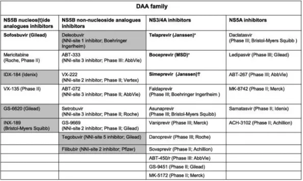
et al., 2007; Soriano et al., 2011). These compounds are globally called direct-acting antiviral (DAA) agents and target different viral non-structural proteins, including the NS3/4A protease, the NS5B polymerase, and the NS5A protein (Table 1).
Table 1. Direct acting antiviral (DAA) agents approved or in more advanced stages of clinical development.
The dark background identifies those compounds for which developments has been stopped. ∗ Approved by the FDA and EMA in 2011.
†
Approved by the FDA in November 2013.
ψ
Approved by the FDA and EMA in December 2013 and January 2014, respectively.
Two safer and more effective compounds have been recently approved for HCV treatment: the protease inhibitor simeprevir and the nucleotide polymerase inhibitor sofosbuvir. The current quest of HCV therapy development is to find the most effective, tolerable and affordable DAA combination with the least pill burden and highest viral resistance threshold that can cure people infected with HCV in the shortest period of time.
HCV interferon-dependent therapies rely on host factors such as IL28B polymorphism, liver fibrosis stage, and prior pegIFNα-RBV history to predict treatment response (Pawlotsky, 2013; Asselah and Marcelin, 2013; Poveda et al., 2012). While these factors may also affect responses to IFN-free DAA therapies; their impact diminishes as the potency of DAA regimens increases. One key component of the potency of DAA regimens is the resistance barrier for compounds in the regimen and the overall regimen itself. HCV is an RNA virus with an error-prone RNA polymerase, for which some analogies to HIV reverse-transcriptase can be drawn. Drug resistance frequently emerges in HIV patients treated with antiretrovirals and therefore limits the efficacy of these therapies. Given the known high virion production of HCV (100-fold higher than that of HIV) and error rate of the RNA polymerase (10–4 substitutions per base per year, approximately 10-fold higher than that of HIV reverse transcriptase), the potential for the existence of baseline resistant polymorphism and/or the short-term resistance development following PI exposure is greater than HIV (Neumann et al., 1998; Martell et al., 1992). However, one key difference is the presence of an extremely long-lived viral reservoir in the case of HIV; such a reservoir does not exist for HCV (Soriano et al., 2008).
As we are just entering the era of IFN-free DAA therapy for hepatitis C, many key questions regarding these therapies and resistance remain. What is the clinical significance of baseline DAA resistance polymorphisms? If significant, do these baseline resistance polymorphisms impact different classes of DAAs equally? Is PI cross-resistance a consideration for patients who failed prior HCV PI regimens? If so, for how long, and does resistance testing play any role in determining this? In this review, we present the DAAs according to their mechanism of action, discuss important clinical differences among licensed PI, review the relevant drug resistance profile for each class, according to available in vitro and in vivo data, and address clinical implications, where appropriate. Finally, we discuss the utility of performing baseline resistance testing to detect baseline DAA polymorphism and discuss specific situations where its use could be clinically meaningful.
2. Main HCV resistance patterns and mutations for DAA agents
Resistance to DAAs is driven by the selection of mutations at different positions in the NS3 protease, NS5B polymerase and NS5A protein (Sarrazin and Zeuzem, 2010; Kieffer et al., 2010; Vermehren and Sarrazin, 2012; Poveda and Soriano, 2012). Each compound or drug family displays a specific mutation profile that may be influenced by the genotype/subtype. Furthermore, each class of DAAs is characterized by a difference in the genetic barrier to resistance; though this general characterization differs for individual agents in the class. Cross-resistance between compounds in the same inhibitor class is of most concern for NS3 protease and NS5A inhibitors.
2.1. HCV protease inhibitors
confer significant loss of inhibitory activity (Kieffer et al., 2012; Barnard et al., 2012; Ogert et al., 2013; Halfon and Locarnini, 2011).
In April 2011, telaprevir and boceprevir were the first generation of HCV PIs approved for the treatment of genotype 1-infected patients in combination with pegIFN-RBV. Both drugs are orally bioavailable, linear ketoamide inhibitors which bind covalently but reversibly to the protease catalytic site (Kieffer et al., 2012; Barnard et al., 2012; Ogert et al., 2013; Halfon and Locarnini, 2011).
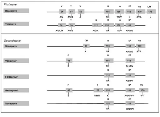
Several mutations in different positions at the NS3 protease have been associated with loss in susceptibility to PIs (Fig. 1). Resistance to first-generation PIs is characterized by selection of mutations at positions 36, 54, 55, 155, 156, and 170 and the resistance mutation profile is influenced by genotype subtypes (Sarrazin and Zeuzem, 2010; Kieffer et al., 2010; Kieffer et al., 2012; Vermehren and Sarrazin, 2012; Poveda and Soriano, 2012; Barnard et al., 2012; Ogert et al., 2013; Halfon and Locarnini, 2011; Poveda and García, 2013). Patients infected with HCV genotype subtype 1a mainly select mutations at positions 36 and 155. However, individuals with HCV subtype 1b select changes at codons 54, 55, 156 and 170. This difference in resistance pathways has been explained by the number of nucleotide changes needed at position 155 by genotype 1a and 1b. For example, subtype 1b needs two nucleotide changes at position 155 to produce resistance (R155K: CGG to AAG) whereas only one is needed for subtype 1a (R155K: AGG to AAG). This difference in nucleotide changes required combined with the higher relative fitness of the R155 variants compared to those selected in 1b, such as A156, explains the clinical observation that patients infected with genotype subtype 1b have a greater barrier to resistance than those with subtype 1a (Sarrazin and Zeuzem, 2010; Kieffer et al., 2010; Kieffer et al., 2012; Vermehren and Sarrazin, 2012; Poveda and Soriano, 2012; Barnard et al., 2012; Ogert et al., 2013; Halfon and Locarnini, 2011; Poveda and García, 2013).
In December 2013, simeprevir was approved by the FDA for the treatment of patients infected with genotype 1 and represents the second wave of the first generation of PIs. The second wave of PIs includes agents with improved potency and dosing but with resistance profiles that are similar to telaprevir and boceprevir (Lenz et al., 2013a). Both first and second-wave of first generation of PIs are characterized by a low genetic barrier for resistance and broad cross-resistance between compounds (Table 2) (Poveda and García, 2013). It is proposed that the term “second-generation” PIs be used for agents that have an improved resistance profile, such as several that are currently in development (e.g., ABT-450 and MK-5172) (De Nicola and Aqhemo, 2014).
Table 2. Main characteristics of the genotype activity and resistance of DAA classes.
Genotype activity Cross-resistance Key resistance mutations
NS3 protease
High First PI generation WHO, 2013; European Association for the Study of the Liver, 2011; McHutchison et al., 2009; Pawlotsky et al.,
High Sofosbuvir* Martell et al. (1992): G1a: S282T+(I434M)
Across all genotypes (1b > 1a) High G1a Ogert et al. (2013) and Osinusi et al. (2013): M28T, Q30E/R, L31F/M/V, Y93C/H/N G1b Ogert et al. (2013) and Osinusi et al. (2013): L31F/M/V, Y93C/H/N
2.1.1. Which advantages enhance second-wave and second-generation of protease inhibitors?
First, in contrast to telaprevir or boceprevir, second-wave protease inhibitors represent a significant improvement for dose administration, being administered once daily and are generally better tolerated (Summa et al., 2012). Second, while the first generation PIs are most active against genotype 1, the second wave of protease inhibitors generation are active against all genotypes with the exception of genotype 3, due to the presence of the natural polymorphism D168Q that confers resistance to available PIs (Lenz et al., 2013a). Third, although broad cross-resistance exists between PIs mainly due to the selection of mutations at positions 155 and 156 (the first wave of the first generation) and 168 (second wave or second generation), resistance to the first wave of PIs (telaprevir and boceprevir) does not completely overlap with the second wave or second generation, such as simeprevir, ABT-450, faldaprevir or asunaprevir (Fig. 1) (Kieffer et al., 2010; Kieffer et al., 2012; Vermehren and Sarrazin, 2012; Poveda and Soriano, 2012; Barnard et al., 2012; Ogert et al., 2013; Halfon and Locarnini, 2011; Poveda and García, 2013; Lenz et al., 2013a; De Nicola and Aqhemo, 2014).
MK-5172 is also a second generation PIs that is administered as a once a day pill that seems to be very potent with a broader HCV genotype coverage. In vitro, MK-5172 is very potent and retains activity against HCV viruses that harbor resistance mutations to other HCV PIs, such as V36A/M, T54A/S, R155K/Q/T, A156S, V36M+R155K or T54S+R155K. Moreover, MK-5172 is expected to be broadly active against multiple HCV genotypes (Summa et al., 2012). Recently, results from a phase 2 clinical trial showed promising results with MK-5172-based therapy with sustained viral response (SVR) rates of 89–100% in HCV genotype 1 patients (Manns et al., 2013a).
2.1.2. What is the impact of the natural polymorphism Q80K on the SVR?
Table 3. Prevalence of key polymorphisms at NS3/4A, NS5B polymerase and NS5A protein sequences associated with
* In combination with mutations Y448H, D559G or Y555C. 1. Bae et al. (2010); 2. Mcphee et al. (2012a,b); 3. Lenz et al. (2013); 4. Lawitz et al. (2010); 5. Troke et al. (2012); 6. Lawitz et al. (2012); 7. Fridell et al. (2011); 8. Gao (2013).
In vitro studies have shown that Q80K reduces susceptibility to simeprevir but not to other second wave PIs such as sovaprevir, asunaprevir or faldaprevir. Having a Q80K reduces 10-fold susceptibility to simeprevir (Bae et al., 2010) and confers ⩽5-fold reduction in the replicon susceptibility to sovaprevir and minimally increases the EC50 to asunaprevir (3-fold increase) (Mcphee et al., 2012a).
In vivo studies support the in vitro observations. The Phase II ASPIRE trial evaluated the impact of Q80K polymorphism on the virologic response to simeprevir (Lenz et al., 2013c). Patients infected with HCV genotype 1a that harbored the Q80K polymorphism had a significant decrease in the SVR to simeprevir (100 mg dose) compared to patients without Q80K (22% vs. 70%, respectively). However, an increase in the simeprevir doses to 150 mg was enough to achieve similar rates of SVR irrespective of Q80K status (Q80K 61% vs. Q80Q 66%).
The Phase III clinical trials QUEST-1 and QUEST-2 assessing the safety and efficacy of simeprevir in combination with pegIFN-RBV in patients infected with genotype 1 refined our understanding of the impact of Q80K on SVR rates to simeprevir. Compared with those patients infected with HCV genotype 1a without Q80K, lower SVR rates were observed among patients with the baseline Q80K treated with simeprevir (58% vs. 84%, respectively) (Jacobson et al., 2013).
Recent studies have defined at least two clades (clade 1 and clade 2) for subtype 1a, which display different geographic distribution and prevalence of the Q80K polymorphisms. Clade 1 is more frequently observed in NS3 sequences coming from the Americas and is associated with a high prevalence of Q80K (48.9%). Conversely, clade 2 is more frequent in sequences coming from Europe and has a lower frequency of Q80K (De Luca et al., 2013; Pickett et al., 2011). These findings have recently been corroborated with data from simeprevir Phase IIB/III studies (PILLAR, ASPIRE, QUEST-1, QUEST-2 and PROMISE) which showed a higher prevalence of Q80K among genotype 1a protease sequences from North America, compared with those from Europe (48.1% vs. 19.4%, respectively) (Lenz et al., 2013b).
The package insert and the most recent American HCV Guidelines for the treatment of chronic hepatitis C (AAS, 2014) recommend performing baseline resistance testing for Q80K in genotype 1a patients with consideration of an alternative to simeprevir if this mutation is present.
2.2. HCV nucleos(t)ide analog polymerase inhibitors
This family of compounds blocks HCV RNA synthesis by inhibitory competition with the physiologic nucleotide triphosphates for binding to the catalytic site of the enzyme (Ranjith-Kumar et al., 2006). Nucleos(t)ides are also called chain terminators because following incorporation the subsequent nucleotide triphosphate cannot be added to the RNA strand. Nucleos(t)ide analogs display a uniquely high barrier to resistance and possess antiviral activity across all genotypes (Table 2). In vitro studies demonstrated that drug resistance mutations to this class are selected within or near the polymerase catalytic site. Thus, the enzymatic activity appears to be impaired to such a large degree that viral replication is seriously compromised. The S282T is the in vitro signature resistance mutation to this class. However, the S282T mutation has rarely been detected in patients who failed treatment with nucleos(t)ide polymerase inhibitors in clinical trials. Several nucleotide analogs have shown very promising results and sofosbuvir is the first DAA in this family to gain regulatory approval ( Gerber et al., 2013; Pockros, 2013; Soriano et al., 2012).
2.2.1. Is there any influence of HCV genotype/subtype on the resistance profile to nucleotide analogs?
In contrast to NS3 protease-, NS5B non-nucleoside- and NS5A-inhibitors where resistance mutations are subtype-dependent, little is known about NS5B nucleos(t)ide analogs genotype and subtype-dependent resistance mutations.
2.2.2. In vitro resistance data for sofosbuvir
Sequence analyses also revealed differences in the resistance profile to nucleoside analogs among genotypes/subtypes. For genotype 1b, S282T was the only change selected after sofosbuvir exposure while for genotype 1a an additional mutation I434M was observed in combination with S282T. In the case of genotype 2a replicons, at least five additional mutations (T179A, M289L, I293L, M434T, and H479P) were selected prior to and after the emergence of S282T.Sepecifically, S282T together with mutations from both the finger (T179A) and palm (M289L and I293L) domains was essential to conferring resistance to sofosbuvir, while changes at the surface of the thumb domain (M434T and H479T) act as compensatory mutations improving the fitness of S282T variants (Lam et al., 2012).
Other in vitro studies evaluated the impact of the S282T mutation on the replication capacity of different genotypes/subtypes in the presence of sofosbuvir. Fitness assays demonstrated that the genotype 1a S282T replicon was the least fit compared with the wild type (3%) followed by genotype 1b S282T (12%), while genotype 2a S282T replicon was the most fit (30%) (Gerber et al., 2013).
2.2.3. In vivo data related to sofosbuvir resistance
The S282T mutation has been found infrequently in patients failing sofosbuvir-based regimens. First, in a patient infected with HCV genotype 2b who had detectable HCV RNA (virologic relapse) after 12 weeks of sofosbuvir monotherapy (Gene et al., 2013), second, in a HCV genotype 1 patient who relapsed in the SPARE trial after treatment with sofosbuvir/ribavirin for 24 weeks (Osinusi et al., 2013) and finally in another patient infected with HCV genotype 1 who had a virologic relapse after 8 weeks of treatment with sofosbuvir and the NS5A inhibitor ledipasvir (Lawitz et al., 2014).
2.2.4. In vitro data related to mericitabine resistance
Mericitabine is a prodrug of a cytosine nucleoside analog. It is active against all genotypes (1– 6) but it has been most extensively studied against genotype 1. The S282T mutation resulted in a moderate 3- to 6-fold reduction in susceptibility to mericitabine but significantly impacted replication capacity with a reduction to 15% compared to wild type replication levels. Similarly to what was seen in genotype 2a with sofosbuvir, S282T can be accompanied by the selection of other mutations (K81R, S84S/P, I239L, A300A/T, L320F/L/C, A421V, and Y586C) that appear to enhance replication capacity (Pawlotsky et al., 2012; Ali et al., 2008).
2.2.5. In vitro data related to mericitabine resistance
To date, there is no evidence of selection of S282T in studies of patients exposed to mericitabine in combination with pegIFN α-RBV (JUMP-C trial). However, during the INFORM-1 trial which evaluated the safety and efficacy of mericitabine in combination with the protease inhibitor danoprevir plus ribavirin, one genotype 1a patient who experienced a viral breakthrough had dual resistance to mericitabine (S282T) and danoprevir (R155K) Gane et al., 2012. Recently, a double mutant L159F/L320F with impaired replication capacity was identified in one genotype 1b-infected patient failing mericitabine plus pegIFN α-RBV in the PROPEL and JUMP-C trials (Tong et al., 2014).
2.2.6. Is the antiviral activity of nucleos(t)ide analogs inhibitors influenced by genotype/subtype?
Unlike many other DAA, HCV nucleos(t)ide analog inhibitors display pangenotypic antiviral activity. This feature, together with its high resistance barrier and lack of primary resistance makes these drugs very attractive as key components of future DAA interferon-free regimens. However, initial data from sofosbuvir clinical trials (FISSION, POSITRON and FUSSION trials) have demonstrated lower frequencies of SVR against genotype 3 compared to genotype 2. In fact, the rate of SVR for patients infected with genotypes 3 under sofosbuvir/ribavirin therapy varied between 30% for treatment experienced patients (FUSION trial) to 56% for interferon-naïve patients (FISSION) compared with SVR rates of 86% and 97% in genotypes 2, respectively (Table 2) (Asselah, 2013). Subsequent data from two new studies (VALENCE and LONESTAR-2) have shown increased cure rates for HCV genotype 3 patients under sofosbuvir/ribavirin therapy by increasing treatment duration from 12 to 24 weeks, or by the addition of pegIFN α (85% and 83%, respectively) (Zeuzem et al., 2013; Lawitz et al., 2013b).
Regarding subtypes 1a and 1b, no significant differences were observed in SVR rates to sofosbuvir. The NEUTRINO trial evaluated the efficacy of sofosbuvir in combination with pegIFN α-RBV in naïve patients. This study reported rates of SVR of 92% and 82% for HCV genotypes 1a and 1b, respectively, similarly to that found in recent data from the ATOMIC and NEUTRINO trials (Foster et al., 2014). For genotype 2 interferon-naïve or treatment-experienced patients, sofosbuvir-ribavirin provided excellent results showing rates of SVR of 97% and 86%, respectively. Among genotypes 4 and 5/6 the SVR rates were up to 96%; however, these results must be taken with caution, since only 35 patients infected with these genotypes were included in the trial (Lawitz et al., 2013a).
The efficacy of mericitabine in combination with pegIFN α-RBV in the JUMP-C trial was similar between genotypes 1a and 1b. However, during the INFORM-1 study, which evaluated the interferon-free regimen of mericitabine + danoprevir + ribavirin a significant difference was observed in rates of SVR between genotypes 1a and 1b (26% vs. 71%, respectively) (Gane et al., 2012). In this case, different SVR between 1a and 1b are probably due to lower rates of response to danoprevir for genotypes 1a compared with 1b. Finally, of the five patients receiving mericitabine in the JUMP-C trial, three (60%) achieved SVR (Pockros et al., 2013).
2.3. HCV non-nucleoside polymerase inhibitors
Non-nucleoside polymerase inhibitors (NNI) are non-competitive blockers of HCV-RNA synthesis that interact with the HCV polymerase at allosteric sites outside the catalytic site and prevent conformational changes in the polymerase that are critical to its function. At least five different allosteric binding sites have been identified as targets for non-nucleoside inhibitors (NNI-1, NNI-2, NNI-3, NNI-4, and NNI-5) as determined by their unique resistance patterns (Pauwels et al., 2007). These sites are located at the thumb (sites 1 and 2) and palm (sites 3, 4 and 5) of a right-hand modeled polymerase enzyme (Bressanelli et al., 2002; De Francesco and Carfi, 2007).
2.3.1. Resistance to NNI-site 1 inhibitors
Resistance mutations to deleobuvir are selected at residues 495, 496 and 499 of the NS5B polymerase in genotype 1b patients. The most predominant variants detected at failure were P495L or P495S, which decrease susceptibility to deleobuvir 123–130-fold and 91-fold, respectively. V499A, a polymorphism detected in the majority (>96%) of genotypes 1a, 2, 3 and 4, confers 5.6-fold increase in the EC50 to deleobuvir in genotype 1b. Mutation A421V caused 5.8-5.6-fold increase resistance to deleobuvir in genotype 1a (Larrey et al., 2013). The clinical development of deleobuvir has been recently halted due to its lower efficacy against genotype 1a.
2.3.2. Resistance to NNI-site 2 inhibitors
Filibuvir binds to the thumb 2 domain of the NS5B polymerase. Mutations at residue M423I/L/T represent the predominant pathway to filibuvir resistance displaying high-level resistance and reduced replicative capacity relative to the wild-type (Troke et al., 2012). For VX-222, another NNI-site 2 inhibitor in phase II development; breakthrough was associated with the selection of mutations at codons L419, R422, and M423. In the case of GS-9669, a NNI-site 2 inhibitor in early clinical development, substitution at codons L419, R422, and A486 were commonly selected in genotype 1a and 1b following 3 days of GS-9669 monotherapy (Lawitz et al., 2012).
2.3.3. Resistance to NNI-site 3 inhibitors
Setrobuvir is under Phase II investigation. In vitro studies have associated the selection of mutations at positions M414, G554, and D559 with setrobuvir resistance. ABT-072 and ABT-333 are also NNI-site 3 inhibitors currently under evaluation in combination with other DAA (i.e., ABT-450, a protease inhibitor) and RBV in IFN-free regimens. In vitro studies have revealed mutations to these inhibitors at the following positions: C316, S368, M414, Y448, and S556 ( Lawitz et al., 2010).
2.3.4. Resistance to NNI-site 5 inhibitors
Finally, tegobuvir binds to the β-hairpin in the thumb domain of NS5B, representing the first-in-class site 5 inhibitor. The most commonly detected mutation in patients failing tegobuvir-based therapy in combination with pegIFN α-RBV was Y448H. Tegobuvir was evaluated in double, triple and quadruple regimens with the protease inhibitor GS-9256, with or without RBV and/or pegIFN α (Zeuzem et al., 2012). Finally, because tegobuvir was associated with pancytopenia when administered with GS-9256 plus pegIFN-ribavirin, its development has been halted (Gerber et al., 2013).
2.4. NS5A inhibitors
The NS5A protein plays a crucial role in regulating HCV replication and host-cell interactions as an essential component of the viral replication complex. It has a three domain organization: domain I (amino acids 1–213) located at the N-terminal region, domain II (amino acids 250–342) and domain III (amino acids 356–447) at the C-terminal region. A new family of antivirals against HCV was recently found to inhibit the NS5A replication complex by unclear mechanisms involving interaction with NS5A (Tellinghuisen et al., 2004).
Daclatasvir is the first compound in this class, a small-molecule inhibitor that targets NS5A domain I. It has demonstrated potent antiviral activity against HCV replicons from different genotypes. However, higher rates of virological responses to daclatasvir have been observed among genotype 1b patients, compared to genotype 1a. These findings might be explained by a higher barrier to resistance for daclatasvir in genotype 1b than 1a (Table 2) (Sulkowski et al., 2014). Indeed, in vitro studies have highlighted that the loss of susceptibility in genotype 1a by the selection of resistance mutations ranged from 233 to 3350-fold, whereas mutations at similar positions only resulted in a 3–28-fold loss in susceptibility to genotype 1b (Gao et al., 2010; Fridell et al., 2010).
Overall, resistance substitutions identified in the in vitro replicon system correlate well with those observed in the clinic. Several amino acid changes at the N terminus of NS5A domain I at positions M28, Q30, L31 and Y93 for HCV genotype 1a and L31 and Y93 for genotype 1b have been associated with daclatasvir resistance. Daclatasvir is characterized by a low genetic barrier to resistance, especially for HCV genotype 1a in which the selection of a single mutation is enough to lose susceptibility to daclatasvir. Diminished replication capacity has been associated with resistance substitutions at residue Y93 ( Fridell et al., 2011; Gao, 2013).
Second-wave NS5A inhibitors (i.e., ledipasvir (GS-5885), BMS-766, ACH-3102, MK-8742, and IDX-719) displaying improved potency in vitro against resistant variants selected by daclatasvir are in development. As an example, the mutation Q30E frequently selected in genotype 1a confers high level resistance to daclatasvir (∼7500-fold), but only confers 5–50-fold resistance to BMS-766, ledipasvir and MK-8742. However, a broad cross-resistance between NS5A inhibitors is expected by the selection of mutations at codons 31 and/or 93 causing a loss in susceptibility to the majority of these compounds (Gao, 2013).
2.4.1. Resistance to daclatasvir
Natural polymorphisms in NS5A at positions that may influence susceptibility to daclatasvir are less frequently observed in genotypes 1a and 3, with the exception of the polymorphism Q30A found in almost all NS5A sequences from genotype 3 (Plaza et al., 2012). However, Q30A is not a relevant change for resistance to NS5A inhibitors. Mutation L31M is frequently observed among NS5A sequences from genotype 2 (range 50–85%), one of the key mutations to this family among genotypes 1a and 1b. However, the impact of this mutation has recently been assessed during a clinical trial evaluating the antiviral activity of daclatasvir against genotypes 2 and 3. This study demonstrated that the presence of L31M does not predict failure in genotype 2 patients receiving short-term treatment (12 weeks) with daclatasvir in combination with pegIFN α-RBV (Dore et al., 2013).
associated with daclatasvir resistance was assessed in the COMMAND-1 study which evaluated the efficacy of daclatasvir plus pegIFN α-RBV in genotype 1 treatment-naïve patients. For genotype 1a patients, from 32.7% of failures, 10% had pre-existing variants associated with resistance to NS5A inhibitors (M28T, Q30R/H, L31M/V/I, Y93H/N/C/S/T). At failure, predominant emergent NS5A resistance mutations were at codons 30, 31 and/or 93. Among genotypes 1b, from 14.5% of failures, 20% had baseline mutations associated with resistance to daclatasvir. Of note, in this study the prevalence of Y93H was 10% among genotypes 1b. The most prevalent resistance mutations selected at the time of failure were Y93H, Q30H-Y93H, L31V-Y93H, L31I-Y93H, L31M-Y93H (McPhee et al., 2012b).
Subsequently, the impact of baseline polymorphisms associated with loss of susceptibility to NS5A inhibitors was evaluated in a Phase III trial of daclatasvir combined with asunaprevir. This study highlighted that the presence of mutations at amino acids L31 and Y93 may reduce the barrier to resistance and influence virologic outcome for those patients who carry these polymorphisms at baseline (McPhee et al., 2013). Very recent data from the HALLMARK-dual study suggested that even in genotype 1b patients, the rate of SVR in patients with baseline resistance associated polymorphisms to asunaprevir/daclatasvir was only 40% compared to over 80% with no baseline resistance (Manns et al., 2013b). More recently, the results of safety and efficacy for the combination daclatasvir plus sofosbuvir for previously treated or untreated chronic HCV-infected patients have been published. In this study, although the prevalence of baseline polymorphisms associated with daclatasvir resistance was around 8% for genotype 1 NS5A sequences, all but one patient achieved SVR (Sulkowski et al., 2014).
In addition, recent data have pointed out that due to massive protein–protein interactions in the HCV replication complex the emergence of changes at NS3, NS4B and NS5B proteins might modulate the susceptibility to NS5A inhibitors (Yang et al., 2013).
2.4.2. Resistance to ledipasvir
Ledipasvir is a new NS5A inhibitor with demonstrated antiviral activity against HCV genotypes 1a and 1b. Very high rates of sustained virological response (93–100%) have been reported from Phase II and Phase III clinical trials in combination with sofosbuvir among untreated and previously treated patients (LONESTAR and ION-1 & ION-2 trials, respectively) (Lawitz et al., 2014; Afdhal et al., 2014a; Afdhal et al., 2014b). Although virological failure is rare using this DAA combination, baseline polymorphisms associated with loss of susceptibility to NS5A inhibitors have been found in half of the patients who relapsed (Afdhal et al., 2014a; Afdhal
et al., 2014b). Cross-resistance is expected between daclatasvir and ledipasvir, mainly due to the presence of mutations at positions L31 and Y93 (Gao, 2013).
3. Clinical implications of DAA resistance: Lessons from clinical trials
3.1. The importance of HCV genotype/subtype
Clinical trials evaluating the efficacy of DAA have shown higher rates of SVR among genotype 1b than genotypes 1a. Uniformly, genotypes 1b displayed a higher barrier to resistance than genotypes 1a for protease inhibitors, non-nucleosides inhibitors and NS5A inhibitors (Sarrazin and Zeuzem, 2010; Kieffer et al., 2010). These findings have highlighted the need for a correct subtype identification in genotypes 1 for an optimized clinical management of patients (European Association for the Study of the Liver, 2011).
In vitro studies have also demonstrated the lack of efficacy of the majority of protease inhibitors against genotypes 3 (with the exception of MK-5172), mainly due to the presence of the natural polymorphism D168Q among HCV genotype 3 protease sequences (Lenz et al., 2013a). Data from sofosbuvir clinical trials demonstrated lower efficacy against genotypes 3 showing SVR rates which ranged from 30% to 56%, compared with rates of 86% and 97% for genotype 2. However, these sub-optimal cure rates have been partially offset by increasing treatment duration from 12 to 24 weeks or by the addition of pegIFN α (Asselah, 2013). Interestingly, this difference in efficacy for genotype 3 is not clearly related to a different antiviral potency or barrier to resistance for sofosbuvir.
3.2. IL28B genotype
Single nucleotide polymorphisms (SNP) in the IL28B gene were identified in 2009 as strong predictors of treatment response to pegIFN α-RBV in HCV-infected patients. The SNP most strongly linked to SVR was rs12979860. Individuals carrying one (CT) or two copies (TT) of the T allele had a higher probability of failure compared with individuals carrying genotype CC (Ge et al., 2009). More recently, data obtained from different clinical trials evaluating the efficacy of several DAA compounds in combination with pegIFN α-RBV also point out the role of the IL28B polymorphism in the virologic response to these new therapies. In fact, in patients receiving a DAA-based therapy in combination with pegIFN α-RBV, virologic failure is more frequent in genotype 1a non-CC IL28B patients.
In this context, it has been observed that the presence of baseline polymorphisms and/or resistance-associated variants might be clinically relevant in IFN non-responder patients, genotypes 1a and non-CC IL28 patients (Barnard et al., 2012; Dore et al., 2013; McPhee et al., 2012b; McPhee et al., 2013). However, as new clinical guidelines are moving rapidly away from interferon-based combinations (AAS, 2014), the clinical utility of IL-28 testing is likely to be limited, particularly with more potent, all-DAA regimens.
3.3. Baseline natural polymorphisms
The impact of natural polymorphisms at positions involved in DAA resistance may be negligible in the context of combination therapies when other compounds of the regimen retain full activity. Using DAA-based therapies in combination with pegIFN α-RBV the presence of baseline polymorphisms or resistance associated variants might negatively influence the virologic response in poorly interferon-responsive patients (i.e., genotypes 1a and non-CC IL-28B) (Barnard
In theory, for interferon-free regimens the presence of baseline polymorphisms and/or resistance associated variants may have a clinically significant impact. The low genetic barrier to resistance for many DAAs, with the exception of nucleos(t)ide analogs, might facilitate the on-therapy emergence of resistance variants in patients harboring baseline polymorphisms and/or resistance mutations. Indeed, baseline polymorphisms associated with resistance to NS5A inhibitors has a non-negligible prevalence (10–15%) and their presence has been associated with lower rates of virologic response in some daclatasvir-based regimens (Dore et al., 2013; McPhee
et al., 2012b; McPhee et al., 2013).
3.4. Resistance variants for re-treatment strategies
In the case of telaprevir- and boceprevir-experienced patients, the selection of resistance mutations in the HCV protease might be relevant, if re-treatment with another protease inhibitors (e.g., simeprevir, faldaprevir) is considered. It has been demonstrated that after telaprevir or boceprevir discontinuation, resistance mutations tend to disappear after a median follow-up of 30 months in most patients (>85%) (Sherman et al., 2011). However, there are limited data evaluating re-treatment strategies with protease inhibitors. The C219 study examined the efficacy of a re-treatment strategy with telaprevir/pegIFN α-RBV in 9 patients who had received 14 days of telaprevir monotherapy a mean of 5.7 years prior to re-exposure. In this case, 5/9 had SVR with telaprevir/pegIFN α-RBV (Sarrazin et al., 2013). The OPERA study evaluated the efficacy of re-treatment with simeprevir/pegIFN α-RBV in 5 patients who had been exposed to a short course of simeprevir monotherapy 1.5 years prior. While 3 patients achieved SVR, in the other 2, persistence of low-level resistant variants may have contributed to the observed virologic failure (Lenz et al., 2012). Therefore, due to the potential risk of persistence of protease-resistant variants at low frequencies, re-treatment strategies with low-resistance barrier protease inhibitors in combination with pegylated interferon and ribavirin would not be advisable (AAS, 2014).
Conversely, very recent data from the LONESTAR trial demonstrated the success of a retreatment strategy with sofosbuvir/ledipasvir/RBV in one patient who previously had failed 8 weeks of sofosbuvir/ledipasvir. This favorable response was seen despite the presence of mutations associated with resistance to both NS5A inhibitors (Q30L, L31M, and Y93H) and the S282T mutation associated with sofosbuvir resistance at the onset of re-treatment (Lawitz et al., 2014). Whether this is result is generalizable is not known and requires confirmation from ongoing studies.
4. Current role of HCV drug resistance in clinical practice
4.1. Treatment-naïve patients and re-treatment after pegIFN α-RBV treatment failure
In these patient populations, baseline resistance testing for Q80K among genotype 1a is recommended, and alternative treatments to simeprevir should be considered if this mutation is present (AAS, 2014). Although there is a link between baseline resistance variants to NS5A inhibitors and treatment failure, in the context of very potent DAA combinations (e.g., sofosbuvir/daclatasvir) failures seem not to be associated with the presence of pre-existing resistance variants.
4.2. Re-treatment after failure of conventional telaprevir or boceprevir/pegIFN α-RBV
Since cross-resistance exists between first- and second-generation protease inhibitors, and due to the potential risk of persistence of protease resistance variants at low frequencies, re-treatment strategies with protease inhibitors would not be advisable.
Transparency declarations
None to declare.
References
AAS, 2014. The AASLD/IDSA Clinical Guidance for Hepatitis C. March 21, 2014. Available from: <http://www.hcvguidance.org>.
Afdhal et al., 2014a. N. Afdhal, K. Reddy, D. Nelson, et al. Ledipasvir and sofosbuvir for previously treated HCV genotype 1 infection. N. Engl. J. Med., 370 (2014), pp. 1483–1493.
Afdhal et al., 2014b. N. Afdhal, S. Zeuzem, P. Kwo, et al. Ledipasvir and sofosbuvir for untreated HCV genotype 1 infection. N. Engl. J. Med. (2014).
Ali et al., 2008. S. Ali, V. Leveque, S. Le Pogam, et al. Selected replicon variants with low-level in vitro resistance to the hepatitis C virus NS5B polymerase inhibitor PSI-6130 lack cross-resistance with R1479. Antimicrob. Agents Chemother., 52 (2008), pp. 4356–4369.
Asselah, 2013. T. Asselah. Sofosbuvir-based interferon-free therapy for patients with HCV infection. J. Hepatol (2013) Available from: <http://dx.doi.org/10.101016/j.jhep.2013.07.023>.
Asselah and Marcelin, 2013. T. Asselah, P. Marcelin. Interferon free therapy with direct acting antivirals for HCV. Liver Int., 33 (Suppl. 1) (2013), pp. 93–104.
Bacon et al., 2011. B. Bacon, S. Gordon, E. Lawitz, et al. Boceprevir for previously treated chronic HCV genotype 1 infection. N. Engl. J. Med., 364 (2011), pp. 1207–1217.
Bae et al., 2010. A. Bae, S.C. Sun, X. Qi, et al.. Susceptibility of treatment-naïve hepatitis C virus (HCV) clinical isolates to HCV protease inhibitors. Antimicrob. Agents Chemother., 54 (2010), pp. 5288–5297. Barnard et al., 2012. R.J. Barnard, J.A. Howe, R.A. Ogert, et al. Analysis of boceprevir resistance associated amino acid variants (RAVs) in two phase 3 boceprevir clinical trials. Virology, 444 (2012), pp. 329–336. Bartels et al., 2008. D. Bartels, Y. Zhou, E. Zhang, et al.. Natural prevalence of HCV variants with decreased sensitivity to NS3-4A protease inhibitors in tratment-naïve subjects. J. Infect. Dis., 198 (2008), pp. 800– 807.
Berger et al., 2013. K.L. Berger, L. Lagacé, I. Triki, et al. Viral resistance in hepatitis C genotype-1 infected patients receiving the NS3 protease inhibitor faldaprevir (BI 2011335) in a phase 1b multiple rising-dose study. Antimicrob. Agents Chemother., 57 (2013), pp. 4928–4936.
Bressanelli et al., 2002. S. Bressanelli, L. Tomei, F. Rey, et al. Structural analysis of the hepatitis C virus RNA polymerase in complex with ribonucleotides. J. Virol., 76 (2002), pp. 3482–3492.
De Francesco and Carfi, 2007. R. De Francesco, A. Carfi. Advances in the development of new therapeutic agents targeting NS3-4A serine protease or the NS5b RNA-dependent RNA polymerase of the hepatitis C virus. Adv. Drug Deliv. Rev., 59 (2007), pp. 1242–1262.
De Luca et al., 2013. A. De Luca, S. Di Giambenedetto, M. Prosperi, et al. Two distinct HCV genotype 1a clades: geographical distribution and association with natural resistance mutations to HCV NS3/4A inhibitors. Antivir. Ther., 18 (Suppl. 1) (2013), p. A47.
Dore et al., 2013. Dore, G.J., Lawitz, E., Hezode, C., et al., 2013. Daclatasvir combined with peginterferon alfa 2a-ribavirin for 12 or 16 weeks in patients with hepatitis C virus genotype 2 or 3 infection: COMMAND GT 2/3 study. In: 48th Annual Meeting of the European Association for the Study of the Liver, 24–28 April 2013, Amsterdam, Belgium. (Abstract 1418).
European Association for the Study of the Liver, 2011. European Association for the Study of the Liver. EASL clinical practice guidelines: management of hepatitis C virus infection. J. Hepatol., 55 (2011), pp. 245–264.
Fabrycki et al., 2012. Fabrycki, J., Patel, D., Yang, G., et al., 2012. Characterization of HCV NS3 protease variants from patients enrolled in a 28-day phase 2a trial of ACH-1625 daily dosing plus pegIFN-alpha 2a/RBV. In: 47th Annual Meeting of the European Association for the Study of the Liver, 18–22 April 2012, Barcelona, Spain. (Abstract 1182).
Foster et al., 2014. Foster, G., et al., 2014. Sofosbuvir-based regimens are associated with high SVR rates across genotypes and among patients with multiple negative predictive factors. In: 49th Annual Meeting of the European Association for the Study of the Liver, 9–13 April, 2014, London, UK. (Abstract 066). Fridell et al., 2010. R. Fridell, D. Qiu, C. Wang, L. Valera, M. Gao. Resistance analysis of the hepatitis C
virus NS5A inhibitor BMS-790052 in an in vitro replicon system. Antimicrob. Agents Chemother., 54 (2010), pp. 3641–3650.
Fridell et al., 2011. R.A. Fridell, C.F. Wang, J.H. Sun, et al. Genotypic and phenotypic analysis of variants resistant to hepatitis C virus nonstructural protein 5A replication complex inhibitor BMS-790052 in humans: in vitro and in vivo correlations. Hepatology, 54 (2011), pp. 1924–1935.
Gane et al., 2012. E.J. Gane, et al. Interferon-free treatment with a combination of mericitabine and danoprevir/r with or without ribavirin in treatment-naive HCV genotype-1 infected patients. J. Hepatol, 56 (Suppl. S2) (2012), pp. S555–S556.
Gane et al., 2012. E.J. Gane, P. Pockros, S. Zeuzem, et al. Interferon-free treatment with a combination of mericitabine and danoprevir/r with or without ribavirin in treatment-naïve HCV genotype 1-infected patients. J. Hepatol., 56 (2012), pp. S555–S556.
Gao, 2013. M. Gao. Antiviral activity and resistance of HCV NS5A replication complex inhibitors. Curr. Opin. Virol., 3 (2013), pp. 1–7.
Gao et al., 2010. M. Gao, R. Nettles, M. Belema, et al. Chemical genetics strategy identifies an HCV NS5A inhibitor with a potent clinical effect. Nature, 465 (2010), pp. 96–100.
Ge et al., 2009. D. Ge, J. Fellay, A. Thompson, et al. Genetic variation in IL28B predicts hepatitis C treatment-induced viral clearance. Nature, 461 (2009), pp. 399–401.
Gene et al., 2013. E.J. Gene, C.A. Stedman, R.H. Hyland, et al. Nucleotide polymerase inhibitor sofosbuvir plus ribavirin for hepatitis C. N. Engl. J. Med., 368 (2013), pp. 34–44.
Gerber et al., 2013. L. Gerber, T. Welzel, S. Zeuzem. New therapeutic strategies in HCV: polymerase inhibitors. Liver Int., 33 (Suppl. 1) (2013), pp. 85–92.
Halfon and Locarnini, 2011. P. Halfon, S. Locarnini. Hepatitis C virus resistance to protease inhibitors. J. Hepatol., 55 (2011), pp. 192–206.
Jacobson et al., 2011. I. Jacobson, J. McHutchison, G. Dusheiko, et al. Telaprevir for previously untreated chronic hepatitis C virus infection. N. Engl. J. Med., 364 (2011), pp. 2405–2416.
Jacobson et al., 2013. Jacobson, I., Dore, G., Foster, G., et al., 2013. Simeprevir (TMC435) with
peginterferon/ribavirin for treatment of chronic HCV genotype 1 infection in treatment-naïve patients: efficacy in difficult-to-treat patient sub-population in the QUEST-1 and 2 Phase III trials. In: 64th Annual Meeting of the American Association for the Study of Liver Disease. 1–5 November, 2013, Washington DC, USA. (Abstract 1122).
Kieffer et al., 2010. T.L. Kieffer, A.D. Kwong, G.R. Picchio. Viral resistance to specifically targeted antiviral therapies for hepatitis C (STAT-Cs). J. Antimicrob. Chemother., 65 (2010), pp. 202–212.
Kieffer et al., 2012. T. Kieffer, S. De Meyer, D. Bartels, et al. Hepatitis C Viral evolution in genotypes 1 treatment-naïve and treatment-experienced patients receiving telaprevir-based therapy clinical trials. PLoS One, 7 (2012), p. e34372.
Kuntzen et al., 2008. T. Kuntzen, J. Timm, A. Berical, et al. Naturally occurring dominant resistance mutations to HCV protease and polymerase inhibitors in treatment-naive patients. Hepatology, 48 (2008), pp. 1769–1778.
Lam et al., 2012. A.M. Lam, C. Espiritu, S. Bansal, et al. Genotype and subtype profiling of PSI-7977 as a nucleotide inhibitor of hepatitis C virus. Antimicrob. Agents Chemother., 56 (2012), pp. 3359–3368. Larrey et al., 2013. D. Larrey, A. Lohse, C. Trepo, et al. Antiviral effect, safety, and pharmacokinetics of
5 days’oral administration of deleobuvir (BI 207127), and investigational HCV RNA polymerase inhibitor, in patients with chronic hepatitis C. Antimicrob. Agents Chemother., 57 (2013), pp. 4727–4735. Lawitz et al., 2010. E. Lawitz, M. Rodriguez-Torres, V.K. Rustgi, et al. Safety and antiviral activity of
Lawitz et al., 2012. Lawitz, E., Hazan, L., Gruener, D., et al., 2012. GS-9669, a novel NS5B non-nucleoside thumb site II inhibitor, demonstrates potent antiviral activity, favorable safety profile and potential for once-daily. In: 47th Annual Meeting of the European Association for the Study of the Liver, 18–22 April 2012, Barcelona, Spain. (Abstract 1189).
Lawitz et al., 2013a. Lawitz, E., Wyles, D., Davis, M., et al., 2013a. Sofosbuvir + peginterferon + ribavirin for 12 weeks achieves 90% SVR12 in genotype 1, 4, 5, or 6 HCV infected 90% SVR12 in genotype 1, 4, 5, or 6 HCV infected patients: the NEUTRINO study. In: 48th Annual Meeting of the European Association for the Study of the Liver, 24–28 April, Amsterdam, Belgium. (Abstract 1411).
Lawitz et al., 2013b. Lawitz, E., Poordad, F., Brainard, D.M., et al., 2013b. Sofosbuvir in combination with peginterferon and ribavirin for 12 weeks provides high SVR rates in HCV-infected genotype 2 or 3 treatment experienced patients with and without compensated cirrhosis: results from the LONESTAR-2 study. In: 64th Annual Meeting of the American Association for the Study of Liver Disease, 1–5 November, 2013, Washington DC, USA. (Abstract LB-4).
Lawitz et al., 2014. E. Lawitz, F. Poordad, P.S. Pang, et al. Sofosbuvir and ledipasvir fixed-dose combination with and without ribavirin in treatment-naïve and previously treated patients with genotype 1 hepatitis C virus infection (LONESTAR): an open-label, randomized, phase 2 trial. Lancet, 383 (2014), pp. 515–523. Lenz et al., 2012. O. Lenz, J. Bruijne, L. Vijgen, et al. Efficacy of re-treatment with TMC435 as combination
therapy in hepatitis C virus-infected patients following TMC435 monotherapy. Gastroentorology, 143 (2012), pp. 1176–1178.
Lenz et al., 2013a. O. Lenz, L. Vijgen, J.M. Berke, et al. Virologic response and characterization of HCV genotype 2–6 in patients receiving TMC435 (stydy TMC435-C202). J. Hepatol., 58 (2013), pp. 445–451. Lenz et al., 2013b. Lenz, O., Fevery, B., Verbinnen, T., et al., 2013b. Resistance analyses of HCV isolates
from patients treated with simeprevir in Phase IIB/III studies. In: 64th Annual Meeting of the American Association for the Study of Liver Disease. 1–5 November, 2013, Washington DC, USA. (Abstract 1101).
Lenz et al., 2013c. Lenz, O., Fevery, B., Vijgen, L., et al., 2013. TMC-435 in patients infected with HCV genotype 1 who failed previous pegylated interferon/ribavirin treatment: virologic analyses of the ASPIRE trial. In: Presented at 49th Annual Meeting of the European Association for the Study of the Liver, 18–22 April 2013, Barcelona, Spain. (Abstract 1329).
Manns et al., 2013a. Manns, M., Vierling, J.M., Bacon, B.R., et al., 2013. High sustained viral response at 12- and 24-week follow-up of MK-5172 with pegylated interferon alfa-2b and ribavirin in HCV genotype 1 treatment-naïve non-cirrhotic patients. In: 48th Annual Meeting of the European Association for the Study of the Liver (EASL 2013), 24–28 April, 2013, Amsterdam, Belgium. (Abstract 66).
Manns et al., 2013b. Manss, M., Pol, S., Jacobson, I.M., et al., 2013. All-oral dual therapy with daclatasvir and asunaprevir in patients with HCV genotype 1b infection: phase 3 HALLMARK-DUAL study results. In: 49th Annual Meeting of the European Association for the Study of the Liver, 9–13 April 2013, London, UK. (Abstract O166).
Martell et al., 1992. M. Martell, J.I. Esteban, J. Quer, J. Genesca, A. Weiner, R. Esteban, et al. Hepatitis C virus (HCV) circulates as a population of different but closely related genomes: quasispecies nature of HCV genome distribution. J. Virol., 66 (1992), pp. 3225–3229.
McCormick et al., 2013. A.L. McCormick, L. Wang, A. Garcia, et al. Development of a panel of genotype-specific resistance assays for the detection of amino acid changes in domain 1 of hepatitis C virus (HCV) NS5A associated with drug resistance. Antivir. Ther., 18 (Suppl. 1) (2013), p. A56.
McHutchison et al., 2009. J. McHutchison, E. Lawitz, M. Shiffman, et al. Peginterferon alfa-2b or alfa-2a with ribavirin for treatment of hepatitis C infection. N. Engl. J. Med., 361 (2009), pp. 580–593.
Mcphee et al., 2012a. F. Mcphee, J. Friborg, S. Levine, et al. Resistance analysis of the hepatitis C virus NS3 protease inhibitor asunaprevir. Antimicrob. Agents Chemother., 56 (2012), pp. 3670–3681.
McPhee et al., 2012b. F. McPhee, D. Hernandez, F. Yu, et al. A description of virologic escape in HCV genotype 1-infected patients treated with daclatasvir (BMS-790052) in combination with ribavirin and peginterferon alfa-2a or peinterferon alfa-2b. J. Hepatol., 56 (2012), p. S473.
McPhee et al., 2013. McPhee, F., Toyota, J., Chayama, D., et al., 2013. Analysis of HCV resistance variants in a phase 3 trial of daclatasvir combined with asunaprevir for Japanese patients with genotype 1b infection. In: 64th Annual Meeting of the American Association for the Study of Liver Disease, 1–5 November, 2013, Washington DC, USA. (Abstract 1111).
Neumann et al., 1998. A.U. Neumann, N.P. Lam, H. Dahari, et al. Hepatitis C viral dynamics in vivo and the antiviral efficacy of interferon-alpha therapy. Science, 282 (1998), pp. 103–107.
Ogert et al., 2013. R.A. Ogert, J.A. Howe, J.M. Vierling, et al. Resistance-associated amino acid variants associated with boceprevir plus pegylated interferon-2b and ribavirin in patients with chronic hepatitis C in the SPRINT-1 trial. Antivir. Ther., 18 (2013), pp. 387–397.
Pauwels et al., 2007. F. Pauwels, W. Mostmans, L. Quirymen, et al. Binding-site identification and genotypic profiling of hepatitis C virus polymerase inhibitors. J. Virol., 81 (2007), pp. 6909–6919.
Pawlotsky, 2013. J.M. Pawlotsky. Treatment of chronic hepatitis C: current and future. Curr. Top. Microbiol. Immunol., 369 (2013), pp. 321–342.
Pawlotsky et al., 2007. J.M. Pawlotsky, S. Chevaliez, J. McHutchison. The hepatitis C virus life cycle as a target for new antiviral therapies. Gastroenterology, 132 (2007), pp. 1979–1998.
Pawlotsky et al., 2012. J.M. Pawlotsky, I. Najera, I. Jacobson. Resistance to mericitabine, a nucleoside analogue inhibitor of HCV RNA-dependent RNA polymerase. Antivir. Ther., 17 (2012), pp. 411–423. Pickett et al., 2011. B.E. Pickett, R. Striker, E.J. Lefkowitz. Evidence for separation of HCV subtype 1a into
two distinct clades. J. Viral Hepat., 18 (2011), pp. 608–618.
Plaza et al., 2011. Z. Plaza, V. Soriano, M.M. Gonzales, et al. Impact of antiretroviral therapy on the variability of the HCV NS5B polymerase in HIV/HCV co-infected patients. J. Antimicrob. Chemother., 66 (2011), pp. 2838–2842.
Plaza et al., 2012. Z. Plaza, V. Soriano, E. Vispo, et al. Prevalence of natural polymorphisms at the HCV NS5A gene associated with resistance to daclatasvir, an NS5A inhibitor. Antivir. Ther., 17 (2012), pp. 921–926.
Pockros, 2013. P. Pockros. Nucleoside/nucleotide analogue polymerase inhibitors in development. Clin. Liver Dis., 17 (2013), pp. 123–128.
Pockros et al., 2013. P.J. Pockros, D. Jensen, N. Tsai, et al. JUMP-C: a randomized trial of mericitabine plus pegylated interferón alpha-2a/ribavirin for 24 weeks in treatment-naive HCV genotypes 1/4 patients. Hepatology, 58 (2013), pp. 514–523.
Poordad et al., 2011. F. Poordad, J. McCone, B. Bacon, et al. Boceprevir for untreated chronic HCV genotype 1 infection. N. Engl. J. Med., 364 (2011), pp. 1195–1206.
Poveda and García, 2013. E. Poveda, F. García. Telaprevir resistance. Enferm. Infecc. Microbiol. Clin., 31 (Suppl 3) (2013), pp. 26–32.
Poveda and Soriano, 2012. E. Poveda, V. Soriano. Drug resistance testing in hepatitis C therapy. Future Virol., 7 (2012), pp. 1–13.
Poveda et al., 2012. E. Poveda, E. Vispo, P. Barreiro, et al. Predicted challenges using direct acting antivirals in the current HIV-HCV coinfected population in Spain. Antivir. Ther., 17 (2012), pp. 571–575. Ranjith Kumar et al., 2006. C.T. Ranjith-Kumar, C. Cheng Kao. Biochemical activities of the HCV NS5B
RNA-dependent RNA polymerase. S.L. Tan (Ed.), Hepatitis C Viruses: Genomes and Molecular Biology (1st ed.), Horizon Bioscience, Norfolk (2006), pp. 293–310.
Sarrazin and Zeuzem, 2010. C. Sarrazin, S. Zeuzem. Resistance to direct antiviral agents in patients with hepatitis C virus infection. Gastroenterology, 138 (2010), pp. 447–462.
Sarrazin et al., 2013. C. Sarrazin, H. Reesink, S. Zeuzem, et al. Treatment with telaprevir/PEG-IFN/RBV after 14 days telaprevir exposure in phase I studies: results from the phase IIIB C219 rollover study. J. Hepatol., 58 (2013), pp. S369–S370.
Sherman et al., 2011. Sherman, K., Sulkowski, M., Zoulim, F., et al., 2011. Follow-up of SVR durability and viral resistance in patients with chronic hepatitis C treated with telaprevir-based regimens: interim analysis of the extend study. Hepatology 54, 485A–486A.
Soriano et al., 2008. V. Soriano, A. Perelson, F. Zoulim. Why are there different dynamics in the selection of drug resistance in HIV and hepatitis B and C viruses?. J. Antimicrob. Chemother., 62 (2008), pp. 1–4. Soriano et al., 2011. V. Soriano, E. Vispo, E. Poveda, et al. Directly acting antivirals against hepatitis C virus.
J. Antimicrob. Chemother., 66 (2011), pp. 1673–1686.
Soriano et al., 2012. V. Soriano, E. Vispo, C. de Mendoza, et al. Hepatitis C therapy with HCV NS5B polymerase inhibitors. Expert Opin. Pharmacother., 14 (2012), pp. 1161–1170.
Sulkowski et al., 2014. M. Sulkowski, D. Gardiner, M. Rodriguez-Torres, et al. Daclatasvir plus sofosbuvir for previously treated or untreated chronic HCV infection. N. Engl. J. Med., 370 (2014), pp. 211–221. Summa et al., 2012. V. Summa, S.W. Ludmerer, J.A. McCauley, et al. MK-5172, a selective inhibitor of
hepatitis C virus NS3/4A protease with broad activity across genotypes and resistant variants. Antimicrob. Agents Chemother., 56 (2012), pp. 416–417.
Tellinghuisen et al., 2004. T.L. Tellinghuisen, J. Marcotrigiano, A.E. Gorbalenya, et al. The NS5A protein of hepatitis C virus is a zinc metalloprotein. J. Biol. Chem., 279 (2004), pp. 48576–48587.
Tong et al., 2014. X. Tong, S. Le Pogam, L. Li, K. Haines, K. Piso, V. Baronas, J.M. Yan, S.S. So, K. Klumpp, I. Nájera. In vivo emergence of a novel mutant L159F/L320F in the NS5B polymerase confers low-level resistance to the HCV polymerase inhibitors mericitabine and sofosbuvir. J. Infect. Dis., 209 (2014), pp. 668–675.
Treviño et al., 2011. A. Treviño, C. de Mendoza, P. Parra, et al. Natural polymorphisms associated with resistance to new antivirals against hepatitis C virus (HCV) in newly diagnosed HIV/HCV-coinfected patients in Spain. Antivir. Ther., 16 (2011), pp. 413–416.
Troke et al., 2012. P. Troke, M. Lewis, P. Simpson, et al. Characterization of resistance to the nonnucleoside NS5B inhibitor filibuvir in hepatitis C virus-infected patients. Antimicrob. Agents Chemother., 56 (2012), pp. 1331–1339.
Vermehren and Sarrazin, 2012. J. Vermehren, C. Sarrazin. The role of resistance in HCV treatment. Best Pract. Res. Clin. Gastroenterol., 26 (2012), pp. 487–503.
WHO, 2013. World Health Organization. Hepatitis C factsheet No. 164. Updated July 2013. Available from: <http://www.who.int/mediacentre/factsheets/fs164/en/>.
Yang et al., 2013. G. Yang, Y. Zhao, D. Patel, et al. Impact of viral sequences beyond HCV NS5A domain I on viral susceptibility to NS5A inhibitors. Antivir. Ther., 18 (Suppl. 1) (2013), p. A59.
Zeuzem et al., 2011. S. Zeuzem, P. Andreone, S. Pol, et al. Telaprevir for retreatment of HCV infection. N. Engl. J. Med., 364 (2011), pp. 2417–2428.
Zeuzem et al., 2012. S. Zeuzem, P. Buggisch, K. Agarwal, et al. The protease inhibitor, GS-9256, and non-nucleoside polymerase inhibitor tegobuvir alone, with ribavirin or pegylated interferon plus ribavirin in hepatitis C. Hepatology, 55 (2012), pp. 749–758.