TítuloInfluence of ghrelin and growth hormone deficiency on AMP activated protein kinase and hypothalamic lipid metabolism
19
0
0
Texto completo
(2) Data gleaned in the last decade have demonstrated that hypothalamic fatty acid metabolism plays a major role in the modulation of energy balance. Anatomical data show that central enzymes involved in fatty acid metabolism, such as acetyl-CoA carboxylase (ACC), fatty acid synthase (FAS), malonyl-CoA decarboxylase (MCD), carnitine palmitoyltransferase 1 (CPT1), AMP-activated protein kinase (AMPK) and Ca2+/calmodulin-dependent protein kinase kinase 2 (an upstream kinase of AMPK), are expressed at particularly high levels in several hypothalamic nuclei modulating energy balance, such as the arcuate (ARC), dorsomedial (DMH), paraventricular (PVH) and ventromedial (VMH) nuclei (1,2). Pharmacologic and genetic evidence demonstrate that altered levels and activity of these enzymes can modulate feeding (2–5). Importantly, physiological data have also shown that regulation of hypothalamic fatty acid metabolism is a bona fide component of the energy homeostasis system. Whereas fasting activates hypothalamic AMPK and inhibits ACC and FAS, refeeding induces opposite changes (1,2). Moreover, hypothalamic fatty acid metabolism integrates peripheral signals, such as hormones [i.e. leptin, ghrelin, insulin, glucagon-like peptide-1 (GLP-1), cannabinoids, glucocorticoids and adiponectin] (1,6–10) and nutrients/metabolites (i.e. glucose, α-lipoic acid and citrate) (11,12), with the hypothalamic networks modulating energy balance, in particular with the ARC populations expressing agouti-related protein/neuropeptide Y (AgRP/NPY) and cocaine- and amphetamine-regulated transcript/pro-opiomelanocortin (CART/POMC). Among the different hormones modulating hypothalamic fatty acid metabolism, much attention has been focused on ghrelin, a secreted 28-residue peptide hormone from the stomach. Ghrelin is the endogenous ligand that binds to the growth hormone (GH) secretagogue receptor (GHS-R) (13), which in the hypothalamus, is expressed in NPY/AGRP-, POMC-, and growth hormone releasing hormone-containing neurones (14,15). Plasma levels of ghrelin are regulated by food intake, rising during fasting and immediately prior to meals and falling after food intake (16,17). Compelling evidence has demonstrated that central and peripheral acute ghrelin administration activates AMPK (1,6,10,18). As a direct result of this effect, ACC activity is reduced, which results in decreased hypothalamic levels of malonyl-CoA and increased CPT1 activity (1), leading to changes in hypothalamic mitochondrial oxidation and production of reactive oxygen species, which are dependent on uncoupling protein 2 (10). This activation of the mitochondrial oxidative machinery is critical for ghrelin-induced activation of NPY/AgRP neurones and for ghrelininduced food intake; indeed, pharmacological or genetic inhibition of AMPK or CPT1 blunts ghrelin-induced food intake (1,10). Despite this evidence linking hypothalamic fatty acid metabolism with ghrelin in acute settings, no data have been reported about the effect of chronic central ghrelin administration on the hypothalamic AMPK-malonyl-CPT1 axis. Current evidence has demonstrated that central chronic treatment with ghrelin increases adiposity by stimulation of the lipogenic program in the white adipose tissue (WAT) via the sympathetic nervous system, in a food intake-independent fashion (19,20) and diminishes beta-oxidation in liver in a GH-dependent fashion (20). In particular, central ghrelin administration elicited the mRNA expression of several fat storagepromoting enzymes in WAT, such as lipoprotein lipase, ACC, FAS and stearoyl-CoA desaturase-1 (SCD-1) (19,20). Whether a similar mechanism takes place in the hypothalamus is currently unresolved. Thus, the present study first aimed to investigate the effect of chronic central ghrelin administration on hypothalamic fatty acid metabolism. Fasting is a natural situation characterised in rats by high levels of ghrelin (17) and low levels of GH (21). In addition, ghrelin is a potent GH secretagogue in both rodents and humans (22). Moreover, it is known that the GHS-R expression is affected by GH status (23) and that GH receptor deficiency results in blunted ghrelin-induced feeding response in mice (24). For these reasons, we also explored the GH-dependency of both fasting and ghrelin-induced effect on hypothalamic fatty acid metabolism by using the spontaneous dwarf rat (SDR), a classical model for the study of GH-deficiency (25)..
(3) Materials and methods Animals We utilised two male rat models, wild-type (controls) and GH-deficient (dwarf) Lewis rats (HsdOla:dw-4, 2–3 months old; body weight: 365 ± 4 g and 222 ± 5 g, respectively, Harlan, Bicester, UK). Rats were housed in a temperature controlled room, under a 12 : 12 h light/dark cycle (lights on 08.00 h). The animals were sacrificed at 16.00 h. All experiments and procedures involved in the present study were reviewed and approved by the Ethics Committee of the USC, in accordance with EU normative for the use of experimental animals. Study of hypothalamic fatty acid metabolism in wild-type and GH-deficient Lewis rats To study the possible differences in hypothalamic fatty acid metabolism and neuropeptides levels as a result of GH deficiency, we sacrificed wild-type and GH-deficient Lewis rats without any kind of treatment. We used eight rats per group. Fasting experiments To study hypothalamic metabolism in a natural situation with high levels of ghrelin, normal and GH-deficient rats were fasted for 48 h. Groups comprised six to eight rats. Infusion of ghrelin into lateral ventricle in wild-type and GH-deficient Lewis rats To assess the chronic effects of i.c.v. ghrelin treatment on hypothalamic fatty acid metabolism in presence or absence of GH, normal and GH-deficient rats were infused into lateral ventricle with saline as vehicle (controls) or acyl-ghrelin (Bachem catalogue number H-4864; Bachem AG, Bubendorf, Switzerland) for 8 days. Chronic i.c.v. cannulae were implanted under ketamine/xylazine anaesthesia as previously described (1,20,26). The correct location of the cannulae in the lateral ventricle was confirmed by methylene blue staining. Animals were individually caged and allowed to recover for 1 week before the experiment. During the postoperative recovery period, the rats were handled regularly under nonstressful conditions. A catheter tube was connected from the brain infusion cannulae to an osmotic mini-pump flow moderator (model 2001D or 2ML2; Alzet Corp., Palo Alto, CA, USA). A subcutaneous pocket on the dorsal surface of the animal was created using blunt dissection, and the osmotic mini-pump was inserted. The incision was closed with sutures, and the rats were kept warm until fully recovered. The rats were then infused with either vehicle alone (saline) or vehicle containing acylghrelin; the pumps released the solutions at a rate of 1 μl/h and 20 μg ghrelin/day. Animals were treated for 8 days. Groups comprised six to eight rats. Tissue dissection Rats were killed by cervical dislocation and trunk blood was extracted. The hypothalamus [for real-time polymerase chain reaction (PCR)] or the whole brain (for studies of in situ hybridisation) were dissected and stored at −80 °C until further processing and analysis. The hypothalamus was defined by the posterior margin of the optic chiasm and the anterior margin of the mammillary bodies to the depth of approximately 2 mm. Fresh hypothalami were homogenised with a motor pestle in 500 μl ice-cold buffer: 20 mm Tris-HCl (pH 7.4), 250 mm sucrose, 1 mm ethylenediaminetetraacetic acid, 1 mm dithiothreitol, 100 mm NaF and protease inhibitor cocktail (Roche, Indianapolis, IN, USA). Subsequently, homogenates were centrifuged for 10 min at.
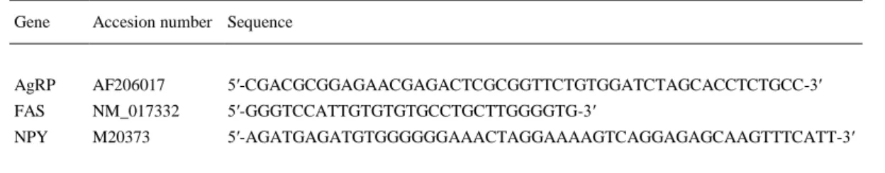
(4) 1000 g and the supernatant was extracted and part used for studies of CPT1 activity. The remainder was frozen at −80 °C for further analysis by western blotting and assay of the enzymatic activities of FAS, glucose-6-phosphate dehydrogenase (G6PDH) and 6-phosphogluconate dehydrogenase (6PGDH). Real-time quantitative PCR Expression of mRNA levels of ACCα, CPT1M (muscle type isoform) CPT1L (liver type isoform), CPT1C (brain type isoform), FAS, SCD-1 and sterol-regulatory-element-binding protein-1c (SREBP1c) in hypothalamus were studied using real-time PCR (TaqMan®; Applied Biosystems, Foster City, CA, USA) with specific primers and probes (Table 1) as previously described (1,26). All reactions were carried out using the cycling parameters: 50 °C for 2 min, 95 °C for 10 min followed by 40 cycles of 95 °C for 15 s and 60 °C for 1 min. For data analysis, the input value of the target gene was standardised to the 18S value for each sample. Data are expressed in comparison with the average value for the control group. Groups comprised six rats.. Table 1. Primer Sequences Used for Real Time Reverse Transcriptase-Polymerase Chain Reaction. Gene. Accesion number. ACCα. NM_022193. Forward 5′-TGGGCGGGATGGTCTCTTT-3′ Reverse 5′-AGTCGCAGAAGCAGCCCATT-3′ Probe. CPT1C. FAM-5′-ACCTTTGAAGATTTCGTCAGGATCTTTGATGA-3′-TAMRA. XM_001078512.1 Forward 5′-CGCTCCTGGAAAAGGAATCTC-3′ Reverse 5′-CGGGACCACACCAGCAA-3′ Probe. CPT1L. NM_031559. FAM-5′-CGTGTCTGGAATGACTTT-3′-TAMRA. Forward 5′-ATGACGGCTATGGTGTCTCC -3′ Reverse 5′-TCATGGCTTGTCTCAAGTGC -3′ Probe. FAM-5′-TGAGACAGACTCAGACCGCT-3′-TAMRA. CPT1M. NM_013200.1. Forward Commercial primers provided by Applied Biosystems (Ref. Rn01407782_g1). FAS. NM_017332. Forward 5′-GACATTTCATCAGGCCACC-3′ Reverse 5′-CCTCTAGCAGCCGCACCTC-3′ Probe. SCD-1. NM_139192.2. FAM-5′-CTGCCCAGGACAGGAACCG-3′-TAMRA. Forward 5′-TGCCAGAGGGAATAGGGAAA-3′ Reverse 5′- CTCTCCCATCCTTACTTACAAACCA-3′ Probe. SREBP1c XM_213329. FAM-5′-TCACCTTGAGAGAAGAATTAGCACGCACGG-3′-TAMRA. Forward 5′-CTCATCAACAACCAAGACAG-3′ Reverse 5′-CCTGTCTCACCCCCAGCAT-3′ Probe. 18S. V01270.1. FAM-5′-CCCTGGCCTATTTGATGCC-3′-TAMRA. Forward 5′-CGGCTACCACATCCAAGGAA-3′ Reverse 5′-GCTGGAATTACCGCGGCT-3′ Probe. FAM-5′-GACGGCAAGTCTGGTGCCAGCA-3′-TAMRA.
(5) Western blotting Total protein lysates from hypothalamus (40 μg) were subjected to sodium dodecyl sulphatepolyacrylamide gel electrophoresis, electrotransferred on a polyvinylidene difluoride membrane and probed with the indicated antibodies: ACC, phosphoACC-Ser79 (pACC), AMPKα1 and AMPKα2 (Upstate, Lake Placid, NY, USA); phospho-AMPKα-Thr172 (pAMPKα) (Cell Signaling, Danvers, MA, USA); β-actin (Abcam, Cambridge, UK); CPT1M, FAS, SREBP1c and CPT1L (Santa Cruz Biotechnology, Santa Cruz, CA, USA); CPT1C (Proteintech Europe, Manchester, UK), as previously described (1). Protein detection was performed using horseradish peroxidase-conjugated secondary antibodies and chemiluminescence (Amersham Biosciences, Little Chalfont, UK). Groups comprised six to eight rats and the protein levels were normalised to β-actin for each sample. In situ hybridisation Coronal hypothalamic sections (16 μm) from frozen brain were cut on a cryostat, mounted on slides and immediately stored at −80 °C until hybridisation. Specific oligos for AgRP, FAS and NPY detection were used (Table 2) as previously described (1). These probes were 3′-end labelled with 35S-αdATP using terminal deoxynucleotidyl transferase (Amersham Biosciences). In situ hybridisations were performed as previously described (1). The slides from all experimental groups were exposed to the same autoradiographic film. All sections were scanned and the specific hybridisation signal was quantified by densitometry using a digital imaging system (ImageJ, version 1.33; NIH, Bethesda, MD, USA). The optical density of the hybridisation signal was determined and subsequently corrected by the optical density of its adjacent background value. Groups comprised seven rats.. Table 2. Antisense Oligonucleotides for In Situ Hybridisation Analysis. Gene. Accesion number Sequence. AgRP. AF206017. 5′-CGACGCGGAGAACGAGACTCGCGGTTCTGTGGATCTAGCACCTCTGCC-3′. FAS. NM_017332. 5′-GGGTCCATTGTGTGTGCCTGCTTGGGGTG-3′. NPY. M20373. 5′-AGATGAGATGTGGGGGGAAACTAGGAAAAGTCAGGAGAGCAAGTTTCATT-3′. Enzyme assays Enzyme activities of FAS, CPT1, G6PDH and 6PGDH were determined by spectrophotometry using a microplate reader (Tecan, Sunrise, Switzerland). The reactions were started by the addition of homogenates (30 μl) and substrates (20 μl, omitted in controls) to the reaction mixture (final volume 0.25 ml), and allowing the reactions to proceed at 37 °C for pre-established times (5– 15 min). FAS (27) G6PDH, 6PGDH (28) and CPT1 (1, 26) activities were measured using methods previously described. CPT1 activity was assayed in fresh tissue preserving mitochondrial integrity so as to avoid contamination with CPT2. Groups comprised six to eight rats..
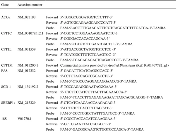
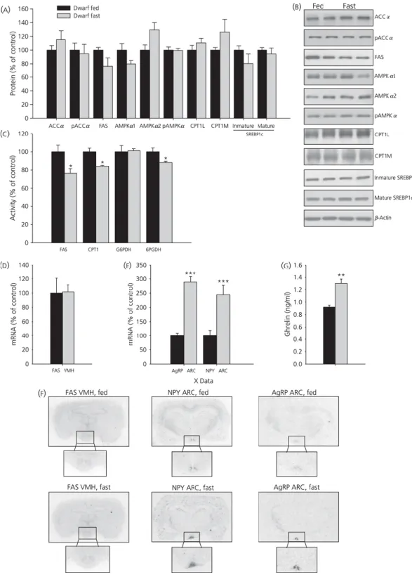
(6) Statistical analysis Data are expressed as percentage of wild-type rats, fed rats or those infused with saline (control groups). Data are expressed as the mean ± SEM. Statistic significance was determined by Student’s t-test. P < 0.05 was considered statistically significant.. Results Effects of GH deficiency on hypothalamic lipid metabolism and neuropeptide levels Gene expression, protein and activity levels of key enzymes involved in the regulation of lipid metabolism in hypothalamus of untreated wild-type and GH-deficient Lewis rats are shown in Fig. 1(a–g). GH deficiency markedly diminished mRNA levels of FAS and CPT1C (Fig. 1a). Protein levels of pACCα, FAS and SREBP1c were lower in dwarf rats compared to normal Lewis rats and both phosphorylated and total protein levels of AMPK were higher in GH deficient Lewis rats compared to controls (Fig. 1b, c). In dwarf rats, CPT1 and FAS activities were lower compared to controls (Fig. 1d). Similar results were observed for G6PDH activity. Because recent data have shown that FAS is specifically modulated in the VMH (1, 26), we examined the effect of GHdeficiency at this level. The data obtained showed that FAS mRNA levels in VMH were markedly lower in dwarf rats compared to wild-type Lewis rats (Fig. 1e, g). Similar data were obtained when studying the mRNA levels of AgRP and NPY in the ARC (Fig. 1f, g)..
(7) Figure 1. Hypothalamic mRNA (a), protein (b, c) and activity levels (d) of lipid metabolism-related enzymes in wild-type and growth hormone-deficient Lewis rats. Fatty acid synthase (FAS) mRNA levels in ventromedial nucleus of the hypothalamus (VMH) (e, g) and neuropeptide Y (NPY) and agouti-related protein (AgRP) mRNA levels (g) in the arcuate nucleus (ARC) (f, g). Values are expressed as the mean ± SEM. *, **, ***P < 0.05, 0.01 and 0.001, respectively, versus wild-type Lewis..
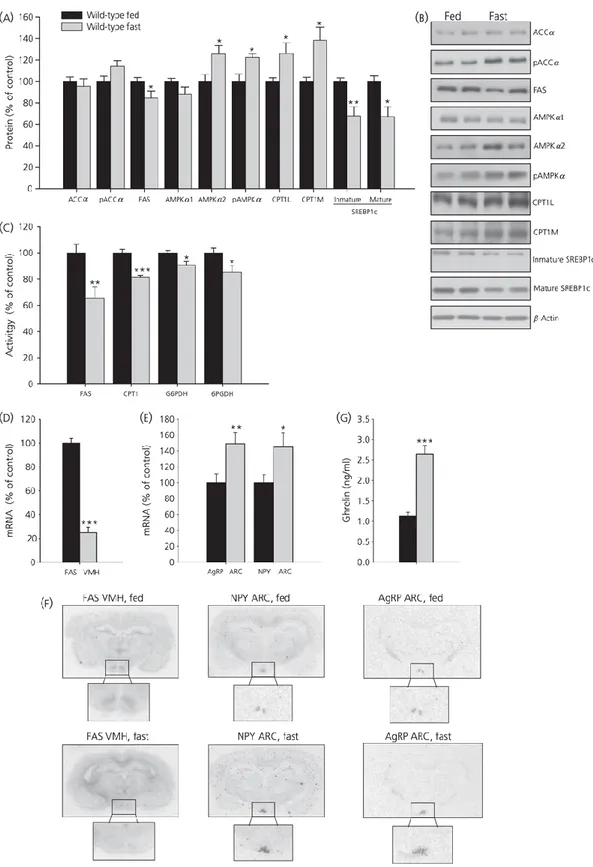
(8) Effects of fasting on hypothalamic lipid metabolism in wild-type and GH-deficient Lewis rats Next, we studied the fasting-induced effect in the hypothalamus of wild-type and GH-deficient Lewis rats; the results obtained are shown in Figs 2(a–f) and 3(a–f). In general, fasting exerted a marked effect on fatty acid metabolism in normal rats, which was not as clearly evident in GHdeficient dwarf rats. Food deprivation diminished protein levels of FAS and SREBP1c and increased the levels of pAMPKα, AMPKα2, CPT1L and CPT1M in wild-type Lewis rats (Fig. 2a, b) but not in dwarf rats (Fig. 3a, b). However, FAS, CPT1 and 6PGDH activities significantly diminished after fasting in both models of rats (Figs 2c and 3c). Finally, fasting decreased FAS mRNA levels in VMH only in wild-type rats (Figs 2d–f and 3d–f); inversely, fasting enhanced NPY and AgRP mRNA levels in ARC in both strains of rat (Figs 2e, f and 3e, f)..
(9) Figure 2. Hypothalamic protein (a, b) and activity levels (c) of lipid metabolism-related enzymes and fatty acid synthase (FAS) mRNA levels in ventromedial nucleus of the hypothalamus (VMH) (d, f), neuropeptide Y (NPY) and agouti-related protein (AgRP) mRNA levels in the arcuate nucleus (ARC) (e, f) and plasma ghrelin levels (G) in fed and fasted wild-type Lewis rats. Values are expressed as the mean ± SEM. *, **, ***P < 0.05, 0.01 and 0.001, respectively, versus fed..
(10) Figure 3. Hypothalamic protein (a, b) and activity levels (c) of lipid metabolism-related enzymes and fatty acid synthase (FAS) mRNA levels in ventromedial nucleus of the hypothalamus (VMH) (d, f), neuropeptide Y (NPY) and agouti-related protein (AgRP) mRNA levels in the arcuate nucleus (ARC) (e, f) and plasma ghrelin levels (G) in fed and fasted growth hormone-deficient Lewis rats. Values are expressed as the mean ± SEM. *, **, ***P < 0.05, 0.01 and 0.001, respectively, versus fed..
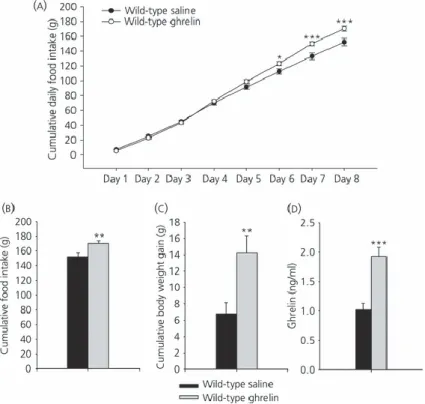
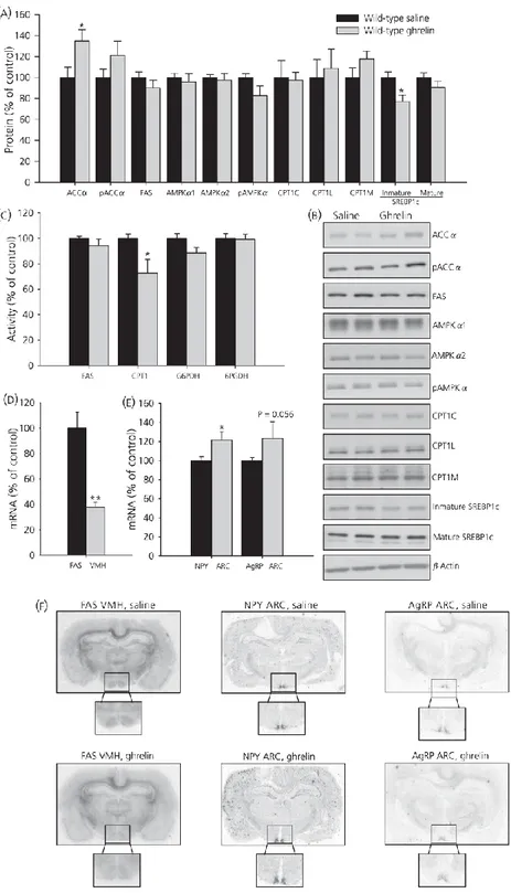
(11) Effects of fasting on plasma ghrelin levels in wild-type and GH-deficient Lewis rats Plasma ghrelin levels are shown in Figs 2(g) and 3(g). In wild-type Lewis rats fasted for 48 h, plasma ghrelin levels increased by 80% compared to the fed group, whereas levels in the dwarf group increased by only 40%. Effects of chronic central ghrelin treatment on food intake, plasma ghrelin levels and hypothalamic lipid metabolism in wild-type Lewis rats To shed light on the mechanism involved in fasting induced changes in lipid metabolism in normal and GH-deficient rats, we assessed the central effects of ghrelin in both experimental models. As expected, chronic i.c.v. ghrelin treatment increased food intake and body weight gain (Fig. 4a–c) during the 8-day experimental period in wild-type Lewis rats compared to their salinetreated controls. As previously reported (19), i.c.v. ghrelin administration elicited an increase in plasma ghrelin levels in wild-type Lewis rats (Fig. 4d). To assess the central effect of ghrelin on hypothalamic lipid metabolism, protein and activity levels of enzymes involved in the synthesis and oxidation of lipids were measured in wild-type Lewis rats (Fig. 5a–c). By contrast to the major changes in hypothalamic fatty acid metabolism after acute ghrelin treatment (1, 6, 10, 18), chronic i.c.v. ghrelin infusion only increased ACCα and diminished SREBP1c protein levels in the hypothalamus (Fig. 5a, b), as well as CPT1 activity (Fig. 5c). Similar to the acute setting (1) central chronic ghrelin treatment induced a decrease of FAS mRNA levels in the VMH (Fig. 5d, f). Finally, both mRNA levels of NPY and AgRP in ARC were enhanced by ghrelin infusion (Fig. 5e, f).. Figure 4. Effect of 8-day i.c.v. ghrelin treatment on cumulative daily food intake (a), cumulative food intake (b), cumulative body weight gain (c) and plasma ghrelin levels (d) in wild-type Lewis rats. Values are expressed as the mean ± SEM. *, **, ***P < 0.05, 0.01 and 0.001, respectively, versus saline..
(12) Figure 5 Effect of 8-day i.c.v. ghrelin treatment on hypothalamic protein (a, b) and activity levels (c) of lipid metabolism-related enzymes. Fatty acid synthase (FAS) mRNA levels in ventromedial nucleus of the hypothalamus (VMH) (d, f) and neuropeptide Y (NPY) and agoutirelated protein (AgRP) mRNA levels in the arcuate nucleus (ARC) (e, f) in wild-type Lewis rats. Values are expressed as the mean ± SEM. *, **P < 0.05 and 0.01, respectively, versus saline..
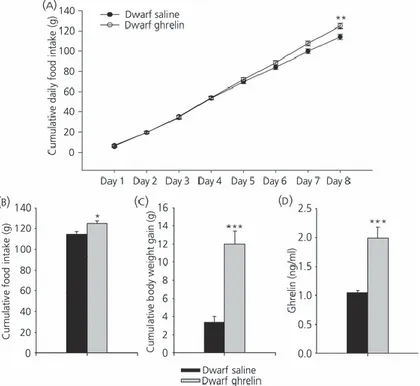
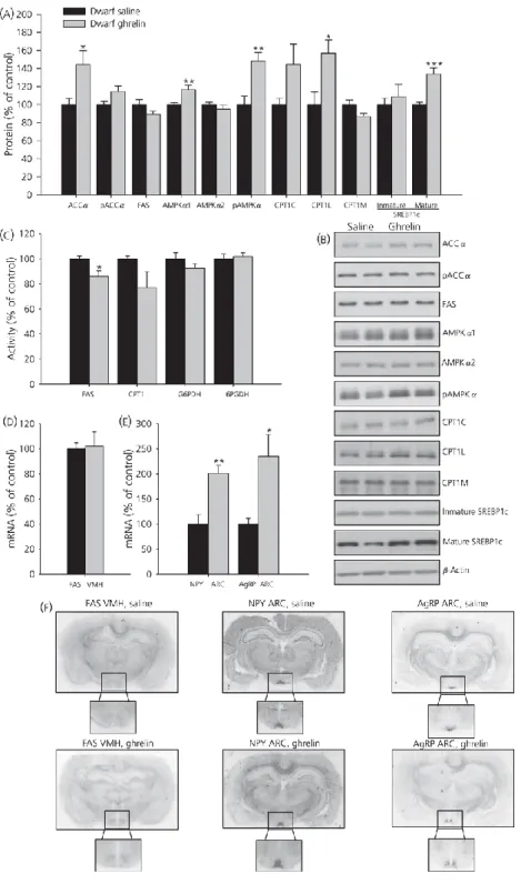
(13) Effects of chronic central ghrelin treatment on food intake, plasma ghrelin levels and hypothalamic lipid metabolism in GH-deficient Lewis rats As observed in wild-type Lewis rats, chronic i.c.v. ghrelin treatment increased food intake, body weight gain in dwarf rats (Fig. 6a–c) and plasma ghrelin levels (Fig. 6d). By contrast to normal rats, ghrelin treatment exerted a profound effect on hypothalamic fatty acid metabolism in GH-deficient dwarf rats. Chronic ghrelin administration markedly enhanced protein levels of ACCα, AMPKα1, pAMPKα1 CPT1L and SREBP1c (Fig. 7a, b). FAS activity was significantly diminished in ghrelin-treated rats (Fig. 7c); however, mRNA levels in VMH did not change in those animals (Fig. 7d). In line with the increased food intake in ghrelin-treated rats, NPY and AgRP levels in ARC were markedly enhanced in the ARC of dwarf rats after central chronic ghrelin treatment (Fig. 7e, f).. Figure 6. Effect of 8-day i.c.v. ghrelin treatment on cumulative daily food intake (a), cumulative food intake (b), cumulative body weight gain (c) and plasma ghrelin levels (d) in growth hormone-deficient Lewis rats. Values are expressed as the mean ± SEM. *, **, ***P < 0.05, 0.01 and 0.001, respectively, versus saline..
(14) Figure 7. Effect of 8-day i.c.v. ghrelin treatment on hypothalamic protein (a, b) and activity levels (c) of lipid metabolism-related enzymes. Fatty acid synthase (FAS) mRNA levels in ventromedial nucleus of the hypothalamus (VMH) (d, f) and neuropeptide Y (NPY) and agoutirelated protein (AgRP) mRNA levels in the arcuate nucleus (ARC) (e, f) in growth hormonedeficient Lewis rats. Values are expressed as the mean ± SEM. *, **, ***P < 0.05, 0.01 and 0.001, respectively, versus saline..
(15) Discussion The central nervous system (CNS), specifically the hypothalamus, monitors energy needs by assessing current energy surplus/deficit and responding by modulating appetite and peripheral energy expenditure. Recent data suggest that key regulatory enzymes and intermediates in the fatty acid metabolism pathway act as hypothalamic sensors that monitor peripheral signals (such as hormones and nutrients/metabolites) to inform about energy status. This in turn acts as a mechanism allowing organisms to adjust their food intake and energy expenditure accordingly (1,7,8,10,29). Current evidence has also highlighted the key physiological importance of hypothalamic fatty acid metabolism in the control of glucose homeostasis and food intake. Thus, activation of AMPK in several hypothalamic nuclei, such as the VMH, ARC and PVH, appears to play a prominent role in hypoglycaemia-sensing (30), mediating counter-regulatory responses. In line with this, central administration of glucose suppresses AMPK activity in the hypothalamus (5). α-Lipoic acid, a cofactor of mitochondrial enzymes with antioxidant and anorectic properties, also inhibits AMPK activity in the hypothalamus(31). Lastly, i.c.v. administration of citrate elicits an anorexigenic response associated with the inhibition of AMPK, activation of ACC and the subsequent increase in malonyl-CoA (12). In addition to metabolic regulation, peripheral hormones also modulate de novo lipogenesis in the hypothalamus. The evidence for this is that anorectic hormones, such as leptin, insulin, GLP-1, ciliary neurotrophic factor and melanocortin receptor agonists, including melanotan II, inhibit hypothalamic AMPK (5,6,8,29). On the other hand, orexigenic signals, such as cannabinoids, glucocorticoids, thyroid hormones, adiponectin, ghrelin and AgRP, activate hypothalamic AMPK, decrease malonyl-CoA levels and increase feeding (1,6,9,32–34). Overall, this evidence demonstrates that hypothalamic fatty acid metabolism is a bona fide mechanism modulating energy homeostasis at the whole body level. In the present study, we first studied the effect of GH-deficiency on fatty acid pathway in the hypothalamus. The data obtained showed that GH-deficiency induces reductions in both de novo lipogenesis and β-oxidation pathways in the hypothalamus. Thus, dwarf rats display reductions in FAS mRNA expression both in the VMH and whole hypothalamus, as well as in FAS protein and activity and CPT1 activity, probably because of increased malonyl-CoA levels as a result of FAS inhibition (1,35,36). Diminished mRNA, protein and activity levels of FAS correlate with reduced protein levels of mature SREBP1c, a transcription factor that regulates the expression of several genes involved in cellular fatty acid synthesis in the peripheral tissues (37). On the other hand, total and pAMPK protein levels were higher in dwarf rats compared to controls. These results agree with other studies showing that dwarf rats and GH receptor knockout mice presented total and pAMPKα protein levels that were significantly higher compared to their littermate controls in peripheral organs, such as the liver (38,39). By contrast, pAMPKα and AMPKα in muscle and liver are reduced in mice overexpressing GH compared to their wild type counterparts (40,41). We also studied the effect of GH-deficiency on neuropeptide levels in ARC. The selective absence of GH, in dwarf rats, results in a decrease in AgRP and NPY mRNA in the ARC. These observations are consistent with the results previously described in other studies (23,42) and also with the fact that the majority of NPY neurones in the ARC express GH receptor mRNA and the binding of GH to its receptor induces the expression of NPY mRNA (43). Next, we examined the effect of GH-deficiency on fasting-induced effects on hypothalamic fatty acid metabolism. As expected, starvation induced a marked, AMPK-dependent inactivation of hypothalamic fatty acid synthesis in normal rats, a decrease in CPT1 and FAS activities and a specific decrease in FAS mRNA levels in the VMH (1,5). Interestingly, fasting induced a different pattern in dwarf rats, which show reduced FAS and CPT1 activities, but unaltered AMPK signalling and FAS mRNA expression in the VMH (because it happened after chronic ghrelin administration; see below)..
(16) Three recent studies have proposed that short-term ghrelin action on the AMPK-malonyl-CPT1 axis acts as an acute signal, triggering the beginning of feeding after a period of fasting, coincident with increased ghrelin levels and increased AgRP and NPY expression in the ARC (1,10,18). Although these data provide convincing evidence of short-term ghrelin effects on hypothalamic fatty acid metabolism, its chronic effects are less clear. Current evidence has demonstrated that central chronic treatment with ghrelin directly increases adiposity by stimulation of the lipogenic program in the WAT (19,20). These data suggest that central ghrelin action is of physiological relevance in the control of adipocyte metabolism. In this setting, ghrelin, in addition to its orexigenic effect, could trigger meal preparation processes in the CNS that stimulate metabolic pathways in the WAT to allow more efficient storage of calories. However, no data have been reported describing chronic effects of ghrelin on hypothalamic fatty acid metabolism. In the present study, we first demonstrate that chronic central ghrelin administration does not induce major changes in all the enzymes of hypothalamic fatty acid metabolism in wild-type Lewis rats, but specifically decreases FAS mRNA expression in the VMH and hypothalamic CPT1 activity. Considering that chronic ghrelin treatment showed a marked orexigenic action and parallel changes in AgRP and NPY expression in the ARC, the relevance of these findings is quite intriguing. First, they suggest that, in the long-term setting, AMPK-induced changes in hypothalamic fatty acid metabolism plays no role in feeding control; indeed, this idea has been proposed before, and fatty acid metabolism in the hypothalamus has been suggested as a regulatory mechanism designed to preserve energy balance only in situations of starvation (1,10). Second, quite opposite to short-term ghrelin action, chronic injection decreased CPT1 activity in the hypothalamus, which indicates that, in the long-term setting, ghrelin blocks hypothalamic beta oxidation, similar to the results obtained in liver after long-term ghrelin treatment (20). The physiological relevance of this action is unclear, although we believe that it could be a compensatory mechanism that attempts to ‘brake’ the massive orexigenic signal elicited by central hyperghrelinaemia. We based this idea on previous data showing that pharmacological inhibition or genetic ablation of hypothalamic CPT1 activity (3,4) or overexpression of MCD (44) induce the cytoplasmatic accumulation of fatty acyl-CoA and reduce feeding and body weight. Besides its role in the regulation of energy balance, ghrelin also regulates the somatotrope axis, acting as a potent GH secretagogue in both rodents and humans (13). Although several studies in GH-deficient rats have demonstrated that weight gain and adiposity caused by ghrelin are independent of its ability to modulate GH secretion (13,16,45), GH receptor deficiency results in blunted ghrelin-induced feeding response in mice (24). Thus, we aimed to evaluate whether GHdeficiency had any effect on ghrelin-induced alterations in hypothalamic fatty acid metabolism. We evaluated the effect chronic ghrelin treatment on hypothalamic fatty acid metabolism in dwarf rats. The data obtained showed an increase in pAMPK levels in GH-deficient dwarf rats, alongside a nonsignificant decrease in CPT1 activity and no change in FAS mRNA and protein expression. These results suggest that lack of GH leads to enhanced sensitivity of AMPK signalling to the central actions of ghrelin. The molecular underpinnings of this effect are unclear, although it could be related to increased hypothalamic GHS-R expression in GH deficiency. Supporting this idea, recent studies have demonstrated that, in normal animals, an increased expression of GHS-R mRNA was found in several nuclei of hypothalamus and that GHS-R expression was affected by GH status, increasing in dwarf rats compared to normal rats from the same strain (23). Increased hypothalamic GHS-R expression in GH deficiency could lead to enhanced sensitivity to the central actions of ghrelin. On the other hand, wild-type Lewis rats may exhibit desensitisation after continuous ghrelin treatment, an effect that was observed after chronic exposure to GHS with respect to GH release (16). Whatever the case, the obtained results indicate that chronic ghrelin treatment decreased both de novo lipogenesis and beta oxidation in the hypothalamus of dwarf rats. Overall, these data suggest that the actions of ghrelin on hypothalamic fatty acid metabolism are GH-independent. Considering the important role of GH on peripheral lipid metabolism, the physiological relevance of these data is very challenging. In keeping with these observations, we have recently demonstrated that central ghrelin effects on adipose and hepatic liver effect do not show GH-dependency either (20)..
(17) Recent data from our group have demonstrated that the ghrelin-and fasting-induced decrease in FAS levels in the VMH is a physiological adaptive mechanism that helps to prevent malonyl-CoA from decreasing to deleteriously low levels in the hypothalamus, as a consequence of ghrelin and fasting-induced inactivation of ACC by AMPK (1). Because malonyl-CoA plays a critical regulatory role in this system by acting both as a substrate for fatty acid biosynthesis and an inhibitor of CPT1 (1,46), extremely low levels of malonyl-CoA would compromise neuronal viability, by preventing lipid biosynthesis and allowing potentially harmful neuronal fatty acid oxidation during fasting, a situation of low energy surplus. Because our data demonstrate that this regulatory mechanism is lost in dwarf rats, it is possible that GH-deficiency might elicit altered fatty acid fluxes in the hypothalamic neurones that could lead to the accumulation of toxic lipid species and lipotoxicity-associated phenomena, such as endoplasmatic-reticulum stress (47,48), which could compromise normal hypothalamic function in these animals. Further work will be necessary to address these issues (49). In summary, we demonstrate that both chronic ghrelin and fasting decreased FAS mRNA expression in the VMH of normal rats but not dwarf rats, suggesting GH-dependency. In addition, the results obtained in the present study provide a clear demonstration that, contrary to short-term administration, chronic central ghrelin treatment does not induce major changes in the overall fluxes of hypothalamic fatty acid metabolism, and this effect is independent of GH. Overall, these results suggest that ghrelin plays a dual time-dependent role in modulating hypothalamic lipid metabolism. As a result of this effect, short-term central ghrelin elicits a global AMPK-mediated inactivation of fatty acid metabolism pathway, and thus promotes food intake (1,10,16). By contrast, in the long-term setting, ghrelin does not induce AMPK-dependent changes in hypothalamic fatty acid metabolism, suggesting a lack of role for the ghrelin-AMPK interaction in the long-term feeding control. Understanding the molecular mechanism underlying the interplay between GH and ghrelin on hypothalamic lipid metabolism will allow new strategies for the design and development of suitable drugs for the treatment of GH-deficiency, obesity and its comorbidities.. Acknowledgements This work was supported by grants from Xunta de Galicia (C.D.: PGIDIT06PXIB208063PR; M.L.: GRC2006/66; F.C.: PS07/12), Fondo Investigationes Sanitarias (M.L.: PI061700 and PS09/01880; F.C.: PI051024 and PI070413), Ministerio de Educacion y Ciencia (C.D.: BFU2008; M.L.: RyC-2007-00211) and European Union (CD and M.L. : Health-F2-2008-223713). CIBER de Fisiopatología de la Obesidad y Nutrición is an initiative of ISCIII.. References 1. López M, Lage R, Saha AK, Pérez-Tilve D, Vázquez MJ, Varela L, Sangiao-Alvarellos S, Tovar S, Raghay K, Rodríguez-Cuenca S, Deoliveira RM, Castañeda T, Datta R, Dong JZ, Culler M, Sleeman MW, Álvarez CV, Gallego R, Lelliott CJ, Carling D, Tschop MH, Diéguez C, Vidal-Puig A. Hypothalamic fatty acid metabolism mediates the orexigenic action of ghrelin. Cell Metab 2008; 7: 389– 399. 2. López M, Lelliott CJ, Tovar S, Kimber W, Gallego R, Virtue S, Blount M, Vázquez MJ, Finer N, Powles TJ, O’Rahilly S, Saha AK, Diéguez C, Vidal-Puig AJ. Tamoxifen-induced anorexia is associated with fatty acid synthase inhibition in the ventromedial nucleus of the hypothalamus and accumulation of malonyl-CoA. Diabetes 2006; 55: 1327–1336. 3. Obici S, Feng Z, Arduini A, Conti R, Rossetti L. Inhibition of hypothalamic carnitine palmitoyltransferase1 decreases food intake and glucose production. Nat Med 2003; 9: 756–761. 4. Wolfgang MJ, Lane MD. The role of hypothalamic malonyl-CoA in energy homeostasis. J Biol Chem 2006; 281: 37265–37269. 5. Minokoshi Y, Alquier T, Furukawa N, Kim YB, Lee A, Xue B, Mu J, Foufelle F, Ferre P, Birnbaum MJ, Stuck BJ, Kahn BB. AMP-kinase regulates food intake by responding to hormonal and nutrient signals in the hypothalamus. Nature 2004; 428: 569–574..
(18) 6. Andersson U, Filipsson K, Abbott CR, Woods A, Smith K, Bloom SR, Carling D, Small CJ. AMPactivated protein kinase plays a role in the control of food intake. J Biol Chem 2004; 279: 12005–12008. 7. Wolfgang MJ, Cha SH, Sidhaye A, Chohnan S, Cline G, Shulman GI, Lane MD. Regulation of hypothalamic malonyl-CoA by central glucose and leptin. Proc Natl Acad Sci USA 2007; 104: 19285– 19290. 8. Seo S, Ju S, Chung H, Lee D, Park S. Acute effects of glucagon-like peptide-1 on hypothalamic neuropeptide and AMP activated kinase expression in fasted rats. Endocr J 2008; 55: 867–874. 9. Kola B, Hubina E, Tucci SA, Kirkham TC, Garcia EA, Mitchell SE, Williams LM, Hawley SA, Hardie DG, Grossman AB, Korbonits M. Cannabinoids and ghrelin have both central and peripheral metabolic and cardiac effects via AMP-activated protein kinase. J Biol Chem 2005; 280: 25196–25201. 10. Andrews ZB, Liu ZW, Walllingford N, Erion DM, Borok E, Friedman JM, Tschop MH, Shanabrough M, Cline G, Shulman GI, Coppola A, Gao XB, Horvath TL, Diano S. UCP2 mediates ghrelin’s action on NPY/AgRP neurons by lowering free radicals. Nature 2008; 454: 846–851. 11. McCrimmon RJ, Shaw M, Fan X, Cheng H, Ding Y, Vella MC, Zhou L, McNay EC, Sherwin RS. Key role for AMP-activated protein kinase in the ventromedial hypothalamus in regulating counterregulatory hormone responses to acute hypoglycemia. Diabetes 2008; 57: 444–450. 12. Stoppa GR, Cesquini M, Roman EA, Prada PO, Torsoni AS, Romanatto T, Saad MJ, Velloso LA, Torsoni MA. Intracerebroventricular injection of citrate inhibits hypothalamic AMPK and modulates feeding behavior and peripheral insulin signaling. J Endocrinol 2008; 198: 157–168. 13. Kojima M, Hosoda H, Date Y, Nakazato M, Matsuo H, Kangawa K. Ghrelin is a growth-hormonereleasing acylated peptide from stomach. Nature 1999; 402: 656–660. 14. Zigman JM, Jones JE, Lee CE, Saper CB, Elmquist JK. Expression of ghrelin receptor mRNA in the rat and the mouse brain. J Comp Neurol 2006; 494: 528–548. 15. Howard AD, Feighner SD, Cully DF, Arena JP, Liberator PA, Rosenblum CI, Hamelin M, Hreniuk DL, Palyha OC, Anderson J, Paress PS, Diaz C, Chou M, Liu KK, McKee KK, Pong SS, Chaung LY, Elbrecht A, Dashkevicz M, Heavens R, Rigby M, Sirinathsinghji DJ, Dean DC, Melillo DG, Patchett AA, Nargund R, Griffin PR, DeMartino JA, Gupta SK, Schaeffer JM, Smith RG, Van der Ploeg LH. A receptor in pituitary and hypothalamus that functions in growth hormone release. Science 1996; 273: 974–977. 16. Tschop M, Smiley DL, Heiman ML. Ghrelin induces adiposity in rodents. Nature 2000; 407: 908–913. 17. Cummings DE, Purnell JQ, Frayo RS, Schmidova K, Wisse BE, Weigle DS. A preprandial rise in plasma ghrelin levels suggests a role in meal initiation in humans. Diabetes 2001; 50: 1714–1719. 18. Kola B, Farkas I, Christ-Crain M, Wittmann G, Lolli F, Amin F, Harvey-White J, Liposits Z, Kunos G, Grossman AB, Fekete C, Korbonits M. The orexigenic effect of ghrelin is mediated through central activation of the endogenous cannabinoid system. PLoS ONE 2008; 3: e1797. 19. Theander-Carrillo C, Wiedmer P, Cettour-Rose P, Nogueiras R, Perez-Tilve D, Pfluger P, Castaneda TR, Muzzin P, Schurmann A, Szanto I, Tschop MH, Rohner-Jeanrenaud F. Ghrelin action in the brain controls adipocyte metabolism. J Clin Invest 2006; 116: 1983–1993. 20. Sangiao-Alvarellos S, Vázquez MJ, Varela L, Nogueiras R, Saha AK, Cordido F, López M, Diéguez C. Central ghrelin regulates peripheral lipid metabolism in a growth hormone-independent fashion. Endocrinology 2009; 150: 4562–4574. 21. Bruno JF, Olchovsky D, White JD, Leidy JW, Song J, Berelowitz M. Influence of food deprivation in the rat on hypothalamic expression of growth hormone-releasing factor and somatostatin. Endocrinology 1990; 127: 2111–2116. 22. Peino R, Baldelli R, Rodríguez-García J, Rodríguez-Segade S, Kojima M, Kangawa K, Arvat E, Ghigo E, Diéguez C, Casanueva FF. Ghrelin-induced growth hormone secretion in humans. Eur J Endocrinol 2000; 143: R11–R14. 23. Bennett PA, Thomas GB, Howard AD, Feighner SD, Van Der Ploeg LH, Smith RG, Robinson IC. Hypothalamic growth hormone secretagogue-receptor (GHS-R) expression is regulated by growth hormone in the rat. Endocrinology 1997; 138: 4552–4557. 24. Egecioglu E, Bjursell M, Ljungberg A, Dickson SL, Kopchick JJ, Bergstrom G, Svensson L, Oscarsson J, Tornell J, Bohlooly YM. Growth hormone receptor deficiency results in blunted ghrelin feeding response, obesity, and hypolipidemia in mice. Am J Physiol Endocrinol Metab 2006; 290: E317–E325. 25. Charlton HM, Clark RG, Robinson IC, Goff AE, Cox BS, Bugnon C, Bloch BA. Growth hormonedeficient dwarfism in the rat: a new mutation. J Endocrinol 1988; 119: 51–58. 26. Vázquez MJ, Gónzalez CR, Varela L, Lage R, Tovar S, Sangiao-Alvarellos S, Williams LM, Vidal-Puig A, Nogueiras R, López M, Diéguez C. Central resistin regulates hypothalamic and peripheral lipid metabolism in a nutritional-dependent fashion. Endocrinology 2008; 149: 4534–4543. 27. Saggerson ED, Greenbaum AL. The regulation of triglyceride synthesis and fatty acid synthesis in rat epididymal adipose tissue. Effects of altered dietary and hormonal conditions. Biochem J 1970; 119: 221– 242..
(19) 28. Tian WN, Braunstein LD, Apse K, Pang J, Rose M, Tian X, Stanton RC. Importance of glucose-6phosphate dehydrogenase activity in cell death. Am J Physiol 1999; 1: C1121–C1131. 29. Steinberg GR, Watt MJ, Fam BC, Proietto J, Andrikopoulos S, Allen AM, Febbraio MA, Kemp BE. Ciliary neurotrophic factor suppresses hypothalamic AMP-kinase signaling in leptin-resistant obese mice. Endocrinology 2006; 147: 3906–3914. 30. McCrimmon RJ, Fan X, Ding Y, Zhu W, Jacob RJ, Sherwin RS. Potential role for AMP-activated protein kinase in hypoglycemia sensing in the ventromedial hypothalamus. Diabetes 2004; 53: 1953–1958. 31. Kim MS, Park JY, Namkoong C, Jang PG, Ryu JW, Song HS, Yun JY, Namgoong IS, Ha J, Park IS, Lee IK, Viollet B, Youn JH, Lee HK, Lee KU. Anti-obesity effects of alpha-lipoic acid mediated by suppression of hypothalamic AMP-activated protein kinase. Nat Med 2004; 10: 727–733. 32. Kubota N, Yano W, Kubota T, Yamauchi T, Itoh S, Kumagai H, Kozono H, Takamoto I, Okamoto S, Shiuchi T, Suzuki R, Satoh H, Tsuchida A, Moroi M, Sugi K, Noda T, Ebinuma H, Ueta Y, Kondo T, Araki E, Ezaki O, Nagai R, Tobe K, Terauchi Y, Ueki K, Minokoshi Y, Kadowaki T. Adiponectin stimulates AMP-activated protein kinase in the hypothalamus and increases food intake. Cell Metab 2007; 6: 55–68. 33. Shimizu H, Arima H, Watanabe M, Goto M, Banno R, Sato I, Ozaki N, Nagasaki H, Oiso Y. Glucocorticoids increase neuropeptide Y and agouti-related peptide gene expression via adenosine monophosphate-activated protein kinase signaling in the arcuate nucleus of rats. Endocrinology 2008; 149: 4544–4553. 34. Ishii S, Kamegai J, Tamura H, Shimizu T, Sugihara H, Oikawa S. Triiodothyronine (T3) stimulates food intake via enhanced hypothalamic AMP-activated kinase activity. Regul Pept 2008; 3: 164–169. 35. Loftus TM, Jaworsky DE, Frehywot GL, Townsend CA, Ronnett GV, Lane MD, Kuhajda FP. Reduced food intake and body weight in mice treated with fatty acid synthase inhibitors. Science 2000; 288: 2379– 2381. 36. Hu Z, Cha SH, Chohnan S, Lane MD. Hypothalamic malonyl-CoA as a mediator of feeding behavior. Proc Natl Acad Sci USA 2003; 100: 12624–12629. 37. Espenshade PJ. SREBPs: sterol-regulated transcription factors. J Cell Sci 2006; 6: 973–976. 38. To K, Yamaza H, Komatsu T, Hayashida T, Hayashi H, Toyama H, Chiba T, Higami Y, Shimokawa I. Down-regulation of AMP-activated protein kinase by calorie restriction in rat liver. Exp Gerontol 2007; 42: 1063–1071. 39. Al-Regaiey KA, Masternak MM, Bonkowski M, Sun L, Bartke A. Long-lived growth hormone receptor knockout mice: interaction of reduced insulin-like growth factor i/insulin signaling and caloric restriction. Endocrinology 2005; 146: 851–860. 40. Wang Z, Masternak MM, Al-Regaiey KA, Bartke A. Adipocytokines and the regulation of lipid metabolism in growth hormone transgenic and calorie-restricted mice. Endocrinology 2007; 148: 2845– 2853. 41. Olsson B, Bohlooly YM, Fitzgerald SM, Frick F, Ljungberg A, Ahren B, Tornell J, Bergstrom G, Oscarsson J. Bovine growth hormone transgenic mice are resistant to diet-induced obesity but develop hyperphagia, dyslipidemia, and diabetes on a high-fat diet. Endocrinology 2005; 146: 920–930. 42. Kamegai J, Unterman TG, Frohman LA, Kineman RD. Hypothalamic/pituitary-axis of the spontaneous dwarf rat: autofeedback regulation of growth hormone (GH) includes suppression of GH releasinghormone receptor messenger ribonucleic acid. Endocrinology 1998; 139: 3554–3560. 43. Chan YY, Steiner RA, Clifton DK. Regulation of hypothalamic neuropeptide-Y neurons by growth hormone in the rat. Endocrinology 1996; 137: 1319–1325. 44. He W, Lam TK, Obici S, Rossetti L. Molecular disruption of hypothalamic nutrient sensing induces obesity. Nat Neurosci 2006; 9: 227–233. 45. Wren AM, Small CJ, Ward HL, Murphy KG, Dakin CL, Taheri S, Kennedy AR, Roberts GH, Morgan DG, Ghatei MA, Bloom SR. The novel hypothalamic peptide ghrelin stimulates food intake and growth hormone secretion. Endocrinology 2000; 141: 4325–4328. 46. Wolfgang MJ, Kurama T, Dai Y, Suwa A, Asaumi M, Matsumoto S, Cha SH, Shimokawa T, Lane MD. The brain-specific carnitine palmitoyltransferase-1c regulates energy homeostasis. Proc Natl Acad Sci USA 2006; 103: 7282–7287. 47. Zhang X, Zhang G, Zhang H, Karin M, Bai H, Cai D. Hypothalamic IKKbeta/NF-kappaB and ER stress link overnutrition to energy imbalance and obesity. Cell 2008; 135: 61–73. 48. Ozcan L, Ergin AS, Lu A, Chung J, Sarkar S, Nie D, Myers MG Jr, Ozcan U. Endoplasmic reticulum stress plays a central role in development of leptin resistance. Cell Metab 2009; 9: 35–51. 49. Martínez de Morentin PB, Varela L, Ferno J, Nogueiras R, Diéguez C, López M. Hypothalamic lipotoxicity and the metabolic syndrome. Biochim Biophys Acta 2010; 3: 350–361..
(20)
Figure
+6
Documento similar
E protein conductance in planar membranes made of different mix- tures of neutral DPhPC and negatively charged DPhPS lipids in 1 M KCl (top panel) and 30 mM KCl solutions (bottom
This reduced fertilization capacity was accompanied by increased levels of lipid peroxidation in both the testes and sperm, indicating that selenium deficiency induced
ApoAV reduces plasma triglycerides by inhibiting very low density lipoprotein-triglyceride (VLDL-TG) production and stimulating lipoprotein lipase-mediated VLDL-TG hydrolysis. 156)
3.3 Lipid indices of the fatty acid profile of insects and insect extracts 346. Taking into account the significant modification of the fatty acid profile observed
Intraperitoneal administration of the potent Nrf2 activator sulforaphane (SFN) increased Nrf2 protein levels in the basal ganglia and led to upregulation of phase II antioxidant
The mechanistic target of rapamycin (mTOR) signaling pathway is an evolutionarily conserved nutrient-sensing protein kinase that regulates cell growth and metabolism
Effects of hormones on the mRNA levels of thiol synthetase genes (A), homoglutathione synthetase (hGSHS) activity (B), and thiol contents (C) in roots of Lotus japonicus..
In the context of lipid metabolism, we identified an expression signature: ColoLipidGene (ABCA1 (ATP- Binding Cassette Subfamily-A Member), ACSL1 (Acyl- CoA Synthetase
