AUTOR: Irene Navarro Lobato
http://orcid.org/0000-0002-7866-3192
EDITA: Publicaciones y Divulgación Científica. Universidad de Málaga
Esta obra está bajo una licencia de Creative Commons Reconocimiento-NoComercial-SinObraDerivada 4.0 Internacional:
Cualquier parte de esta obra se puede reproducir sin autorización pero con el reconocimiento y atribución de los autores.
No se puede hacer uso comercial de la obra y no se puede alterar, transformar o hacer obras derivadas.
http://creativecommons.org/licenses/by-nc-nd/4.0/legalcode
FACULTAD DE MEDICINA
COVER
Ph.D. Thesis
Programa de Doctorado: Neurociencia y sus Aplicaciones Clínicas
RGS14
414-mediated prevention of an episodic
memory loss: a study of molecular mechanism
Irene Navarro Lobato
Lab. Neurobiología, CIMES, UMA
Thesis supervisor: Dr. Zafaruddin Khan
SUPERVISOR CERTIFICATE
Dr. Zafaruddin Khan, Director del laboratorio de Neurobiología del Centro de Investigaciones Médico Sanitarias y Profesor del departamento de Medicina y Dermatología en la facultad de Medicina de la Universidad de Málaga,
INFORMA
Que Doña Irene Navarro Lobato, Licenciada en Biología por la Universidad de Málaga, ha realizado bajo su dirección el trabajo experimental que ha llevado a la redacción de la presente memoria de Tesis Doctoral, titulada “RGS14414-mediated prevention of an episodic memory loss: a study of molecular mechanism”. Considerando que constituye trabajo de Tesis Doctoral, se autoriza su presentación para optar al Grado de Doctor.
Y para que así conste y surta los efectos oportunos, se firma el presente documento en Málaga, a 20 de octubre de 2015.
AGRADECIMIENTOS
ACACKNOWLEDGEMENTS
Los años de formación que he vivido culiminan con este manuscrito que sin quitarle importancia, tan sólo es una parte de todo el trabajo que encierran sus páginas, de muchos consejos y la magnífica y útil ayuda de muchas personas que han sido imprescindibles en todo su desarrollo y en el mío personal. En este pequeño espacio mi intención no es agradecer, sino dejar bien claro que son ellos también sus dueños. Todo lo que me habéis aportado a distintos niveles es mi más preciado tesoro que me ha ayudado a crecer y madurar como profesional y como persona, porque somos lo que somos por la gente que nos rodea. Yo he tenido la suerte de cruzarme con ‘GRANDES’.
En primer lugar, quiero agradecer a mi director de tesis, el Dr. Zafaruddin Khan, por su confianza en mí a la hora de acogerme en su laboratorio y darme la oportunidad de sumergirme en este mundo sacrificado, y a veces no tan fructífero como desearíamos, que en el fondo nos aporta más de lo que seríamos capaces de percibir. Gracias por guiar mis pasos y permitir que llegase al destino. Espero haber estado a la altura.
En segundo lugar, mostrar mi más sincera gratitud a los grupos con los que realicé mis estancias predoctorales por haber hecho hecho muy fructífero el timpo que pasé con vosotros aportando resultados importantes en este trabajo de tesis:
-A la Dra. Diana Frechilla y al Dr. Alberto Mediavilla del CIMA (Pamplona) y sus miembros (Esther, María y Ana María) cuya colaboración en el trabajo de Alzheimer ha sido crucial. Quiero agradecer muy especialmente a Ana María, una gran amiga, el dedicarme su tiempo enseñándome las técnicas de ORT de ratón, las inmunos, el haberme hecho sentir como en casa, sus valiosos consejos sobre el mundo de la ciencia y de la vida en general y el haberme enseñado algunos rincones de Pamplona y San Sebastian.
-A la Dra. Antonia Vlahou y al Dr. Ieronymos-Jerome Zoidakis,“Makis”, así como a todo su grupo Manoussos, Alexander, Vassiliki, Maria Frantzi, Anggeliki y al Dr. Konstantinos Vougas del Biomedical Research Foundation of Athens; tuvieron mucha paciencia con aquella inexperta en proteómica que se presentó en su laboratorio. Gracias a vuestra magnífica experiencia y buen hacer aprendí muchísimo en tan sólo 5 meses y todo el trabajo salió a adelante. Quiero hacer una mención especial a Makis, que me dedicó tantas horas, gracias por tu optimismo y positivismo y por poseer esa “locura” particular que me amenizó todo el proceso de aprendizaje. También a Idili que sin ser parte de este grupo compartió laboratorio conmigo especialmente aquel agosto en Atenas. Eres una amiga muy especial a la que tengo que agradecer muchísimo, fuiste mi apoyo donde no tenía a nadie más y me abriste las puertas de tu casa y de tu maravillosa familia sin más. Eternamente te estaré agradecida.
Mi agradecimiento también al departamento de Fisiología Humana y de la Educanción Físico Deportiva y en especial a su director, el Dr. Marc Stefan, tutor de mi DEA, que siempre se ha mostrado muy servicial y amable cuando lo he necesitado en toda la burocracia que supone la defensa de una tesis doctoral.
Mil gracias a los miembros de distintos grupos de investigación con los que hemos colaborado o nos han aportado su conocimiento, su ayuda, sus consejos, así como material necesario durante la realización de experimentos. Sois todos grandísimos profesionales y mejores personas con las que ha sido y es un lujo contar:
investigación en colaboración. Gracias al Dr. Juan José Canales y sus discípulos, Toni y Clara por acogerme en Valencia e introducirme en el estudio de la memoria de miedo, aportando siempre muy buenas ideas para continuar. Muchas gracias al Dr. Juan Carlos López y a Esperanza de la Universidad de Sevilla por enseñarnos la técnica de aversión condicionada al sabor.
Mi agradecimiento también a todos aquellos que a diario nos allanáis el camino, el personal del estabulario (nuestro preciado e imprescindible veterinario, Ricardo, a Ana, Eva, Isa, Conchi, Marivi, Vanessa, Sole, Loli y nuestra querida Soraya), los técnicos de cultivos celulares y de biología molecular del SCAI (Casimiro y Reme), los bedeles y miembros de seguridad del CIMES (Miguel, María Jesús, Antonio, Jaime, Cárdenas, Raquel, Guerra, Ricardo…). A Gema que siempre está pendiente de Mariam y de mí y se alegra mucho de vernos terminar por fin esta etapa.
Agradecer profundamente a aquellos con los que día a día a lo largo de esta etapa he compartido laboratorio, gracias por en mayor o en menor medida dejar una huella en mí:
A Manuel, por enseñarme entre risas al principio de los tiempos, por tu meticulosidad en la realización de los experimentos, por sentar las bases de este trabajo y enseñarme las técnicas de ORT y estereotaxia en rata, así como por tu participación en el proyecto de aging y Alzheimer.
A Eduardo, por tu humor irónico y por lo que me has aportado en el campo de la psicología y del estudio conductual con animales.
A Elisa por enseñarme la técnica de Morris aunque, por ser una buena profesional, que pesar de todo lo que lleva para adelante siempre sabe buscarte un hueco cuando lo necesitas. Además, agradecerte las horas que has dedicado a la corrección de este manuscrito y a la preparación de la presentación. Tu minuciosidad me ha sido de gran ayuda.
A Sinforiano, por compartir con nosotros su experiencia en qRT-PCR y ayudarme con los experimentos relacionados con ese tema, también por iniciarme en las técnicas de epigenética que retomaré seguro más adelante.
A mi queridísima Gloria, gracias por tu buen trabajo y ayuda con el estudio de arborización neuronal, tu paciencia y el cariño que le pones a todo incluyendo a las personas que te rodeamos es lo que te hace única y lo que hace que te adoremos. Gracias por tu amistad, tus consejos y tu preocupación, amiga.
A Juan, al que debo tanto, ese “hermano mayor” al que considero me ha enseñado la mayor parte de lo que soy profesionalmente, desde mis inicios en el labo como alumna interna hasta mis conocimientos en las distintas tecnologías que conozco, biología molecular, cultivos, inmunohistoquímica, Western blot... Con tu gran experiencia y sabiduría has sabido disminuir mi “petardismo” particular. Gracias por todo, por ser como eres y por preocuparte siempre. Sin ti las cosas hubiesen sido mucho más difíciles o imposibles.
A Mariam, mi mejor amiga, trabajar codo con codo contigo ha sido un verdadero placer; sin ti no sé si hubiese llegado a la meta. Gracias por no sólo por tu profesionalidad y tu minuciosidad, sino también por tu bondad y generosidad, siempre has estado cuando te he necesitado, ¿quién me iba a entender mejor que tú? Te admiro como amiga y como colega, y lo sabes. Sé que llegarás muy alto porque sabes pelear y no te conformas con un porque sí, al igual que cualquier GRANDE haría. Compañera de viaje, al final nuestro apoyo mutuo ha merecido la pena, y lo mejor de todo es que he ganado la mejor amiga que hubiese podido imaginar.Y contigo también a Zouhir, sois magníficos y seréis muy felices, amigos.
También a las chicas que llegaron al laboratorio en la fase final del trabajo, Inma, Lucía y María Elena, gracias por vuestro apoyo moral, por haceros cargo del labo mientras Mariam y yo escribíamos para evitar nuestra distracción; gracias por los momentos de risa y por escucharme. Así como a todos los demás, María, Marta, Carlos y José.
La última parte de este apartado de agradecimientos lo reservo para aquellos que forman parte de mi vida por circunstancias ajenas a la realización de mi tesis, pero que también me acompañaron durante el viaje y han sido y son muy importantes para mí:
A mis amigas de toda la vida (Ana, Eli y Rosa) y a mis ‘amigos políticos’ (Óscar, Grajales, Burra, Ángel, Pepi, Beli, Patri, Sandra) que siempre se alegran de cada paso y con los que me lo paso muy bien celebrándolo; tenésis el don de estar siempre que se os necesita. También, a mis amigas de la “flacu”: Carmen, María Victoria y en especial a Isa y Andrea.
Infinitas gracias a esa persona que sabe disimular muy muy bien la molestia que supone aguantar mi mal humor y mis quejas, mis tardanzas y mis ausencias. Pedro, muchas gracias por apoyarme en todo, y estar ahí siempre que te necesito sin esperar nada más que mi felicidad. Tu abrazo es la mejor terapia para los malos momentos y han sido imprescidibles para seguir día a día.
Y por supuesto agradecer a mi familia, mis padres, mi hermanita, con la que ni contigo ni sin ti, mis abuelos, tíos y primos, esa gran familia que te lo da todo sin esperar nada a cambio porque te quieren de corazón. Esos que por el simple hecho de saber que se sentirán orgullosos de tí merece la pena levantarse cada mañana. Gracias por vuestro cariño y vuestra confianza en mí ¡Qué haría sin vosotros! A Cristobal, ese cuñado tan apañado que tengo que siempre me ayuda con el photoshop y con toda su paciencia y profesionalidad me ha preparado la portada de este trabajo. No obstante, quiero agradecerle especialmente a mi madre, muchas gracias mamá por todo lo que haces no sólo por mí sino por todos, tu carácter lleno de amor nos conduce por el buen camino. Este trabajo va dedicado especialmente a ti, porque sé la ilusión que te hace.
DEDICATION
A mis padres y a mi hermana
CITATION
“[…], the new biology posits that consciousness is a biological process that will eventually be
explained in terms of molecular signaling pathways used by interacting populations of nerve
cells[…].”
ABBREVIATIONS
AD: Alzheimer´s disease
AKT: Protein kinase B
aMCI: Amnestic mild cognitive impairment
Ampr: Ampicillin resistance gene
ANOVA: Analysis of variance
AP: Anteroposterior
hAPP: Human amyloid protein precursor
BDNF: Brain-derived neurotrophic factor
BSA: Bovine serum albumin
CA1: Cornu ammonis area 1
CamKII: Ca2+ / calmodulin - dependent protein kinase II
cDNA: Complementary DNA
CFU: Colony-forming unit
CRE: cAMP response element
CREB: cAMP response element-binding protein
Ct: Threshold cycle
DABCO: 1,4-Diazabicyclo [2.2.2] octane
DAB: 3,3’-diaminobenzidine
DI: Discrimination index
DNA: Deoxyribonucleic acid
DV: Dorsoventral
ERK: Extracellular signal-regulated kinase
FBS: Fetal bovine serum
FGF2: Fibroblast growth factor 2
FGFR: Fibroblast growth factor receptor
GPCR: G protein-coupled receptor
GPR domain: G protein regulatory domain
GTP: Guanosine triphosphate
LB: Lennox broth
LTD: Long-term depression
LTM: Long-term memory
LTP: Long-term potentiation
MALDI-TOF: Matrix-assisted laser desorption/ionization Time-of-Flight
MAPK: Mitogen-activated protein kinase
MAPKK (MKK): Mitogen-activated protein kinase kinase
MCS: Multiple-cloning site
MEM: Eagle's minimum essential medium
ML: Mediolateral
mRNA: Messenger ribonucleic acid
MTL: Medial temporal lobe
NGF: Nerve growth factor
O.D: Optical density
ORM: Object recognition memory
PBS: Phosphate buffered saline
PBST: PBS Tween-20
PCR: Polymerase chain reaction
PDGF: Platelet-derived growth factor B chain promoter
PFC: Prefrontal cortex
Puror: Puromycin resistance gene
p-value: Error probability value
PVDF: Polyvinylidene difluoride
qRT-PCR: Quantitative reverse transcription PCR
r: Correlation coefficient
RACK1: Activated protein kinase 1
RBD domain: Raf-like Ras binding domains
RGS: Regulator of G protein signaling
RNA: Ribonucleic acid
Rpl19: Ribosomal protein L19
RT: Reverse transcription reaction
ssDNA: Single-stranded DNA
STM: Short-term memory
TGS: Tris-glycine-SDS buffer
TBE: Tris-boric acid-EDTA buffer
TrkA receptor: Tropomyosin receptor kinase A
TrkB receptor: Tropomyosin receptor kinase B
Tukey's HSD test: Tukey’s honest significant difference test
V2: Secondary visual cortex
WB: Western blot
TABLE OF CONTENTS
I. INTRODUCTION ... 1
1 Regulator of G protein signaling 14 (RGS14) ... 3
1.1 RGS14414, a spliced variant of RGS14 ... 4
2 Object recognition memory and RGS14414 ... 6
2.1 Object recognition memory (ORM) ... 6
2.2 RGS14414 in ORM enhancement ... 6
3 Memory loss in aging and Alzheimer´s disease ... 7
3.1 Aging ... 7
3.2 Alzheimer’s disease ... 7
4 Memory processing in brain ... 8
4.1 Neuronal structural remodeling ... 8
4.2 Neurotrophic factors ... 9
4.2.1 Fibroblast growth factor 2 (FGF2) ... 9
4.2.2 Nerve growth factor (NGF) ... 10
4.2.3 Brain-derived neurotrophic factor (BDNF) ... 10
4.3 14-3-3ζ protein ... 11
II.OBJECTIVES ... 13
III. MATERIALS & METHODS ... 17
1 First block of experiments. Effect of RGS14414 gene treatment on prevention of an episodic memory loss in aging and Alzheimer’s disease ... 19
1.1 Experimental design ... 19
1.1.1 Effect of RGS14414 on memory loss in aging ... 20
1.1.2 Effect of RGS14414 on memory loss in Alzheimer’s disease ... 20
1.2 Methods ... 21
1.2.1 Preparation of RGS14-lentivirus ... 21
1.2.2 Production of lentivirus ... 30
1.2.3 Animals ... 33
1.2.4 Stereotaxic surgery ... 34
1.2.5 Test of ORM ... 36
1.2.6 Data analysis ... 37
1.2.7 Immunohistochemistry ... 38
2 Second block of experiments. Determination of a correlation between RGS-mediated enhanced object recognition memory and neuronal arborization ... 38
2.1 Experimental design ... 39
2.2 Methods ... 39
2.2.1 RGS14414 treatment and brain dissection ... 39
3.2.1 Brain extraction ... 43
3.2.2 qRT-PCR ... 43
3.2.3 Determination of BDNF protein level by Western blot ... 47
3.2.4 Data analysis ... 49
4 Fourth block of experiments. Identification of proteins implicated in RGS-mediated enhanced memory processing ... 50
4.1 Experimental design ... 50
4.2 Methods ... 51
4.2.1 Tissue homogenization and protein estimation ... 51
4.2.2 Two-dimensional electrophoresis (2-DE) ... 51
4.2.3 Blue silver gel staining and analysis of protein spots ... 53
4.2.4 Identification of proteins ... 53
4.2.5 Western blot analysis to elucidate a relationship of 14-3-3 ζ protein with RGS-mediated memory enhancement ... 54
4.2.6 Data analysis ... 54
IV. RESULTS ... 55
1 RGS14414 gene treatment prevents memory loss in aging and Alzheimer’s disease ... 57
1.1 Study in aging rats ... 57
1.2 Study in Alzheimer´s disease mice ... 58
2 RGS14414 gene treatment promotes cortical neuronal arborization ... 60
3 RGS14414 gene treatment boosts expression of brain derived neurotrophic factor (BDNF) ... 64
3.1 A selective increase in both mRNA and protein levels of BDNF ... 64
3.2 A dynamic BDNF protein expression during ORM processing ... 65
4 An implication of 14-3-3ζ protein in RGS-mediated memory enhancement ... 67
4.1 Proteomic profiling revealed an elevated expression of 14-3-3ζ in RGS-treated animals ... 67
4.2 Participation of 14-3-3ζ protein in ORM processing ... 69
V.DISCUSSION ... 71
1 Prevention of memory loss ... 73
2 BDNF in neuronal arborization and ORM ... 74
3 Regulation of BDNF levels by 14-3-3ζ ... 76
4 RGS-14-3-3ζ-BDNF pathway in aging and Alzheimer´s disease ... 78
VI. CONCLUSIONS ... 79
VII. RESUMEN ... 83
1 INTRODUCCIÓN Y OBJETIVOS ... 85
1.2 Importancia de la plasticidad estructural y los factores neurotróficos en la
formación de la memoria ... 88
2 MATERIALES Y MÉTODOS ... 91
2.1 Primer bloque experimental. Efecto del tratamiento del gen RGS14414 sobre la prevención de la pérdida de memoria episódica que aparece en la enfermedad de Alzheimer y en el envejecimiento ... 91
2.2 Segundo bloque experimental. Determinación de la correlación entre la mejora de la ORM mediada por la proteína RGS14414 y la arborización neuronal ... 96
2.3 Tercer bloque experimental. Determinación de la relación entre la mejora de la memoria mediada por RGS14414 y los factores neurotróficos. ... 98
2.4 Cuarto bloque experimental. Identificación de proteínas implicadas en el procesamiento de la mejora de la memoria mediada por el tratamiento con RGS14414... 103
3 RESULTADOS ... 107
3.1 El tratamiento con el gen RGS14414 previene la pérdida de memoria asociada al envejecimiento y a la enfermedad de Alzheimer ... 107
3.2 El tratamiento con el gen RGS14414 promueve la arborización dendrítica de neuronas corticales ... 109
3.3 El tratamiento con el gen RGS14414 aumenta la expresión de BDNF ... 110
3.4 Implicación de la proteína 14-3-3ζ en la potenciación de la memoria mediada por RGS14414 ... 112
4 DISCUSIÓN ... 115
4.1 Prevención de la pérdida de memoria. ... 115
4.2 Papel de la proteína BDNF en la arborización neuronal y en la ORM en el modelo RGS... 116
4.3 Regulación de los niveles de la proteína BDNF mediante la proteína 14-3-3ζ ... 119
4.4 Vía de RGS-14-3-3ζ-BDNF en el envejecimiento y en la enfermedad de Alzheimer ... 120
VIII.REFERENCES ... 123
IX. APPENDICES ... 137
1 Appendix 1. Molecular Biology ... 139
2 Appendix 2. Cell Culture... 142
3 Appendix 3. ORM Tests ... 144
4 Appendix 4. Histology ... 145
1
Regulator of G protein signaling 14 (RGS14)
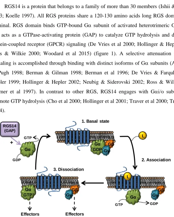
RGS14 is a protein that belongs to a family of more than 30 members (Ishii & Kurachi 2003; Koelle 1997). All RGS proteins share a 120-130 amino acids long RGS domain at N-terminal. RGS domain binds GTP-bound G subunit of activated heterotrimeric G proteins and acts as a GTPase-activating protein (GAP) to catalyze GTP hydrolysis and disrupt G-protein-coupled receptor (GPCR) signaling (De Vries et al 2000; Hollinger & Hepler 2002; Ross & Wilkie 2000; Woodard et al 2015) (figure 1). A selective attenuation in GPCR signaling is accomplished through binding with distinct isoforms of G subunits (Arshavsky & Pugh 1998; Berman & Gilman 1998; Berman et al 1996; De Vries & Farquhar 2002; Hepler 1999; Hollinger & Hepler 2002; Neubig & Siderovski 2002; Ross & Wilkie 2000; Tesmer et al 1997). In contrast to other RGS, RGS14 engages with Gi/o subfamily to promote GTP hydrolysis (Cho et al 2000; Hollinger et al 2001; Traver et al 2000; Traver et al 2004). Gβ Gγ L GTP GDP Gα Gα GDP Gβ Gγ 1. Basal state
L 2. Association Effectors Gβ Gγ Effectors Gα GTP 3. Dissociation RGS14 (GAP) GDP GTP Gα +
Figure 1. Heterotrimeric G-protein signaling. Classically defined G protein signaling begins with a heterotrimeric G protein (Gαβγ) bound to a G protein-coupled receptor (GPCR). GPCR activation promotes GDP release and subsequent GTP binding to activated Gα. Activation of Gα leads to dissociation of the heterotrimeric complex and allows Gα and Gβγ to interact with downstream effectors. As a GTPase, the α-subunit then rapidly initiates its own inactivation through GTP-hydrolysis and returns to its basal state (Gα-GDP), which leads the reassociation of the three subunits with a GPCR. RGS proteins with GAP activity accelerates GTP hydrolysis of Gα subunit regulating the G protein signaling (Oldham & Hamm 2008).
avertion of its activation and targeting to the plasma membrane (Kimple et al 2001; Kimple et al 2002; Mittal & Linder 2004; Shu et al 2007; Willard et al 2004). Phosphorylation of RGS14 at threonine 497 by protein kinase A (PKA) enhances its GDI activity (Hollinger et al 2003). In addition, through its RBD1 domain, RGS14 binds activated forms of H-Ras and Rap2 allowing RGS14 to engage H-Ras signaling pathways such as Ras/Raf/MAP kinase (Formstecher et al 2005; Kiel et al 2005; Mittal & Linder 2006; Shu et al 2010; Traver et al 2000; Willard et al 2009; Wohlgemuth et al 2005). At present, binding partners of RBD2 are unknown. Due to the presence of several binding domains, RGS14 has been considered as a scaffold protein with multiple functions. This idea is further strengthened by a dynamic spatial and temporal distribution pattern of RGS14 across the whole brain and even in
different cellular compartments (Evans et al 2014; Lopez-Aranda et al 2006; Shu et al 2007).
Northern blot experiments (Snow et al 1997), in situ hybridization studies (Grafstein-Dunn et al 2001), and quantitative polymerase chain reaction (qPCR) (Larminie et al 2004) have independently reported that RGS14 mRNA is present in rat and human brain tissue. Similarly, immunohistochemical studies (Lopez-Aranda et al 2006) and immunoblot experiments (Hollinger et al 2001) have shown that RGS14 protein is enriched in rat and monkey brain.
1.1 RGS14414, a spliced variant of RGS14
NH3 RGS RBD1 RBD2 COOH
G
o
L
o
c
o
1-31 149-220 222-292 346-368
67-184 302-373 375-445
NH3 RGS RBD1 RBD2 COOH
G
o
L
o
c
o
498-521
RGS14566 protein
RGS14414 protein
Figure 2. A graphic representation of human RGS14 protein.
RGS
RGS14544 MPGKPKHLGVPNGRMVLAVSDGELTSTSGSQAQGEGRGSSLSIHSLPSGPSSPFSTDEQPVAS
RGS14414 ---
RGS14544 WAQSFERLLQDPRGLAYFTEFLKKEFSAENVTFWQACERFQQIPASDTKQLAQEAHNIYHEF
RGS14414 ---
RGS14544 LSSQALSPVNIDRQAWLSEEVLAQPRPDMFRAQQLQIFNLMKFDSYARFVKSPLYQECLLAE
RGS14414 ---MFRAQQLQIFNLMKFDSYARFVKSPLYRECLLAE
RGS14544 AEGRPLREPGSSHLGSPDTARKKPKLKPGKSLPLGVEELGQLPLAEG---RPLRKSFRREMPG
RGS14414 AEGRPLREPGSSRLGSPDATRKKPKLKPGKSLPLGVEELGQLPPVEGPGGRPLRKSFRREL-G
RBD1
RGS14544 GTASVNSALRRESQGSLNSSASLDLGFLAFVSSKSESHRKSLGSGEGESESRPGKYCCVYLPD
RGS14414 GTA--NAALRRESQGSLNSSASLDLGFLAFVSSKSESHRKSLGSTEGESESRPGKYCCVYLPD
RGS14544 GTASLALARPGLTIRDMLAGICEKRGLSLPDIKVYLVGKEQKALVLDQDCTVLADQEVRLENR
RGS14414 GTASLALARPGLTIRDMLAGICEKRGLSLPDIKVYLVGNEQKALVLDQDCTVLADQEVRLENR
RBD2
RGS14544 ITFQLELVGLERVVRISAKPTKRLQEALQPILAKHGLSLDQVVLHRPGEKQLVDLENLVSSVA
RGS14414 ITFELELTALERVVRISAKPTKRLQEALQPILEKHGLSPLEVALHRPGEKQPLDLGKLVSSVA
RGS14544 SQTLVLDTLPDAKTREASSIPPCRSQGCLPRTQTKDSHLPPLSSSLSVEDASGSTGKRQTCDI
RGS14414 AQRLVLDTLPGVKISKARDKSPCRSQGCPPRTQDKATHPPPASPSSLVKVPSSATGKRQTCDI
GoLoco
RGS14544 EGLVELLNRVQSSGAHDQRGLLRKEDLVLPEFLQLPSQRPGSQEAPP---
RGS14414 EGLVELLNRVQSSGAHDQRGLLRKEDLVLPEFLQLPAQGPSSEETPPQTKSAAQPIGGSLNST
RGS14544 ---
2
Object recognition memory and RGS14
4142.1 Object recognition memory (ORM)
Recognition memory is one of the most studied examples of episodic memory. Recognition memory is a process of identifying an object, a person, a place or an event that has been encountered previously. Recognition memory is widely viewed as consisting of two components: recollection and familiarity (Diana et al 2007; Yonelinas 2001; Yonelinas et al 2010). Interest in this distinction greatly increased when Brown and Aggleton (Brown & Aggleton 2001) proposed a neuroanatomical basis for these two processes. Their proposal was that recollection depends on the hippocampus, whereas familiarity relies on the adjacent perirhinal cortex. However, other researchers argue that recognition memory is a single process dependent on both the hippocampus and adjacent cortex (Donaldson 1996; Haist et al 1992; Squire et al 2004; Squire et al 2007).
Over the years, it has been shown that medial temporal lobe (MTL) is central to ORM processing. However, different subregions of the MTL make distinct contributions (Aggleton & Brown 2006; Eichenbaum et al 2007; Yonelinas et al 2002). Novel objects recognition on basis of simple features, such as size, shape or color relies on perirhinal cortex (Aggleton et al 1997; Barker et al 2007; Bussey et al 1999; Meunier et al 1993; Mumby & Pinel 1994), whereas hippocampus contributes to object recognition within familiar environment, such as location or context (Bachevalier et al 2015; Barbosa et al 2012; Barker & Warburton 2011; Bussey et al 2000; Hunsaker et al 2008; Lee et al 2005). In addition to MTL, other brain structures, such as medial prefrontal cortex (PFC) (Morici et al 2015) and area V2 of visual cortex (Lopez-Aranda et al 2009), have also been implicated in ORM processing.
2.2 RGS14414 in ORM enhancement
information of two objects, while RGS-rats were able to keep up to six objects. These findings suggest that RGS14414 is a robust memory enhancer and this might be useful in treatment against memory loss.
3
Memory loss in aging and Alzheimer´s disease
Deficits in memory function has been observed not only in aging but also in many neurological and neurodegenerative diseases. However, here, we will describe two most studied conditions of memory loss: aging and Alzheimer´s disease (AD), because they will be part of this thesis work.
3.1 Aging
It has consistently been shown that in aging, there is substantial loss in episodic memories, such as ORM (Nilsson 2003), however in contrast, semantic memory, implicit or procedural memory (unconscious automated actions and movements) are relatively unaffected (Churchill et al 2003). Nevertheless, there is a disagreement on what kind of recognition memory is impaired in aging (Koen & Yonelinas 2014). Some studies have reported that aging and amnestic mild cognitive impairment (aMCI) are associated with specific recollection impairment but not familiarity-based episodic memory (Anderson et al 2008; Luo et al 2007; Westerberg et al 2013; Westerberg et al 2006; Yonelinas & Levy 2002). Whereas others have found that aging and aMCI are associated with declines in both recollection- and familiarity-based episodic memory (Friedman et al 2010; Peters & Daum 2008; Wang et al 2012; Wolk et al 2011). It is widely believed that this episodic memory loss in aging is not due to neurodegeneration in areas related to learning and memory (Burke & Barnes 2006; Morrison & Hof 1997; Rapp & Gallagher 1996; Rasmussen et al 1996; Wickelgren 1996), but it is caused by decrease in soma size (de Brabander et al 1998; Wong et al 2000), loss of dendrites and dendritic spines (Duan et al 2003; Jacobs et al 1997; Page et al 2002; Peters et al 1998), loss of synapses (Chen et al 1995; Wong et al 1998), aberration in neuronal networks and diminished synaptic activity (Khan et al 2014; Rizzo et al 2014; Wilson et al 2006).
due to a disruption in the communications between neurons caused by a neuronal synaptic loss and pruning of dendrites in temporal lobe and diencephalon. This neurodegeneration, specially in cholinergic neurons (Mufson et al 2008), has been related to different causes: high concentration of amyloid-β peptide (Kamenetz et al 2003; Puzzo et al 2008; Puzzo et al 2012; Shankar et al 2008) promoting formation of amyloid-β plaques; hyperphosphorylation of tau protein causing aggregation of neurofibrillary tangles (Alonso et al 2001) and disruption in microtubule assembly (Lindwall & Cole 1984). Microtubule assembly is necessary for the intracellular trafficking of neurotrophins and other functional proteins and a disruption in this process causes decrease in synaptic availability of molecular components crucial for memory processing (Salehi et al 2003).
4
Memory processing in brain
There are generally two kind of memories: short-term memory (STM), which lasts for
minutes or longer (Castellucci et al 1989; Xia et al 1998); long-term memory (LTM), which
can persist for days, months, even a lifetime (Bailey et al 1996; Castellucci et al 1989;
Reissner et al 2006; Sangha et al 2003). Unlike STM that is usually mediated by posttranslational modifications, LTM requires protein synthesis and long-lasting changes in synaptic transmission, including long-term potentiation (LTP) and depression (LTD). It has been argued that these underlying processes of LTM produces structural changes for the formation of neural circuits that encode representations of memory (Bailey & Kandel 2008; Bosch et al 2014; Caroni et al 2012; Lamprecht & LeDoux 2004). There are large number of molecules and pathways that have been implicated in memory; however, we will focus here on some that are pertaining to this thesis work.
4.1 Neuronal structural remodeling
thought to be a critical substrate for the acquisition and consolidation of long-term memories (Bailey & Kandel 1993; Yang et al 2009). In fact, changes in the length of neck of spine, in dendritic spine volume, in dendritic turnover through the loss and/or addition of dendrites have been related with learning and memory (Lamprecht & LeDoux 2004; Segal 2005). In recent years, neurotrophic factors and its signaling cascades have been shown to be linked to
remodeling of neuronal dendrites via dendritic branching (Kopec & Carew 2013; McAllister et al 1999).
4.2 Neurotrophic factors
Neurotrophic factors are secreted molecules which bind membrane-associated
extracellular receptors and activate intracellular signaling cascades that ultimately promotes
cellular survival, neurogenesis, synaptogenesis and activity-dependent pruning, including
axon outgrowth, dendritic growth and dendrite maturation (Dijkhuizen & Ghosh 2005; Horch
& Katz 2002; Ji et al 2005; Park & Poo 2013). Neurotrophic factors have been shown to be
involved in induction of long-lasting synaptic plasticity (Conner et al 2009; Chao 2003; Egan
et al 2003; McAllister et al 1999; Poo 2001) and protein synthesis (Tanaka et al 2008),
activities fundamental for the formation of LTM. Considering that there is a large list of
neurotrophic factors (Kopec & Carew 2013), we have restricted to some of them which are
found abundant in brain and have been shown to be related to structural plasticity and memory, and are pertaining to this thesis work.
4.2.1 Fibroblast growth factor 2 (FGF2)
4.2.2 Nerve growth factor (NGF)
NGF has been found in hippocampus, cerebral cortex, olfactory regions and forebrain (Korsching et al 1985; Large et al 1986; Levi-Montalcini & Angeletti 1968). It binds to tropomyosin receptor kinase A (TrkA) and induces neuronal survival, neurite outgrowth and synaptic plasticity (Barrett 2000; Chao & Bothwell 2002; Dechant & Barde 2002; Miller & Kaplan 2001). NGF contributes to changes in neuronal networks by promoting formation of newer dendritic spines in mature brain (Alleva & Aloe 1989). In fact, NGF not only is indispensable for the survival of basal forebrain cholinergic neurons (Allard et al 2012; Chen et al 1997; Niewiadomska et al 2011; Van der Zee et al 1995), but also is crucial for cholinergic connections in insular cortex and subsequent acquisition of new memories associated to conditioned taste aversion and contextual fear conditioning (Gutierrez et al 1997). Also, septal NGF and its receptor TrkA in CA1 are required for hippocampal LTP (Conner et al 2009), spatial memory (Conner et al 2009) and fear memory consolidation (Woolf et al 2001).
4.2.3 Brain-derived neurotrophic factor (BDNF)
BDNF and its receptor tropomyosin receptor kinase B (TrkB) are widely expressed in brains of rodent and human, with higher expression in hippocampus and cerebral cortex (Aid et al 2007; Maisonpierre et al 1990; Timmusk et al 1994). BDNF is secreted from neurons and glial cells, and binding of this neurotrophic factor with its receptor TrKB activates several signaling pathways, including Ras/mitogen-activated protein kinase (Ras/MAPK), phospholipase C-gamma (PLC-γ) and phospatidyl inositol-3 kinase/protein kinase B (PI3K/AKT) (Andero et al 2014; Chao 2003). Many works have demonstrated the function of BDNF in dendritic remodeling of cortical neurons; in regulation of dendritic outgrowth and branching; in increment of proximal dendrite growth and spine density; in rise in number of pyramidal and non-pyramidal neurons during development and in adult brain (Horch 2004; Horch et al 1999; Jin et al 2003; Wirth et al 2003). Also, BDNF expression has been revealed to be essential for the maintenance of dendritic structure and the size of cortical neurons in adult brain (Gorski et al 2003).
plasticity in a synapse-specific manner (Bramham & Messaoudi 2005). BDNF is required for both STM and LTM formation of inhibitory avoidance learning (Alonso et al 2002). In addition, this neurotrophic factor contributes to acquisition and storage of long-lasting memories (Andero et al 2014; Bekinschtein et al 2008a; Bekinschtein et al 2014) and to spatial and contextual fear memory (Alonso et al 2002; Koponen et al 2004; Linnarsson et al 1997; Mizuno et al 2000; Tyler et al 2002). BDNF is required at early time points during formation of inhibitory avoidance LTM, for regulation of phosphorylated state of synaptic proteins and their persistence through new protein synthesis (Bekinschtein et al 2007; Bekinschtein et al 2008b). Furthermore, an increase in BDNF was observed within 2 h of the acquisition of object information (Romero-Granados et al 2010). A BDNF V(66)M polymorphism, which significantly hampers memory performance in human, has been shown to be linked to an impairment in episodic memory (Kauppi et al 2013). In contrary, exercise boosts spatial memory through an increase in BDNF transcripts I and IV in hippocampus (Intlekofer et al 2013).
4.3 14-3-3ζ protein
First evidence that related 14-3-3ζ with memory was done in Drosophila melanogaster where leonardo gene, a homologous of vertebrate 14-3-3ζ, is abundantly expressed in mushroom body neurons. Mutant Drosophila that lacked leonardo gene, showed significant decrease in capacity for olfactory memory but not for olfactory sensory (Philip et al 2001; Skoulakis & Davis 1996; 1998). Similarly, a study using mutant mice with deletion of 14-3-3ζ, displayed remarkably reduced capacity of spatial memory and ORM compared to their wild-type siblings (Cheah et al 2012). Furthermore, functional knockout mice of 14-3-3 in hippocampus presented impairments in associative memory and a deficit in LTP (Qiao et al 2014).
Previously, we have shown that RGS14414 is a robust memory enhancer and it produces an enduring effect on memory. We found that RGS14414 gene treatment in area V2 of visual cortex led to a memory enhancement of such extent that it converted an object recognition memory normally lasting for 45 min into long-term memory that could be traced even after many months (Lopez-Aranda et al 2009). Therefore, considering this long-lasting effect of RGS14414 on memory, we planted to study whether memory loss observed in rodent models of aging and Alzheimer´s disease can be prevented by RGS14414 gene treatment or not. Next, we explored through various biological processes in brain to provide explanation of RGS-mediated memory enhancement in rodents. Following objectives were considered:
Objective 1: Examine the effect of RGS14
414gene treatment on prevention
of an episodic memory loss in aging and Alzheimer’s disease.
A number of psychiatric and neurological disorders are associated with memory impairments; however Alzheimer’s disease and aging related cognitive decline are the most studied examples of it. With the use of normal aging rats and transgenic mice of Alzheimer´s disease, we have evaluated whether RGS14 gene treatment into area V2 could serve as a therapeutic tool in prevention of episodic memory loss seen in aging and in many neurological and neurodegenerative diseases.
Objective 2: Determination of relationship between neuronal arborization
and RGS14-mediated enhancement in ORM.
Considering the enduring effect of RGS14414 protein on memory enhancement, we believe that this long-lasting effect are due to permanent structural change caused by surge in neuronal connections and enhanced neuronal remodeling. Therefore, in this objective, brains of RGS-animals were subjected to analysis of cell body neurites outgrowth in both pyramidal and non-pyramidal neurons and of proliferation in dendritic branching in pyramidal neurons.
factors. Thus, in this objective, we have studied the effect of RGS14 treatment on FGF2, NGF and BDNF, neurotrophic factors that are abundant in brain and are related with structural plasticity and memory.
Objective 4: Explore the implication of 14-3-3ζ in facilitation of
RGS-mediated memory enhancement.
1
First block of experiments. Effect of RGS14
414gene treatment on
prevention of an episodic memory loss in aging and Alzheimer’s
disease
In this block, we have explored whether RGS14414 gene treatment can prevent memory loss in aging and Alzheimer´s disease, because they are two most studied conditions where memory loss has consistently been observed (Drag & Bieliauskas 2010).
1.1 Experimental design
To achieve the objective, we have used rodent models of aging rats and transgenic mice of Alzheimer´s disease. A summary of methodological approach for this block of experiments is described in figure 4:
Lentivirus production
(RGS14 or vehicle)
Data collection
(DI)
Statistical analysis
(RGS14 vs vehicle group)
B. Effect of RGS14414on memory loss in Alzehimer’s disease.
Stereotaxic surgery
(RGS/vehicle treatment in area V2)
10 min
10 min
10 min 10 min
24 h
Test of ORM
4
2 7
Age in months
A. Effect of RGS14 on memory loss in aging.
Delay
3 min 3 min
Test of ORM
Delay
5
3 18 20 22
45 min 45 min and 24h 24 h
Age in months
24 h
Stereotaxic surgery
1.1.1 Effect of RGS14414 on memory loss in aging
Male Wistar Han rats of 3 months and older were obtained from Charles River to be used in this study.
(i) One group of 12 rats was monitored for their ORM statuses from the age of 3 to 18 months to evaluate the age when memory loss emerges.
(ii) For the study of prevention of memory loss, 15 rats of 3 months old were treated with RGS14-lentivirus and 7 rats with vehicle-lentivirus and were further tested for ORM at ages of 5, 18, 20 and 22 months (figure 4.A).
1.1.2 Effect of RGS14414 on memory loss in Alzheimer’s disease
Transgenic mice with Alzheimer’s disease of J20 line (AD-mice) were obtained from Jackson Laboratory. These mice overexpress human β-amyloid precursor protein (hAPP) with Swedish (K670N/M671L) and Indiana (V717F) familial AD mutations under control of platelet-derived growth factor B chain promoter (PDGF) (Mucke et al 2000). The mice were on an inbred C57BL/6J genetic background. For current study, in addition to AD-mice, we have used wild-type mice of C57BL/6J strain as control.
(i) 12 wild-type mice (C57BL/6J strain) and 8 AD-mice were tested for ORM status at ages of 2 and 4 months to monitor the age of appearnace of ORM loss.
1.2 Methods
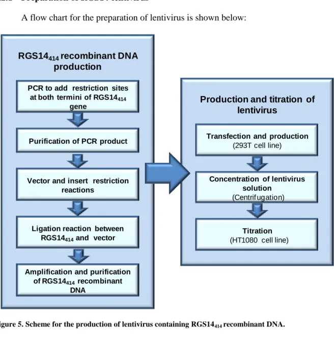
1.2.1 Preparation of RGS14-lentivirus
A flow chart for the preparation of lentivirus is shown below:
RGS14414recombinant DNA
production
PCR to add restriction sites at both termini of RGS14414
gene
Purification of PCR product
Vector and insert restriction reactions
Ligation reaction between RGS14414and vector
Amplification and purification of RGS14414recombinant
DNA
Production and titration of lentivirus
Transfection and production
(293T cell line)
Concentration of lentivirus solution
(Centrif ugation)
Titration
(HT1080 cell line)
Figure 5. Scheme for the production of lentivirus containing RGS14414 recombinant DNA.
1.2.1.1 Construction of recombinant RGS14414
1.2.1.1.1 RGS14414 gene
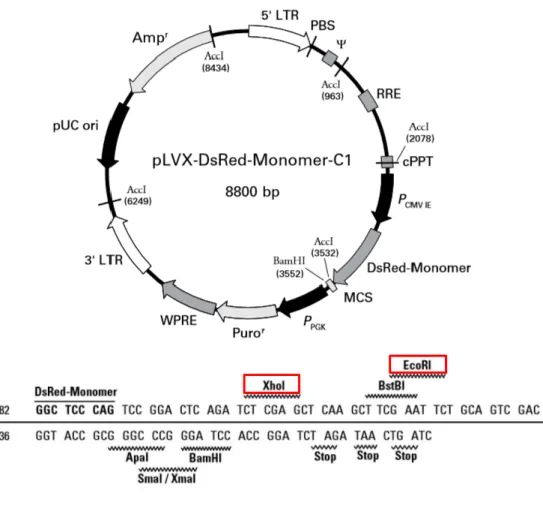
and is 8800 bp long. RGS14414 gene was cloned into multiple-cloning site (MCS) placed in the carboxyl terminus of DsRed-Monomer sequence. This vector allowed the expression of RGS14414 gene fused to DsRed-Monomer, a monomeric mutant of the Discosoma specie red fluorescent protein, and therefore, facilitated the identification of RGS14-protein expression in brain cells.
A
B
1.2.1.2 Amplification of RGS14414 gene
Prior to PCR, a restriction sites analysis of RGS14414 gene was carried out by NEBcutter version 2.0 (New England Biolabs). Considering that both XhoI and EcoRI restriction sites of MCS of pLVX-DsRed-Monomer-C1 (figure 6.B) were absent in RGS gene, both were selected for cohesive end gene insertion.
A PCR was performed to include XhoI and EcoRI restriction sequences at 5’ and 3’ ends, respectively. Both forward (M103-XhoI), and reverse (M104-EcoRI) primers used in this reaction are shown in table 1 and were synthesized by Sigma Aldrich company. In addition to restriction sequence in forward primer, Shine-Dalgarne sequence (a ribosomal binding site in prokaryotic mRNA to initiate protein synthesis in prokaryotes) and Kozak sequence (a consensus sequence of eukaryotic mRNA necessary to initiate the translation process) were added to promote efficient protein synthesis of RGS gene.
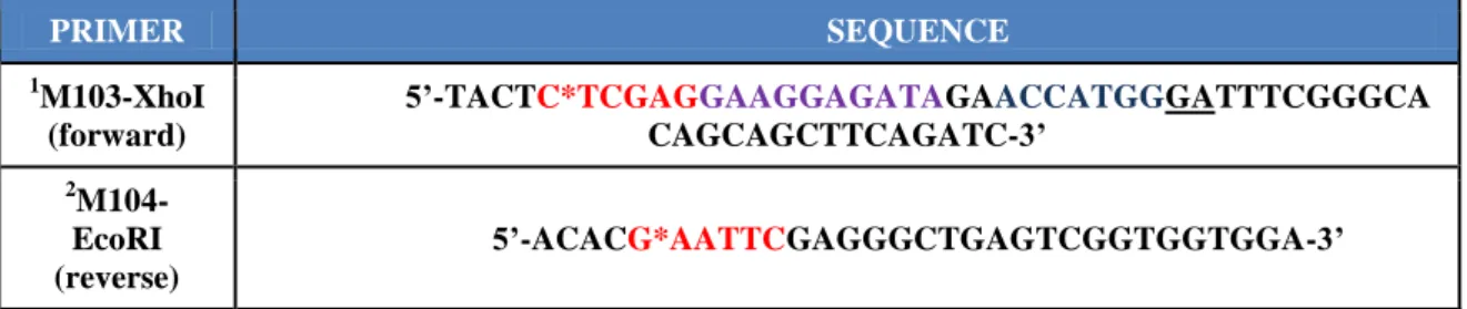
Table 1. Primers to add XhoI and EcoRI restriction sites in RGS14414.
PRIMER SEQUENCE
1M103-XhoI (forward)
5’-TACTC*TCGAGGAAGGAGATAGAACCATGGGATTTCGGGCA CAGCAGCTTCAGATC-3’
2 M104-EcoRI (reverse)
5’-ACACG*AATTCGAGGGCTGAGTCGGTGGTGGA-3’
The PCR was carried out using reagents from Promega (table 2) in Sprint thermocycler (Thermo Electro Corporation)as shown in table 3.
1Xho target sequence in red; Shine Dalgarne sequence in purple and Kozak sequence in blue. Two
underlined nucleotides were added to keep the open reading frame.
2 EcoRI target sequence in red.
Table 2. PCR reaction.
1
2 mM of triphosphate deoxynucleotides (dNTPs) stock solutions means a final concentration of each dNTPs (dATP, dGTP, dCTP and dTTP).
Table 3. Amplification cycles and temperatures.
STEP TEMPERATURE TIME
Initial denaturation 95 ºC 3 min
30 cycles
Denaturation 95 ºC 1 min
Primer annealing 57 ºC 1 min
Extension 72 ºC 3 min
Hold 4 ºC --
1.2.1.2.1 Gel purification of PCR product
The PCR product from above was purified by 1 % agarose gel electrophoresis (see appendix 1.A.2). Samples mixed with 30 % glycerol in proportion 4:1 were loaded on gel, and separated with Tris-borate-EDTA (TBE) buffer (appendix 1.A.1) by applying 100 V power (Power-Pac 300, Bio-Rad). The DNA band was visualized under a long-wavelength UV light (Bio-Rad equipment Gel Doc 2000) (figure 7) and was excised using a sterile
REAGENT VOLUME FINAL
CONCENTRATION
5X Go Taq® Flexi Buffer Mg free
(Promega, Cat. No. M890A) 10 µl 1 X
MgCl2 solution (25 mM)
(Promega, Cat. No. A351B) 2 µl 1 mM
1PCR Nucleotide Mix (2 mM)
(Promega, Cat No. C1141) 5 µl 0.2 mM
M103-XhoI ( primer forward) (Sigma-Aldrich) 2 µl 0.4 µM
M104-EcoRI (primer reverse) (Sigma-Aldrich) 2 µl 0.4 µM
RGS14414 cDNA (560 ng/μl) 0.5 µl 280 ng
GoTaq® g2 Flexi DNA polymerase(5 U/µl)
(Promega, Cat. No. M78OA) 0.5 µl 2.5 U
Nuclease-Free water (Gibco, Cat. No. 10977) 28 µl n/a
scalpel and transferred to a previously weighted 1.5 ml tube. The weight of the excised band was 350 mg.
RGS DNA purification from excised gel was carried out following manufacture’s protocol of Wizard® SV Gel and PCR Clean-Up System kit (Promega, Cat. No. A9281) with some modifications (see appendix 1.C.1). Recovered DNA solution (about 40 μl) with a final concentration of 55.63 ng/μl was stored at -20 ºC.
1.2.1.2.2 Restriction and ligation reactions
Compatible cohesive ends between RGS14414 gene and pLVX-DsRed-Monomer-C1 were generated by XhoI and EcoRI endonuclease enzymes reaction as is shown in table 4. The reaction was carried out at 37 ºC in a thermostatic bath (SW22, Julabo) for 16 h in case of RGS14414 gene and 4 h in case of vector. The endonucleases were inactivated by heating at 65 ºC in a dry block thermostat (Bio TDB-100, Boeco) for 20 min.
1 2 3
1636 bp 1018 bp
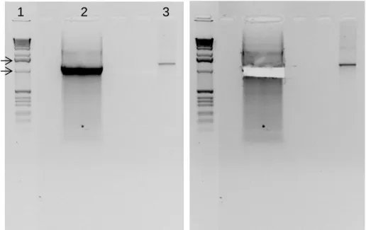
Figure 7. Electrophoresis of RGS14 DNA with restriction sites for XhoI and EcoRI to purify. (1) Molecular weight ladder, (2) RGS14414 cDNA with endonuclease target sequence resulting from PCR (1283 bp)
The DNAs resulting from restriction reactions were purified using agarose gel (figure 8) as explained in previous section (III.1.2.1.2.1) and their concentrations were determined by absorbance at 260/280 nm. The final concentration was 6.06 ng/µl in both cases.
Table 4. Restriction reaction of RGS14414 and pLVX lentiviral vector.
REAGENT VOLUME RGS14414 (55,63 ng/µl) pLVX (0,5µg/µl)
DNA 1 μg (18 µl) 0.5 μg (1 µl)
XhoI (20000 U/ml)
(New England Biolabs, Cat. No. R0146S)
1.5 µl (30U/μg DNA)
0.25 µl (10U/μg DNA)
EcoRI (20000 U/ml)
(New England Biolabs, Cat. No. R0101S)
1.5 µl (30U/μg DNA)
0.25 µl (10U/μg DNA)
NEBuffer 2.1 (10X)
(New England Biolabs, Cat. No. B7202S) 5 µl 2.5 µl
Nuclease-Free water
(Gibco, Cat. No. 10977) 24 µl 25 µl
TOTAL VOLUME 50 μl 25 μl
To insert gene into the vector, a ligation reaction using the T4 DNA ligase (Life Technologies, Cat. No. 15224) was performed as it is detailed in table 5. All reagents, excluding the T4 ligase enzyme, were mixed previously and incubated at 45 ºC in the dry block thermostat (Bio TDB-100, Boeco) for 5 minutes to prevent non-specific binding between cohesive ends. After addition of ligase enzyme, the reaction was performed at 24 ºC for 1 hour. At the end of reaction, ligase enzyme was inactivated by incubation at 70 ºC for 10 min.
Table 5. Ligation reaction.
REAGENT VOLUME
5X reaction buffer
(Life technologies Cat. No. 46300-18) 4 µl
Gen RGS14414 (6.06 ng/µl) 9.9 µl (60 ng)
pLVX vector (6.06 ng/μl) 3.3 µl (20 ng)
T4-Ligase enzyme (1 U/μl)
(Life technologies, Cat. No. 15224) 1 µl
Nuclease-Free water
(Gibco, Cat. No. 10977) 1.8 µl
4072 bp 12216 bp
1 3 4
1636 bp 1018 bp
B.
Vector pLVX.1 2
1636 bp 1018 bp
A.
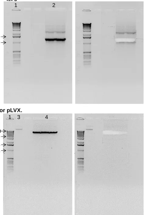
RGS14414gene.Figure 8. Electrophoresis of the RGS14 (A) and plasmid pLVX (B) cDNAs cut with XhoI and EcoRI. The gels on the right show the bands cut. (1) Molecular weight ladder, (2) RGS14414 cDNA (1283 bp) restriction
reaction, (3) pLVX cDNA without endonuclease cutting and (4) pLVX vector resulting from restriction reaction.
1.2.1.2.3 Transformation into E.coli and test of RGS gene insert
(Life Technologies, Cat. No. C8540-03) by heat shock according to the manufacturer’s protocol. In summary, 4 µl of the ligation product diluted 1:20, were added to a vial containing 50 µl of competent cells and gently mixed. The mixture was incubate on ice for 30 min and cells were heat-shocked for 30 s at 42 °C without shaking at the thermostatic bath SW22 (Julabo) to facilitate the entrance of DNA into E. coli. The vial was immediately returned to ice for 2 min and 250 µl of pre-warmed SOC medium was added to the transformed bacteria. This vial was incubated for 90 min at 37 ºC with shaking at 225 rpm in an orbital shaker incubator (Optic Ivymen System).
Aliquots of transformed bacteria (20, 50 and 200 µl) were spread on LB-Agar plates prepared with 100 µg/ml ampicillin (appendix 1.B.1) and were incubated for 18 h at 37 °C at an incubator (Incudigit, J.P Selecta). The ampicillin resistant colonies were then processed for miniprep to identify colonies expressing correct size of gene insert.
1.2.1.2.3.2 Miniprep
Among the grown colonies, four (Col. 1-4) were selected for the test of gene insert. Each colony was inoculated in 2 ml of LB liquid medium with 100 µg/ml ampicillin for 16 h at 37 ºC while shaking at 225 rpm in an orbital shaker incubator (Optic Ivymen System). An aliquot of each bacterial culture was spread on 90 mm diameter Petri plate of LB-agar with ampicillin. The plates were incubated for 24 h at 37 ºC and stored at 4 ºC as stock for further maxiprep preparations.
The rest of bacterial culture was processed to extract and purify recombinant DNA by using StrataPrep plasmid miniprep kit (Agilent technologies, Cat. No. 400761), following manufacturer’s protocol with some modifications (appendix 1.C.2).
Table 6. Restriction reaction with XhoI and EcoRI endonucleases of DNA from miniprep.
REAGENT VOLUME
(for each colony)
DNA purified from Miniprep 5 µl
XhoI (20000 U/ml)
(New England Biolabs, Cat. No. R0146S)
1.5 µl (30U/μg DNA)
EcoRI (20000 U/ml)
(New England Biolabs, Cat. No. R0101S)
1.5 µl (30U/μg DNA)
NEBuffer 2.1 (10X)
(New England Biolabs, Cat. No. B7202S) 2 µl
Nuclease-Free water
(Gibco, Cat. No. 10977) 10 µl
TOTAL VOLUME 20 μl
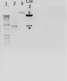
The restriction products were loaded in a 1 % agarose gel (appendix 1.A.2) to visualize the result (figure 9.A), similar to as described in previous section (see III.1.2.1.2.1). As shown in figure 9.A, colonies 2 and 3 presented two main bands of 8.8 and 1.28 kbp, which corresponds to the vector pLVX and the RGS14414 gene respectively. These results indicate that at least, colonies 2 and 3 retain the characteristics of vector as well as of RGS gene insert. For our future experiments, colony 2 was selected to proceed with the maxiprep and obtain a bigger amount of RGS14414 recombinant DNA.
1.2.1.2.3.3 Maxiprep
Stored stock of bacteria of colony 2 was inoculated in 5 ml of LB liquid medium with 100 µg/ml ampicillin (appendix 1.B.1) for 8 h at 37 ºC with shaking at 225 rpm and then, this culture was added into a flask containing 300 ml of LB medium with ampicillin and further incubated at 37 ºC for 15 h, shaking at 225 rpm. A stock of bacteria with RGS14414 recombinant DNA was prepared at this stage and stored at -80 ºC (appendix 1.B.2) for long-term use.
Maxiprep was done by using Wizard ® Plus Maxiprep DNA Purification System kit
restriction reaction as described previously (section III.1.2.1.2.3.2, table 6) (figure 9.B) and additionally by 5´and 3´ sequencing. The recombinant DNA obtained from colony 2 showed correct size and orientation of RGS gene insert. In addition, whole sequence was intact and showed no mutation (DNA sequencing was done by STAB VIDA; http://www.stabvida.net).
* *
1 2 3 Colony 1 2 3 4
4072 bp 12216 bp
1636 bp 1018 bp
A
1 2 3
*
Col. 2
B
1.2.2 Production of lentivirus
1.2.2.1 Transfection of 293T cells and lentivirus collection
The lentivirus stock was produced in the 293T cell line (Clontech, Cat. No. 632180), a subclone of the transformed human embryonic kidney cell line, HEK 293, with type-5 human adenovirus. These eukaryotic cells are highly transfectable and support high levels of lentiviral protein expression (Pear et al 1993). A cryogenic tube (Nunc, Cat. No. 375418) containing 2.0 x 10⁶ 293T cells was thawed (appendix 2.C) and seeded in a 75-cm2 culture flask (Nunc, Cat. No. 156499) with 10 ml of complete culture medium composed of: (i) 90 % Dulbecco’s Modified Eagle’s Medium (DMEM) with high glucose (4.5 g/L), 4 mM L-glutamine, 0.1 mM non-essential amino acids, 3.7 g/L sodium bicarbonate (Sigma-Aldrich, Figure 9. XhoI and EcoRI restriction reaction of DNA resulting of miniprep and maxiprep to prove the presence of RGS14414 recombinant DNA. (A) Restriction reaction of miniprep from colonies 1-4,
demonstrated that only, colonies 2 and 3 had included RGS14414 recombinant. Thus, the restriction reaction
generated two main bands of 8.8 and 1.280 kbp which correspond to the vector pLVX and the RGS14414 gene
respectively. Colonies 1 and 4 only had included the vector without the insert. (B) Restriction reaction of maxiprep from colony 2. Arrows indicate the vector pLVX and asterisks the RGS14414 gene. Numbers 1, 2 and 3
indicate the molecular weight ladder, RGS14414 as positive control and pLVX vector without endonucleases
Cat. No. D5796) and 1 mM sodium pyruvate (Sigma-Aldrich, Cat. No. S8636); (ii) 10 % Fetal Bovine Serum (Tet System Approved FBS); (iii) 1 % penicillin-streptomycin antibiotic (appendix 2.A.1). To facilitate cell attachment, flasks were pre-treated with a 0.3 % (w/v) gelatin solution (appendix 2.B). Cells were maintained in a 37 °C humidity incubator (5 % CO2 and 21 % O2). Complete culture medium was exchanged every 2 days and subcultures were performed when cells reached 80 %-90 % confluence (about 3-4 days; see appendix 2.E). A stock of 293T cell was prepared after first subculture and stored in liquid nitrogen for future use (appendix 2.D).
At the sixth subculture, 293T cells were seeded in two T175-cm2 culture flasks (Nunc, Cat. No. 159910) with 25 ml of complete medium, with goal to obtain enough amounts of cells required for transfection with recombinant DNA. Both RGS-lentivirus and vehicle-lentivirus of empty vector were prepared by using Lenti-XTM Packaging System (Clontech, Cat. No. 631247) according to manufacturer’s protocol. All steps were carried out according to biosafety level 2 instructions. In brief, 4.5 x 10⁶ cells per plate were seeded in twelve 100-mm Petri plates (Corning, 734-1815) containing 10 ml of complete medium but without antibiotic. Here 6 plates were for control and other 6 were for RGS14. Cells were incubated in 5 % CO2 incubator at 37 ºC for 24 h. Transfection solutions were prepared as shown in table 7. The polymer solution (tube 2) was mixed with the DNA solution (tube 1) and incubated for 10 min at room temperature to allow complex formation. Mixed solution of 1200 µl was added dropwise to cell culture plate and then, was gently rocked before incubating at 37 ºC in 5 % CO2.
Table 7. Preparation of transfection solutions (volume per plate).
Tube 1 (DNA solution) Tube 2 (polymer solution)
557µl Xfect Reaction Buffer (Clontech, 631317)
592.5µl Xfect Reaction Buffer (Clontech, 631317)
36 µl Lenti-X HTX Packaging Mix (Clontech, 631260)
7.5µl Xfect Polymer (Clontech, 631317)
11 µl RGS14414-pLVX (0.635 μg/µl) or
14 µl vector pLVX DsRed Monomer C1 0.5 μg/µl (Clontech, 632153)
---
After 24 h, transfection medium was replaced with 10 ml fresh complete cell culture medium and cell culture plates were incubated at 37 °C for an additional 48 h. During this period, transfected cells produce and release non-replicant lentivirus to medium. Therefore, lentiviral supernatants were harvested and pooled. Cellular debris was removed by centrifugation at 500 x g for 15 min at 4 ºC in CS15R Beckman centrifuge, followed by filtration of supernatants through 0.45 µm polysulfonic filter (Sarstedt, 831826). The lentiviral supernatants were maintained on ice during all the procedure.
Finally, to concentrate, lentiviral solutions were ultracentrifuged (Ichim & Wells 2011; Reiser 2000) at 25,000 rpm for 90 min at 4 ºC in Ultra-Clear tubes (Beckman Coulter, 344058) using Beckman XL-90 ultracentrifuge and SW29 rotor. Supernatants were discarded and 150 μl of sterile saline serum was added to pellet and placed at 4 ºC for overnight to facilitate the resuspension of lentivirus. Aliquots of 3.5 μl were prepared and stored at -80 ºC.
1.2.2.2 Determination of lentivirus titer in HT1080 cell line
The lentivirus titer was determined using HT1080 cells (ATCC, CCL-121), a cell line derived from human fibrosarcoma cell line. A cryogenic tube containing 1.1 x 10⁶ HT1080 cells was thawed (appendix 2.C) and seeded in a 75-cm2 culture flask (Nunc, Cat. No. 156499) with 10 ml of complete culture medium composed of: (i) 90 % (v/v) Eagle's Minimum Essential Medium supplemented with 1.5 g/L sodium bicarbonate (Sigma-Aldrich, Cat. No. D5796) and 1 mM sodium pyruvate (Sigma-Aldrich, Cat. No. S8636); (ii) 10 % (v/v) Fetal Bovine Serum; and 1 % (v/v) penicillin-streptomycin antibiotic (appendix 2.A.2). Cells were maintained in a CO2 incubator at 37 °C. Complete medium was exchanged every 2 days and subcultures were performed when cells were 80 %-90 % of confluence (usually every 3 days) (appendix 2.E). At the second subculture, a stock of HT1080 cells was prepared and stored in liquid nitrogen for future use (appendix 2.D).
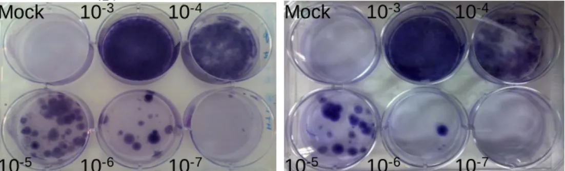
from 6-well plate was removed and replaced by 2 ml of complete HT1080 growth medium containing 1 μg/μl puromycin (Clontech, Cat. No. 631306). Since vector pLVX contains Puror gene, transfected cells expressing this gene are expected to show resistance against this drug. In continuation, cells were incubated in this drug for 10 days with change of culture medium every two days. Survived cell colonies were stained in 1 % (w/v) crystal violet (Sigma Aldrich, Cat. No. C3886), 10 % (v/v) ethanol (JT Baker, Cat. No. 8025) solution. Cells were rinsed twice for 1 min in 1 ml of phosphate buffered saline (PBS) pH 7.4 (Gibco, Cat. No. 10010) to eliminate cellular debris, and then incubated in the stain solution for 10 min at room temperature. To remove staining solution, 3 washes of 1 min in PBS were done.
The titer in terms of colony-forming units (CFU) of lentivirus stock was estimated by calculating mean of the number of colonies generated by two least concentrated dilutions, multiplied by the dilution factor. As shown in figure 10, RGS-lentivirus titer was 1.75 x 10⁷ CFU/ml, because 15 colonies were observed in well of 10-6 dilution and 2 colonies in 10-7 dilution. In contrast to RGS-lentivirus, vehicle-lentivirus titer was 2.75 x 10⁶ CFU/ml.
Mock 10-3 10-4
10-5 10-6 10-7
B.
Vehicle-LentivirusA.
RGS14414-LentivirusMock 10-3 10-4
10-5 10-6 10-7
Figure 10. Titration of lentivirus stocks. (A) RGS14414-lentivirus shows a titer of 1.75 x
107 CFU/ml and (B) vehicle-lentivirus presents a titer of 2.75 x 10⁶ CFU/ml.
1.2.3 Animals
1.2.4 Stereotaxic surgery
Approximately 4000 CFU in 2 µl (for rats) or 2000 CFU in 1 µl (for mice) of RGS-lentivirus or vehicle-RGS-lentivirus were delivered into area V2 of visual cortex by stereotaxic surgery (Cetin et al 2006; Cooley & Vanderwolf 2005; Fornari et al 2012), Before proceeding, animals were deeply anesthetized by administering intraperitoneally 75 mg/kg ketamine (Imalgene 1000; Merial Laboratorios) and 1 mg/kg (rats) or 0.5 mg/Kg (mice) of medetomidine (Domtor, Pfizer).
During surgery, animal body temperature was maintained with an electric blanket. Animals head was placed on the stereotaxic apparatus (Stoelting) as indicated in figure 11. For the surgery of mice, an adaptor (Cunningham mouse, Harvard Apparatus) was coupled.
The coordinates for injection in area V2 (see table 8) were taken from The Mouse Brain in Stereotaxic Coordinates (Paxinos & Franklin 2001) and The Rat Brain in Stereotaxic Coordinates (Paxinos & Watson 1998). In our experiments, anteroposterior (AP) and mediolateral (ML) coordinates are from bregma and dorsoventral (DV) from dura mater. To access the dura mater, the skull was perforated using a drill (Dremel), and the dura mater was punctured to prevent the carpule deviation.
Table 8. Rodent injection coordinates for area V2.
Rodent Anteroposterior
(AP)
Mediolateral (ML)
Dorsoventral (DV)
RAT + 4.3 - 2.1 - 1.9
Figure 11. Illustration of the experimental setup for intracraneal lentivirus microinjection in rodents. (A) A picture representing a rat placed in the stereotactic frame. (B) A picture showing the bregma position (indicated by an arrow) in the rat skull. (C) A picture representing a mouse placed in the stereotactic frame. 1: stereotaxic apparatus; 2: infusion system; 3: drill; 4: anesthesia; 5: surgical instruments; 6: electric blanket; 7: cold light; 8: dry heat sterilizer; 9: dental carpule; 10: mouse stereotaxic adaptor.
The injection was carried out by a 30 G dental carpule (Heraeus Kulzer Iberia) connected to a 10 µl Hamilton syringe (MicroliterTM 701, Harvard Apparatus) through a plastic tube connector (Plastic One). Lentivirus was infused into area V2 at the rate of 10 μl/h using an infusion pump (11 Plus, Harvard Apparatus). Once the lentivirus injection was finished the carpule was maintained into the brain area for five more minutes to facilitate complete diffusion.