TítuloHyperfine coupling constants on inner‐sphere water molecules of GdIII‐based MRI contrast agents
23
0
0
Texto completo
(2) Introduction Gadolinium(III) complexes with poly(aminocarboxylate) ligands have attracted considerable interest during the last two decades since they are commonly used as contrast agents (CAs) in magnetic resonance imaging (MRI).1,2 CAs are paramagnetic probes that enhance the image contrast by preferentially influencing the longitudinal and/or transverse relaxation times of water molecules in the vicinity of the complex. Commercially available GdIII‐based contrast agents contain one water molecule coordinated to the metal ion that exchanges rapidly with the bulk water. The efficiency of a CA is evaluated in vitro in terms of its relaxivity, which is defined as the longitudinal relaxation rate enhancement of water proton nuclei per mM concentration of gadolinium ions. The observed relaxivity is accounted for by the presence of two different contributions: outer‐sphere relaxation, which is the result of free diffusion of water molecules in the vicinity of the metal center, and inner‐sphere relaxation, which is the result of the exchange between the coordinated water molecule(s) and the bulk water. The inner‐sphere contribution to relaxivity is proportional to the number of water molecules coordinated to the metal ion (q). The 1H longitudinal relaxation rate of the inner‐sphere water molecules is dominated by the dipolar interaction, which is proportional to 1/r6, where r is the distance between the metal ion and the inner‐sphere water proton nuclei. Additionally, the inner‐sphere contribution depends on four correlation times: the residence time of a water proton in the inner coordination sphere (τm), the rotational correlation time of the Gd⋅⋅⋅H vector (τR), and the longitudinal and transverse electronic relaxation times of the metal ion (T1e and T2e).3 These correlation times show temperature dependence, and as a result the inner‐sphere contribution to relaxivity depends on a relatively large number of parameters. Besides this, the outer‐sphere contribution depends on the distance of closest approach of an outer‐sphere water molecule to GdIII and the diffusion coefficient for the translational diffusion of a water molecule away from the GdIII chelate, which also varies with temperature.4 Thus, there are a large number of parameters determining the relaxivity of a given compound, and it is difficult to determine them from relaxivity measurements without obtaining independent information of at least some of the most important parameters.5 Variable‐temperature 17O NMR measurements of chemical shifts and longitudinal and transverse relaxation rates constitute a valuable tool to investigate the parameters influencing relaxivity in MRI CAs. 1 17O NMR data provide information on the water exchange kinetics of the complex as well as on the rotational correlation time.6 A combined analysis of the 1H relaxivity and 17O NMR data has been applied successfully to evaluate the parameters governing relaxivity in many GdIII complexes.7 Both the 17O NMR chemical shifts and relaxation rates depend on the hyperfine coupling constant A/ħ between the electron spin of the metal ion and the 17O nuclear spin. For the GdIII aqua‐ion [Gd(H2O)8]3+ A/ħ was determined to be 5.3×10⁶ rad s−1;5 the values measured for many small GdIII chelates fall within a relatively narrow range of 3.9±0.3×10⁶ rad s−1.5,7,8 Some examples exist in the literature where significantly lower hyperfine coupling constants have been reported.9 These examples include [Gd(BPEDA)(H2O)]− and [Gd(HP‐DO3A)(H2O)], for which A/ħ values of 2.3×10⁶ rad s−1 and 2.9×10⁶ rad s−1 have been determined (Scheme 1).10,11 However, these results should be taken with care, since unusually low fitted A/ħ values could also be the result of a hydration number lower than expected, for instance due to the presence in solution of an equilibrium between complex species with different hydration numbers. Indeed, different examples of small GdIII complexes that present hydration equilibria in solution have been reported in the literature.12,13 Although high‐level correlated wave‐function‐based ab initio computational methods have successfully been applied to calculate hyperfine coupling constants in small organic radicals,14 the scaling behaviour of these methods with system size prevent their application to larger systems with the currently available computational resources. In the last decade, methods based on density functional theory (DFT) have become an attractive tool for the calculation of hyperfine coupling constants (HFCCs) due to the high accuracy that can be achieved at relatively low computational cost. Indeed, DFT calculations have been shown to provide accurate HFCCs for both small organic radicals15 and transition metal complexes.16 Yazyev et al..
(3) investigated the hyperfine interactions of 1H and 17O nuclei of inner‐sphere water molecules in [Gd(H2O)8]3+ and [Gd(DOTA)(H2O)]− complexes.17 The approach used to calculate the hyperfine interactions involved either classical or Car–Parrinello molecular dynamics (MD) simulations. From the trajectories of these simulations clusters of molecules were extracted, and then investigated by using DFT calculations, which provided 17O hyperfine coupling constants in good agreement with the experimental values.18 The main drawback of this approach is that GdIII complexes with relevance in MRI can be hardly handled with Car–Parrinello MD calculations due to the excessive computational cost, and thus HFCCs must be calculated on clusters taken from classical MD simulations, which might be problematic due to the lack of parametrization of many GdIII complexes in commonly available force fields. On the other hand, MD simulations have the advantage that allows one to perform a meaningful sampling of different configurations, as the observed HFCC corresponds to the mean value of all configurations in solution.. Scheme 1. Ligands discussed herein.. The aim of this work is to develop a simple methodology based on DFT for the calculation of 17O hyperfine coupling constants of coordinated water molecules in GdIII complexes, and to investigate the reasons for the particularly low hyperfine coupling constants determined experimentally for some of them. For this purpose we have selected different GdIII complexes such as [Gd(DOTA)(H2O)]− (gadoterate meglumine, Dotarem; Guerbet, Aulnaysous‐Bois, France), [Gd(DTPA)(H2O)]2− (gadopentetate dimeglumine, Magnevist; Schering, Berlin, Germany), [Gd(DTPA‐BMA)(H2O)] (gadodiamide, Omniscam; Nycomed, Oslo, Norway) and [Gd(HP‐DO3A)(H2O)] (Gadoteridol, ProHance; Bracco, Milan, Italy) currently used in clinical practice as MRI CAs (Scheme 1). Additionally, we have performed calculations on [Gd(BPEDA)(H2O)]− as a representative example of a complex for which a low A/ħ value has been reported.10 The fitting of the.
(4) experimental 17O NMR data often requires one to consider an outer‐sphere contribution to the observed chemical shifts. Herein we present a set of calculations performed on molecular clusters, explicitly including second‐sphere water molecules, that allows one to obtain information about the scalar outer‐sphere contribution to the 17O NMR chemical shifts. Finally, we have also calculated 1H hyperfine coupling constants to confirm that the scalar mechanism to the proton relaxation enhancement can be safely neglected.. Theory The hyperfine coupling tensor for the nucleus N consists of three contributions, which are the isotropic Fermi contact (FC) and the anisotropic spin–dipolar contributions and the spin–orbit contribution. Herein we focus on the calculation of isotropic FC contribution (Aiso), which is given by Equation (1):19. 𝐴iso (𝑁) =. 4π 3𝑆. 𝛽e 𝛽N 𝑔e 𝑔N 𝜌α−β (𝑅N ). (1). where βN and βe are the nuclear and Bohr magnetons, respectively, gN and ge are nuclear and free‐ electron g values, S is the total electron spin of the system, and ρα–β(RN) represents the difference between majority spin (α) and minority spin (β) densities at the position of the nucleus N. Thus, Aiso is proportional to the value of the spin density at the position of nucleus N, which may be transmitted directly through the bonds by spin delocalization and/or by spin polarization. Experimental values of isotropic 17O HFCCs have been determined for many GdIII chelates from 17O NMR chemical shifts and as absolute values from transverse relaxation rates. The reduced transverse relaxation rates and chemical shifts, 1/T2r and ωr, observed on bulk water nuclei may be written as in Equations (2)– (3),20 where 1/T2m is the relaxation rate of the bound water, τm is the residence time of water molecules in the inner sphere, Δωm is the chemical shift difference between bound and bulk water, and ΔωOS is the outer sphere contribution to the chemical shift.. 1 𝑇2r. =. −2 +𝜏−1 𝑇 −1 +Δ𝜔2 1 𝑇2m m 2m m. (2). −1 +𝑇 −1 )2 +Δ𝜔2 𝜏m (𝜏m m 2m. Δ𝜔r =. Δ𝜔m 2 −1 2 Δ𝜔2 (1+𝜏m 𝑇2m ) +𝜏m m. + Δ𝜔os. (3). Δωm is governed by the hyperfine or scalar interaction according to Equation (4), where B represents the magnetic field, S is the electron spin and gL is the isotropic Landé g factor.21. Δ𝜔m =. 𝑔L 𝜇B 𝑆(𝑆+1) 𝐴 3𝑘B 𝑇. ℏ. (4).
(5) It must be pointed out that for f 7 ions such as GdIII, the ligand field splitting is zero under first‐order conditions, and therefore no pseudocontact contribution to the hyperfine shifts is expected. However, higher‐ order effects may provoke the splitting of the J=7/2 level, and therefore a small pseudocontact shift. It has been estimated that such high‐order effects may result in shifts of ∼0.25 ppm,22 while Equation (4) predicts a 17O contact shift of 2545 ppm at 298 K for a typical A/ħ value of 3.9×10⁶ rad s−1. Thus, pseudocontact contributions to the 17O NMR shifts of coordinated water molecules in GdIII complexes can be safely neglected. It has been suggested that the outer‐sphere contribution to Δωr may be represented by an empirical constant COS as given by Equation (5):5. Δ𝜔OS = 𝐶OS Δ𝜔m. (5). The transverse 17O NMR relaxation of bound water molecules is dominated by the scalar contribution, 1/T2sc, as given in Equation (6):23. 1 𝑇2m. ≅. 1 𝑇2sc. =. 𝑆(𝑆+1) 𝐴 2 3. (ℏ ) 𝜏𝑠1. (6). where 1/τs1 is the sum of the exchange rate constant kex=1/τm and the electron spin relaxation rate 1/T1e. Thus, both the 17O NMR chemical shifts and transverse relaxation rates depend on the HFCC A/ħ. In Equations (4) and (6) A/ħ is expressed in rad s−1, and therefore equals 2 π⋅Aiso as defined in Equation (1).. Computational methods All calculations presented herein were performed employing the Gaussian 09 package (Revision B.01).24 An important issue for the adequate computational description of LnIII complexes is the treatment of relativistic effects.25 In general, two different approaches have been developed to handle relativistic effects in systems containing heavy elements, namely the use of all‐electron relativistic approaches such as the DKH2 and ZORA approximations,26 and the use of relativistic effective core potentials (RECP). The most widely used approximation to treat relativistic effects in LnIII complexes is the RECP approach, in which only the chemically relevant valence electrons are treated explicitly and relativistic effects are implicitly accounted for by a proper adjustment of free parameters in the valence model Hamiltonian.27 In the present work we employed the energy‐consistent quasirelativistic ECPs and associated basis sets of Dolg and coworkers,28,29 for which two different core definitions have been developed: “large‐core” (LC), in which the 4f electrons are included in the core, and “small‐core” (SC), which treats the four‐, five‐ and six‐shell electrons explicitly. The LCRECP includes 46+4f 7 electrons in the core for GdIII, while the outermost eight electrons are treated explicitly with a [5s4p3d]‐GTO valence basis set. For the SCRECP, which includes 28 electrons in the core, we selected the associated ECP28MWB_GUESS basis set. 30 Full geometry optimizations of the [Gd(BPEDA)(H2O)]−⋅x H2O, [Gd(DOTA)(H2O)]−⋅x H2O, [Gd(DTPA)(H2O)]2−⋅x H2O, [Gd(DTPA‐BMA)(H2O)]⋅x H2O and [Gd(HP‐DO3A)(H2O)]⋅x H2O systems (x=0, 1, or 2) were performed in aqueous solution employing DFT within the hybrid meta generalized gradient approximation (hybrid meta‐ GGA), with the TPSSh exchange–correlation functional.31 Input geometries of the [Gd(BPEDA)(H2O)]− and [Gd(DOTA)(H2O)]− systems for geometry optimization were taken from previous computational.
(6) studies.10,32 For the DOTA4− complex, which is known to exist in solution as a mixture of square antiprismatic (SAP) and twisted‐square antiprismatic (TSAP) isomers, we performed our calculations for both isomers.33 The input geometry of [Gd(HP‐DO3A)(H2O)] was constructed from that of the DOTA4− analogue, and only the SAP isomer was considered. The starting geometry of [Gd(DTPA)(H2O)]2− was taken from the X‐ray structure of the MS‐325 analogue,34 while that of [Gd(DTPA‐ BMA)(H2O)] was constructed from the optimized geometry of the DTPA analogue by adding the methylamide groups. Thus, only one of the four diasteroisomers of the complex observed in aqueous solution has been investigated (the cis isomer).35 For geometry optimization purposes we used the LCRECP for Gd, while the standard 6‐31G(d) basis set was used for C, H, N and O atoms. No symmetry constraints have been imposed during the optimizations. The use of LCRECPs requires a separate potential for each oxidation state or 4f subconfiguration, which precludes the modeling of f–f centered processes and the treatment of spin–orbit coupling. However, this approach avoids many difficulties associated to the computational treatment of open‐shell systems, and despite their approximate nature, it is an efficient computational tool that has proven to give good results in studies that focus on the structural features or the estimates of relative energies for LnIII complexes at both the HF and DFT level.36 The stationary points found on the potential energy surfaces as a result of the geometry optimizations have been tested to represent energy minima rather than saddle points via frequency analysis. Isotropic 17O HFCCs in the [Gd(H2O)8]3+ model system were calculated in aqueous solution with unrestricted DFT methods by employing different functionals within the LSDA approximation (SVWN 37,38 and SPL39), GGA (BLYP,40,41 G96LYP,41,42,43 mPWLYP41,44), meta‐GGA (BB95,40,45 TPSS31), hybrid‐GGA (B3LYP,41,46 BH&HLYP,47 PBE1PBE1,48 and B3PW9146,49) and hybrid meta‐GGA (TPSSh31). The geometry of the [Gd(H2O)8]3+ system is the same as described in a previous paper,50 obtained from geometry optimizations in aqueous solution (PCM model) at the B3LYP/LCRECP/6‐31+G(d) level. The coordination environment around the metal ion in [Gd(H2O)8]3+ corresponds to a slightly distorted square antiprismatic geometry of D4d symmetry with a Gd―O distance of 2.433 Å. Both the mean Gd―O distance and the metal coordination environment are in good agreement with previous theoretical investigations. 51 For HFCC calculation purposes we used both the SCRECP approach and the all‐electron second‐order Douglas–Kroll– Hess (DKH2) method as implemented in Gaussian 09,52 which employs a Gaussian nuclear model. For DKH2 calculations, the all‐electron scalar relativistic basis set of Pantazis and Neese was used for Gd.53 DFT investigations of HFCCs require the use of specifically developed basis sets with extra flexibility in the core region.54 Thus, for the description of water molecules of [Gd(H2O)8]3+ in both DKH2 and SCRECP calculations we used the EPR‐II and EPR‐III basis sets of Barone,55 which were optimized for the computation of HFCCs by DFT methods. EPR‐II is a double‐zeta basis set with a single set of polarization functions and an enhanced s part, while EPR‐III is a triple‐zeta basis set including diffuse functions, double d polarizations and a single set of f‐polarization functions. Both basis sets contain an improved s part to better describe the nuclear region. The isotropic 17O and 1H HFCCs in [Gd(BPEDA)(H2O)]−⋅x H2O, [Gd(DOTA)(H2O)]−⋅x H2O, [Gd(DTPA)(H2O)]2−⋅x H2O, [Gd(DTPA‐BMA)(H2O)]⋅x H2O and [Gd(HP‐ DO3A)(H2O)]⋅x H2O (x=0, 1 or 2) were calculated by using the TPSSh functional and the SCRECP and DKH2 approaches, with the EPR‐III basis set to describe the ligand atoms. The highest spin state was considered as the ground state (octuplet, 4f 7) in all cases. Since the calculation of HFCCs was performed by using an unrestricted model, spin contamination56 was assessed by a comparison of the expected difference between S(S+1) for the assigned spin state [S(S+1)=15.75 for the mononuclear GdIII complexes investigated here] and the actual value of <S2>.57 The results obtained for [Gd(H2O)8]3+ indicate that spin contamination is negligible for all functionals tested in this work [<S2>−S(S+1)<0.0060]. A similar situation holds for all GdIII complexes with polyaminocarboxylate ligands investigated by using the TPSSh functional [<S2>−S(S+1) <0.0090]. Convergence of the SCF procedure during the calculations of 17O HFCCs was.
(7) problematic in some cases, and therefore a quadratically convergent SCF procedure was used when first‐ order SCF did not achieve convergence (by using the scf=xqc keyword in Gaussian 09). The default values for the integration grid (75 radial shells and 302 angular points) and the SCF energy convergence criteria (10−8) were used in all calculations. Throughout this work solvent effects were included by using the polarizable continuum model (PCM), in which the solute cavity is built as an envelope of spheres centered on atoms or atomic groups with appropriate radii. In particular, we used the integral equation formalism (IEFPCM) variant as implemented in Gaussian 09.58. Results and discussion Calculation of 17O HFCCs in [Gd(H2O)8]3+ Due to its small size, DFT calculations on the [Gd(H2O)8]3+ complex are relatively undemanding, and therefore we used this system to evaluate the performance of twelve commonly available density functionals (SVWN, SPL, BLYP, G96LYP, mPWLYP, BB95, TPSS, B3LYP, BH&HLYP, B3PW91, PBE1PBE and TPSSh) to predict isotropic 17O HFCCs. The main results of our calculations are shown in Figure 1 and Table 1. Experimental Aiso values of 0.7159 and 0.7650 MHz have been obtained from 17O NMR shift data, while a value of 0.845 MHz was determined from the simultaneous analysis of 17O NMR chemical shifts and 1H relaxivity data.. Figure 1. Absolute deviations of calculated Aiso values [MHz] obtained for [Gd(H2O)8]3+ by using different density functionals with respect to the experimental value (0.84 MHz)..
(8) Table 1. Calculated 17O hyperfine coupling constants (Aiso) [MHz] calculated for [Gd(H2O)8]3+. Type. Functional. Method. Aiso. LSDA. SVWN. RSC28/EPR‐II RSC28/EPR‐III DKH2/Neese/EPR‐III RSC28/EPR‐II RSC28/EPR‐III DKH2/Neese/EPR‐III RSC28/EPR‐II RSC28/EPR‐III DKH2/Neese/EPR‐III RSC28/EPR‐II RSC28/EPR‐III DKH2/Neese/EPR‐III RSC28/EPR‐II RSC28/EPR‐III DKH2/Neese/EPR‐III RSC28/EPR‐II RSC28/EPR‐III DKH2/Neese/EPR‐III RSC28/EPR‐II RSC28/EPR‐III DKH2/Neese/EPR‐III RSC28/EPR‐II RSC28/EPR‐III DKH2/Neese/EPR‐III RSC28/EPR‐II RSC28/EPR‐III DKH2/Neese/EPR‐III RSC28/EPR‐II RSC28/EPR‐III DKH2/Neese/EPR‐III RSC28/EPR‐II RSC28/EPR‐III DKH2/Neese/EPR‐III RSC28/EPR‐II RSC28/EPR‐III DKH2/Neese/EPR‐III. 0.387 0.481 0.626 0.392 0.485 0.623 0.028 0.175 0.399 0.058 0.193 0.424 0.035 0.182 0.401 −0.020 0.116 0.480 0.432 0.542 0.764 0.533 0.640 0.683 0.836 0.907 0.853 0.679 0.776 0.824 0.777 0.861 0.873 0.656 0.766 0.885. SPL. GGA. BLYP. G96LYP. mPWLYP. Meta‐GGA. BB95. TPSS. Hybrid‐GGA. B3LYP. BH&HLYP. B3PW91. PBE1PBE. Hybrid meta‐GGA. TPSSh. The data reported in Table 1 and Figure 1 indicate that LSDA functionals (SVWN and SPL) provide a poor agreement between the experimental and calculated Aiso values, with absolute deviations above 0.2 MHz. The agreement is even worse when using GGA functionals (BLYP, G96LYP or mPWLYP), which deviate by 0.42 to 0.81 MHz from the experimental value of 0.84 MHz. The performance of the meta‐GGA functional BB95 is also poor, while TPSS performs somewhat better, particularly when using DKH2 calculations. These results are in agreement with previous calculations performed on small main‐group.
(9) radicals and 3d and 4d transition metal complexes, which showed that meta‐GGA functionals do not substantially improve the results compared to GGA functionals.60 The use of hybrid‐GGA functionals (B3LYP, BH&HLYP and B3PW91 or PBE1PBE) results in an important improvement of the agreement between the experimental and calculated Aiso values. This is in line with previous investigations on transition metal complexes, which established that hybrid GGA functionals provide the best predictions of HFCCs.61,62 In the case of [Gd(H2O)8]3+ the hybrid GGA functionals BH&HLYP, B3PW91 and PBE1PBE appear to perform somewhat better than B3LYP. As observed previously,63 the hybrid variant of TPSS, TPSSh, provides a clear improvement of the calculated HFCCs in comparison to the non‐hybrid counterpart. Considering the uncertainty of the experimental value of Aiso in [Gd(H2O)8]3+, for which values ranging between 0.71 and 0.84 MHz have been reported, we conclude that hybrid GGA functionals BH&HLYP, B3PW91 and PBE1PBE, as well as the hybrid meta‐GGA functional TPSSh, provide Aiso values in close agreement with the experimental values. The DKH2 and SCRECP approaches give Aiso values in good mutual agreement when using hybrid GGA and hybrid meta‐GGA functionals, while the DKH2 approach provides somewhat better results with LSDA and GGA functionals. The use of the EPR‐III basis set in SCRECP calculations significantly improves the agreement between experimental and calculated values with respect to RSC28/EPR‐II calculations. It must be pointed out that our SCECP calculations used point charges for the nuclei, while DKH2 calculations employed a Gaussian nuclear model. Different studies showed that the use of point charges for the nuclei is not always a suitable approximation, particularly when hyperfine properties for heavy nuclei are considered.64 Thus, the discrepancies in Aiso values calculated with the SCECP and DKH2 approaches, particularly when using LSDA and GGA functionals, might be related to the use of the point charge approximation for the nuclei in SCECP calculations. Molecular geometries of GdIII polyaminocarboxylates Prior to calculation of the 17O Aiso values of the inner‐sphere water molecule in the various Gd complexes, the molecular geometries of these systems were fully optimized in aqueous solution (IEFPCM model) at the TPSSh/LCRECP/6‐31G(d) level. The optimized molecular geometries of the complexes are shown in Figure 2 and the average bond distances of the metal coordination environment are given in Table 2. The results show that our calculations reproduce the distances between Gd and the donor atoms of the ligands observed in the corresponding X‐ray structures fairly well. However, the distances between Gd and the oxygen atom of the inner‐sphere water molecule (2.56–2.67 Å) are clearly longer than those observed in the solid state (2.44–2.52 Å). All calculated Gd―OW distances are also longer than that normally assumed in the analysis of 17O NMR longitudinal relaxation data of nine‐coordinate GdIII complexes (2.50 Å),5 and also longer than those determined experimentally by using ENDOR spectra (2.4–2.5 Å).65 This bond elongation can be attributed, at least in part, to the fact that continuum models of solvation cannot account for specific solvent–solute interactions (i.e. hydrogen‐bonding interactions between inner‐sphere and second‐sphere water molecules). Indeed, it has been shown that continuum dielectric solvent models are often inadequate for investigating ionic solutes that have concentrated charge densities with strong local solute–solvent interactions.66 To overcome this deficiency of continuum solvent models, it has become a common practice to add explicit solvent molecules to the model ionic systems.67 Thus, we performed geometry optimizations by using a mixed cluster/continuum model explicitly including second‐sphere water molecules. The major disadvantage of this approach is that adding extra solvent molecules to the first solvation sphere increases the computational cost. Moreover, the more atoms are included in the system, the larger the number of degrees of freedom and the higher the number of minimum‐energy structures..
(10) Figure 2. Structures of the [Gd(L)(H2O)]+/− systems (L=BPEDA, DOTA, DTPA, DTPA‐BMA or HP‐DO3A) as optimized in water at the TPSSh/LCRECP/6‐31G(d) level. Views are along the Gd―OW axis.. Our calculations performed on the [Gd(L)(H2O)]+/−⋅x H2O systems (L=BPEDA, DOTA, DTPA, DTPA‐BMA or HP‐DO3A) show that the inclusion of one second‐sphere water molecule considerably shortens the Gd―OW distances, while the inclusion of a second water molecule involved in hydrogen‐bonding interaction with the inner‐sphere water provides Gd―OW distances in close agreement with the experimental values (Table 2). Furthermore, the Gd⋅⋅⋅HIS distances (HIS=hydrogen atoms of the inner‐sphere water molecule) calculated for the systems including two second‐sphere water molecules fall within the range of 2.96–3.10 Å, the average value of the five systems investigated amounting to 3.02 Å. These values are in excellent agreement with those determined experimentally with the use of pulsed ENDOR spectroscopy (3.1±0.1.
(11) Å)73 and neutron diffraction measurements.2a In each of the [Gd(L)(H2O)]+/−⋅2 H2O complexes investigated the two Gd⋅⋅⋅HIS distances are quite similar, differing by less than 0.083 Å. As an illustrative example, the optimized geometry of [Gd(DOTA)(H2O)]−⋅x H2O (SAP isomer) is shown in Figure 3. The Gd―OW distances calculated for the SAP isomer of [Gd(DOTA)(H2O)]−⋅x H2O are slightly shorter than those obtained for the TSAP isomer, which is in line with a higher steric compression around the inner‐ sphere water molecule in the latter form.74. Table 2. Averaged bond distances [Å] of the metal coordination environments and ϕ angles [°] calculated at the TPSSh/LCRECP/6‐31G(d) level for [Gd(L)(H2O)]n+/–⋅x H2O complexes.[a] L DOTA/SAP. [b]. DOTA/TSAP. DTPA‐BMA[c]. HP‐DO3A[d]. DTPA[e]. BPEDA[f]. Gd―OC Gd―NAM Gd―OW ϕ Gd―OC Gd―NAM Gd―OW ϕ Gd―OC Gd―OA Gd―NAM Gd―OW ϕ Gd―OC Gd―OH Gd―NAM Gd―OW ϕ Gd―OC Gd―NAM Gd―OW ϕ Gd―OC Gd―OPY Gd―NAM Gd―NPY Gd―OW ϕ. x=0. x=1. x=2. Exp.. 2.383 2.679 2.606 85.7 2.394 2.686 2.646 83.38 2.351 2.551 2.699 2.559 103.8 2.366 2.495 2.666 2.568 88.7 2.407 2.673 2.639 83.6 2.391 2.417 2.768 2.659 2.669 86.4. 2.394 2.703 2.523 104.2 2.396 2.705 2.585 92.9 2.361 2.520 2.730 2.494 125.1 2.366 2.495 2.661 2.544 101.9 2.416 2.711 2.542 102.0 2.380 2.419 2.787 2.682 2.551 113.4. 2.394 2.720 2.494 123.7 2.398 2.727 2.514 121.9 2.371 2.521 2.742 2.483 118.0 2.372 2.533 2.677 2.514 120.2 2.412 2.743 2.501 127.7 2.394 2.433 2.792 2.698 2.516 121.7. 2.365 2.655 2.456. 2.374 2.441 2.672 2.442 2.375 2.330 2.647 2.507 2.400 2.640 2.490 2.46 2.43 2.69 2.61 2.52. [a] OC: coordinated oxygen atoms of acetate groups; OPY: coordinated oxygen atoms of pyridylcarboxylate groups; NAM: amine nitrogen atoms; NPY: pyridyl nitrogen atoms; OA: oxygen atoms of amide groups; OH: oxygen atoms of hydroxyl groups; OW: oxygen atom of inner‐sphere water molecules. [b] Experimental data from ref. 68. [c] Experimental data for the [Gd(DTPA‐ BBA)] complex, which crystallizes in cis conformation, from ref. 69. [d] Experimental data from ref. 70. [e] Experimental data from ref. 71. [f] Experimental data from ref. 72..
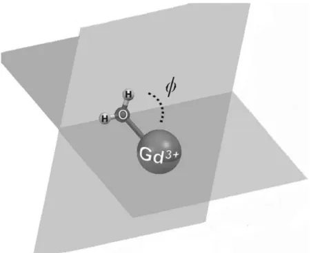
(12) Figure 3. View along the Gd―OW axis of the structure of [Gd(DOTA)(H2O)]−⋅2 H2O (SAP isomer) as optimized in water at the TPSSh/LCRECP/6‐31G(d) level showing hydrogen‐bonding interactions involving inner‐ and second‐ sphere water molecules. Hydrogen atoms, except those of water molecules, are omitted for simplicity.. These results show that the explicit inclusion of a few second‐sphere water molecules is crucial for the computation of accurate Gd―OW distances in complexes with polyaminocarboxylate ligands. Additionally, the inclusion of second‐sphere water molecules also affects the orientation of the inner sphere water molecule with respect to the Gd―O axis, as given by the tilt angle ϕ defined in Figure 4. Indeed, the inner‐ sphere water molecule in [Gd(L)(H2O)]n+/− complexes is tilted due to the hydrogen‐bonding interaction established between hydrogen atoms of the water molecule and the negatively charged oxygen atoms of the ligand, which results in ϕ angles of about 85°. In the [Gd(L)(H2O)]+/−⋅2 H2O systems the inner‐sphere water molecule is involved in hydrogen‐bonding interaction with second sphere water molecules rather than with oxygen atoms of the ligand, thereby increasing ϕ to about 120° (Table 2). As a general trend, the inclusion of second‐sphere water molecules provokes a slight lengthening of the Gd―ligand bonds. The distances calculated for the [Gd(L)(H2O)]+/−⋅2 H2O systems generally differ ≤4 % from the crystallographic reference data of the hydrated complexes. The calculated Gd―O C distances [OC=oxygen atoms of carboxylate groups] present an excellent agreement with the experimental values observed in the solid state. The distances between Gd and the N donor atoms of the ligands are overestimated by our calculations by 0.04–0.10 Å with respect to the X‐ray values, as previously observed for related systems.32,75 The distances between the GdIII ion and the H atoms of second‐sphere water molecules in [Gd(L)(H2O)]+/−⋅2H2O fall within the range 3.32–5.10 Å (Table S1, Supporting Information), with an average value of 4.18 Å. The average distance between Gd and the oxygen atoms of second‐sphere water molecules is slightly longer (4.25 Å), as second‐sphere water molecules tend to orientate their H atoms to negatively charged groups of the ligand. These values could be taken as a reference for the analysis of.
(13) longitudinal 1H and 17O relaxation data of Gd‐based MRI contrast agents containing a high negative charge. For such systems, it has been shown that second‐sphere water molecules provide a substantial contribution to 1H relaxivity and longitudinal 17O relaxation rates, both related to 1/r6.76. Figure 4. Definition of the tilt angle ϕ formed between the inner‐sphere water molecule and the Gd―O axis.. Calculation of 17O HFCCs of the coordinated water molecule in [Gd(DTPA)(H2O)]2−⋅x H2O The 17O Aiso values of the inner‐sphere water molecule in the [Gd(DTPA)(H2O)]2−⋅x H2O systems were calculated with the aid of the TPSSh functional in combination with the DKH2 and SCRECP approaches. The TPSSh functional was selected for this purpose on the basis of the results for [Gd(H2O)8]3+ presented above. Calculations performed on the [Gd(DTPA)(H2O)]2− complex provided 17O Aiso values of ca. 0.10 and 0.08 MHz by using the ECP and DKH2 approaches, respectively. These values deviate by ca. 0.5 MHz from the experimental values of 0.61 and 0.54 MHz obtained from the analysis of the 17O NMR data (Table 3). Inclusion of one second‐sphere water molecule pushes the calculated values (∼0.3 MHz) closer to the experimental one, while the inclusion of two second‐sphere water molecules provides calculated Aiso values in reasonable agreement with the experiment (0.50 and 0.52 MHz for the DKH2 and ECP approaches, respectively). Thus, the explicit consideration of the most important hydrogen‐bonding interactions involving the coordinated water molecule and the second coordination shell appears to be crucial for an accurate calculation of 17O Aiso values of the inner‐sphere water molecule. As pointed out previously,17a an important parameter influencing the 17O HFCCs is the Gd―O distance. Thus, we performed a relaxed potential energy surface scan on the [Gd(DTPA)(H2O)]2− system where the Gd―O distance was varied from 2.41 to 2.79 Å in steps of 0.015 Å. Subsequently, Aiso was calculated for each of these points at the RSC28/EPR‐III level (Figure 5). The variation of the Gd―O distance also provoked a substantial change of the tilt angle ϕ, which increased from 76.9° to 93.5° as the Gd―O distance was reduced from 2.79 to 2.41 Å. Our results show that Aiso increases from 0.028 to 0.33 MHz as the Gd―O distance decreases from 2.79 to 2.41 Å. These results highlight the importance of an accurate calculation of the Gd―O distance to obtain Aiso values in close agreement with the experimental data. However, these.
(14) calculations provide a Aiso value of 0.23 MHz for Gd―O distance of 2.49 Å, which corresponds to the distance observed in the solid state for the [Gd(DTPA)(H2O)]2− complex. Considering the experimental values of Aiso reported in the literature, 0.61 and 0.54 MHz,5,77 we conclude that the Gd―O distance is not the only factor affecting the magnitude of the 17O HFCC of inner‐sphere water molecules of GdIII complexes. Indeed, further calculations performed on the [Gd(DTPA)(H2O)]2− complex show that the orientation of the water molecule with respect to the Gd―O axis has a strong influence on the calculated HFCCs too (Figure 6). For a fixed Gd―O distance of 2.504 Å the calculated Aiso values increase from 0.22 to 0.58 MHz as the ϕ angle increases from 89.6 to 180°. A ϕ value of about 90° implies that the water molecule uses a lone‐pair placed on a p orbital to bind Gd. As the tilt angle increases, the s character of the lone pair is increasing, which increases the probability of the unpaired spin density to sit just on the nucleus, resulting in an increased Aiso.. Table 3. Calculated hyperfine coupling constants (Aiso) [MHz] for [GdL(H2O)]n+/–⋅xH2O complexes (TPSSh model). L DTPA DOTA/SAP DOTA/TSAP DTPA‐BMA HP‐DO3A BPEDA. RSC28/EPR‐III DKH2/Neese/EPR‐III RSC28/EPR‐III DKH2/Neese/EPR‐III RSC28/EPR‐III DKH2/Neese/EPR‐III RSC28/EPR‐III DKH2/Neese/EPR‐III RSC28/EPR‐III DKH2/Neese/EPR‐III RSC28/EPR‐III DKH2/Neese/EPR‐III. x=0. x=1. x=2. Exp.. 0.097 0.077 0.140 0.123 0.083 0.064 0.304 0.286 0.179 0.161 0.007 −0.014. 0.290 0.271 0.371 0.357 0.266 0.245 0.537 0.518 0.279 0.261 0.238 0.220. 0.518 0.499 0.561 0.547 0.509 0.490 0.557 0.543 0.434 0.416. 0.605[a] 0.541[b] 0.590[c]. [f]. 0.368[e]. 0.605[a] 0.462[d]. 0.396. [a] Ref. 5. [b] Ref. 77. [c] Ref. 5. The Gd DOTA complex is known to exist in solution as a mixture of SAP and TSAP isomers, with the SAP form representing ca. 85 % of the overall population (see text). [d] Ref. 11. [e] Ref. 10. [f] SCF convergence problems prevented the calculation of Aiso.. The spin density distribution in a given paramagnetic molecule denotes the difference between the contributions due to electrons with majority spin (α) and minority spin (β), and is the result of two effects: 1) spin delocalization, that is, the transmission of spin density through the bonds towards the observed nucleus; 2) spin polarization, which is the result of an effective attraction of unpaired electrons to the nearby ones of the same spin. Spin delocalization gives always a positive contribution to the spin density in contrast to spin polarization which can lead to a positive or negative contribution.18 Figure 7 shows a spin density map calculated for [Gd(DTPA)(H2O)]2−⋅2 H2O at the RSC28/EPR‐III level. As found previously for [Gd(H2O)8]3+,17a,18 the negative spin density calculated at the coordinated water oxygen indicates domination of the spin‐polarization effect at this location. Most of the positive spin density is placed on the Gd III ion itself. This shows that spin‐polarization effects dominate the 17O Aiso values of the coordinated water molecule in GdIII complexes. It is worth to recall that the negative spin density at the 17O nucleus of the inner‐sphere water molecule corresponds to a positive Aiso value (+0.518 MHz, Table 3) as a consequence of the negative magnetic moment of the 17O nucleus..
(15) Figure 5. Dependence of the 17O Aiso values with the Gd―O distance in [Gd(DTPA)(H2O)]2− as calculated at the RSC28/EPR‐III level. The arrows indicate the data obtained for the equilibrium geometry of [Gd(DTPA)(H2O)]2− (Gd―OW=2.639 Å, Aiso=0.097 MHz), and the Gd―OW distance used to scan ϕ in Figure 6. This distance is close to that found for [Gd(DTPA)(H 2O)]2−⋅2 H2O (2.501 Å).. Figure 6. Dependence of the Aiso values with tilt angle ϕ at a fixed Gd―O distance of 2.504 Å in [Gd(DTPA)(H2O)]2− (RSC28/EPR‐III level). The arrow indicates the torsion angle obtained from geometry optimization when the Gd―O distance is fixed at 2.504 Å..
(16) Figure 7. Contour spin density map obtained for the [Gd(DTPA)(H 2O)]2−⋅2 H2O system at the RSC28/EPR‐III level on the plane defined by the Gd atom, the oxygen atom of the coordinated water molecule and one of its hydrogen atoms [in au−3].. Calculation of 17O HFCCs in related systems Calculations performed on [Gd(L)(H2O)]n+/− complexes without second‐sphere water molecules confirm the poor agreement of calculated Aiso with experimental data. Inclusion of only two second‐sphere water molecules hydrogen‐bonded to the first‐sphere H2O molecule results in calculated HFCCs in close agreement with the experimental data. Furthermore, RSC28/EPR‐III and DKH2/Neese/EPR‐III calculations provide similar results, with the latter giving Aiso values typically 0.02 MHz smaller than the former (Table 3). The results suggest that the small experimental HFCCs determined for [Gd(BPEDA)(H 2O)]− and [Gd(HP‐ DO3A)(H2O)] were correctly determined, and that in some cases the HFCCs might fall out of the generally accepted 3.9±0.3×10⁶ rad s−1 (Aiso=0.67–0.57 MHz) range. A close inspection of the data reported in Tables 2 and 3 shows that the calculated Aiso values roughly correlate with the calculated Gd―OW distances (Figure 8). From Figure 8 it can be seen that Aiso calculated for [Gd(DTPA‐BMA)(H2O)]⋅2 H2O is low, considering its particularly short Gd―OW distance. The offset from the linear correlation can be partially ascribed to the small Gd―O―H―H torsion angle (118.0°) calculated from the optimized structure. The Aiso values obtained for the TSAP isomer of [Gd(DOTA)(H2O)]−⋅2 H2O are ∼10 % lower than those determined for the SAP isomer, in agreement with the shorter Gd―OW distance for the latter. The population of the TSAP isomer of [Gd(DOTA)(H2O)]− in solution is relatively small (ca. 15 %),33 and therefore the 17O HFCC determined experimentally is expected to be dominated by that of the SAP isomer..
(17) Figure 8. Correlation between Gd―O distances and Aiso values calculated for [Gd(L)(H2O)]n+/− 2H2O complexes at the DKH2/Neese/EPR‐III level.. An advantage using snapshots from MD simulations for the calculation of HFCCs is the inclusion of solution dynamics on the calculated Aiso values.18 However, the data shown in Figures 5 and 6 indicate an approximately linear dependence of Aiso around the equilibrium configuration with respect to both the Gd―OW distance and the ϕ angle. The measured HFCCs are weighted averages (<Aiso>) of individual values for different configurations present in solution. It is therefore not expected that <Aiso> differs substantially from the value corresponding to the equilibrium geometry. The good agreement between the experimental and calculated HFCCs given in Table 3 are in line with this reasoning. Furthermore, previous molecular dynamics studies performed on the [Gd(DOTA)(H2O)]− system pointed to a relatively narrow distribution width of Aiso (0.58±0.11 MHz).18 The calculations performed on the [Gd(L)(H2O)]n+/−⋅x H2O systems allow to evaluate 17O HFCCs of second‐ sphere water molecules, which could provide a non‐negligible scalar contribution to the observed 17O NMR chemical shifts. Indeed, different experimental works assumed that the outer‐sphere contribution can be accounted for by an empirical constant COS [Eq. (5)], with fitted values of 0.0–0.2. Thus, according to these studies, the outer‐sphere contribution to the observed chemical shifts may be responsible of up to 20 % of the observed chemical shifts. The Aiso values obtained for second‐sphere water molecules (𝐴ss iso , Table 4) are close to zero, and their absolute values amount to 1.5–3.6 % of those calculated for the coordinated water molecule. Again, both the RSC28/EPR‐III and DKH2/Neese/EPR‐III approaches provide results in good mutual agreement. Furthermore, our calculations provide negative 𝐴ss iso values, which suggest that the scalar contribution of second‐sphere water molecules to the overall observed 17O chemical shifts might be negligible, or result in slightly reduced observed chemical shifts..
(18) Table 4. Calculated 1H HFCCs of inner‐sphere water molecules (Aiso, MHz) and 17O HFCCs of second‐sphere water molecules (𝐴ss iso ) [MHz] in [GdL(H2O)]n+/–⋅2 H2O complexes (TPSSh model). Type. Method. 17 𝐴ss iso , O. Aiso, 1H. DTPA. RSC28/EPR‐III. −8.4×10⁻³ −6.0×10⁻³ −0.010 −7.9×10⁻³ −0.013 −2.3×10⁻³ −0.015 −4.0×10⁻³ −4.7×10⁻³ −7.4×10⁻³ −6.3×10⁻³ −8.7×10⁻³ −7.8×10⁻³ −0.013 −9.0×10⁻³ −0.015 −0.013 −0.013 −0.014 −0.015. 0.056 0.039 0.070 0.055 0.051 0.085 0.067 0.099 0.054 0.056 0.070 0.070 0.054 0.076 0.073 0.092 0.073 0.099 0.085 0.108. [a]. [a]. −9.5×10⁻³ 2.7×10⁻³. 0.058 0.068. DKH2/Neese/EPR‐III DOTA/SAP. RSC28/EPR‐III DKH2/Neese/EPR‐III. DOTA/TSAP. RSC28/EPR‐III DKH2/Neese/EPR‐III. DTPA‐BMA. RSC28/EPR‐III DKH2/Neese/EPR‐III. HP‐DO3 A. RSC28/EPR‐III DKH2/Neese/EPR‐III. BPEDA. RSC28/EPR‐III DKH2/Neese/EPR‐III. [a] SCF convergence problems prevented the calculation of Aiso.. 1. H HFCCs of the coordinated water molecules. As expected, the calculated 1H isotropic HFCCs for the systems investigated herein are quite small. For [Gd(H2O)8]3+ our calculations provide 1H Aiso values of −0.031 (RSC28/EPR‐III) and −0.012 (DKH2/Neese/EPR‐III), which compare well with the experimental values (−0.015 to 0.04 MHz) obtained from EPR studies.78 Previous DFT calculations performed by Helm and coworkers provided a small 1H Aiso, which was however found to be positive (+0.025).17a A positive value was also found for [Gd(H2O)8]3+ from ENDOR experiments (0.03±0.02 MHz).73 The 1H Aiso values calculated for the [Gd(L)(H2O)]n+/−⋅2 H2O complexes were found to be positive (Table 4), in agreement with the value obtained for the complex of HP‐ DO3 A from ENDOR experiments (+0.04±0.06) MHz.73 The small absolute values and different signs obtained both experimentally and computationally for 1H HFCCs in different GdIII complexes are attributed to the fact that the hydrogen atoms lie close to the node of the spin density, as illustrated in Figure 7 for [Gd(DTPA)(H2O)]2−⋅2 H2O. All experimental and calculated 1H Aiso values of coordinated water molecules are close to zero, which confirms that the scalar contribution to 1H relaxivity can be safely neglected..
(19) Conclusions We have evaluated the performance of twelve commonly available functionals for the calculations of 17O HFCCs in [Gd(H2O)8]3+. Our results show that both hybrid GGA functionals (BH&HLYP, B3PW91 and PBE1PBE) and the hybrid meta‐GGA functional TPSSh provide Aiso values in close agreement with the experimental data, while functionals based on the LSDA, GGA and meta‐GGA approximations perform poorly. Calculations performed on [Gd(L)(H2O)]n+/− complexes (L=BPEDA, DOTA, DTPA, DTPA‐BMA or HP‐DO3A) gave 17O Aiso values deviating up to 100 % from the experimental data obtained from 17O NMR shifts and relaxation data. However, the use of a mixed cluster/continuum approach with the explicit inclusion of two second‐sphere water molecules during geometry optimization allowed us to calculate accurate HFCCs for these complexes. The DKH2/Neese/EPR‐III and RSC28/EPR‐III approaches were shown to provide calculated HFCCs of essentially the same quality. The theoretical investigation reported here shows that the 17O HFCCs on inner‐sphere water molecules are sensitive not only to the Gd―O distances, but also to the orientation of the coordinated water molecule plane and the Gd―O vector. Small changes of these structural parameters result in relatively important changes on the isotropic 17O HFCCs, which in some particular cases might deviate from the generally accepted range of 0.67–0.57 MHz. Additionally, our calculations suggest that the outer‐sphere contribution to the observed 17O NMR chemical shifts can safely be neglected, as well as the scalar contribution to 1H relaxivity, which is clearly dominated by the dipolar mechanism. We believe that the independent calculation of isotropic 17O HFCCs following the methodology presented herein will allow a more reliable fitting of experimental 17O NMR data, thereby providing more accurate values of the parameters governing the relaxivity in Gd complexes with potential application in MRI.. Acknowledgements The authors thank Ministerio de Educación y Ciencia (MEC, CTQ2009‐10721), Fondo Europeo de Desarrollo Regional (FEDER, CTQ2009‐10721) and Xunta de Galicia (IN845B‐2010/063) for financial support. The authors are also indebted to Centro de Supercomputación de Galicia (CESGA) for providing the computer facilities.. Supporting information Supporting information for this article is available online: https://doi.org/10.1002/cphc.201200417.. References [1]. The Chemistry of Contrast Agents in Medical Magnetic Resonance Imaging (Eds: A. E. Merbach, É. Tóth), Wiley, New York, 2001.. [2]. a) P. Caravan, J. J. Ellinson, T. J. McMurry, R. B. Lauffer, Chem. Rev. 1999, 99, 2293–2352; b) L. M. De Leon‐Rodriguez, A. J. M. Lubag, C. Malloy, G. V. Martinez, R. J. Gillies, A. D. Sherry, Acc. Chem. Res. 2009, 42, 948–957; c) K. W.‐Y. Chan, W.‐T. Wong, Coord. Chem. Rev. 2007, 251, 2428– 2451; d) E. Terreno, D. D. Castelli, A. Viale, S. Aime, Chem. Rev. 2010, 110, 3019–3042..
(20) [3]. a) I. Solomon, Phys. Rev. 1955, 99, 559–565; b) I. Solomon, N. Bloembergen, J. Chem. Phys. 1956, 25, 261–266; c) N. Bloembergen, J. Chem. Phys. 1957, 27, 572–573; d) N. Bloembergen, L. O. Morgan, J. Chem. Phys. 1961, 34, 842–850.. [4]. J. H. Freed, J. Chem. Phys. 1978, 68, 4034–4037.. [5]. D. H. Powell, O. M. Ni Dhubhghaill, D. Pubanz, L. Helm, Y. S. Lebedev, W. Schlaepfer, A. E. Merbach, J. Am. Chem. Soc. 1996, 118, 9333–9346.. [6]. a) F. A. Dunand, A. Borel, A. E. Merbach, J. Am. Chem. Soc. 2002, 124, 710–716; b) F. Yerly, K. I. Hardcastle, L. Helm, S. Aime, M. Botta, A. E. Merbach, Chem. Eur. J. 2002, 8, 1031–1039.. [7]. a) G. Dehaen, P. Verwilst, S. V. Eliseeva, S. Laurent, L. Vander Elst, R. N. Muller, W. M. De Borggraeve, K. Binnemans, T. N. Parac‐Vogt, Inorg. Chem. 2011, 50, 10005–10014; b) M. Botta, S. Avedano, G. B. Giovenzana, A. Lombardi, D. Longo, C. Cassino, L. Tei, S. Aime, Eur. J. Inorg. Chem. 2011, 802–810; c) Y.‐H. Chang, C.‐Y. Chen, G. Singh, H.‐Y. Chen, G.‐C. Liu, Y.‐G. Goan, S. Aime, Y.‐M. Wang, Inorg. Chem. 2011, 50, 1275–1287; d) V. Kubíček, A. Hamplová, L. Maribé, S. Mameri, R. Ziessel, E. Tóth, L. Charbonnière, Dalton Trans. 2009, 9466–9474; e) P. Caravan, G. Parigi, J. M. Chasse, N. J. Cloutier, J. J. Ellison, R. B. Lauffer, C. Luchinat, S. A. McDermid, M. Spiller, T. J. McMurry, Inorg. Chem. 2007, 46, 6632–6639; f) J. Rudovský, M. Botta, P. Hermann, A. Koridze, S. Aime, Dalton Trans. 2006, 2323–2333.. [8]. M. C. Alpoim, A. M. Urbano, C. F. G. C. Geraldes, J. A. Peters, J. Chem. Soc. Dalton Trans. 1992, 463–467.. [9]. A. Rodríguez‐Rodríguez, D. Esteban‐Gómez, A. de Blas, T. Rodríguez‐Blas, M. Fekete, M. Botta, R. Tripier, C. Platas‐Iglesias, Inorg. Chem. 2012, 51, 2509–2521.. [10] C. Platas‐Iglesias, M. Mato‐Iglesias, K. Djanashvili, R. N. Muller, L. Vander Elst, J. A. Peters, A. de Blas, T. Rodríguez‐Blas, Chem. Eur. J. 2004, 10, 3579–3590. [11] S. Laurent, L. Vander Elst, R. N. Muller, Contrast Media Mol. Imaging 2006, 1, 128–137. [12] a) N. Graeppi, D. H. Powell, G. Laurenczy, L. Zekany, A. E. Merbach, Inorg. Chim. Acta 1995, 235, 311–326; b) E. Tóth, O. M. Ni Dhubhghaill, G. Besson, L. Helm, A. E. Merbach, Magn. Reson. Chem. 1999, 37, 701–708. [13] a) C. Platas‐Iglesias, D. M. Corsi, L. Vander Elst, R. N. Muller, D. Imbert, J.‐C. G. Bünzli, E. Tóth, T. Maschmeyer, J. A. Peters, Dalton Trans. 2003, 727–737; b) M. Mato‐Iglesias, A. Roca‐Sabio, Z. Palinkas, D. Esteban‐Gómez, C. Platas‐Iglesias, E. Tóth, A. de Blas, T. Rodríguez‐Blas, Inorg. Chem. 2008, 47, 7840–7851; c) E. Balogh, M. Mato‐Iglesias, C. Platas‐Iglesias, E. Tóth, K. Djanashvili, J. A. Peters, A. de Blas, T. Rodríguez‐Blas, Inorg. Chem. 2006, 45, 8719–8728. [14] A. R. Al Derzi, S. Fau, R. J. Bartlett, J. Phys. Chem. A 2003, 107, 6656–6667. [15] V. Barone, P. Cimino, A. Pedone, Magn. Reson. Chem. 2010, 48, S11–S22. [16] a) A. C. Saladino, S. C. Larsen, Catal. Today 2005, 105, 122–133; b) J. Schraut, A. V. Arbuznikov, S. Schinzel, M. Kaupp, ChemPhysChem 2011, 12, 3170–3179. [17] a) O. V. Yazyev, L. Helm, V. G. Malkin, O. L. Malkina, J. Phys. Chem. A 2005, 109, 10997–11005; b) O. V. Yazyev, L. Helm, J. Chem. Phys. 2007, 127, 084506. [18] O. V. Yazyev, L. Helm, Eur. J. Inorg. Chem. 2008, 201–211..
(21) [19] F. Neese, Coord. Chem. Rev. 2009, 253, 526–563. [20] a) J. R. Zimmerman, W. E. Brittin, J. Phys. Chem. 1957, 61, 1328–1333; b) T. J. Swift, R. E. Connick, J. Chem. Phys. 1962, 37, 307–320; c) J. S. Leigh, Jr., J. Magn. Reson. 1971, 4, 308–311; A. C. McLaughlin, J. S. Leigh, Jr., J. Magn. Reson. 1973, 9, 296–304. [21] A. D. McLachlan, Proc. R. Soc. London Ser. A 1964, 280, 271–288. [22] B. Bleaney, J. Magn. Reson. 1972, 8, 91–100. [23] R. V. Southwood‐Jones, W. L. Earl, K. E. Newman, A. E. Merbach, J. Chem. Phys. 1980, 73, 5909– 5918. [24] Gaussian 09 (Revision B.1), M. J. Frisch, G. W. Trucks, H. B. Schlegel, G. E. Scuseria, M. A. Robb, J. R. Cheeseman, G. Scalmani, V. Barone, B. Mennucci, G. A. Petersson, H. Nakatsuji, M. Caricato, X. Li, H. P. Hratchian, A. F. Izmaylov, J. Bloino, G. Zheng, J. L. Sonnenberg, M. Hada, M. Ehara, K. Toyota, R. Fukuda, J. Hasegawa, M. Ishida, T. Nakajima, Y. Honda, O. Kitao, H. Nakai, T. Vreven, J. A. Montgomery, Jr., J. E. Peralta, F. Ogliaro, M. Bearpark, J. J. Heyd, E. Brothers, K. N. Kudin, V. N. Staroverov, R. Kobayashi, J. Normand, K. Raghavachari, A. Rendell, J. C. Burant, S. S. Iyengar, J. Tomasi, M. Cossi, N. Rega, J. M. Millam, M. Klene, J. E. Knox, J. B. Cross, V. Bakken, C. Adamo, J. Jaramillo, R. Gomperts, R. E. Stratmann, O. Yazyev, A. J. Austin, R. Cammi, C. Pomelli, J. W. Ochterski, R. L. Martin, K. Morokuma, V. G. Zakrzewski, G. A. Voth, P. Salvador, J. J. Dannenberg, S. Dapprich, A. D. Daniels, Ö. Farkas, J. B. Foresman, J. V. Ortiz, J. Cioslowski, D. J. Fox, Gaussian, Inc., Wallingford, CT, 2009. [25] P. Pyykkö, Chem. Rev. 1988, 88, 563–594. [26] C. van Wüllen, J. Comput. Chem. 1999, 20, 51–62. [27] a) H. Stoll, B. Metz, M. Dolg, J. Comput. Chem. 2002, 23, 767–778; b) P. Schwerdtfeger, ChemPhysChem 2011, 12, 3143–3155. [28] M. Dolg, H. Stoll, A. Savin, H. Preuss, Theor. Chim. Acta 1989, 75, 173–194. [29] M. Dolg, H. Stoll, H. Preuss, J. Chem. Phys. 1989, 90, 1730–1734. [30] Energy‐consistent ECPs and associated basis sets are available at http://www.theochem.uni‐ stuttgart.de/index.en.html. [31] J. M. Tao, J. P. Perdew, V. N. Staroverov, G. E. Scuseria, Phys. Rev. Lett. 2003, 91, 146401. [32] F. Mayer, C. Platas‐Iglesias, L. Helm, J. A. Peters, K. Djanashvili, Inorg. Chem. 2012, 51, 170–178. [33] S. Aime, M. Botta, M. Fasano, M. P. M. Marques, C. F. G. C. Geraldes, D. Pubanz, A. E. Merbach, Inorg. Chem. 1997, 36, 2059–2068. [34] Z. Tyeklár, S. U. Dunham, K. Midelfort, D. M. Scott, H. Sajiki, K. Ong, R. B. Lauffer, P. Caravan, T. J. McMurry, Inorg. Chem. 2007, 46, 6621–6631. [35] a) H. Lammers, F. Maton, D. Pubanz, M. W. van Laren, H. van Bekkum, A. E. Merbach, R. N. Muller, J. A. Peters, Inorg. Chem. 1997, 36, 2527–2538; b) U. Cosentino, D. Pitea, G. Moro, V. Barone, A. Villa, R. N. Muller, F. Botteman, Theor. Chem. Acc. 2004, 111, 204–209..
(22) [36] a) C. Platas‐Iglesias, A. Roca‐Sabio, M. Regueiro‐Figueroa, D. Esteban‐Gómez, A. De Blas, T. Rodríguez‐Blas, Curr. Inorg. Chem. 2011, 1, 91–116; b) C. Platas‐Iglesias, Eur. J. Inorg. Chem. 2012, 2023–2033. [37] J. C. Slater, The Self‐Consistent Field for Molecular and Solids, Quantum Theory of Molecular and Solids, Vol. 4, McGraw‐Hill, New York, 1974. [38] S. H. Vosko, L. Wilk, M. Nusair, Can. J. Phys. 1980, 58, 1200–1211. [39] J. P. Perdew, A. Zunger, Phys. Rev. B 1981, 23, 5048–5079. [40] A. D. Becke, Phys. Rev. A 1988, 38, 3098–3100. [41] C. Lee, W. Yang, R. G. Parr, Phys. Rev. B 1988, 37, 785–789. [42] P. M. W. Gill, Mol. Phys. 1996, 89, 433–445. [43] C. Adamo, V. Barone, J. Comput. Chem. 1998, 19, 418–429. [44] C. Adamo, V. Barone, J. Chem. Phys. 1998, 108, 664–675. [45] A. D. Becke, J. Chem. Phys. 1996, 104, 1040–1046. [46] A. D. Becke, J. Chem. Phys. 1993, 98, 5648–5652. [47] A. D. Becke, J. Chem. Phys. 1993, 98, 1372–1377. [48] C. Adamo, V. Barone, J. Chem. Phys. 1999, 110, 6158–6169. [49] J. P. Perdew, K. Burke, Y. Wang, Phys. Rev. B 1996, 54, 16533–16539. [50] K. Djanashvili, C. Platas‐Iglesias, J. A. Peters, Dalton Trans. 2008, 602–607. [51] a) J. Ciupka, X. Cao‐Dolg, J. Wiebke, M. Dolg, Phys. Chem. Chem. Phys. 2010, 12, 13215–13223; b) B. Kvamme, M. C. F. Wander, A. E. Clark, Int. J. Quantum Chem. 2009, 109, 2474–2481; c) U. Cosentino, A. Villa, D. Pitea, G. Moro, V. Barone, J. Phys. Chem. B 2000, 104, 8001–8007. [52] a) M. Barysz, A. J. Sadlej, J. Mol. Struct.: THEOCHEM 2001, 573, 181–200; b) M. Reiher, Theor. Chem. Acc. 2006, 116, 241–252. [53] D. A. Pantazis, F. Neese, J. Chem. Theory Comput. 2009, 5, 2229–2238. [54] M. Orio, D. A. Pantazis, F. Neese, Photosynth. Res. 2009, 102, 443–453. [55] N. Rega, M. Cossi, V. Barone, J. Chem. Phys. 1996, 105, 11060–11067. [56] J. F. Stanton, J. Gauss, Adv. Chem. Phys. 2003, 125, 101–146. [57] A. Montoya, T. N. Truong, A. F. Sarofim, J. Phys. Chem. A 2000, 104, 6108–6110. [58] J. Tomasi, B. Mennucci, R. Cammi, Chem. Rev. 2005, 105, 2999–3093. [59] J. Reuben, D. Fiat, J. Chem. Phys. 1969, 51, 4909–4917. [60] A. V. Arbuznikov, M. Kaupp, V. G. Malkin, R. Reviakine, O. L. Malkina, Phys. Chem. Chem. Phys. 2002, 4, 5467–5474..
(23) [61] M. Munzarová, M. Kaupp, J. Phys. Chem. A 1999, 103, 9966–9983. [62] K. J. de Almeida, Z. Rinkevicious, H. W. Hugosson, A. C. Ferreira, H. Agren, Chem. Phys. 2007, 332, 176–187. [63] S. Koβmann, B. Kirchner, F. Neese, Mol. Phys. 2007, 105, 2049–2071. [64] a) J. Autschbach, S. Patchkovskii, B. Pritchard, J. Chem. Theory Comput. 2011, 7, 2175–2188; b) E. Malkin, M. Repisky, S. Komorovsky, P. Mach, O. L. Malkina, V. G. Malkin, J. Chem. Phys. 2011, 134, 044111. [65] A. M. Raitsimring, A. V. Astashkin, D. Baute, D. Goldfarb, P. Caravan, J. Phys. Chem. A 2004, 108, 7318–7323. [66] J. R. Pliego, J. M. Riveros, J. Phys. Chem. A 2001, 105, 7241–7247. [67] a) V. S. Bryantsev, M. S. Diallo, W. A. Goddart III, J. Phys. Chem. B 2008, 112, 9709–9719; b) V. S. Bryantsev, M. S. Diallo, W. A. Goddart III, J. Phys. Chem. A 2009, 113, 9559–9567. [68] C. A. Chang, L. C. Francesconi, M. F. Malley, K. Kumar, J. Z. Gougoutas, M. F. Tweedle, D. W. Lee, L. J. Wilson, Inorg. Chem. 1993, 32, 3501–3508. [69] S. W. A. Bligh, A. H. M. S. Chowdhury, M. McPartlin, I. J. Scowen, R. A. Bulman, Polyhedron 1995, 14, 567–569. [70] K. Kumar, C. A. Chang, L. C. Francesconi, D. D. Dischino, M. F. Malley, J. Z. Gougoutas, M. F. Tweedle, Inorg. Chem. 1994, 33, 3567–3575. [71] H. Gries, H. Miklautz, Physiol. Chem. Phys. Med. NMR 1984, 16, 105–112. [72] N. Chatterton, C. Gateau, M. Mazzanti, J. Pecaut, A. Borel, L. Helm, A. E. Merbach, Dalton Trans. 2005, 1129–1135. [73] A. V. Astashkin, A. M. Raitsimring, P. Caravan, J. Phys. Chem. A 2004, 108, 1990–2001. [74] M. Woods, Z. Kovacs, S. Zhang, A. D. Sherry, Angew. Chem. 2003, 115, 6069–6072; Angew. Chem. Int. Ed. 2003, 42, 5889–5892. [75] M. Purgel, Z. Baranyai, A. de Blas, T. Rodríguez‐Blas, I. Banyai, C. Platas‐Iglesias, I. Toth, Inorg. Chem. 2010, 49, 4370–4382. [76] J. Kotek, P. Lebduskova, P. Hermann, L. Vander Elst, R. N. Muller, C. F. G. C. Geraldes, T. Maschmeyer, I. Lukes, J. A. Peters, Chem. Eur. J. 2003, 9, 5899–5915. [77] S. Laurent, L. Vander Elst, S. Houzé, N. Guérit, R. N. Muller, Helv. Chim. Acta 2000, 83, 394–406. [78] R. De Beer, F. Biesboer, D. Van Ormondt, Phys. B 1976, 83, 314–317.. i. MRI: Magnetic resonance imaging..
(24)
Figure
![Figure 1. Absolute deviations of calculated A iso values [MHz] obtained for [Gd(H 2 O) 8 ] 3+](https://thumb-us.123doks.com/thumbv2/123dok_es/7244016.434056/7.892.131.767.595.950/figure-absolute-deviations-calculated-iso-values-mhz-obtained.webp)
![Table 1. Calculated 17 O hyperfine coupling constants (A iso ) [MHz]](https://thumb-us.123doks.com/thumbv2/123dok_es/7244016.434056/8.892.188.708.142.954/table-calculated-o-hyperfine-coupling-constants-iso-mhz.webp)
![Figure 2. Structures of the [Gd(L)(H 2 O)] +/− systems (L=BPEDA, DOTA, DTPA, DTPA‐BMA or HP‐DO3A) as optimized in water at the TPSSh/LCRECP/6‐31G(d) level](https://thumb-us.123doks.com/thumbv2/123dok_es/7244016.434056/10.892.140.756.76.874/figure-structures-systems-bpeda-dtpa-optimized-tpssh-lcrecp.webp)
![Table 2. Averaged bond distances [Å] of the metal coordination environments and ϕ angles [°] calculated at the TPSSh/LCRECP/6‐31G(d) level for [Gd(L)(H 2 O)] n+/– ⋅x H 2 O complexes](https://thumb-us.123doks.com/thumbv2/123dok_es/7244016.434056/11.892.145.748.323.955/table-averaged-distances-coordination-environments-calculated-lcrecp-complexes.webp)
+7
![Figure 3. View along the Gd―O W axis of the structure of [Gd(DOTA)(H 2 O)] − ⋅2 H 2 O (SAP isomer) as optimized in water at the TPSSh/LCRECP/6‐31G(d) level showing hydrogen‐bonding interactions involving inner‐ and second‐](https://thumb-us.123doks.com/thumbv2/123dok_es/7244016.434056/12.892.202.698.81.539/structure-optimized-lcrecp-showing-hydrogen-bonding-interactions-involving.webp)
![Table 3. Calculated hyperfine coupling constants (A iso ) [MHz]](https://thumb-us.123doks.com/thumbv2/123dok_es/7244016.434056/14.892.137.755.433.721/table-calculated-hyperfine-coupling-constants-iso-mhz.webp)
![Figure 5. Dependence of the 17 O A iso values with the Gd―O distance in [Gd(DTPA)(H 2 O)] 2− as calculated at the RSC28/EPR‐III level](https://thumb-us.123doks.com/thumbv2/123dok_es/7244016.434056/15.892.197.689.88.487/figure-dependence-values-distance-dtpa-calculated-rsc-level.webp)
![Figure 7. Contour spin density map obtained for the [Gd(DTPA)(H 2 O)] 2− ⋅2 H 2 O system at the RSC28/EPR‐III level on the plane defined by the Gd atom, the oxygen atom of the coordinated](https://thumb-us.123doks.com/thumbv2/123dok_es/7244016.434056/16.892.155.746.125.576/figure-contour-density-obtained-dtpa-defined-oxygen-coordinated.webp)
![Figure 8. Correlation between Gd―O distances and A iso values calculated for [Gd(L)(H 2 O)] n+/− 2H 2 O complexes at the DKH2/Neese/EPR‐III level](https://thumb-us.123doks.com/thumbv2/123dok_es/7244016.434056/17.892.201.680.84.491/figure-correlation-distances-values-calculated-complexes-neese-level.webp)
Documento similar
The initial state of a scheduling grammar is a state S 0 = ⟨G 0 , F ES 0 , 0 ⟩, where G 0 is the grammar initial graph, zero is the simulation start time, and F ES 0 con- tains
While calculated 0 þ 2 energies are continuously decreasing in the UNEDF1 TBMF approach, the Gogny TBMF calculations predict rather sudden changes of the 0 þ 2 energy from isotope
de se convertir en chaux par la calcination- L a formation de ces pierres nous paroît due, en grande partie , au détritus des coquillages : Tidentité des
On the other hand, for high values of intolerance H 0 , and at early stages, any strategist will have, on average, a fraction µ SJ of actions belonging to the set {CC, DD} in his
The Blow Up Issue for Navier-Stokes Equations If the solution of the Euler equations with initial data u (0) is smooth on a time interval [0, T ] then the solutions of the
The Global Compact for Safe, Orderly and Regular Migration (Global Compact for Migration hereinafter) has been considered the first instrument to comprehensively address all
• Truth values (in fuzzy logic) or membership values (in fuzzy sets) belong to the range [0, 1], with 0 being absolute Falseness and 1.. being
[r]
