Innovative Food Science & Emerging Technologies
Volume 62, June 2020, 102366
PECTIN METHYLESTERASE INACTIVATION BY PULSED LIGHT
José Antonio Pellicera, Patricia Navarroa, Vicente M. Gómez-Lópezb*
a Departamento de Ciencia y Tecnología de Alimentos, Universidad Católica de Murcia (UCAM), Campus de los Jerónimos 135, Guadalupe 30107, Murcia, Spain.
b Cátedra Alimentos para la Salud, Universidad Católica de Murcia (UCAM), Campus de los Jerónimos 135, Guadalupe 30107, Murcia, Spain.
https://doi.org/10.1016/j.ifset.2020.102366
© <2020>. This manuscript version is made available under the CC-BY-NC-ND 4.0 license http://creativecommons.org/licenses/by-nc-nd/4.0/
PECTIN METHYLESTERASE INACTIVATION BY PULSED LIGHT
José Antonio Pellicera, Patricia Navarroa, Vicente M. Gómez-Lópezb*
a Departamento de Ciencia y Tecnología de Alimentos, Universidad Católica de Murcia (UCAM), Campus de los Jerónimos 135, Guadalupe 30107, Murcia, Spain.
b Cátedra Alimentos para la Salud, Universidad Católica de Murcia (UCAM), Campus de los Jerónimos 135, Guadalupe 30107, Murcia, Spain.
José Antonio Pellicer ([email protected]).
Patricia Navarro ([email protected]).
* Corresponding author:
Vicente M. Gómez-López, phone: + 34 968 278 638, [email protected]
ABSTRACT
Pectinmethylesterase (PME) is a degradative enzyme of the cloudiness of fruit juices. Pulsed light (PL) is a non-thermal technology able to inactivate enzymes. This work aimed to assess the potential inactivation of PME by PL and to explain how it occurs. To this, a suspension of the enzyme was treated with PL up to 128 J/cm2. The inactivation curve was built and structural changes were determined by far-UV circular dichroism, intrinsic and extrinsic fluorescence and spectrophotometry. Furthermore, the presence/absence of unfolding intermediaries was assessed by phase-diagram analysis. PL was able to inactivate PME with a first-order kinetic rate of 0.034 cm2/J. The inactive protein seemed to have a molten-globule structure, due to the conserved secondary structure and modified tertiary structure. Phase diagram data fitted a single line, which is consistent with absence of unfolding intermediaries. A significant increase in absorbance at 420 nm (p<0.05) showed that PL promoted enzyme aggregation.
Keywords: pulsed light; pectinmethylesterase; enzyme inactivation; protein structure;
inactivation kinetics, fruit juices.
1. Introduction
Pectinmethylesterase (PME, E.C.: 3.1.1.11) is an enzyme that hydrolyses the ester linkage between methanol and galacturonic acid in esterified pectin (Christgau et al., 1996). It can be of microbial or plant origin and there are slight differences in the structure of the PME depending on the source. A commercial suspension of recombinant PME from Aspergillus aculeatus has been commonly used for experiments about PME inactivation by different technologies (Dirix, Duvetter, Van Loey, Hendrickx, & Heremans, 2005; Duvetter et al., 2005; Plaza et al., 2008), which structure consists in a single polypeptide chain with 331 amino acid residues, including one free Cys and one disulfide bridge (Christgau et al., 1996). This PME has a reported molecular mass between 36.2 and 41 kDa depending on the authors (Christgau et al., 1996; Duvetter et al., 2005; Plaza et al., 2008; Vandevenne et al., 2011). The spatial structure of PMEs as other pectinolytic enzymes consist in a right-handed parallel β-helix (Dirix et al., 2005).
The global market of fruit juices is in expansion, it reached 45.4 billion liters in 2018 and it is projected to reach a volume of 50.6 billion liters by 2024 (Imarc Group, 2019). Cloudy juices are a part of this market; this includes juices made from apple, orange, pineapple; likewise, there are also cloudy juices made from carrots. The inactivation of PME is necessary to preserve cloudy juices since cloudiness is a determinant of the purchase decision of consumers in this type of drinks (Beveridge, 2002) and to avoid the jellification of concentrated juices. Additionally, juice cloud provides flavor, aroma and characteristic colour (Baker & Cameron, 1999). Cloudy juices own their turbid appearance to a colloid suspension of pectin. The precipitation of cloud is due a series of events initiated by PME. The free acid groups of pectins produced by PME activity can be cross-linked by divalent cations, leading to precipitation of pectins and consequent loss of cloudiness (Cameron, Baker, & Grohmann, 1998). Besides its importance for the quality of this kind of juices, the cloud can have healthy properties; there is some evidence pointing that cloudy apple juice can have anticarcinogenic (Barth et al., 2005) and antidiabetic properties (Krajka-
Kuźniak et al., 2015) related to phytochemicals that are absent in clear juice due to their loss during the clarification process.
PME has shown to be more resistant than other important enzymes present in cloudy juices such as polyphenol oxidase and peroxidase to conventional and novel treatments like microwave-assisted pasteurization (Siguemoto, Pereyra, & Gut, 2018), high pressure carbon dioxide (Illera et al, 2018a), thermosonication (Illera et al., 2018b), high electric field pulses (Van Loey, Verachetert, & Hendrickx, 2001) and high hydrostatic pressure (Terefe, Buckow, &
Versteeg, 2014).
It is widely acknowledged that thermal treatments designed to preserve foods inactivate enzymes but also impair the quality of the products, therefore, non-thermal methods have been developed in order to provide to the food industry with techniques able to preserve foods while keeping their fresh-like attributes. Pulsed light (PL) is a non-thermal method for food preservation based in the application of bursts of high-power broad-spectrum light, including UV-C light (Gómez-López, Ragaert, Debevere, & Devlieghere, 2007). PL was initially intended for microbial inactivation but its fields of application has been expanded to goals such as enzyme inactivation (Manzocco, Panozzo, & Nicoli, 2013a), mycotoxin degradation (Chen et al., 2019), enhancing vitamin D content (Koyyalamudi, Jeon, Pang, Teal, & Biggs, 2011), etc.
One of the most important limitations of UV-based technologies is the poor penetration of light in opaque fluids. However, this limitation can be coped by using UV reactors that promote suitable fluid mixing such as a Dean vortex reactor (Müller et al., 2013) or a Taylor-Couette reactor (Forney, Ye, & Koutchma, 2008). Enzyme inactivation by PL has been reported in liquids as opaque as raw milk when suitable treatment conditions are used (Innocente et al., 2014). UV light from wide-spectrum mercury lamps (emission range 250-740 nm), which is a technology related to PL, can inactivate enzymes such as polyphenol oxidase and peroxidase in grape musts
(Falguera, Garza, Pagán, Garvín, & Ibarz, 2013) and apple and pear juices (Falguera, Pagán, &
Ibarz, 2011; Falguera, Garvín, Garza, Pagán, & Ibarz, 2014).
Given the importance of PME inactivation in the food industry and the positive prospects of the use of PL technology, the aim of this research was to study the feasibility of using PL to inactivate PME as well as explain why is inactivated by this technology. The inactivation of this enzyme is explained based on determinations of changes in secondary, tertiary and quaternary structures.
2. Materials and Methods 2.1. Reagents
Recombinant Aspergillus aculeatus pectinmethylesterase (E.C. 3.1.1.11) suspension was purchased from Novoshape (Novozymes, Bagsvaerd, Denmark). Pectin, anilino-8- naphthalenesulfonate (ANS) and buffers were from Sigma-Aldrich (St. Louis, MO, USA), bromocresol green from Scharlau (Barcelona, Spain), bovine serum albumin from Acros Organics (Geel, Belgium) and Bradford reagent from Alfa Aesar (Kandel, Germany).
2.2. Pulsed light treatment
Pulsed light treatment was carried out at 25 ºC in XeMaticA-Basic-1L unit (Steribeam, Germany) worked at 2.5 kV, which generated a broad spectrum light of known emission spectrum (Izquier
& Gómez-López, 2011) from UV to infrared and a fluence on sample surface of 2.14 J/cm2. 20- mL of PME suspension diluted 1:10 in phosphate buffer (pH 4.8, 3 mM) were placed in a Petri dish without cover below the centre of the lamp. Samples were withdrawn at regular intervals up to 60 pulses (128 J/cm2) at one pulse per minute. Treatments were carried out in triplicate.
The temperature of the samples was monitored by using an infrared thermometer (ScanTemp 410, TFA, Germany) according to a standardized procedure (Pellicer & Gómez-López, 2017).
2.3. Measurement of enzymatic activity
PME activity was measured based in the method by Reignault, Mercier, Bompeix, and Boccara (1994) with some modifications. The PME substrate was prepared by dissolving 0.5 g of pectin from citrus peel in 100 mL of phosphate buffer pH 4.8, 3 mM. The enzyme activity was determined by mixing 667 µL of pectin, 100 µL of bromocresol green (0.04%), 200 µL of buffer and 25 µL of enzyme extract. PME activity was monitored spectrophotometrically (UV-1700, Shimadzu, Japan) at 25 ºC and 620 nm for 5 minutes. A blank was prepared replacing the enzyme extract by buffer.
2.4. Far-UV circular dichroism
The effect of PL on the secondary structure of PME was monitored by far-UV circular dichroism using a PiStar-180 Spectrometer (Applied Photophysics, Leatherhead, United Kingdom) and a 1- mm path-length rectangular quartz cuvette, at 20 °C. The number of samples per wavelength was automatically adjusted by the equipment according to a signal-to-noise ratio of 0.01. Slits were set at 5 nm. In order to transform millidegrees to mean residual molar ellipticity, it was used a molar mass of 35.7 kDa (Vandevenne et al., 2011) and a number of aminoacids of 331 (Christgau et al., 1996). Protein concentration was determined by using Bradford assay (Bradford, 1976). Data was deconvoluted by the BestSel software (Micsonai et al., 2015).
2.5. Intrinsic fluorescence
Changes in the tertiary structure of PME were studied by tryptophan fluorescence spectroscopy (Spectrofluorimeter RF-Shimadzu, Japan) using an excitation wavelength of 293 nm and collecting the spectra in the emission range 300-450 nm. The cuvette had a 1-cm optical path.
Results are reported as fluorescence spectra and data were also analyzed by phase-diagram (Kuznetsova, Turoverov, & Uversky, 2004) and the center of spectral mass (Silva, Miles, & Weber, 1986).
2.6. ANS fluorescence
The effect of PL on PME tertiary structure was also studied by measuring fluorescence changes using the ANS probe. To this, PME samples were incubated for 30 min at 25 ºC in the dark with suitable amount of ANS, in 0.1 M phosphate buffer, pH 6.5. ANS fluorescence was excited at 385 nm and spectra between 400 and 600 nm were registered. Measurements in the absence of PME were also carried out in order to visualize fluorescence intensities from unbound ANS.
Results are reported as percentage of fluorescence increase at 493 nm with respect to fluorescence of unbound ANS.
2.7. Protein aggregation
The aggregation of PME during the course of PL inactivation was followed by spectrometry.
Turbidity was assessed by absorbance at 420 nm (Ju & Kilara, 1998) and the aggregation indexes were calculated according to Katayama et al. (2005).
2.8. Data analysis
Inactivation curves were processed with Excel 2016 (Microsoft, USA). ANOVA and Tukey’s test were used to evaluate statistically significant differences with p ≤ 0.05 using SPSS Statistics 24 (IBM, USA). All the experiments were repeated thrice.
3. Results and Discussion
3.1. Inactivation kinetics of PME by pulsed light
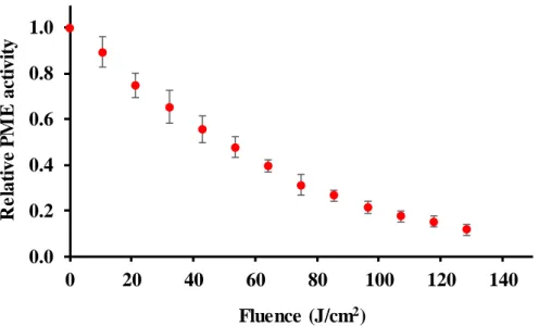
The potential of PL technology to inactivate PME was tested. Results (fig. 1) show that nearly 90 % inactivation can be accomplished by applying 128 J/cm2. This level of residual activity is considered acceptable for some kind of fruit juices; even 75 % inactivation has been found enough to maintain the cloudiness of orange juice for three months (Carbonell, Navarro, Izquierdo, & Sentandreu, 2013). PL could be useful for the industry of cloudy juices for inactivation of PME in place of thermal treatment avoiding the damage caused by heat to juices.
PL technology has the advantages of being a non-thermal and fast treatment that uses lamps filled with xenon, an inert gas that is more environmentally friendly than the conventional UV lamps filled with mercury. Yet, a study on the inactivation of PME by PL in cloudy juices and a comprehensive evaluation of the effect of PL on these food matrixes including microbial inactivation and sensory analysis is required before a final recommendation can be made.
The spectrum emission of PL contains UV-C and UV-B, therefore, it can inactivate enzymes because of the absorption of UV light (250-298 nm) by aromatic residues (tryptophan, tyrosine and phenylalanine), which triggers electron ejection from their side chains (Neves-Petersen, Gajula, & Petersen, 2012). The inactivation follows a first-order kinetic with a rate of 0.034 cm2/J (R2=0.99), this value is in the range reported in previous studies on the inactivation of other enzymes by PL (0.026-0.043 cm2/J) (Pellicer & Gómez-López, 2017; Pellicer, Navarro, & Gómez- López, 2018, 2019). Experiments were carried out at 25 ºC and temperature increased less than 3 ºC at the end of the treatments, which allows to rule out any photothermal effect.
The current research was carried out in a model suspension, which has a low absorbance and no suspended solids. However, turbid juices have a high absorbance and content suspended solids that reduce the efficacy of light-based treatments because of light absorption and scattering. Therefore, a higher fluence would be required to inactivate PME by using this technology. In this regard, the inactivation of microorganisms by PL in apple, melon, orange and strawberry juices was found to be negatively correlated with the absorbance in the UV range of the juices (Ferrario, Alzamora, & Guerrero, 2013); a similar finding has been reported for the inactivation of Escherichia coli O157:H7 by UV light in orange, apple and multifruit juices (Oteiza, Peltzer, Gannuzzi, & Zaritzky, 2005). Likewise, the antimicrobial efficacy of UV light is lower in turbid than in clear grape juice (Kaya & Unluturk, 2016) and the inactivation of polyphenol oxidase is lower in apple and grape juices than in buffer (Müller, Noack, Greiner, Stahl, & Posten,
2014). Further determinations were carried out in order to elucidate which the effects of PL are on PME structure that may help to explain how it is inactivated.
Figure 1. Inactivation kinetics of PME by pulsed light.
3.2. Effect of pulsed light on the secondary structure of PME
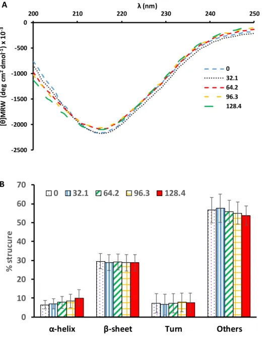
Potential changes in the secondary structure of PME due to PL treatment were studied by far- UV circular dichroism. The shape of the spectrum of the native protein (fig. 2A) in the range of 200-250 nm exhibits the single negative peak at 216 nm typical of proteins with a predominant β-sheet structure (fig. 2B), which is in line with previous reports (Dirix et al., 2005; Plaza et al., 2008). The spectrum was stable during the course of the inactivation (fig. 2A) and the statistical analysis of the results corresponding to the different proportions of types of secondary structure showed no differences (p>0.05) evidencing that PL was unable to change the secondary structure of this enzyme under the study conditions. This result contrasts with previous studies in which the inactivation by PL of the α-helix enzymes peroxidase (Pellicer et al., 2017; Wang et al., 2017) and lipoxygenase (Pellicer, Navarro, Hernández-Sánchez, & Gómez-López, 2019)
0.0 0.2 0.4 0.6 0.8 1.0
0 20 40 60 80 100 120 140
Relative PME activity
Fluence (J/cm2)
induced significant loss of secondary structure. The extreme baroresistance of PME has been associated to the stability of its secondary structure, which requires hydrostatic pressures higher than 1 GPa to be changed, and has been associated to the high content of β-helices (Dirix et al., 2005). The same reasoning might be used to hypothesize that the stability of the secondary of the PME to PL treatment is related to its β-helix structure. In this case, it must be noticed that it is the β-helix structure and not the β-sheet composition what it seems related to the stability of PME secondary structure, since other β-sheet proteins or protein mixtures loss secondary structure upon PL treatment; namely, β-lactoglobulin (Fernández et al., 2012) and whey protein concentrated (Siddique, Maresca, Pataro, & Ferrari, 2016).
Figure 2. Far-UV circular dichroism of PME during the course of PL inactivation. A: spectra, B:
structural percentage at increasing fluences (J/cm2). Bars represent standard deviation.
3.3. Effect of pulsed light on the tertiary structure of PME
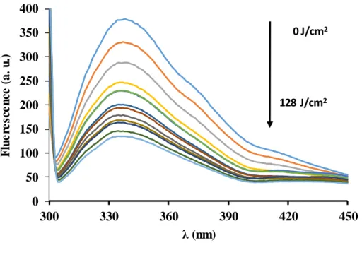
Potential changes of the tertiary structure of PME during PL treatment were monitored by tryptophan intrinsic fluorescence and ANS fluorescence (Fig. 3). The loss of tryptophan
-2500 -2000 -1500 -1000 -500 0
200 210 220 230 240 250
[θ]MRW (deg cm2dmol-1) x 10-3
λ (nm)
0 32.1 64.2 96.3 128.4
A
0 10 20 30 40 50 60 70
α-helix β-sheet Turn Others
% strucure
0 32.1 64.2 96.3 128.4 B
fluorescence with the progress of the PL treatment can be interpreted to be a consequence of the progressive exposure of tryptophan residues, initially fully or partially buried in the inner part of the protein, to the hydrophilic environment, where its quantum yield is reduced (Eskandari, & Ghourchian, 2012). This means that PME gets unfolded during the PL treatment.
The ANS fluorescence increased with PL treatment, especially after 60 J/cm2, which is also consistent with protein unfolding. No spectral shifts were observed in the tryptophan fluorescence spectra neither by direct analysis of fluorescence spectra nor by analysis of the center of spectral mass, the latter did not change during treatment and was in the range 360- 363 nm.
Figure 3. Evolution of intrinsic tryptophan fluorescence during the progress of pulsed light treatment.
The fluorescence of the extrinsic probe ANS increased significantly (p<0.05) during the PL treatment and peak fluorescence underwent a blue shift from 519 nm of the unbound ANS to 491 nm at the end of the treatment (fig. 4). These results corroborated those found by intrinsic
0 50 100 150 200 250 300 350 400
300 330 360 390 420 450
Fluerescence (a. u.)
λ (nm)
0 J/cm2
128 J/cm2
fluorescence measurements indicating that PME unfolds during PL treatment. ANS is essentially non-fluorescent in a polar environment, but increases fluorescence and experiences blue-shift when bound to the non-polar residues that get exposed during unfolding (Stryer, 1965).
Figure 4. Evolution of ANS fluorescence during the course of PME inactivation by pulsed light. abc Points with different letters represent statistically different means, n= 3 (p<0.05).
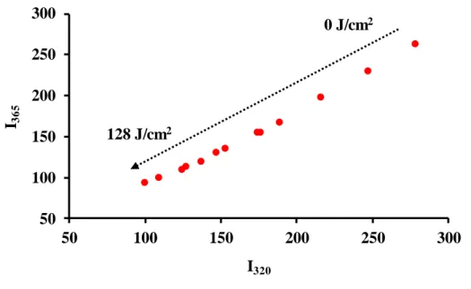
The combined study of results from circular dichroism and fluorescence leads to hypothesize that PME changes from its native conformation to a molten globule state, which is characterized by loss of tertiary structure but conservation of secondary structure (Stojanovski, Breydo, Uversky, & Ferreira, 2016). The phase diagram (Stojanovski et al., 2016) (fig. 5) shows decreasing values with the progress of PL treatment without any indication of formation of intermediaries, which implies that the inactivation of PME by PL occurs without intermediate conformation states (all-or-none process). Therefore, it seems that PL drives PME from its native state directly to a molten-globule state, which is enough to cause the observed loss of activity without reaching a completely unfolded state. This situation differs from changes observed during the inactivation of peroxidase (Pellicer & Gómez-López, 2017) and lipoxygenase (Pellicer et al., 2019)
0 2 4 6 8 10 12 14 16
0 20 40 60 80 100 120 140
% relative ANS fluorescence
Fluence (J/cm2)
a a a
b
c
by PL, where the inactivation is also an all-or-none process but conducting to a completely unfolded state.
Figure 5. Phase diagram of the inactivation of PME by pulsed light. The arrow indicates the progress of the inactivation treatment.
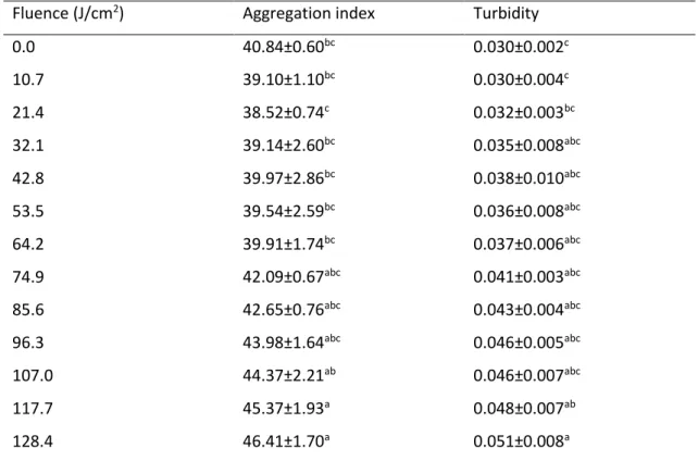
3.4. Protein aggregation in PME after pulsed light treatment
Protein aggregation during PL treatment was evaluated spectrophotometrically by determining the aggregation index and turbidity (table 1). Both parameters exhibited a similar trend, with higher values at the end than at the beginning of the PL treatment, which are an evidence of protein aggregation. The linear adjust of data was poor for the aggregation index (R2=0.80) but high for turbidity (R2=0.96). The aggregation likely occurs via hydrophobic interaction between PME molecules that have had their hydrophobic residues buried inside the inner part of the protein in their native state that are exposed when protein unfolds during inactivation. As demonstrated by intrinsic fluorescence and ANS data, PL disrupts the tertiary structure of the enzyme; therefore, the exposure of hydrophobic residues lead to intermolecular hydrophobic interactions causing that leads to the formation of aggregates (Siddiqui & Bano, 2018). The
50 100 150 200 250 300
50 100 150 200 250 300
I365
I320 128 J/cm2
0 J/cm2
aggregation phenomenon has also been observed during the PL treatment of lipoxygenase (Pellicer et al., 2019) and other type of proteins (Manzocco, Panozzo, & Nicoli, 2013b; Siddique, Maresca, Pataro, & Ferrari, 2017).
Table 1. Aggregation index and turbidity of PME treated by pulsed light at increasing fluences.
Fluence (J/cm2) Aggregation index Turbidity
0.0 40.84±0.60bc 0.030±0.002c
10.7 39.10±1.10bc 0.030±0.004c
21.4 38.52±0.74c 0.032±0.003bc
32.1 39.14±2.60bc 0.035±0.008abc
42.8 39.97±2.86bc 0.038±0.010abc
53.5 39.54±2.59bc 0.036±0.008abc
64.2 39.91±1.74bc 0.037±0.006abc
74.9 42.09±0.67abc 0.041±0.003abc
85.6 42.65±0.76abc 0.043±0.004abc
96.3 43.98±1.64abc 0.046±0.005abc
107.0 44.37±2.21ab 0.046±0.007abc
117.7 45.37±1.93a 0.048±0.007ab
128.4 46.41±1.70a 0.051±0.008a
abc Within the same parameter, means with different superscripts are statistically different (p<0.05).
4. Conclusions
Results show that pulsed light technology is able to inactivate pectinmethylesterase. The inactivation followed a first-order kinetic. Measurements of far-UV circular spectra showed that the secondary structure of the protein was not affected by the PL treatment, while changes in tryptophan and ANS fluorescence were compatible with protein unfolding and spectrophotometric results were compatible with protein aggregation. The phase diagram showed no indication of formation of intermediates during the inactivation process. Therefore, the analysis of the structural changes of the enzyme during the treatment allowed us to conclude that the inactivation of pectinmethylesterase by PL was congruent with the formation
of a molten-globule state in all-or-none process where PME losses tertiary structure and aggregates, while the secondary structure was conserved.
Declarations of Interest: none.
Acknowledgments
Dr. César Flores (ACTI, Universidad de Murcia) for his assistance in circular dichroism determinations. This work was supported by Universidad Católica de Murcia, grant PMAFI/29/14.
References
Baker, R. A., & Cameron, R. G. (1999). Clouds of citrus juices and juice drinks. Food Technology, 53, 64-69.
Barth, S. W, Fähndrich, C., Bub, A., Dietrich, H., Watzl, B., Will, F., Briviba, K., & Rechkemmer, G.
(2005). Cloudy apple juice decreases DNA damage, hyperproliferation and aberrant crypt foci development in the distal colon of DMH-initiated rats. Carcinogenesis, 26(8): 1414-1421.
https://doi.org/10.1093/carcin/bgi082
Beveridge, T. (2002). Opalescent and cloudy fruit juices: formation and particle stability. Critical
Reviews in Food Science and Nutrition, 42, 317–337.
https://doi.org/10.1080/10408690290825556
Bradford, M. M. (1976). A rapid and sensitive method for the quantitation of microgram quantities of protein utilizing the principle of protein-dye binding. Analytical Biochemistry, 72, 248-254. https://doi.org/10.1016/0003-2697(76)90527-3
Cameron, R. G., Baker, R. A., & Grohmann, K. (1998). Multiple forms of pectinmethylesterase from citrus peel and their effects on juice stability. Journal of Food Science, 63, 253-256.
https://doi.org/10.1111/j.1365-2621.1998.tb15720.x
Carbonell, J. V., Navarro, J. L., Izquierdo, L., & Sentandreu, E. 2013. Influence of high pressure homogenization and pulp reduction on residual pectinmethylesterase activity, cloud stability and acceptability of Lane Late orange juice: A study to obtain high quality orange juice with extended shelf life. Journal of Food Engineering, 119, 696–700.
http://dx.doi.org/10.1016/j.jfoodeng.2013.06.041.
Chen, D., Chen, P., Cheng, Y., Peng, P., Liu, J., & Ma, Y. (2019). Deoxyvalenol decontamination in raw and germinating barley treated by plasma-activated water and intense pulsed light. Food and Bioprocess Technology, 12, 246-254. https://doi.org/10.1007/s11947-018-2206-2
Christgau, S., Kofod, L.V., Halkier, T., Andersen, L.N., Hockauf, M., Dörreich, K., Dalbøge, H., &
Kauppinen, S. (1996). Pectin methyl esterase from Aspergillus aculeatus: expression cloning in yeast and characterization of the recombinant enzyme. Biochemical Journal, 319, 705-712.
https://doi.org/10.1042/bj3190705
Dirix, C., Duvetter, T., Van Loey, A., Hendrickx, M., & Heremans, K. (2005). The in situ observation of the temperature and pressure stability of recombinant Aspergillus aculeatus pectin methylesterase with Fourier transform IR spectroscopy reveals an unusual pressure stability of β-helices. Biochemical Journal, 392, 565-571. https://doi.org/10.1042/bj20050721
Duvetter, T., Van Loey, A., Smout, C., Verlent, I., Nguyen, B. L., & Hendrickx, M. (2005).
Aspergillus aculeatus pectin methylesterase: study of the inactivation by temperature and pressure and the inhibition by pectin methylesterase inhibitor. Enzyme and Microbial Technology, 36, 385-390. https://doi.org/10.1016/j.enzmictec.2004.01.014
Eskandari, K., & Ghourchian, H. (2012). Structural changes of glucose oxidase upon interaction with gold-coated nano-particles. International Journal of Biological Macromolecules, 51, 998- 1002. https://doi.org/10.1016/j.ijbiomac.2012.08.001
Falguera, V., Pagán, J., & Ibarz, A. (2011). Effect of UV irradiation on enzymatic activities and physicochemical properties of apple juices from different varieties. LWT-Food Science and Technology, 44, 115-119. https://doi.org/10.1016/j.lwt.2010.05.028
Falguera, V., Garza, S., Pagán, J., Garvín, A., & Ibarz, A. (2013). Effect of UV–vis irradiation on enzymatic activities and physicochemical properties of four grape musts from different varieties.
Food and Bioprocess Technology, 6, 2223-2229. https://doi.org/10.1007/s11947-012-0781-1 Falguera, V., Garvín, A., Garza, S., Pagán, J., & Ibarz, A. (2014). Effect of UV–vis photochemical processing on pear juices from six different varieties. Food and Bioprocess Technology, 7, 84-92.
https://doi.org/10.1007/s11947-013-1069-9
Fernández, E., Artiguez, M. L., Martínez de Marañón, I., Villate, M., Blanco, F.J., & Arboleya, J. C.
(2012). Effect of pulsed-light processing on the surface and foaming properties of β-lactoglobulin.
Food Hydrocolloids, 27, 154-160. https://doi.org/10.1016/j.foodhyd.2011.08.001
Ferrario, M., Alzamora, S. M., & Guerrero, S. (2013). Inactivation kinetics of some microorganisms in apple, melon, orange and strawberry juices by high intensity light pulses.
Journal of Food Engineering, 118, 302-311. http://dx.doi.org/10.1016/j.jfoodeng.2013.04.007.
Forney, L. J., Ye, Z., & Koutchma, T. (2008). UV Disinfection of E. coli between concentric cylinders:
effects of the boundary layer and a wavy wall. Ozone: Science and Engineering, 30, 405–412.
https://doi.org/10.1080/01919510802473872
Gómez-López, V. M., Ragaert, P., Debevere, J., & Devlieghere, F. (2007). Pulsed light for food decontamination: a review. Trends in Food Science and Technology, 18, 464-473.
https://doi.org/10.1016/j.tifs.2007.03.010
Imarc Group (2019). Fruit Juice Market: Global Industry Trends, Share, Size, Growth, Opportunity and Forecast 2019-2024. https://www.imarcgroup.com/fruit-juice-manufacturing-plant.
Accessed on January 08, 2020.
Innocente, N., Segat, A., Manzocco, L., Marino, M., Maifreni, M., Bortolomeoli, I., Ignat, A., &
Nicoli, M. C. (2014). Effect of pulsed light on total microbial count and alkaline phosphatase activity of raw milk. International Dairy Journal, 39, 108-112.
https://doi.org/10.1016/j.idairyj.2014.05.009
Illera, A. E., Sanz, M. T., Benito-Román, O., Varona, S., Beltrán, S. et al. (2018a). Effect of thermosonication batch treatment on enzyme inactivation kinetics and other quality parameters of cloudy apple juice. Innovative Food Science and Emerging Technologies, 47, 71- 80. https://doi.org/10.1016/j.ifset.2018.02.001.
Illera, A. E., Sanz, M. T., Beltrán, S., Melgosa, R., Solaesa, A. G., & Ruiz, M. O. (2018b). Evaluation of HPCD batch treatments on enzyme inactivation kinetics and selected quality characteristics
of cloudy juice from Golden delicious apples. Journal of Food Engineering, 221, 141-150.
https://doi.org/10.1016/j.jfoodeng.2017.10.017.
Izquier, A., & Gómez-López, V. M. (2011). Modelling the pulsed light inactivation of microorganisms naturally occurring on vegetable substrates. Food Microbiology, 28, 1170-1174.
https://doi.org/10.1016/j.fm.2011.03.010
Jeon, M. -S., Park, K. -M., Yu, H., Park, J. -Y., & Chang, P. -S. (2019). Effect of intense pulsed light on the deactivation of lipase: Enzyme deactivation kinetics and tertiary structural changes by fragmentation. Enzyme and Microbial Technology, 124, 63-69.
https://doi.org/10.1016/j.enzmictec.2019.02.001
Ju, Z. Y., & Kilara, A. (1998). Aggregation induced by calcium chloride and subsequent thermal gelation of whey protein isolate. Journal of Dairy Science, 81, 925-931.
https://doi.org/10.3168/jds.S0022-0302(98)75652-8
Katayama, D. S., Nayar, R., Chou, D. K., Campos, J., Cooper, J., Vander Velde, D. G., Villarete, L., Liu, C.P., & Cornell Manning, M. (2005). Solution behavior of a novel type 1 interferon, interferon ‐ τ . Journal of Pharmaceutical Sciences, 94, 2703-2715.
https://doi.org/10.1002/jps.20461
Kaya, Z., & Unluturk, S. (2016). Processing of clear and turbid grape juice by a continuous flow UV system. Innovative Food Science and Emerging Technologies, 33, 282-288.
http://dx.doi.org/10.1016/j.ifset.2015.12.006.
Koyyalamudi, S. R., Jeon, S. C., Pang, G., Teal, A., & Biggs, T. (2011). Concentration of vitamin D2 in white button mushroom (Agaricus bisporus) exposed to pulsed UV light. Journal of Food Composition and Analysis, 24, 976-979. https://doi.org/10.1016/j.jfca.2011.02.007
Krajka-Kuźniak, V., Szaefer, H., Ignatowicz, E., Adamska, T., Markowski, J., & Baer-Dubowska, W.
(2015). Influence of cloudy apple juice on n-nitrosodiethylamine- induced liver injury and phases I and II biotransformation enzymes in rat liver. Acta Poloniae Pharmaceutica, 72(2):267-76.
Kuznetsova, I., Turoverov, K.K., & Uversky, V.N. (2004). Use of the phase diagram method to analyse the protein unfolding-refolding reactions: fishing out the “invisible” intermediates.
Journal of Proteome Research, 3, 485-494. https://doi.org/10.1021/pr034094y
Manzocco, L., Panozzo, A., & Nicoli, M.C. (2013a). Inactivation of polyphenol oxidase by pulsed light. Journal of Food Science, 78, E1183-E1187. https://doi.org/10.1111/1750-3841.12216 Manzocco, L., Panozzo, A., & Nicoli, M. C. (2013b). Effect of pulsed light on selected properties of egg white. Innovative Food Science and Emerging Technologies, 18, 183-189.
https://doi.org/10.1016/j.ifset.2013.02.008
Micsonai, A., Wien, F., Kernya, L., Lee, Y. H., Goto, Y., Réfrégiers, M., & Kardos, J. (2015). Accurate secondary structure prediction and fold recognition for circular dichroism spectroscopy.
Proceedings of the National Academy of Sciences, 112 (24), E3095-E3103.
https://doi.org/10.1073/pnas.1500851112
Müller, A., Briviba, K., Gräf, V., Greiner, R., Herrmann, C., Kuballa, T., & Stahl, M. R. (2013). UV- C treatment using a Dean vortex technology — impact on apple juice enzymes and toxicological potential. Innovative Food Science and Emerging Technologies, 20, 238-243.
https://doi.org/10.1016/j.ifset.2013.07.010
Müller, A., Noack, L., Greiner, R., Stahl, M. R., & Posten, C. (2014). Effect of UV-C and UV-B treatment on polyphenol oxidase activity and shelf life of apple and grape juices. Innovative Food Science and Emerging Technologies, 26, 498-504. http://dx.doi.org/10.1016/j.ifset.2014.05.014.
Neves-Petersen, M. T., Gajula, G. P., & Petersen, S. B. (2012). UV Light Effects on Proteins: From Photochemistry to Nanomedicine. In: Molecular Photochemistry - Various Aspects, Satyen Saha (Ed.). InTech, Available from: https://doi.org/10.5772/37947.
Oteiza, J. M., Peltzer, M., Gannuzzi, L., & Zaritzky, N. (2005). Antimicrobial efficacy of UV radiation on Escherichia coli O157:H7 (EDL 933) in fruit juices of different absorptivities. Journal of Food Protection, 68, 49-58. http://dx.doi.org/10.4315/0362-028x-68.1.49.
Pellicer, J. A., & Gómez-López, V. M. (2017). Pulsed light inactivation of horseradish peroxidase and associated structural changes. Food Chemistry, 237, 632-637.
https://doi.org/10.1016/j.foodchem.2017.05.151
Pellicer, J. A., Navarro, P., & Gómez-López, V. M. (2018). Pulsed light inactivation of mushroom polyphenol oxidase: a fluorometric and spectrophotometric study. Food and Bioprocess Technology, 11, 603-609. https://doi.org/10.1007/s11947-017-2033-x
Pellicer, J. A., Navarro, P., & Gómez-López, V. M. (2019). Pulsed light inactivation of
polygalacturonase. Food Chemistry, 271, 109-113.
https://doi.org/10.1016/j.foodchem.2018.07.194
Pellicer, J. A., Navarro, P., Hernández Sánchez, P., & Gómez-López, V. M. 2019. Structural changes associated with the inactivation of lipoxygenase by pulsed light. LWT-Food Science and Technology, 113, 108332. https://doi.org/10.1016/j.lwt.2019.108332.
Plaza, L., Duvetter, T., Van der Plancken, I., Meersman, F., Van Loey, A., & Hendrickx, M. (2008).
Influence of environmental conditions on thermal stability of recombinant Aspergillus aculeatus
pectinmethylesterase. Food Chemistry, 111, 912-920.
https://doi.org/10.1016/j.foodchem.2008.05.004.
Reignault, P., Mercier, M., Bompeix, G., & Boccara, M. (1994). Pectin methylesterase from Botrytis cinerea: physiological, biochemical and irnmunochernical studies. Microbiology, 140, 3249-3255. https://doi.org/10.1099/13500872-140-12-3249
Siddique, M. A. B., Maresca, P., Pataro, G., & Ferrari, G. (2016). Effect of pulsed light treatment on structural and functional properties of whey protein isolate. Food Research International, 87, 189-196. https://doi.org/10.1016/j.foodres.2016.07.017
Siddique, M. A. B., Maresca, P., Pataro, G., & Ferrari, G. (2017). Influence of pulsed light treatment on the aggregation of whey protein isolate. Food Research International, 99, 419-425.
https://doi.org/10.1016/j.foodres.2017.06.003.
Siddiqui, M. F., & Bano, B. 2018. A biophysical insight into the formation of aggregates upon trifluoroethanol induced structural and conformational changes in garlic cystatin.
Spectrochimica Acta Part A: Molecular and Biomolecular Spectroscopy, 204, 7–17.
https://doi.org/10.1016/j.saa.2018.06.009.
Siguemoto, E. S., Pereyra, L. J., & Gut, J. A. W. (2018). Inactivation kinetics of pectin methylesterase, polyphenol oxidase, and peroxidase in cloudy apple juice under microwave and conventional heating to evaluate non-thermal microwave effects. Food and Bioprocess Technology, 11, 1359-1369. https://doi.org/10.1007/s11947-018-2109-2.
Silva, J. L., Miles, E. W., & Weber, G. (1986). Pressure dissociation and conformational drift of the β dimer of tryptophan synthase. Biochemistry, 25, 5780-5786.
https://doi.org/10.1021/bi00367a065.
Stojanovski, B. M., Breydo, L., Uversky, V. N., & Ferreira, G. C. (2016). The unfolding pathways of the native and molten globule states of 5-aminolevulinate synthase. Biochemical and Biophysical Research Communications, 480, 321-327. http://dx.doi.org/10.1016/j.bbrc.2016.10.037.
Stryer, L. (1965). The interaction of a naphthalene dye with apomyoglobin and apohemoglobin a fluorescent probe of non-polar binding sites. Journal of Molecular Biology, 13, 482-495.
https://doi.org/10.1016/s0022-2836(65)80111-5
Terefe, N. S., Buckow, R., & Versteeg, C. (2014). Quality-related enzymes in fruit and vegetable products: effects of novel food processing technologies, part 1: high-pressure processing. Critical
Reviews in Food Science and Nutrition, 54, 24-63.
https://doi.org/10.1080/10408398.2011.566946.
Vandevenne, E., Van Buggenhout, S., Peeters, M., Compernolle, G., Declerck, P. J., Hendrickx, M., Van Loey, A., & Gils, A. (2011). Development of an immunological toolbox to detect endogenous and exogenous pectin methylesterase in plant-based food products. Food Research International, 44, 931-939. https://doi.org/10.1016/j.foodres.2011.01.056
Van Loey, A., Verachtert, B., & Hendrickx, M. (2001). Effects of high electric field pulses on enzymes. Trends in Food Science and Technology, 12, 94-102. https://doi.org/10.1016/S0924- 2244(01)00066-8.
Wang, B., Zhang, Y., Venkitasamy, C., Wu, B., Pan, Z., & Ma, H. (2017). Effect of pulsed light on activity and structural changes of horseradish peroxidase. Food Chemistry, 234, 20-25.
https://doi.org/10.1016/j.foodchem.2017.04.149.