This publication provides an overview of analytical applications of SR resources in the fields of cultural heritage, forensics and materials science, and presents relevant research projects carried out by 14 IAEA Member States. This publication has been prepared from original material submitted by the authors and has not been edited by the IAEA Editorial Board.
Currently
The low destructiveness of the methods based on light-matter interactions (from IR to X-rays) that enable protocols involving complementary characterization, essential due to the complexity and heterogeneity of the samples studied;. The abundance of information that can be achieved (from the atomic to the structural levels), also due to the multimodal character and the interaction between the different user communities that take place on the same instrument;.
To be explored
Synchrotron beamlines used for archeology and cultural heritage with more than five resulting publications, based on a literature review during the period 1986–2010.
Instrumental and methodological needs
- Full-field X ray (XRF, XAS) imaging
- Scanning tomography
- µCT and laminography capabilities
- Macro-scanning
- New energy ranges
- Processing and analysis of multi-/hyper spectral data
- Databases of reference materials
- New beam lines with a program connected to Cultural Heritage
If micrometer-level XRF tomography could be realized in the near future under stable conditions using full-field setups avoiding scanning, there is still a clear interest in higher resolution work. Methodological developments in the hard X-ray field are also relevant to the field, for example for XRF/XRD mapping and 3D imaging.
Support needs
- Support for travel for synchrotron experiments
- Synchrotron schools including hands-on training
- Experience in equivalent laboratory techniques for preliminary characterization and training (µXRF, PIXE,
- Support to international conferences
- Support to regional conferences
- Documentation
P63 (PETRA III), a general purpose microprobe beamline (XRF, XRD, XAS) and macro-scanning for cultural heritage artefacts. High sensitivity and high resolution preservation XRFC characterizing cultural heritage materials [26], [27] CT and transmission X-ray microscopy (TXM).
Research areas and methods covered
Currently
To be explored
Instrumental and methodological needs
- Machine performance
- Optics and diagnostics
- Multi-element spectroscopic detectors
- Energy resolving imaging detectors
- µCT and laminography capabilities
In-situ, non-invasive beam intensity and position monitoring especially for small size foci will be mandatory. For complementary (imaging) methods, interchangeable sample containers with well-defined confidence markers will be needed in the future.
Needs in analytical methodologies
Data mining for imaging
Theoretically supported data evaluation for XAS
Validation of the theoretical data evaluation
Traceability
ISO17025
Support needs
Support for travel for synchrotron experiments
Synchrotron schools including hands-on training
Experience in equivalent laboratory techniques for preliminary characterization
8] COTTE, M., SUSINI, J., DIK, J., JANSSENS, K., Synchrotron-based X-ray absorption spectroscopy for art conservation: looking back and looking forward, Acc. 37] COTTE, M., SUSINI, J., DIK, J., JANSSENS, K., Synchrotron-based X-ray absorption spectroscopy for art conservation: looking back and looking forward, Accounts Chem.
JANSSENS, L. MONICO, M. RADEPONT, M. ALFELD, J. DIK and M. COTTE
The painting was investigated at Beamline L of the Hamburger Synchrotron Strahlungslabor (HASYLAB) at the Deutsches Elektronen Synchrotron (DESY) in Hamburg, Germany. In Table 1, an overview of the characteristics of the different versions of a mobile MA-XRF instrument is presented.

BERTRAND
The low invasiveness of the methods based on light-matter interactions (from IR to X-rays); Samples and standards are irradiated together in the nuclear grid of the Portuguese Research Reactor (ITN, Sacavém) for 2 minutes (short irradiation) and seven hours (longer irradiation). The Temple of the Emerald Buddha is located within the grounds of the Royal Palace.
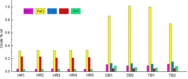
Photographs of the ordination hall enshrining the Emerald Buddha (upper panel), mirror decorations on the wooden panels of the royal thrones (lower, left, and middle panels), and mirror decorations on the base of the statuses of King Rama I, II, and III (panel bottom, right ). Thus, the color in the glass is related to the oxidation states (OS) of the transition metals. 2] KRAIRIKSH, P., The Grand Palace and The Temple of the Emerald Buddha: A Handbook for Guides, Bureau of the Royal Household, Bangkok (1988) 52 p.
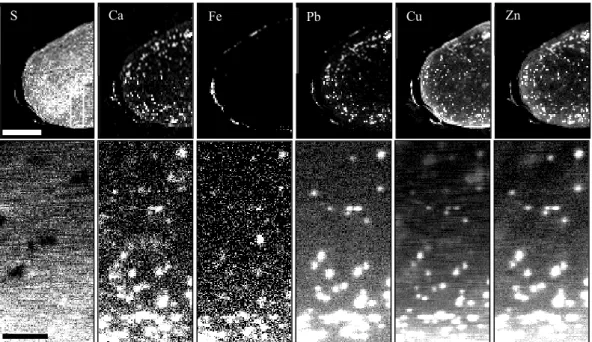
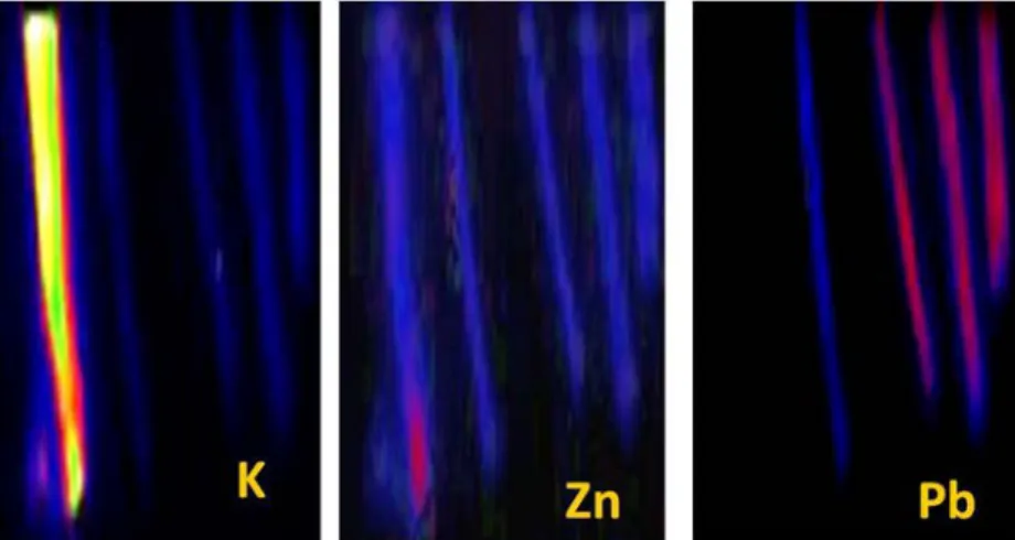
In the case of the hair analyzed for gold (above), the rate of hair growth was known. The chemical shift values imply that Cu(II) is in the 2+ oxidation state in all compounds. New third-generation synchrotron radiation sources (SRS) allow sub-micron resolution to be achieved when operating in the X-ray range.
This parameter must be matched to the size of the radiation generating area of the converter.

Cotte , , Radepont, E. Pouyet, Salome, J. SUSINI
I. DIAS
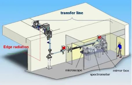
But it is also possible to get a sufficiently clear picture of the hidden portrait in the images obtained with a mobile scanner. While head of the X-ray microprobe D15 at the LURE facility (Orsay, France), Pierre Chevallier was another highly influential scientist in the use of synchrotron techniques for synchrotron from ancient materials. Synchrotron beamlines used for archeology and cultural heritage with more than 5 resulting publications, based on a review of the literature known to the author during the period 1986–2010.
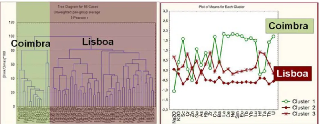
The distribution of the main beamlines used for Cultural heritage and Archeology clearly outlines the main methods of interest (in the past...) to the user community. Evolution in the annual output of the 5 main synchrotron facilities used for Archeology and Cultural heritage based on a review of the literature in the 1999-2010 period. In total, these 5 facilities represent 76% of the total number of publications in the field during this period.
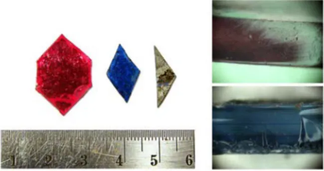
The spread of Chinese porcelain in Europe is a result of the Portuguese naval expansion to the East. CHARACTERIZATION OF THE ANCIENT DECORATIVE MIRRORS FROM THE GRAND PALACE BANGKOK BY SR-BASED TECHNIQUES.
KLYSUBUN, P. KLYSUBUN, P. SONGSIRIRITTHIGUL
M. KEMPSON
Application of XRF analysis of hair to toxicology is provided with the case study of the famous racehorse, Phar Lap. Importantly, the decay in the arsenic profile can test the consistency of the decay rate with known pharmacokinetics. As such, particularly laborious experiments must be performed to derive confidence in the results of the new experimental approach.
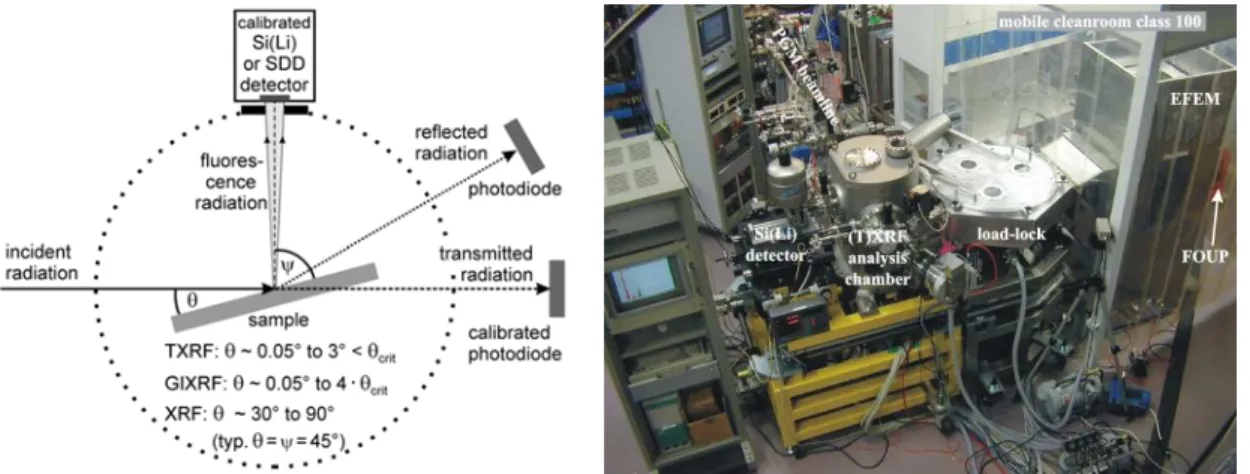
By measuring fluorescent signals at different angles of incidence, GIXRF provides information about the depth distribution and the total dose of the elements in the layers. By measuring fluorescent signals at different angles, GIXRF provides information about the depth distribution and the total dose of the elements in the implanted layers. GIXRF provides information about the depth distribution and the total dose of the elements, even at a depth of a few nanometers.
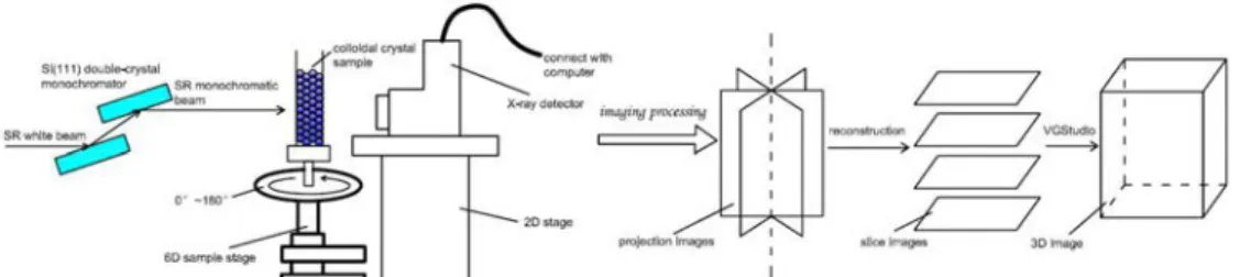
N. FU, H.L. XIE, B. DENG, G.H. DU, T.Q. XIAO
Combining PCI and computed tomography (CT) could show the 3D array structure of PS colloidal crystal. In this paper, the colloidal PS crystal arrays were prepared by gravity sedimentation and membrane filtration, and their 3D structures were characterized in situ by SR X-ray phase-contrast CT technology. The PS spheres in a capillary are observable with clear edges, allowing the 3D arrangement structure of the colloidal crystal to be confirmed from several consecutive slices.
Images of PS colloidal crystal prepared by gravity sedimentation: (a) SR projection image; (b) (c) section view corresponding to the position of the two lines in (a); (d) 3D volume rendering segment in the marked part between the two lines in (a). 5 images of PS colloidal crystal prepared by membrane filtration: (a) SR projection image; (b) (c) slice image corresponding to the position of the line in (a); (d) 3D volume rendering of the colloidal PS crystal without the substrate. The slice image and 3D volume rendering were obtained from the projection images of the colloidal PS crystal by gravity sedimentation and membrane filtration method.
BECKHOFF
With regard to the geometry of the XRS beam, special attention is paid to defining the angles of incidence and observation, as well as the fixed detection angle. A known aperture placed at a given distance from the sample in front of the energy dispersive Si(Li) or SDD detector defines the effective detection angle. The use of SR has led to the advanced nondestructive analysis and speciation of both wafer surface contamination and nanolayered materials by reference-free XRS.
Reference-free XRS can contribute to the assessment of not only surface contamination [1], but also the thickness and composition of micro- and nanolayers [12,13] as well as the composition of bulk samples, e.g. Reference-free GIXRF can also contribute to the analysis of near-vertical sidewalls of semiconductor test structures [3] and of ultra-flat junctions, i.e. Reference-free GIXRF is a promising method for non-destructive access to the compositional depth profile of CIGSe absorber layers [5] and high k nanolayers [25–27] .
H. MENK
From two images taken on the half-slopes of the RC, an absorption equivalent image and a refraction image can be extracted (DEI), respectively. These were investigated using the ABI system at HASYLAB, which was installed within the framework of the European Phasey project [50]. Right panel: ABI CT of the Keshi bead with the reconstructions of the absorption image and the refractive image.
The artifact was found in the Hunsruck proposal of the Rhine region near Bundenbach, Germany. The section is taken in the middle part, at the level of the f holes. 29] INGAL, V.N., BELIAECSKAYA, E.A., Observation of phase-contrast X-ray plane wave topography from a non-crystalline object, J.

K. DEB
RADISAVLJEVIĆ, N. NOVAKOVIĆ, N. ROMČEVIĆ, N. IVANOVIĆ
Although the theory of EXAFS is well understood, a fully quantitative treatment of the XANES is still challenging. Their advantage is in self-consistency which enables more accurate treatment of the electronic structure. However, a large difference was observed in the XANES region of the Yb LIII edge absorption spectra between the experiment, and the model spectrum of Yb placed on regular PbTe lattice position (see Fig. 4.).
Cd1-xMnxTe is one of the first materials from this class to be studied by XAFS. The low concentrations of impurity atoms ensure preservation of the monophasic zinc-blend structure (Td symmetry) of the host CdTe. Closer inspection of the pre-edge structure appearing in the studied systems Fe K edge XANES spectra (see inset in Fig. 1.) revealed the existence of the Fe ion in the mixed valence 2+/3+ configuration [39].
![FIG. 2. Determination of the oxidation state of Mn in Pb 0.9 Mn 0.1 Te [21] from the comparison of its Mn K edge position to that in metal manganese, MnO, Mn 2 O 3 , MnO 2 and KMnO 4 , with Mn valence 0, +2, +3, +4 and +7,](https://thumb-us.123doks.com/thumbv2/123pdforg/7645353.38143/159.892.174.604.94.403/determination-oxidation-state-comparison-position-metal-manganese-valence.webp)
SOKARAS
Three-dimensional technical drawing of the high-resolution X-ray spectroscopy end station at beamline 6-2 at the Stanford Synchrotron Radiation Lightsource (side perspective and top view). Note that the effective X-ray generation area seen by the X-ray optical system changes as a function of the angle α (between the normal to the target and the ion beam axis) and the angle β (between the normal to the target and the axis of the X-ray optical system). The beam passes along the axis of the target unit and falls at an angle of 45° on the copper water-cooled transducer.
All aberration coefficients in equation (5) depend on the vector τ = (а, λ, g, Leff, ra)T defining the ion-optical system parameters, where a is the distance between the object plane and the effective field boundary of the first lens ; λ is the distance between the effective field boundaries of the lenses; g is the working distance (drift space between the effective field boundary of the last lens and the image plane); Leff is the effective length of the lenses, and ra. Ion spot size on the transducer along mutually perpendicular X and Y axes (lx, ly) as a function of the working distance for the EQL doublet. Initial parameters of the particles were obtained from the measured emittance of the beam produced by the microanalytical facility [17].