La infección persistente por el virus de la hepatitis C (VHC) altera la reactividad de las células citotóxicas VHC específicas mediante el desequilibrio entre Mcl 1 y Bim debido a la disminución de la expresión de CD127
258
0
0
Texto completo
(2)
(3) La infección persistente por virus de la hepatitis C (VHC) altera la reactividad de las células citotóxicas VHC específicas mediante el desequilibrio entre Mcl-1 y Bim debido a la disminución de la expresión de CD127.. Tesis presentada para la obtención del grado de Doctor, realizada por. Dª Megha. Uttam Lokhande, Grado en Microbiología y Master en Microbiología. Universidad de Shivaji. India.. y dirigida por. Dr. Juan Ramón Larrubia Marfil FEA de Aparato Digestivo y Cordinador de la Unidad de Hepatología Translacional del Hospital Universitario de Guadalajara. Profesor Asociado en Ciencias de la Salud del Departamento de Medicina y Especialidades Médicas de la Universidad de Alcalá. Guadalajara, 06 de Junio de 2013.
(4)
(5) Table of Contents Acknowledgements. i. Abbreviations. iii. Index of Figures. vi. Index of Tables. viii. I Resumen/Abstract. 1. II Introduction.. 6. II.1 HCV Structure. 8. II.2 Life cycle of HCV. 12. II.3 Natural history of HCV. 15. II.4 Immune response. 16. II.4.1. Innate immune response during acute HCV infection. 17. II.4.2. Adaptive immune response. 23. II.4.3. Humoral immune response. 23. II.4.4. Cellular immune response. 25. II.4.5 Adaptive cellular response during acute HCV infection. 25. II.4.6 Adaptive cellular response during chronic HCV infection. 27. II.5. Apoptosis. 33. II.5.1. Extrinsic pathway. 37. II.5.2. Intrinsic pathway. 38. II.6. Bcl-2 interacting mediator (BIM). 43. II.6.1 Death of activated T cells by Bim. 45. II.6.2. BIM in Hepatitis. 46. II.7 Myeloid cell leukemia-1 (Mcl-1). 49. II.7.1 T cell survival by Mcl-1. 50. II.7.2 Mcl-1 in Hepatitis. 51.
(6) III. Hypothesis and Objectives. 55. IV. Design, Material and Methods. 58. IV.1 Study Design. 59. IV.1.1 Type of study. 59. IV.1.2 Selection of study subjects. 59. IV.1.4 Study Variables. 62. IV.2 Quasi experimental Study. 67. IV.3 Subjects, Material And Experimental Methods. 69. IV.3.1 Samples selected for the study. 69. IV.3.2 Sample and clinical data collection. 70. IV.4 Processing of peripheral blood samples V. Results. 73 82. V.1 Study Variables V.1.1 Clinical Characteristics of the Groups. 86 86. V.2 Viral replication and liver damage according to CD127 expression on pentamer+ cells V.3 Reactivity of pentamer+ cells according to CD127 expression level. 88 93. V.4. Interferon-! production by pentamer+ cells according to CD127 phenotype. 99. V.5. Mcl-1 and Bim expression according to CD127 phenotype. 101. VI Discussion. 111. VII Conclusiones/Conclusion. 120. VIII Rerefences. 123. IX Grants and Publications. 152.
(7) ACKNOWLEDGEMENTS. I would like to thank Dr Juan Ramon Larrubia for supporting me during last three years throughout my PhD. Juan is the person who never let you down. He is one of the smartest people I know. Apart from his guidance related to the work, he was really helpful at personal front, which made my life comfortable right from day one in Spain. He gave me freedom in work; this gave me opportunity to shape up myself as a better researcher.. I warmly thank the people from Guadalajara University Hospital: S. García-Garzon, J. Miquel, A. Gonzalez-Praetorius, T. Parra-Cid and E. Sanzde-Villalobos for their help for my lab work and contribution to my work, which helped me gain good publication record. In parallel, I would like to give special thanks to the all the hospital nurses who helped me in collecting blood samples time to time.. I would like to express my sincere gratitude to the patients who were willing to participate in the study, without whom, I would not have managed any work done.. I would like to give special thanks to my Spanish friends Laura, Elia, Carmen, Raquel, María, Elizabeth, Mónica, Jacinta, Cesar who made my stay in Spain enjoyable and to learn Spanish.. i.
(8) Thanks to Abhijeet Patil for his constant encouragement through last five years. Also I want to thank other friends, Kiran, Shakuntala, Sonali, Pady, Dhanshri, Divakar and Sagar for their constant support.. Special thanks reserved to my family Mammi, Pappa, Aaji, Anil, Amol Dhanu and Lokesh. Their hardwork and working in adverse conditions just to see me happy can’t be just thanked once. I owe my life to them!. Also, I would like to thank my in-laws, Bappa, Aai, and family members Bandu, Sanju, Varsha, Pooja, Arya and Avantika, who gave me support time to time during last few years.. Last but not the least, my best thanks are reserved for my love Vijay, whose love, care, friendship, affection and confidence in me made this happen. You never complained, doubted my decisions and happily stayed away from me to support me. It is always hard to stay away from you; I don’t know how I managed it.. To all of you, Thank You Very Much again from the bottom of my heart.. ii.
(9) Abbreviations 2"-5" OAS. 2"-5" oligoadenylate synthetase. ALT. Alanine amino-transferase. Apaf-1. Apoptosis protease-activating factor-1. Bcl-2. B cell lymphoma 2. BH. Bcl-2 homology. Bim. Bcl-2 interacting mediator. CHC. Chronic Hepatitis C. CTL. Cytotoxic T lymphocyte. DC. Dendritic cells. dsRNA. Double stranded RNA. FCS. Fetal calf serum. FITC. Fluorescein isothiocyanate. FOX03A. Fork-head box transcription factor. HCC. Hepatocellular Carcinoma. HCV. Hepatitis C virus. HCVpp. HCV pseudo typed particles. IFN-!. Interferon-!. IL-7R. IL-7 receptor. IPS-1. IFN- # promoter stimulator protein 1. IQR. Interquartile range. IRES. Internal Ribosome Entry Side. IRF3. IFN regulatory factor 3. ISGs. IFN-stimulated genes. IU/L. International units per liter. iii.
(10) KIR. Killer cell–Ig-like receptors. LCMV. Lymphocytic choriomeningitis virus. LSP. linfocitos de sangre periférica. LTC. linfocitos T citotóxicos. MAVS. Mitochondrial antiviral signaling protein. Mcl-1. Myeloid cell leukemia-1. mDC. Myeloid DCs. MFI. Mean fluoresence Intensity. Nabs. Neutralizing antibodies. ORF. Open reading Frame. PAMP. Pathogen Associated Molecular Patterns. PBL. Peripheral Blood Lymphocytes. PBMC. Peripheral blood mononuclear cells. pDC. Plasmocytoid Dendritic Cells. PDL-1. Programmed Death ligand-1. PE. phycoerythrin. Pe-Cy5. phycoerythrin Cyanine-5. PKC. protein kinase C. PKR. Protein kinase R. PP2A. Protein phosphatase 2A. pSTAT-5. STAT-5 phosphorylation. RIG-1. Retinoic acid–Inducible Gene I. RPMI. Roswell Park Memorial Institute. RT. room temperature. SR-B1. Scavenger receptor class B member I. iv.
(11) SVR. Sustained virologic responder. TCR. T cell receptor. TGF-#. Transforming growth factor -#. TLR3. Toll like receptor-3. TRAF-1. TNFR-associated factor 1. Tregs. Regulatory T cells. UTR. Un-translated Region. VHC. Virus de la hepatitis C. VISA. Virus-induced signaling adapter. v.
(12)
(13) Index of Figures Figure II.1: Hepatitis C virus: model structure and genome organization Figure II.2: HCV life cycle Figure II.3: Natural history of HCV infection Figure II.4: Evasion of Innate immune response by HCV Figure II.5: Collective illustration of the hepatic cells with inflammatory and tolerance activities by stimulation of different molecules or receptors. Figure II.7: Caspase activation Figure II.8: Apoptosis: Extrinsic pathway Figure II.9: Apoptosis: Intrinsic pathway Figure II.10: Models for intrinsic cell death. Figure II.11: Balance between co-stimulatory/ apoptotic molecules and viralspecific cytotoxic T lymphocytes reactivity according to infection outcome Figure II.12: Cell survival marker CD127 modulates Bim and myeloid cell leukemia sequence-1 expression on hepatitis C virus-specific cytotoxic T lymphocytes after cognate antigen stimulation. Figure IV.1: Histogram of a HLA-A2 negative and positive sample Figure IV.2: Outline of HLA-A2 / peptide pentameric complex NS31406-1415. Figure V.1: Dot plots showing the specificity of NS31406-1415 pentameric complexes to detect NS31406-1415-specific cytotoxic T cells. Figure. V.2: Liver damage and viral control according to CD127 expression on hepatitis C virus (HCV)-specific cytotoxic T cells Figure. V.3: Representative FACS dot plots and MFI histograms of peripheral T cells stained with labeled mAb against CD8 and CD127 and with pentameric HLA-A2/peptide PE-labeled complexes against NS31406 and NS31073 HCV epitopes from cases with different degrees of liver damage and viral control Figure. V.4: Box plots showing the directly ex vivo CD127 MFI on total CD8+ and pentamer+ cells in the CD127 expression level groups of the study. Figure. V.5: Representative FACS dot plots and mean fluorescence intensity (MFI) histograms showing the Mcl-1 and Bim expression on peripheral. vi.
(14) CD8+/pentamer+ cells, directly ex vivo with regard to the CD127 expression level on those cells Figure. V.6: Representative FACS dot plots and mean fluorescence intensity (MFI) histograms showing the Mcl-1 and Bim expression on peripheral CD8+/pentamer+ cells, after specific in vitro challenge, with regard to the CD127 expression level on those cells. Figure. V.7: Reactivity of CD8+/pentamer+ cells according to CD127 expression level: Box plots showing the frequency of CD8+/ pentamer+ cells directly ex vivo and after specific in vitro challenge in the presence of either zVAD-fmk or #-galactosidase as control in high and low CD127-expressing pentamer+ cells. Figure. V.8 (A) Representative FACS dot plots and mean fluorescence intensity (MFI) histograms showing directly ex vivo interferon-! (IFN-!) production on pentamer+ cells according to CD127 expression. (B) Box plots showing the percentage of IFN-producing pentamer+ cells, in relation to CD127 expression level. Figure. V.9: Bar plots showing the percentage of experiments with CD8+/pentamer+ cell proliferation after specific in vitro challenge in the presence of either z-VAD-fmk or #-galactosidase as control, with regard to CD127 expression level. Figure. V.10: Representative FACS dot plots and MFI histograms from CD127high-expressing pentamer+ cells, with CD127 expression on CD8+/pentamer+ cells directly ex vivo and after specific in vitro challenge. MFI, mean fluorescence intensity; ID, patient identification. Figure. V.11: Mcl-1 and Bim expression on CD8+/pentamer+ cells according to CD127 expression level and their relationship with CD8+/pentamer+ cell reactivity after antigen encounter: Figure. V.12: Scatter plots showing the correlation between CD127 expression on CD8+/pentamer+ cells and Mcl-1/ Bim expression on peripheral CD8+/pentamer+ cells Figure.V.13: Scatter plot showing the positive correlation between the Mcl1/Bim expression subtraction and CD127 expression level on peripheral CD8+/pentamer+ cells. Figure. V.14: Box plots describing the CD127 expression level and Mcl-1/Bim expression difference on peripheral CD8+/pentamer+ cells with regard to their reactivity after antigen encounter. vii.
(15) Index of Tables Table II.1 Functions of genetic elements of HCV. Table IV.1. Clinical and virological features of the HCV-specific CTL samples tested in the study. Table IV.2. Mediums and Buffers used in Experiments: Table V.1: Clinical and virological features of the groups of the study. viii.
(16)
(17) I. RESUMEN / ABSTRACT. 1.
(18)
(19) Antecedentes: En la infección persistente por virus de la hepatitis C (VHC), la reactividad de los linfocitos T citotóxicos (LTC) se encuentra alterada y esto afecta al control del VHC. La expresión del receptor de la interleuquina-7 (CD127) en estas células podría regular la reactividad de los LTC a través de la modulación del equilibrio entre Bim/Mcl-1. Bim es una molécula proapotótica bloqueada por la acción de Mcl-1. La expresión de Bim/Mcl-1 y la reactividad de los LTC VHC-específicos se compararon en relación al fenotipo CD127.. Material y Métodos: Se obtuvieron linfocitos de sangre periférica (LSP) de pacientes HLA-A2+ VHC+. Los LTC VHC-específicos se visualizaron mediante tinción de los LSP con Ac-anti CD8 y complejos pentaméricos HLAA2/péptido (pentámeros).. El fenotipo Mcl-1/Bim/CD127/IFN-! en los LTC. VHC-específicos se analizó mediante tinción de las células CD8+/pentámero+ detectables con Ac anti Mcl-1/Bim/CD127/IFN-!. La capacidad proliferativa invitro tras estimulación específica de los LTC VHC específicos se evalúo en presencia y ausencia del inhibidor z-VAD-fmk. Todas las células marcadas se analizaron mediante citometría de flujo.. Resultados: Los LTC VHC-específicos con fenotipo CD127bajo se asociaron a elevada viremia del VHC, mientras que las células fenotipo CD127alto se. 2.
(20) correlacionaron con carga viral indetectable (P < 0.001). Directamente ex vivo, la frecuencia de células pentámero+ fue similar en los grupos con expresión alta y baja de CD127. Sin embargo, la capacidad prolierativa tras estimulación antígeno-específica se encontraba alterada en el grupo con expresión CD127bajo (P<0.05), aunque esto se corrigió mediante tratamiento con z-VAD-fmk (P<0.05). La expresión de Mcl-1 directamente ex vivo fue baja (P<0.01) y la expresión de Bim aumentó tras el encuentro antigénico en las células con fenotipo CD127bajo (P<0.05). La diferencia ex vivo entre la expresión de Mcl-1 y Bim en las células pentámero+ se correlacionó directamente con la expresión de CD127 (P<0.001) y con la reactividad de las células pentámero+ (P<0.05). La frecuencia de células productoras de IFN-! directamente ex vivo fue menor en las células CD127bajo que en las CD127alto (P<0.05).. Conclusiones: Un expresión baja ex vivo de Mcl-1 y una sobre-expresión de Bim tras el encuentro antigénico se encuentran involucradas en la hiporeactividad de los LTC-VHC-específicos con baja expresión de CD127 durante la infección crónica, aunque esto puede ser superado mediante el bloqueo de la apoptosis.. 3.
(21) Background: In persistent hepatitis C virus (HCV) infection, HCV-specific cytotoxic T lymphocyte (CTL) reactivity is impaired and this affects HCV control. Interleukin-7 receptor (CD127) expression on these cells could regulate CTL reactivity through Mcl-1/Bim balance modulation. Bim is a proapoptotic molecule blocked by the action of Mcl-1. Mcl-1/Bim expression and T cell reactivity on HCV-specific CTLs were compared according to CD127 phenotype.. Materials and Methods: Peripheral blood lymphocytes (PBL) from HLA-A2+ HCV+ patients were obtained. HCV-specific CTLs were visualized by staining PBL with anti-CD8 and HLA-A2/peptide pentameric complexes (pentamer). Mcl-1/Bim/CD127/IFN-! phenotype of HCV-specific CTLs was tested by staining detectable CD8+/pentamer+ cells with anti-Mcl-1/Bim/CD127/IFN-! antibodies. HCV-specific CTL proliferation ability after specific in vitro challenge was tested in the presence and absence of pancaspase inhibitor zVAD-fmk. All stained cells were analyzed by flow cytometry.. Results: CD127low-expressing HCV-specific CTLs associated with high HCV viraemia, while CD127high correlated with undetectable viral loads (P < 0.001). Directly ex vivo, pentamer+ cell frequency was similar according to CD127 expression level. Nevertheless, CD127low pentamer+ cell proliferation after. 4.
(22) specific in vitro challenge was impaired (P < 0.05), although this was corrected by z-VAD-fmk treatment (P < 0.05). Mcl-1 expression was low directly ex vivo (P < 0.01), and Bim was up regulated after antigen encounter (P < 0.05) of CD127low pentamer+ cells. The ex vivo difference between Mcl-1 and Bim expression on pentamer+ cells correlated positively with CD127 expression level (P < 0.001) and with pentamer+ cell reactivity (P < 0.05). The frequency directly ex vivo of IFN-!-producing pentamer+ cells was lower in CD127low group than in CD127high group (P<0.05).. Conclusion: A low ex vivo Mcl-1 expression and Bim up-regulation after antigen encounter are involved in CD127low HCV-specific CTL hyporeactivity during chronic infection, but it can be overcome by apoptosis blockade.. 5.
(23) II. INTRODUCTION. 6.
(24)
(25) Hepatitis C virus (HCV) is hepatotropic, non-cytopathic virus able to evade immune system efficiently as mechanism to persist in infected hosts. It has been estimated that more than 170 million people are infected with HCV. This virus is spread by contact with infected blood and body fluids. Approximately 80% of infections succeed in establishing a chronic infection with the potential for developing severe liver diseases such as cirrhosis and hepatocellular carcinoma (HCC) [1, 2].. No effective vaccine against HCV is available till date. Therapy for HCV infection as peg-interferon-alpha and ribavirin [3], has limited efficacy, in particular against the genotype 1 virus [4, 5]. The standard care for HCV genotype 1 is currently protease inhibitor (Boceprevir or Telaprevir), pegInterferon-alpha and ribavirin while for other genotypes double therapy with peg-interferon alpha plus ribavirin is still the standard of care. An extended search for new therapy is progressing, already in phase III studies ready to come in the market in the next two years, but still many chronic HCV patients are not cured with these combinations. For these reasons it is necessary to look for new strategies to control HCV infection. Due to the lack of adequate cell culture systems, HCV studies have been slowed down for a long time, but continuous progress in the last few years it has overcome this obstacle. Invivo model to study the biology of HCV have been significantly restricted due to the limited experimental availability of chimpanzees, the primary model for HCV [6, 7], and difficulties encountered in reproducing true infection in small animals. Two breakthroughs has been an important contribution to the field:. 7.
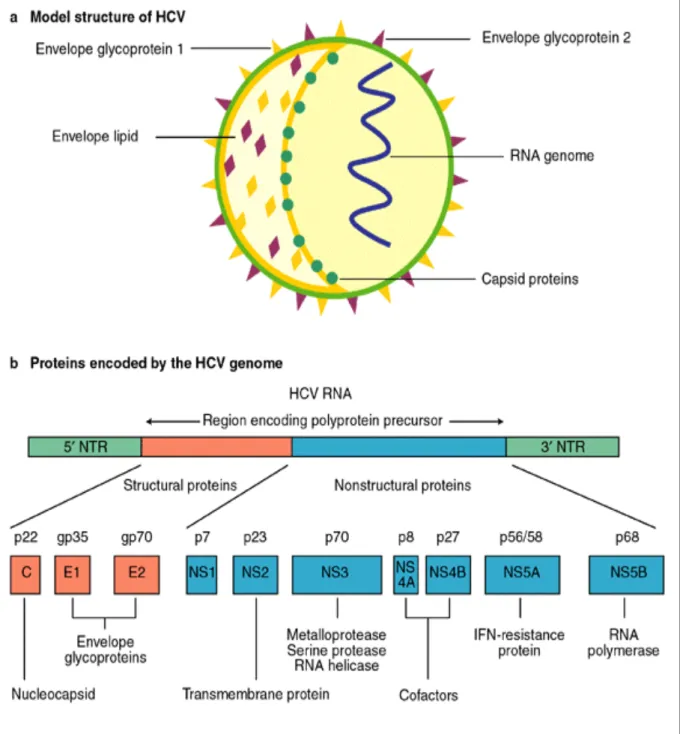
(26) firstly, subgenomic replicons (i.e. without structural genes) [8-10], which are highly permissive for HCV replication [11] and secondly, HCV complete replication in cell culture [12-14]. However, it has long been recognized that these models are complicated by the particularly high error rate of the HCV RNA replicase [15].. Moreover, it is widely accepted that immune-mediated host-virus interactions are responsible for the outcome of HCV and pathogenesis of further severe diseases. Hence, it is interesting to deep in the knowledge of these interactions to find new potential theraputical targets. To be familiar with HCV infection, a brief outline of HCV structure and its life cycle is provided below.. II.1 HCV structure:. The hepatitis C virus (HCV) is an enveloped; positive stranded RNA virus and represents the Hepacivirus genus in the Flaviviridae family. HCV is a small, enveloped, positive sense single-stranded RNA-virus. Spherically shaped with a diameter of between 55–65 nm HCV virion consists of the HCV RNA genome, core and the envelope glycoproteins, E1 and E2. The HCV genetic material (RNA) surrounds by an icosahedral capsid of core proteins, which encased in a lipid membrane, called the viral envelope, in which are anchored the envelope glycoproteins E1 and E2, derived from host cell (Fig II.1) [16].. 8.
(27) HCV Genome:. The positive sense single stranded RNA genome contains two highly conserved non-coding regions (UTR, un-translated region) in the region 5' and 3' which flank a coding region (ORF, open reading frame) that is 9600 base pair long [17]. At the 5' and 3' ends of the RNA are important to translation and replication of the viral RNA instead of translating into proteins. The 5' UTR site that is ribosome-binding site called IRES —Internal ribosome entry site, starts the translation of a very long protein containing about 3,000 amino acids [18]. Structural proteins of the HCV include Core protein, E1 and E2; whereas, NS2, NS3, NS4, NS4A, NS4B, NS5, NS5A, and NS5B as nonstructural proteins are located at 3’ end and function as protease, helicase and polymerase activities necessary for viral replication [19] (Fig. II.1).. 9.
(28) Figure II.1: Hepatitis C virus: model structure and genome organization.. 10.
(29) Table II.1: Functions of genetic elements of HCV.. T. Jake Liang etal Ann Intern Med. 2000.. HCV is very heterogeneous in their genome. This is due to the rapid viral replication, ranging between 1010 to 1012 virions per day (and a predicated viral half-life of 2 to 3 hours) [20] and high error rate of viral RNA polymerase (10-5 errors / nucleotide) without proofreading activity of RNA polymerase [21]. These all reasons are responsible to generate different genotypes, refers to the heterogeneity among different HCV strains isolated in different geographical areas and reflects the accumulation of mutations over a long period of evolution of the virus, and quasispecies, which are the translation of the heterogeneity arising during viral replication in the infected 11.
(30) person [22, 23]. There are six known genotypes (numbered 1 through 6) and more than 50 subtypes (e.g., 1a, 1b, 2a etc) [24].. II.2 Life cycle of HCV. The full replication cycle of HCV can be done by using isolation and engineering of infectious HCV, which have been relatively recent realization in the field, as has the description and characterization of animal models for infection. The development of HCV replicons [8-10, 25], HCV pseudo typed particles (HCVpp) [26] and most recently the infectious HCV cell culture system [12-14] have advanced our understanding of the viral life cycle. Hepatocytes are the primary site of HCV infections. HCV life cycle begins with binding of the virus to cell surface receptors. The putative receptors, the tetraspanin protein CD81 [26-29], the scavenger receptor class B member I (SR-B1) [26, 30-32] and the tight junction proteins claudin-1 [33] and occluding, [34-36] have all been shown to enable HCV entry. In addition, the low-density lipoprotein receptor [28, 37-39], asialoglycoprotein receptor [40], and glycosaminoglycans (heparin sulfate) are also involved, but their exact roles have not been determined. By clathrinmediated endocytosis [41, 42], HCV enters the cell. The virus undergoes an uncoating process by fusion between the viral envelope and endosomal membrane in the acidified endosomal compartment [9, 29, 43-46] via E1/E2-mediated class II fusion [47, 48], to expose the viral genomic RNA to host-cell machinery. About ~9.6 kb viral RNA genome is released into the host cell cytoplasm, to serve as template for the translation of the viral proteins. IRES-mediated translation of. 12.
(31) the HCV genome produces a single ~3,000 amino-acid polyprotein [49], which is processed by cellular and viral proteases into at least 10 different protein products. These products include the structural proteins, which form the viral particle (the virus core and the envelope proteins E1 and E2), and the nonstructural proteins P7, NS3, NS4A, NS4B, NS5A and NS5B [50]. Viral replication is driven by minus strand intermediate. HCV double stranded RNA (dsRNA) is freely exposed in the cytoplasm of infected cell [49], which is recognizable for host innate immune system.. Nucleocapsid is formed by assembling capsid proteins, genomic RNA. The nucleocapsid bud through intracellular membranes into cytoplasmic vesicles. Finally, by secretory pathway, mature enveloped virions release from the cell (Fig.II.2).. 13.
(32) Tibotec, Belgium Figure II.2: HCV life cycle. 14.
(33) II.3 Natural history of HCV:. Acute HCV infection is difficult to diagnose because of asymptomatic behavior of 70%-80% of infected individuals. Most infected persons do not get diagnosed until many years later, due to unaware of their exposure to HCV. Chronic hepatitis C infection is marked by the persistence of HCV RNA in the blood for at least 6 months after onset of acute infection. Self-limiting infection in 15%-25% of patients have been observed. Approximately 75%-85% of infected patients fail to clear the virus by 6 months, and develop chronic hepatitis. Many factors affect the rate of chronic HCV infection, including the age at time of infection, gender, ethnicity, and the development of jaundice during the acute infection.. Therefore, a large proportion of HCV-infected persons, are at risk for advanced liver fibrosis, HCV- related extrahepatic complications, cirrhosis and HCC (Fig II.3). Moreover, a highly variable rates of liver fibrosis progression is the result of the amount of alcohol consumption, age of initial HCV infection, degree of inflammation, HIV or HBV co-infection, and co-mordid conditions. An estimated 10%- 20% of chronic HCV infections develop end-stage liver disease over one or two decades. During chronic HCV infection or cirrhosis, extrahepatic manifestations can appear and HCC can occur only after establishment of cirrhosis.. 15.
(34) Stephen etal, Int. J. Med. Sci. 2006 Figure II.3: Natural history of HCV infection. II.4 Immune response:. To fight against a viral infection the host displays two kinds of immune responses: the innate and adaptive responses. The innate response is the first immunological barrier and it is essential in cytopathic viruses. This response limits viral spreading but also acts as adaptive response activator through antigen presentation to viral specific cells. Adaptive response is the second line in the immunological defense. It plays a major role in non-. 16.
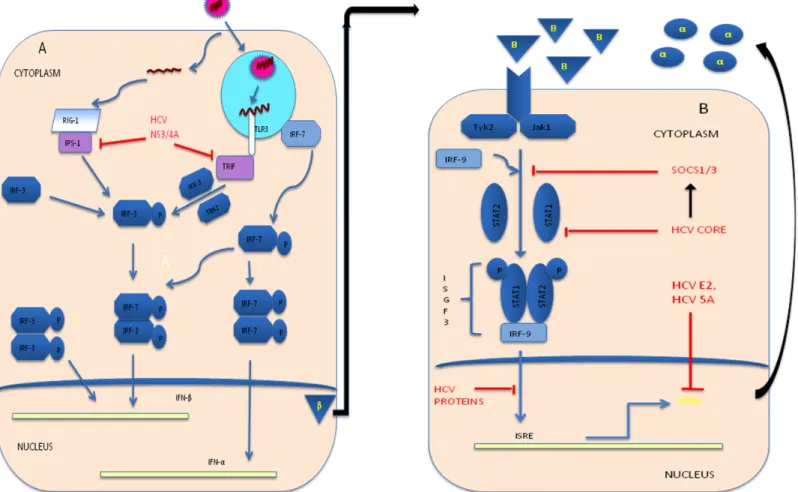
(35) cytopathic viral infections because this type of viruses behaves as an intracellular parasite and they remain occult to the innate system.. But, highly productive and replicative viruses such as, HCV is associated with ineffective antiviral immunity during persistent viral infections. The. complex. ineffective. immune. response. involves. the. functional. deterioration of antiviral responses and contraction of the size of this response (Fig II.3).. II.4.1. Innate immune response during acute HCV infection. The first response to HCV protein is thought to be IFN-# production by infected hepatocytes, which are able to secrete type I IFN. The infected cells are sensed with pathogen associated molecular patterns (PAMP), Toll like receptor-3 (TLR3) [51] and retinoic acid–inducible gene I (RIG-I) [52, 53] by endosomal dsRNA and cytosolic dsRNA respectively, which is an essential intermediate in the HCV replication cycle, and thus, they may be important in the pathogenesis of hepatitis C [54]. RIG-I recruits IFN- # promoter stimulator protein 1 (IPS-1; also called CARD adaptor inducing IFN-# CARDIF), virusinduced signaling adapter (VISA), and mitochondrial antiviral signaling protein (MAVS) [55-57], after ATP-driven activity dependant on recognition of viral proteins [58]. On other hand, TLR3 dimerization, due to leucine-rich repeats [59], recruits the adapter protein, Toll–IL-1 receptor domain– containing adaptor inducing IFN-# (TRIF). Both processes result in downstream signaling, nuclear translocation of IFN regulatory factor 3 (IRF3) and leads to. 17.
(36) stimulation of the transcription of a set of genes including IFN-# [60]. Antiviral state, induced by secreted IFN #, gives an alert to uninfected cells by activation of effector molecules. Binding of IFN -# to cognate receptor complex leads to the activation of JAK/STAT pathway, which results in the induction of IFN-stimulated genes (ISGs) and leads to enhance the IFN response [61] (Fig. II.4).. However, HCV has organized a number of countermeasures not only to inhibit the induction phase, but also interfere with the effector phase of the IFN system (Fig. II.4). It has been confirmed, by in-vitro studies, that HCV serine protease, NS3/4A is enable to cleave MAVS [62], TRIF [63], IPS-1 [64] and oligomerization of MAVS, which is part of signaling process [51, 55, 62, 63, 65, 66]. Disruption of IRF-3 activation occurred by NS3 protein action [67] and it has been shown with different cell lines in-vitro studies [51, 66]. Another key player, HCV core, when over expressed in cell culture, disturbs antiviral activity via interfering in JAK/STAT signaling and ISG expression by inhibition of STAT1 activation. Simultaneously it induces its degradation [68, 69] by induction of inhibitor of the JAK/STAT pathway SOCS3 [70], protein phosphatase 2A (PP2A), which ultimately reduces the transcriptional activity of ISG factor 3 (ISGF3) [71]; and inhibition of ISGF3 interaction to IFNstimulated response elements [61]. HCV NS5A interferes with the function of ISGs by inhibiting 2"-5" oligoadenylate synthetase (2"-5" OAS) and leads to overall ISG expression impairment [72]. Protein kinase R (PKR) can negatively regulate HCV replication noncytolytically in cell cultures [73, 74], which can interacts with HCV NS5A and lost its function. Interestingly, HCV. 18.
(37) E2 acts as distraction target to PKR [75]. To sum up, the main targets of HCV proteins to evade immune response are interference with the induction of IFN synthesis, IFN-induced intracellular signaling and IFN-induced effector mechanisms (Fig.II.4).. Dendritic cells (DC) are professional antigen presenting cells with important functions in antiviral immunity through activation of adaptive immune responses. Type-I IFNs are also produced by plasmocytoid (p) DCs, which derive from the lymphoid lineage. Although, production of IFN alpha/beta, in early phase of infection occurs after recognition of ssRNA and dsRNA by TLR7 and TLR9 respectively, the mechanism is still not clear [76]. The frequency of pDCs in the blood [77] and their production of IFN-$ in HCV infection are reduced after in vitro stimulation [78]. The possible mechanism has been demonstrated in in-vitro studies. First, HCV core and NS3 activate monocytes by TLR2 signaling to produce TNF-$ [79], which in turn inhibits IFN-$ production and induces pDC apoptosis [78]. Second, HCV itself inhibits IFN-$ production of pDCs [80]. However, other studies revealed regular response to TLR stimulation by circulating pDCs of chronically infected individuals [81, 82] and they have high levels of endogenous type I IFNs without immuno-dysfuction [76]. Although this defense mechanism is significant, the host rarely overcomes HCV infection, which suggests several other viral evasion mechanisms that are poorly or not understood yet.. Another group of DCs, myeloid DCs (mDCs) derive from the myeloid lineage [83, 84]. Due to its tolerogenic and stimulatory role [83, 84],. 19.
(38) mDCs have been broadly studied in HCV infection. mDCs have not been observed to be decreased in peripheral blood or dysfunctional in HCV chronic infected individuals in in-vitro studies [85, 86]. Nevertheless, HCV proteins can interact with monocytes/macrophages through TLR2, inducing the IL-10 production, which hampers IL-12 production by mDC and IFN-$ by pDC, or they directly inhibit DC differentiation [87]. IL-12 cytokine production by mDC is decreased in HCV patients in response to stimuli like CD40 L or poly (I:C) [88], which can explain clearly the shift from Th1 to Th2 response in HCV patients. In-vitro studies indicate that DC expressing core and E1 proteins have lower stimulatory ability, which is associated to the lack of maturation after stimulation with TNF-alpha or CD40L [89].. 20.
(39) Figure II.4: Evasion of Innate immune response by HCV: (A) Interference in IFN synthesis: Blocking of TLR 3 and RIG-1 signaling respectively, by cleavage of the adaptor molecule TRIF and IPS-1 via HCV NS3/4A; (B) Interference in IFN-induced Effector mechanisms: Binding of IFN ! and its receptor with TYK2 and JAK1 kinase activation lead to form ISGF3 complex, where this complex interact with IFN stimulated response elements (ISREs) within the promoter and enhancer region of ISGs to induces ISGs (such as 2’, 5’ OAS, PKR, IRF7) production in nucleus. HCV core induce SOCS1/3, which is the inhibitor of the JAK/STAT pathway and inhibits STAT1 phosphorylation, which inhibits assembly of trimeric ISGFs complex. Function of ISGs inhibited by HCVE2 and HCV NS3/4A.. 21.
(40) Other cells involved in the innate response are the NK cells. Functions of these cells include generating a cytotoxic response, regulatory cytokines production and control on DC maturation and amplitude of DC response, which may deeply impact on type of down- stream adaptive immune responses. Response to HCV infection by NK cell is direct apoptosis induction of infected cells with production of antiviral cytokines [90, 91]. Moreover, NK cell depletion or dysfunction favor HCV persistence [90]. The role of interactions between HLA class I and killer cell–Ig-like receptors (KIR) during HCV infection has been shown. KIR can regulate NK cell activities. However puzzling contradictions for this topic in different studies have been revealed [92-94]. The importance of NK cells in the resolution of HCV infection is illustrated by the influence of genetic polymorphisms of KIR and their HLA ligands on the outcome of HCV infection, which was dependent on a homozygous HLA class I ligand background [95-97]. There is need to focus on clear understanding of functional and molecular HLA-KIR interactions to know about the possible way for NK cell- mediated protection in animal models of HCV infection. However, an increased proportion of NK cells expressing activating receptors, enhanced cytotoxicity and defective cytokine production have revealed in chronic HCV infection [98]. Megan et al revealed that IL28A cytokine could significantly inhibit IFN-! production lead to NK cell inactivation [99], which would be important to attenuate chronically activated NK cells. Consequently, the analysis of functional scene between NK cells and type 3 IFN in the immune response to virus will be required to understand the role of the NK in disease progression during HCV infection.. 22.
(41) II.4.2. Adaptive immune response. The second barrier to control HCV infection is the adaptive immunity. This response has two arms to fight against pathogens; humoral and cellular immune response. Humoral immune response, that means neutralizing and non-neutralizing antibodies can endorse antiviral activity and pathogenesis [50]. Cellular immune response shows antiviral immunity by means of virus specific CD8 cytotoxic T lymphocytes (CTLs) and CD4 T helper cells, which play key effector and regulatory roles respectively. These T cells take part in viral pathogenesis of HCV by direct killing of infected cells or producing soluble factors able to clear the virus in a non-cytolytic manner, but also can lead to HCV pathogenic events, favoring direct liver damage and attracting non-specific inflammatory cells to perpetuate the liver inflammation [50].. II.4.3. Humoral immune response Neutralizing antibodies (nAbs) generally play a critical role for controlling initial viremia and protecting from re-infection in viral infections. However, the role of the humoral immune response in the clearance of HCV infection has been in the dark for a long time due to difficulties to determine relative role of antibodies to neutralize HCV. It can exclusively be evaluated by relevant model systems. It is thought that HCV clearance could occur in the absence of nAbs. If they are present alone, these antibodies are inadequate to eradicate HCV in most of the cases in early studies [100-104]. It has been proved that HCV specific T cells may compensate for lack of neutralizing antibodies to obtain HCV clearance [105]. However, due to the development. 23.
(42) of novel model systems [12-14, 26, 106], it is possible to focus on HCV entry into host cells and neutralization process which demonstrated that nAbs are induced by patients who subsequently control [107] or resolve [108] viral infection in the early phase of infection and contrary in chronic infection. This suggests that a strong, early, broad nAbs response may contribute to resolution of HCV in the acute phase of infection while delayed induction of nAbs may contribute to development of chronic HCV infection.. Instead of the rapid, vigorous and multi-specific antiviral host immune responses, chronic patients have been shown to develop a delayed and inefficient neutralizing antibody response [108] due to HCV escape mechanism [109]. Recent studies make evident that entry of HCV can be hampered or modulated by nAbs of chronic HCV patients [110-112], while it is controversial in cell culture study [113]. In addition, although nAbs are incapable to clear the virus in chronic infection, due to selection pressure exerting on viral variants, they contribute to the evolution of the HCV envelope sequences to escape [114, 115]. It has been proposed that HCV stimulates B cells in a B cell receptor-independent manner in chronic infection [116] and may favor the development of lymphoproliferative and autoimmune diseases [50]. Although, in vitro studies make evident that the neutralization ability of HCV-specific. nAbs. is. enhanced. by. complement. activation. against. pseudotyped viruses [116], there is absence of direct experimental evidence about the presence of any of these Ab- mediated functions during natural HCV infection. However, immune complexes are believed to play a pathogenetic. role. in. the. development. 24. of. manifestations. such. as.
(43) cryoglobulinemia,. glomerulonephritis,. porphyria. cutanea. tarda,. and. necrotizing cutaneous vasculitis during chronic HCV infection [117-119].. II.4.4. Cellular immune response. Cytotoxic T lymphocyte (CTL) responses are essential to control HCV infection. Efficiency of antiviral CTL responses depends on where these cells are primed. Efficient antiviral CTL response is observed when it is primed in lymphoid organs, whereas within the liver, priming is more tend to induce T cell inactivation, tolerance or apoptosis [50]. A strong, multispecific and longlasting T-cell immune response emerge to be important for control of viral infection [104, 120]. Persistent HCV unsuccessfully control by T effector cells is due to multiple causes, such as: HCV escape mutant generation, immunosuppressive effects exertion, Tregs induction, or effector T cell exhaustion or apoptosis [121-124].. II.4.5 Adaptive cellular response during acute HCV infection. Vigorous CD4+ and CD8+ T cell responses targeting multiple HCV regions with intrahepatic production of IFN-! emerged in acute hepatitis C infection [101, 123, 125]. Decreasing viral titer correlates precisely with the appearance of HCV-specific T cells and IFN-! expression in the liver [126]. The appearance of HCV-specific T cells can be detectable in the peripheral blood or in the liver compartment several weeks after infection in humans or experimental chimpanzee models [61, 104], respective with primary peak of. 25.
(44) transaminases and irrespective of clinical outcome (resolution or chronicity). Delayed emerging of antigen-specific responses are also essential for the HCV control [61].. The protective function of CD4+ T cells appear to be due to the production of antiviral cytokines, but also their helping nature to antiviral B cells and in CD8+ T cell response. The HCV clearance has been observed and correlated with vigorous proliferation of specific CD4+ T cells [80, 127] with concurrent IL-2 and IFN-! production [128, 129]. The early sustained development of CD4+ T cell response needs to be successful for viral clearance [128], whereas HCV- specific CD4+ T cell responses are not observed in chronic HCV infection. Moreover, the recurrent viremia has been correlated with loss of previous strong CD4+ T cell responses after several months of viral clearance [130, 131]. Studies on the relative importance of CD4 help in spontaneous recovery in acute HCV infection demonstrated that fact [132]. CTL priming in presence of CD4 help is critical factor in protective function [128].. Similarly, Antigen-dependent reactivity of HCV-specific CD8+ T cells has been proved by a rapid decay of CD8+ T cell responses during antiviral therapy [133]. It is evident that, CD8 T cells play a direct role in terminating hepatitis C as shown by prospective studies of acute infection in chimpanzees [134], in which it is revealed that a vigorous, multispecific, and polyclonal intrahepatic CD8 CTL response during early infection correlated with HCV-1 clearance. Furthermore, it is concluded after monitoring the course of HBV. 26.
(45) infection in CD8-depleted in chimpanzees that CD8+ cells are the main effector cells responsible for viral clearance and disease pathogenesis during acute HBV infection, and this study suggests that viral clearance is mediated by both noncytolytic and cytolytic effector functions of the CD8+-T-cell response [135]. In addition, after accidental needlestick exposure, subjects who develop acute HCV infection, the vigor and quality of the antiviral T cell response determines the outcome of acute HCV infection. The subjects who cleared the virus experience a prolonged episode of acute hepatitis that coincided with a CD8+ T cell response to HCV, while chronic infection develops in subjects who fail to produce a significant T cell response [123]. Therefore, significant, broader, multi-specific HCV specific CTLs are important in acute HCV infection control.. The appearance of self-sustaining memory T cells (CD127+ memory HCV-specific CD8+ T cells and CD4+ T cells) are necessary to control HCV infection [101, 123, 128]. In fact, years after HCV control due to anti-HCV treatment it is possible to find HCV traces in association with HCV-specific T cell reactivity. These data suggest that HCV-specific memory T cells are essential to clear HCV infection completely after the initial acute clearing [136].. II.4.6 Adaptive cellular response during chronic HCV infection. Therefore, complete resolved HCV patients exhibit broader CTL responses with higher functional avidity and wider cross-recognition ability. 27.
(46) than patients with persistent HCV infection [137]. There are evidences that demonstrate rapid mutation in HCV genome, T cell exhaustion because of expression of inhibitory molecules, immune regulatory cytokine induction and immune modulatory T reg cell activation, which are the main reasons for HCV persistence in chronically infected patients [121, 138-141]. Like Retrovirus, HCV polymerase has high replication rate and lack of proofreading capacity, which permit a rapid virus escape from emerging humoral and cellular immune responses and lead to persistent infection [139, 142]. Mutation study in early HCV infection in HLA class I restricted epitopes targeted by CD8+ T cells are associated with persistence [143, 144], which proved indirectly that HLA-restricted CD8+ T cells exert selection pressure. Furthermore, the HLA alleles can influence infection outcome [145].. The secretion of certain immuno-regulatory cytokines is also related with HCV persistence. IL-10 cytokine is found to increase during chronic HCV infection [146]. In chronic HCV patients, the suppression of IFN-! production and proliferation of virus- specific CD4+ and CD8+ T cells have been observed in livers with IL-10 –producing HCV- specific CD8+ T cells [147]. IL-10 produced by monocytes or NK cells down-regulates effector T cell responses. For instance, monocytes secrete IL-10 in response to HCV core–mediated TLR2 stimulation in vitro [148]. IL-10 producing HCV-specific CD8+ T cells inhibits IFN-" production [149], but also promotes apoptosis of pDCs [148], and induces liver infiltration of chronically infected individuals, suggesting that they modulate liver immunopathology to favor HCV persistence [147]. In addition, intrahepatic HCV-specific IL-10 producing CD8+ T cells prevent liver. 28.
(47) damage during chronic disease [150]. Moreover, TGF-! is also involved in antiviral immune suppression and chronic HCV infection evolution [151]. To sum up these data, regulatory cytokines such as IL-10 or TGF-beta decrease liver inflammation, after affecting the protective immune response, developing a dual task. First of all, they impair T cell responses to allow viral persistence but also decrease liver damage to extend host survival.. Regulatory T cells (Tregs) are important to control the balance between host damage and viral control produced by specific immune response. In cases of excessive immune response, that could be harmful for the host, these cells can induce immune-tolerance to the viral epitopes. Tregs are derived from natural or induced T cell populations, in which natural CD4+ Tregs are generated during normal T cell development in the thymus, whereas induced Tregs are generated from mature T cells [152]. T cell subset with suppressive function, CD4+ CD25+ FoxP3+ regulatory T (Treg) cells, engages in the control of auto-immunity and immune responses, through various mechanisms including the inhibition of APC maturation and T-cell activation [153]. No difference has been found in the frequency of Treg cells and the extent of suppression irrespective of the outcome of the infection [154]. However, higher Tregs frequency has been observed in chronic HCV infected patients than in resolved patients [155-158]. Interestingly, depletion of CD25+ cells could enhance responsiveness of the remaining HCV-specific effector cells in vitro [155-157], which suggests a fundamental role of Tregs in the establishment of chronic HCV infection. Moreover, Treg cells are induced and proliferate in chronic HCV infection and appeared to alter liver. 29.
(48) inflammation [159]. Conversely, Programmed Death ligand-1 (PDL-1) mediated inhibition limits the expansion of Tregs by controlling STAT-5 phosphorylation (pSTAT-5) [160], which can diminish suppressive function of Tregs, lead to viral load control and ultimately ensure long-lasting survival of the host.. HCV is able to induce the up-regulation of different negative costimulatory molecules in order to provoke an anergic status on HCV-specific T cells. Expression of the inhibitory receptor PD-1 is one of these molecules involved in the generation of a state of exhaustion on HCV-specific CD8+ T cells during chronic HCV infection [161, 162]. Importance of expression of PD1 in HCV-specific T cell failure mechanism has been observed [163, 164], which can hinder by mutation in T cell epitopes [165]. In addition, blocking of PD-1 signaling resulted in the functional restoration of blood-derived HCVspecific CD8+ T cell responses in chronic infection [164, 166]. However, the PD-1 alone is not sufficient in defining exhausted HCV-specific CD8+ T cells during HCV infection. To restore function of HCV-specific T cells isolated from liver biopsies of infected patients, there is need of CTLA4 blockade in addition to PD-1 blockade [167]. In addition, the co-expression of other inhibitory receptors such as 2B4, CD160, Tim-3 and KLRG1 occurred in about half of HCV-specific CD8+ T cell responses and correlate with low or intermediate level of CD127 expression, impaired proliferative capacity, and an intermediate T cell differentiation stage [168]. These data indicates that HCV infection modulates different negative co-stimulatory molecules to favor the development of HCV-specific CD8+ T cell exhaustion.. 30.
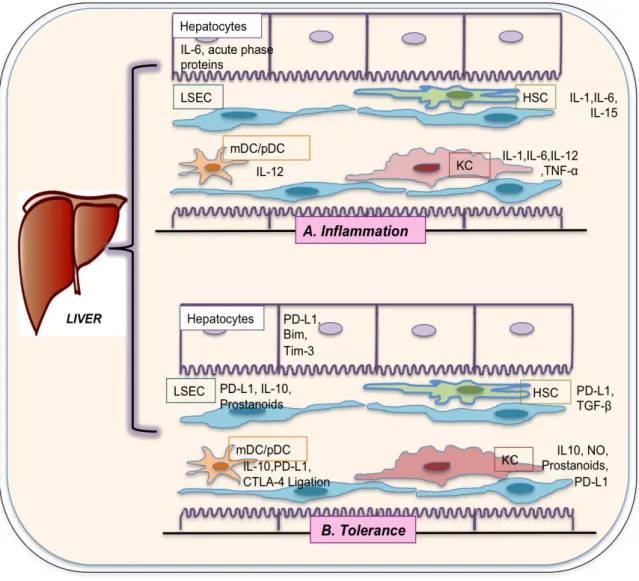
(49) T cell stimulation in the liver encourages tolerance by using mechanisms such as, immune divergence [169], generation of regulatory T cells [170], T cell anergy [171] and T cell death [172]. Undeniably, hepatic tolerance can explain the elevated frequency of viral persistence during hepatotropic virus infections [172]. Although there are evidences showing that most infectious microorganisms are promptly removed from the liver by inflammation, a favorable situation for evading immune responses occurs in some viruses, leading to the triumph of certain pathogens such as HBV and HCV. The Fig I.5 depicted inflammatory and tolerance activity in liver by stimulation of different molecules or receptors. Till date, there are two main mechanisms by which HBV and HCV could successfully escape from CTL action: escape mutant generation, and immunosuppressive effects exertion (effector T cell exhaustion and T cell death by apoptosis) [123, 124, 173, 174].. 31.
(50) Figure II.5: Collective illustration of the hepatic cells with inflammatory and tolerance activities by stimulation of different molecules or receptors. LSEC: Liver sinusoidal endothelial cells; KC: Kuffer cells; DC: Dendritic cells; HSC: Hepatic stellate cells; TNF: Tumor necrosis factor; IL: Interleukin; mDC: Myeloid dendratic cell; pDC: Plasmacytoid dendritic cell; PD-L1: Programmed death ligand-1; Bim: BCL-2 interacting mediator; Tim-3: T cell immunoglobulin mucin-3; CTLA-4: Cytotoxic Tlymphocyte antigen 4; TGF: Transforming growth factor; NO: Nitric oxide.. 32.
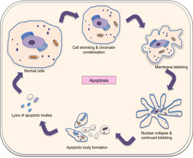
(51) Fig II.6: Apoptosis-programmed cell death.. Among these mechanisms involved in viral hepatitis persistence, new advances on the role of T cell death induction have been obtained recently and will be discussed in the following pages.. II.5. Apoptosis:. A normal cellular process involving physiologically relevant cell death and deletion of unwanted cells is called apoptosis. Apoptosis is essential for cell selection, tissue homeostasis, morphogenesis, and host defense in multicellular organisms. A cell that undergoes apoptosis dies neatly, without. 33.
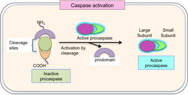
(52) damaging its neighbors. The apoptotic signals give rise to activate various proteins and follow a specific classical caspase chain reaction set activation [175]. Quickly and neatly dismantlement process includes membrane blebbing with shrinking of the cytoplasm and condensation of the nucleus. Phagocytic cells begin to pick up the apoptotic bodies, preventing the release of cellular content and ultimately avoiding inflammation [176] (Fig II.6). Apoptosis occurs by two mechanisms: active and passive mechanism. No presence of antigen gives a signal for termination of immune response by passive apoptotic mechanism (intrinsic pathway). On the other hand, the ligation of Fas (CD95) and TNF receptors-“death receptors” triggered apoptosis lead to active mechanism of apoptosis (extrinsic pathway). Briefly, apoptosis mechanisms involve a family of cysteine proteases, called caspases. These molecules are synthesized in the cell as inactive precursors, or pro-caspases for selfprotection against accidental death, which are usually activated after receiving proper trigger by cleavage (Fig II.7).. Structurally, pro-caspases contain three domains: N terminal prodomain, a large subunit and a small subunit. After activation, the active caspase enzyme is formed by heterodimerization of small and large subunits [177]. Moreover, active caspase molecules are ready to cleave target proteins such as structural or signaling proteins and other effector caspases, preventing other proteins cleavage randomly [176].. 34.
(53) Figure II.7: Caspase activation: Inactive proenzyme (procaspase) activated by proteolytic cleavage by another member of caspase family and cleaved two fragments associate to form the active site of the caspase.. 35.
(54) Figure II.8: Apoptosis: Extrinsic pathway. A: Mitochondria-independent extrinsic pathway: Fas-FasL ligation strikes to recruit pro-caspase 8 activation and induction of caspase cascade by caspase 3 leading to apoptosis; B: Mitochondria-dependent extrinsic pathway: Fas-FasL ligation trigger to activate the pro-caspase 8, which cleave Bid (pro-apoptotic Bcl-2 family molecule) to form truncated Bid (tBid). Then, mitochondrial dependent cell death begins with tBid.. 36.
(55) II.5.1. Extrinsic pathway. The extrinsic pathway initiates from outside the cell through triggering the activation of transmembrane “death receptors” that are members of the TNF receptor gene superfamily. Members of this receptor family bind to extrinsic ligands known as pro-apoptotic ligands [178] and transduce intracellular signals that ultimately result in the destruction of the cell [179, 180]. To date the best characterized ligands of these receptors are FasL, TNF-!, Apo3L and Apo2L and corresponding receptors are FasR, TNFR1, DR3 and DR4/DR5, respectively [184-186]. The signal transduction of active cell death process involves several caspases. Activated caspases have an effect on several cellular functions as part of the process that results in the death of the cells [178]. The signal transduction of mitochondrial-independent active cell death process involves binding of a pro-apoptotic ligand (such as FasL) with its receptors (Fas) on the surface of a target cell. The cytosolic tail of receptors contains a death domain, which when activated, binds to an adaptor protein, which in turn recruits the specific procaspase-8 and -10 and activates them by proteolytic cleavage [181] that finally initiates the proteolytic caspase cascade leading to apoptosis. Activated caspase 8 triggers the caspase cascade via two different pathways, leading to cell death. In type 1 apoptosis, such as in lymphocytes, caspase 8 activates caspase 3 whereas in type 2 apoptosis, like in hepatocytes and pancreatic cells, caspase 8 activate the pro-apoptotic molecule Bid and go ahead for apoptosis via the disruption of mitochondrial membrane and cytochrome C release [182] (Fig II.8). The T cell death by type. 37.
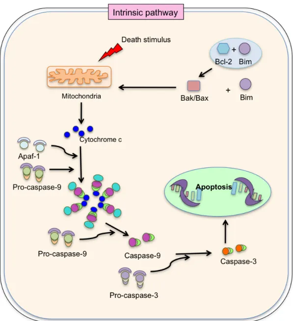
(56) 1 and type 2 Fas induced apoptosis fate is decided by the ratio between proteolytically activated effector caspases, X-chromosome linked inhibitor of apoptosis protein and proto-typical effector caspase substrate inhibitor of caspase-activated. DNase.. Interestingly,. HCV. specific. intrahepatic. lymphocytes contribute to bystander killing via Fas-FasL interaction [183], which support the fact that the liver facilitates liver-trapped activated T cell apoptosis [190].. II.5.2. Intrinsic pathway. The intrinsic or mitochondrial pathway is initiated within the cell, involving non-receptor-mediated intracellular signals and inducing activities in the mitochondria that initiate apoptosis. DNA damage, loss of cell-survival factors or other types of severe cell stress causes the induction signal for the intrinsic pathway. This passive death process pivots on the balance of activity between pro- and anti- apoptotic signals of the B cell lymphoma 2 (Bcl-2) family proteins [184]. This balance is maintained by regulation of the permeability of the mitochondrial membrane and by the pro- or anti-apoptotic signal that will be released inside the cell [185]. Following mitochondrial permeabilization, the intrinsic pathway divides into two pathways: Apoptosis protease-activating factor-1 (Apaf-1) dependent and Apaf-1 independent pathway. In Apaf-1 dependent pathway, release of cytochrome c from mitochondria, by triggering the pro-apoptotic Bcl-2 family member [186], and ATP activate monomer inactive Apaf-1 proteins by a conformational change, leading to form a heptamer of Apaf-1 molecules called apoptosome [187].. 38.
(57) Figure II.9: Apoptosis: Intrinsic pathway. Death stimulation up regulates Bcl-2 interacting mediator leading to the separation from Bcl-2, favoring the activation of Bax, Bak, which form pores in the mitochondrial membrane leading to release of cytochrome c. Cytochrome c with Apaf-1 and procaspase 9 participate in the formation of apoptosome, which activate caspase 9. Caspase-9 activates caspase 3 after cleavage of pro-caspase-3. That caspase-3 triggers to induction of caspase cascade and cell death. Apaf-1: Apoptosis protease-activating factor-1. Bim: Bcl-2 interacting mediator.. 39.
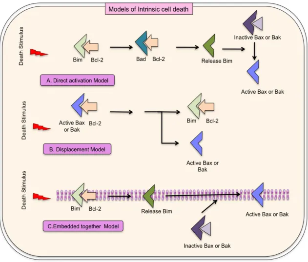
(58) Apoptosome allows activation of pro-caspase 9, which consequently triggers the caspase cascade [188]. On the other hand, in Apaf-1 independent pathway, permeabilization of mitochondrial membrane release DIABLO like proteins, which activates effector caspases by provoking inhibitors of apoptosis proteins [189] and triggers caspase cascade [190, 191] (Fig II.9).. The balance of pro- and anti-apoptotic proteins maintains the apoptotic activity [191]. The Bcl-2 family members regulate mostly neglect or intrinsic pathway. This family is subdivided into three groups of proteins on the basis of their functions and the number of Bcl-2 homology (BH) motifs included in their primary structure; first group: “anti-apoptotic multidomain” members, such as Bcl- xL, have four BH domains (BH1 to BH4) which inhibits apoptotic process. Other two groups of “pro-apoptotic multidomain” members, which are Bax-like proteins and “BH3-only” proteins [192]. Bax-like proteins possess three BH domains (BH1 to BH3), including Bax, Bak, and Bok, which are referred as death effector members. BH3-only members contain BH3 domain, including Bim, Bad, Bik, Puma, Noxa and Bid and are known as messengers of death. In addition, C-terminal transmembrane fragment is thought to confer anchorage to mitochondrial membranes, which is also possessed by most multi-BH members and several BH3-only proteins.. Three models (Fig II.10) have been postulated by which the BH3 family promotes passive cell death in which Bax and Bak bind directly or indirectly with cell death sensitizer (e.g., Bad, Bik) and activators of cell death (e.g., Bim, tBid). The direct activation model proposes that sensitizer BH3-only. 40.
(59) proteins displace the activator BH3-only proteins from the anti-apoptotic proteins to promote apoptosis. Anti-apoptotic proteins inhibit the activator BH3-only proteins but not Bax and Bak to suppress apoptosis.. 41.
(60) Figure II.10: Models for intrinsic cell death. A: Direct activation model postulates Bcl2 inter- acting mediator (Bim) is required for activating Bax and Bak. Anti-apoptotic proteins inhibit BH3-only proteins to suppress apoptosis, but not Bax or Bak. Replacement of Bim to sensitizer BH3-proteins from the anti-apoptotic proteins occurs to promote apoptosis; B: The displacement model proposes that antiapoptotic proteins for cell survival must sequester constitutively active Bax and Bak in cells. Bim inhibits their respective anti-apoptotic proteins by playing sensitizer role to promote apoptosis; C: Embedded together model highlights the active role of the membrane, which is not defined in direct activation model and displacement model. Bcl-2 family proteins insert into and change their conformations that dictate their functions at the membrane. Sensitizer BH3-only proteins relocate the activator BH3only proteins and Bax/Bak from the anti-apoptotic proteins to endorse apoptosis. Activator BH3-only proteins recruit Bax to the membrane to induce mitochondrial outer membrane permeabilization and apoptosis. These reversible interactions are directed by equilibrium constants that are depended on the concentrations and interactions of the proteins with each other and with membranes.. 42.
(61) In the displacement model, Bax and Bak are sequestered by antiapoptotic proteins for cell survival and constitutively active in cells. BH3-only proteins play the sensitizer role and inhibit their respective anti-apoptotic proteins to promote apoptosis. The third model, called embedded together model, highlights the interactions occurring in and on membranes, which were not explained by direct activation and displacement model. In embedded together model, Bcl-2 family proteins insert into and change their conformations according to their functions in membrane [193]. The predominantly studied messenger death molecule, Bcl-2 interacting protein (Bim) will be focused further.. II.6. BIM. Bim/Bod is a pro-apoptotic protein belonging to the BH3-only group of Bcl-2 family members and is being called the “ghost” molecule or “suicide” molecule, which enables cells to expire gracefully. Two independent studies discovered Bim as a Bcl-2 binding protein and Mcl1- binding protein in 1998 [201, 202]. Bim induces apoptosis by binding to and antagonizing antiapoptotic members of the Bcl-2 family. The Bim interactions have been observed with Bcl-2 family members, such as Bcl-2, Bcl-xL, Mcl-1, Bcl-w, etc [194, 195].. Bim is well known pivotal initiator of apoptosis in thymocyte-negative selection. Bim has 19 Bim isoforms including three major isoforms, which have distinct sizes and pro-apoptotic activities in the mammals, caused by. 43.
(62) alternative splicing: BimEL (extra long), BimL (long) and BimS (small) [73]. The shortest form, BimS, is the most potent and is generally only transiently expressed during apoptosis [196]. The other two isoforms are sequestered to the dynein motor complex, and apoptotic activity of these longer isoforms is regulated by phosphorylation [197, 198], which is triggered by environmental stress, resulting in its dissociation from the dynein complex and increasing apoptotic activity.. Expression of Bim is up regulated in human T cells in response to TCRtriggering by protein kinase C and calcineurin pathways [199]. Nevertheless, there are other mechanisms involved in Bim up-regulation during chronic infection, such as the effect of certain cytokines. In fact, in a persistent viral infection animal model, Bim-mediated apoptosis correlates with low IL-7 receptor expression on specific T cells [200].. The regulation of Bim expression at transcriptional level in growth factor deprivation and in endoplasmic reticulum stress has observed by the class O fork-head box transcription factor (FOX03A) and transcriptional factor CEPB-! respectively [201, 202]. Post-transcriptional phosphorylation of Bim can also regulate its function. Phosphorylated Bim is targeted for proteasomal degradation and avoid its interaction with Bax, thus maintaining cell existence [203, 204]. The signaling adaptor TNFR-associated factor 1 (TRAF1) negatively correlates with Bim and it contributes to CD8 T cell-mediated control of chronic viral infections. In addition, linking between survival effects of TRAF1 and TRAF1-dependent Bim down-modulation has been shown in. 44.
(63) CD8 T cells [205-207]. TRAF1 is particularly vanished from virus-specific CD8 T cells during the chronic human immunodeficiency virus and lymphocytic chorio-meningitis virus (LCMV) infection [208].. Bim plays a vital role in the immune system, in bone biology and in tumor-genesis by inducing apoptosis[209]. Bim in T cells, B cells, neurons and many other cell types can trigger apoptosis [209]. Gene targeting in mice for the important region for apoptosis, BH3 region, uncovered the important physiological role in Bim [210]. In fact, in the absence of Bim, leukocytes in blood as well as in LNs, thymus, and spleen were high in number [210]. The role of Bim in apoptosis has been revealed in Bim-/- thymocytes, which were more resistant to apoptosis after different apoptotic treatment such as ionomycin, taxol and !- irradiation [210].. II.6.1 Death of activated T cells by Bim:. The liver is having a property that might explain its role in inducing tolerance due to its recognition as an alternative primary activation of CD8 T cells site. The phenotype of activated CD8 cells in the liver was the same as in lymph nodes. However, liver-activated CD8 T cells displayed poor effector functions and a unique CD25low CD54low phenotype, which was associated with increased expression of the Bim and caspase-3, demonstrating that these cells are programmed to apoptosis following intrahepatic activation. Strikingly, Bim deficient T cells survived following intrahepatic activation [172]. Therefore, the phenotype and fate of naïve CD8 T cells activated by. 45.
(64) hepatocytes in vivo could explain the death penalty role of Bim in chronic hepatotropic viral infection [172]. The distinct phenotype can be due to the lack of co-stimulatory molecule expression on hepatocytes [177]; however the treatment with IL-2 or anti-CD28 antibodies could rescue hepatocyte-activated cells from death [211].. Lymphocyte fate deciding pathways synergize to kill activated T cells in chronic herpes simplex viral immune responses, whereas death of activated T cells in acute immune responses relies only on the mitochondrial pathway involved only Bim with no contribution by Fas, which showed critical overlapping roles for Fas and Bim in T cell death during immune response shutdown, leading to immune tolerance [212].. II.6.2. BIM in Hepatitis:. Bim has been shown to be important for CD8 T cell viability during chronic LCMV infection in mice [213]. In this study, in Bim mutated mice, Bim mutation almost completely blocked the deletion of cognate antigen specific CD8 T cells in liver during chronic viral infection. Bim has a critical role in maintaining naive and memory T cells in LCMV infection [214]. In another study, it has been shown that a defect in apoptosis dramatically not only enhances the antigen-specific memory T cells but also increased the number of virus-specific CD4+ T cells in the lymph nodes following acute LCMV infection, compared to the parental genotypes or wild type mice [215]. Therefore, the loss of both Bim and Fas caused the increase in memory T. 46.
(65) cells in acute LCMV infection [215]. The Bim role has been demonstrated in the development of LCMV-induced, T cell-mediated hepatitis by controlling the apoptosis of both T cells and hepatocytes [216].. Bim attrition of virus specific CTLs during HBV infection has also been confirmed [217, 218]. The gene expression profile in HBV infection showed different patterns of gene expression on HBV-specific CD8+ T cells according to viral control. Bim was one of the up-regulated genes in HBV-specific CD8+ T cells from patients with chronic HBV infection. Blocking Bim-mediated apoptosis improved recovery of HBV-specific CD8+ T cell function [217]. Furthermore, the elevated apoptosis has been observed not only with Bim tolerogenic phenotype, but also with co-inhibitory signals through CTLA-4 [218] or T cell-intrinsic transforming growth factor-! [219].. 47.
(66) Figure II.11: Balance between co-stimulatory/ apoptotic molecules and viral-specific cyto- toxic T lymphocytes reactivity according to infection outcome. Neg.: Negative; Pos.: Positive; CTLs: Cytotoxic T lymphocytes; (+): Possible molecules induced by viral infection; (-): Possible molecules down-regulated by viral infection; BIM: Bcl-2 interacting mediator; Mcl-1: Myeloid cell leukemia sequence-1.. 48.
(67) In HCV chronic infection, HCV specific CD8 cells are depleted by Bim mediated attrition, and remaining cells are functionally exhausted. The cell survival factor CD127 counteracts the induction of apoptosis after antigen encounter through myeloid cell leukemia sequence-1 (Mcl-1) expression and Bim down-regulation [220] after the cognate antigen recognition by TCR. In addition, cleavage of Mcl-1 by caspases modifies its subcellular localization, increases its association with Bim and inhibits its anti-apoptotic function [221]. Therefore, Mcl-1 will be discussed further.. II.7 Myeloid cell leukemia-1 (Mcl-1):. Mcl-1 was originally identified as a gene up-regulated early in the differentiation program of the human myeloid leukemia cell line, ML-1 [222]. Mcl-1 is an anti-apoptotic molecule of Bcl-2 family, which promotes cell viability as anti-apoptotic member of this family [223]. The expression of Mcl-1 affects the programming of differentiation or development and cell viability or death. Moreover, according to the data, Mcl-1 may function by providing short-term enhancement of cell viability [221]. In addition, Mcl-1 deficiency results in periimplantation lethality [220] and genetic studies have demonstrated that Mcl-1 functions as an important anti-apoptotic protein in several different cell types. Deletion of Mcl-1 in mice leads to embryonic lethality owing to a failure of implantation of the blastocyst in the uterus [224]. Mcl-1 also promotes the survival of neutrophils and hematopoietic stem cells [225, 226]. Mcl-1 is a highly regulated protein, suggesting that the ability to closely control Mcl-1 gene expression and protein level is critical for the fine. 49.
(68) tuning of cell fate decisions, more particularly cell death and survival, but also differentiation. Mcl-1 is Bcl-2 family member and therefore, shows sequence similarity, particularly in the carboxyl portion, to Bcl-2 [222]. But, it is distinct from Bcl-2 and Bcl-xL because it lacks a true BH4 domain and is a larger protein that encodes an additional internal PEST domain. Mcl-1 is regulated at the transcriptional, post-transcriptional and translational levels. Full-length Mcl-1 consists of three coding exons, a splice variant; Mcl-1S arises by the juxtaposition of exons 1 and 3. Full-length Mcl-1 contains the BH1–BH4 Bcl-2 homology domains while Mcl-1S contains only the BH3 domain [221].. II.7.1 T cell survival by Mcl-1. Bcl-2 family members including Mcl-1 are expressed in T lymphocytes. It was. recently. uncovered. that. among. signal. transduction. pathways. downstream of TAK1, JNK mediates a survival program through Mcl-1 stabilization downstream of IL-2R in activated T cells and that blockade of TAK1-JNK pathway can eliminate activated T cells by apoptosis [227]. Moreover, IL-7 up-regulates Mcl-1 mRNA expression in T cells and hence, Mcl-1 plays an important role protection of T-cell survival by IL-7, which strongly promotes Mcl-1 stability, possible by controlling lysine-directed ubiquitination [228]. In addition, Mcl-1 is required for survival in developing thymocytes, primary, and mature T lymphocytes [220, 229]. Furthermore, peripheral T cells die upon drug-induced deletion of Mcl-1 in vivo and under different in vitro conditions [220, 230].. 50.
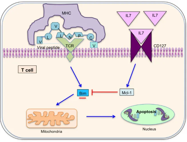
(69) II.7.2 Mcl-1 in Hepatitis. Although it is clear that Mcl-1 is an important molecule for T cell survival, aspects of its role in hepatitis remain to be addressed. Mcl-1 as an antiapoptotic molecule has a pivotal role in impairment of T cell. Hepatitis B virus X protein, which is implicated in pathogenesis of HBV, enhances cisplatininduced hepatotoxicity via a mechanism involving degradation of Mcl-1 [231], which could lead to the T cell apoptosis. In HCV infection, the core protein is a novel BH3-only viral homologue that contributes to the induction of apoptosis and the over expression of Mcl-1 protects against core-induced apoptosis [232]. During LCMV infection, over expression of Mcl-1 protected activated T cells from death, whereas deletion of Mcl-1 during the course of infection leads to a massive loss of LCMV-specific CD4+ and CD8+ T cells [233]. In contrast, additional loss of Bax and Bak completely rescued Mcl-1-deficient effector T-cell number and function, without enhancing T-cell proliferation. Therefore, Mcl-1 is critical for promoting effector T-cell responses, but does so by combating pro-apoptotic molecules (beyond Bim) [233].. The process of T cell death during chronic viral infection is determined by a carefully balanced and complex group of pro- and anti-apoptotic proteins of the Bcl-2 family, such as Bim and Mcl-1 [234] (Fig I11). Interestingly, persistent hepatotropic viral infection is characterized by continuous TCR triggering and CD127 down-regulation on viral-specific CTLs [235], which could favor Bim up-regulation. CD127 plays an essential role in mature lymphocyte survival by counteracting the induction of apoptosis after antigen. 51.
(70) encounter through regulation of some of the B cell lymphoma 2 (Bcl-2) proteins, enhancing IL-2 secretion and life span [236]. Therefore, HCV could modulate CD127 expression on HCV-specific CTLs to impair the quality of the adaptive immune response by IL-7 deprivation, as a survival strategy [121, 166]. Passive T cell death, or death by cytokine deprivation, is controlled by members of the Bcl-2 family, such as Bim and Mcl-1. In addition, it is well known that Bim is clearly involved in intrahepatic specific-CTL apoptosis in animal models [172, 200, 237]. Furthermore, Bim pro-apoptotic effect is blocked by the action of Bcl-2 family anti-apoptotic proteins such as Mcl-1 and Bcl-2 [200, 237]. Interestingly, the co-deletion of Bim fails to prevent the loss of Mcl-1-deficient T cells during LCMV infection [233], clearly pointing out that T cell death also depends on the anti-apoptotic protein expression.. 52.
(71) Figure II.12: Cell survival marker CD127 modulates Bim and myeloid cell leukemia sequence-1 expression on hepatitis C virus-specific cytotoxic T lymphocytes after cognate antigen stimulation. Misbalance of Mcl-1/Bcl-2 interacting mediator (Bim) triggers to apoptosis of hepatitis C virus specific cytotoxic T lymphocytes. TCR: T cell receptor; Mcl-1: Myeloid cell leukemia sequence-1.. 53.
(72) Bearing in mind all these previous facts, this study tries to test a theoretical model to explain specific CTL deletion during persistent hepatotropic viral infection (Fig I.12). This model hypothesizes that CD127 phenotype modulates Bim and Mcl-1 expression on virus-specific CTLs, leading to Mcl-1/Bim imbalance during persistent infection, which impairs T cell reactivity through apotosis induction and suggesting that restoration of T cell function could occur by correcting the levels of Mcl-1 and Bim expression. Therefore, in this study, the correlation between CD127 expression on HCVspecific CTLs and the Mcl-1/Bim phenotype and their effect on T cell reactivity were analyzed.. 54.
(73) III. HYPOTHESIS AND OBJECTIVES. 55.
(74)
(75) III. Hypothesis and Objectives:. Based on the rationale about apoptotic molecules and the impairment of HCVspecific CTL response during chronic hepatitis C infection discussed in previous chapter, we propose the following hypothesis:. Hypothesis:. HCV could modulate CD127 expression on HCV-specific CTLs in order to promote a pro-apoptotic state due to a misbalance between pro(Bim) and anti-apoptotic (Mcl-1) molecules that could alter the ability of T cell response to control viral infection. Modulation of these pathways could restore. the. HCV-specific. cytotoxic. response. in. chronic. patients. characterized by low CD127 expression.. To test this hypothesis, we set out the following study objectives:. General objective:. To correlate the functional features and the apoptotic phenotype (Bim/Mcl-1) of specific cytotoxic response against HCV depending on the CD127 expression level.. 56.
(76) Specific objectives:. -. To correlate CD127 expression level with viral replication and liver damage on HCV specific T cells. -. To analyze the frequency of HCV-specific cytotoxic T cells in peripheral blood according to CD127 expression level.. -. To study the phenotypic and functional characteristics of HCV-specific cytotoxic T cells in peripheral blood according to CD127 expression level:. -. Comparison of the proliferation capacity after specific stimulation with presence or absence of apoptosis inhibitor according to the CD127 expression level.. -. Production of interferon gamma according to CD127 expression level.. -. Bim and Mcl-1 phenotype, directly ex vivo and after specific in vitro stimulation in presence or absence of apoptosis inhibitor, according to CD127 expression level, and correlation of Bim/Mcl-1 balance with HCV-specific T cells reactivity.. 57.
(77) IV. DESIGN, MATERIAL AND METHODS. 58.
(78)
Figure
+7


Documento similar
Cedulario se inicia a mediados del siglo XVIL, por sus propias cédulas puede advertirse que no estaba totalmente conquistada la Nueva Gali- cia, ya que a fines del siglo xvn y en
Where possible, the EU IG and more specifically the data fields and associated business rules present in Chapter 2 –Data elements for the electronic submission of information
The 'On-boarding of users to Substance, Product, Organisation and Referentials (SPOR) data services' document must be considered the reference guidance, as this document includes the
In medicinal products containing more than one manufactured item (e.g., contraceptive having different strengths and fixed dose combination as part of the same medicinal
Products Management Services (PMS) - Implementation of International Organization for Standardization (ISO) standards for the identification of medicinal products (IDMP) in
Products Management Services (PMS) - Implementation of International Organization for Standardization (ISO) standards for the identification of medicinal products (IDMP) in
This section provides guidance with examples on encoding medicinal product packaging information, together with the relationship between Pack Size, Package Item (container)
Package Item (Container) Type : Vial (100000073563) Quantity Operator: equal to (100000000049) Package Item (Container) Quantity : 1 Material : Glass type I (200000003204)
