DEPARTAMENTO DE MICROBIOLOGÍA
ESTUDIO DE CaCwt1 DE CANDIDA ALBICANS
HOMÓLOGO AL FACTOR DE TRANSCRIPCIÓN Rds2 DE
SACCHAROMYCES CEREVISIAE
INMACULADA MORENO GIMENO
UNIVERSITAT DE VALENCIA
Servei de Publicacions
Setembre de 2007 davant un tribunal format per:
- D. Enrique Herrero Perpiñán
- D. Joachim Ernst
- D. Antonio Marcilla Díaz
- D. Juan Carlos Argüelles Ordóñez
- D. Fernando Aniento Company
Va ser dirigida per:
D. Eulogio Valentín Gómez
D. Rafael Sentandreu Ramón
D. Luis Castillo Barahona
©Copyright: Servei de Publicacions
Inmaculada Moreno Gimeno
Depòsit legal:
I.S.B.N.:978-84-370-6999-9
Edita: Universitat de València
Servei de Publicacions
C/ Artes Gráficas, 13 bajo
46010 València
Spain
FACULTAD DE FARMACIA
Departamento de Microbiología y Ecología
Estudio de CaCwt1 de Candida albicans homólogo al factor de
transcripción Rds2 de Saccharomyces cerevisiae.
Memoria que presenta INMACULADA MORENO GIMENO para optar al grado de Doctora en Bioquímica
Parte de los resultados experimentales contenidos en esta Memoria se recogen en las siguientes publicaciones:
- Moreno, I., Pedreño, Y., Maicas, S., Sentandreu, R., Herrero, E., Valentin, E. (2003) Characterization of a Candida albicans gene encoding a putative transcriptional factor required for cell wall integrity. FEMS Microbiol Lett.
226:159-67.
- Maicas, S., Moreno, I., Nieto, A., Gomez, M., Sentandreu, R., Valentin, E. (2005)In silico analysis for transcription factors with Zn(II)2C6 binuclear cluster DNA-binding domains in Candida albicans. Comp Funct Genom. 6:345-56.
- Moreno, I., Castillo, L., Sentandreu, R., Valentin, E. (2007) Global transcriptional profiling of Candida albicans cwt1 null mutant.
βME Beta-mercaptoethanol Beta-mercaptoetanol
BSA Bovine serum albumine Albúmina de suero bovino
cDNA Complemmentary DNA DNA complementario
CSPD Chemiluminiscent substrate Substrato quimioluminiscente
CWP Cell wall proteins Proteínas de pared celular
DBD DNA binding domain Dominio de unión a ADN
DEPC Diethil pyrocarbonate Dietilpirocarbonato
DIG Digoxygenin Digoxigenina
dNTPs Deoxynucleotide triphosphate desoxirribonucleótidos trifosfato DOn Optical density at n nanometers Densidad óptica a n nanómetros
DTT Dithiothreitol Ditiotreitol
ECL Enhanced chimioluminiscence Técnica de quimioluminiscencia
ER Endoplasmic reticulum Retículo endoplásmico
FITC Fluorescein isothiocyanate Isotiocianato de fluoresceína
g Gram Gramo
xg Gravity unit Gravedades
GPI Glycosyl phosphatydil inositol Glicosil fosfatidil inositol
GlcNAc N-acetylglucosamine N-acetilglucosamina
GTP Guanosin triphosphate Guanosina trifosfato
h Hours Horas
IPF Individual protein file kb Kilobase kDa Kilodalton
l Litre Litro
M Molar
min Minute Minuto
mARN Messenger RNA ARN mensajero
NLS Nuclear localization signal Señal de localización nuclear
ORF Open reading frame Pauta abierta de lectura
PAb Polyclonal antibody Anticuerpo policlonal
PAGE Polyacrilamide gel electrophoresis Electroforesis en gel de poliacrilamida
pb Base pairs Pares de bases
PBS Phosphate buffer saline Tampón fosfato salino
PCR Polymerase chain reaction Reacción en cadena de la polimerasa
PDB Protein data bank Banco de datos de proteínas
PEG Polyethilene glycol Polietilenglicol
PMSF Phenylmethylsulphonyl fluoride Fluoruro de fenil-metil sulfonilo
Q-RT-PCR Quantitative RT-PCR RT-PCR semicuantitativa
RMN Nuclear magnetic resonance Resonancia magnética nuclear
RSAT Regulatory sequence analysis tools
RT-PCR Reverse transcriptase-PCR Transcripción reversa-PCR
SDS Sodium dodecyl sulphate Dodecil sulfato sódico
seg Seconds Segundos
TBS Tris buffer saline Tampón tris salino
Tris Tris (hydroxymethyl) aminomethane Tris (hidroximetil) amino metano
TTBS TBS plus Tween-20 TBS con Tween-20
UDP Uridinediphosphoglucose Uridina difosfoglucosa
V Volt Voltio
vol Volume Volumen
Tabla de códigos internacionales de una o tres letras para designar a los aminoácidos.
AMINOÁCIDO 3 LETRAS 1 LETRA PESO MOLECULAR (Da)
Ácido aspártico Asp D 133
Ácido glutámico Glu E 147
Alanina Ala A 89
Arginina Arg R 174
Asparagina Asn N 132
Cisteína Cys C 121
Fenilalanina Phe F 165
Glicina Gly G 75
Glutamina Gln Q 146
Histidina His H 155
Isoleucina Ile I 131
Leucina Leu L 131
Lisina Lys K 146
Metionina Met M 149
Prolina Pro P 115
Serina Ser S 105
Tirosina Tyr Y 181
Treonina Thr T 119
Triptófano Trp W 204
Valina Val V 117
Tabla de uso de codones universal.
2ª base
U C A G U UUU Fenilalanina
UUC Fenilalanina UUA Leucina UUG Leucina UCU Serina UCC Serina UCA Serina UCG Serina UAU Tirosina UAC Tirosina UAA Parada UAG Parada
UGU Cisteína UGC Cisteína UGA Parada UGG Triptófano
C CUU Leucina CUC Leucina CUA Leucina CUG Leucina2
CCU Prolina CCC Prolina CCA Prolina CCG Prolina CAU Histidina CAC Histidina CAA Glutamina CAG Glutamina CGU Arginina CGC Arginina CGA Arginina CGG Arginina
A AUU Isoleucina AUC Isoleucina AUA Isoleucina AUG Metionina1
ACU Treonina ACC Treonina ACA Treonina ACG Treonina AAU Asparagina AAC Asparagina AAA Lisina AAG Lisina AGU Serina AGC Serina AGA Arginina AGG Arginina 1ª b a se
G GUU Valina GUC Valina GUA Valina GUG Valina GCU Alanina GCC Alanina GCA Alanina GCG Alanina
GAU Ác. aspártico GAC Ác. aspártico GAA Ác. glutámico GAG Ác. glutámico
GGU Glicina GGC Glicina GGA Glicina GGG Glicina
1
El primer codón AUG de un mARN indica el sitio de iniciación de la traducción.
2
I. INTRODUCTION 1
I.1- GENERAL ASPECTS AND TAXONOMY OF Candida albicans 2
I.2- C. albicans PATHOGENICITY 2
I.2.1- Types of candidiasis 3
I.2.1.1- Superficial candidiasis 3
I.2.1.2- Internal candidiasis 3
I.2.2- Risk factors 4
I.2.3- Treatment of candidiasis 4
I.2.4- Incidence of candidiasis 5
I.3- VIRULENCE FACTORS 5
I.3.1- Adhesión 6
I.3.2- Secreted hydrolytic enzymes 7
I.3.2.1- Secreted aspartyl proteinases (Saps) 7
I.3.2.2- Phospholipases (PL) 8
I.3.2.3- Lipases (LIP) 8
I.3.3- Morphogenesis 9
I.3.3.1- Outside cues 10
I.3.3.2- Signalling pathways 10
I.3.3.2.1- The cAMP-dependent protein kinase A pathway 11
I.3.3.2.2- The mitogen-activated protein kinase pathway 12
I.3.3.2.3- Repressing factors 13
I.3.3.3- Co-regulation of virulence and morphogenesis genes 13
I.3.4- Phenotypic switching 13
I.3.5- Biofilms formation 14
I.4 Candida albicans CELL WALL 16
I.4.1- Cell wall composition 16
I.4.1.1- β-glucans 18
I.4.1.1.1- β-1,3-glucan 18
I.4.1.1.2- β-1,6-glucan 19
I.4.1.2- Chitin 20
I.4.1.3- Mannoproteins 21
I.4.1.3.1- Classification of cell wall mannoproteins 22
I.4.1.3.1.1- Non-covalently linked (NCL-CWPs) 23
I.4.1.3.1.2- Reducing agents extractable (RAE-CWPs) 23
I.4.1.3.1.3- Glycosyl phosphatydil inositol anchored (GPI-CWPs) 23
I.4.1.3.1.5- Atypical wall proteins 27
I.4.1.3.2- Post-translational modifications of cell wall proteins 28
I.4.1.3.2.1- N-glycosylation 29
I.4.1.3.2.2- O-glycosylation 29
I.4.2- Cell wall remodelling 30
I.4.2.1- Glucan remodelling enzymes 30
I.4.2.2- Chitin remodelling enzymes 30
I.4.2.3- Glucan-chitin cross-linking enzymes 31
I.4.3- Cell wall integrity signalling 31
I.4.3.1- PKC1-MAPK pathway 33
I.4.3.2- Cek1-mediated pathway 34
I.5 TRASNCRIPTIONAL FACTORS 35
I.5.1- Promoter binding sites 35
I.5.1.1- Promoter architecture 35
I.5.1.2- Promoter binding behaviour 36
I.5.2- Zinc finger proteins 37
I.5.3- Zinc cluster proteins Zn(II)2Cys6 38
I.5.3.1- General structure 38
I.5.3.1.1- Nuclear localization signal (NLS) 39
I.5.3.1.1.1- Classical NLS 39
I.5.3.1.1.2- Non-conventional NLS 40
I.5.3.1.2- DNA binding domain (DBD) 40
I.5.3.1.2.1- Zinc finger structure (Zn(II)2Cys6) 40
I.5.3.1.2.2- Linker region 41
I.5.3.1.2.3- Dimerization domain 41
I.5.3.1.3- Regulatory domain (MHR) 41
I.5.3.1.4- Activation domain (AD) 41
I.5.3.2- Binding of zinc cluster transcriptional factors 42
I.5.3.2.1- Binding sites 42
I.5.3.2.2- Mechanism of action 44
I.5.4- Zinc cluster proteins in C. albicans 45
I.6- TRANSCRIPTION FACTORS REGULATING CELL WALL ARCHITECTURE IN C.
albicans
46
I.6.1- Cas5p 46
I.6.2- Rlm1p 46
I.6.3- Ace2p 47
I.6.5- Rim101p 48
I.6.6- Bcr1p 48
II. ANTECEDENTES Y OBJETIVOS 50
III. MATERIALES Y MÉTODOS 53
III.1-MICROORGANISMOS 54
III.1.1- Levaduras 54
III.1.2- Bacterias 54
III.2- MEDIOS Y CONDICIONES DE CULTIVO 55
III.2.1- Cultivo de levaduras 55
III.2.2- Cultivo de bacterias 57
III.3- TAMPONES UTILIZADOS 58
III.4- PLÁSMIDOS 58
III.5- TRANSFORMACIÓN DE CÉLULAS CON ADN 59
III.5.1- Transformación de bacterias 59
III.5.1.1- Obtención de células competentes de E.coli 59
III.5.1.2- Transformación de células competentes de E.coli 60
III.5.2- Transformación de células de C. albicans. 60
III.6- PURIFICACIÓN DE ADN 61
III.6.1- Obtención de ADN plasmídico de E.coli 61
III.6.2- Obtención de ADN genómico de C. albicans 61
III.7. TRATAMIENTO ENZIMÁTICO DEL ADN 62
III.7.1- Digestión con endonucleasas de restricción 62
III.7.2- Ligación de ADN con ligasa de T4 62
III.8- REACCIÓN EN CADENA DE LA POLIMERASA (PCR) 62
III.8.1- Diseño de oligonucleótidos 63
III.8.2- Condiciones de reacción de PCR 63
III.8.3- Transcripción reversa (RT-PCR) 64
III.8.3.1- RT-PCR semicuantitativa 64
III.9- ELECTROFORESIS DE ADN EN GELES DE AGAROSA 65
III.10- PURIFICACIÓN DE FRAGMENTOS DE ADN DE GELES DE AGAROSA 66
III.11- SECUENCIACIÓN DE FRAGMENTOS DE ADN 66
III.12.1- Marcaje no radiactivo de sondas de ADN 67
III.12.2- Detección de secuencias específicas de ADN (Southern Blot) 67
III.12.2.1- Separación y transferencia de los fragmentos de ADN 67
III.12.2.2- Hibridación ADN/ADN 68
III.12.2.3- Detección de la unión ADN / sonda 68
III.13- PURIFICACIÓN DE ARN TOTAL DE C. albicans 69
III.14- CUANTIFICACIÓN DE ÁCIDOS NUCLEICOS 69
III.15- ESTUDIO DEL TRANSCRIPTOMA MEDIANTE MICROMATRICES (MICROARRAYS) DE ADN
70
III.15.1- Marcaje del ADNc 70
III.15.2- Elución, cuantificación y concentración de los ADNc marcados 71
III.15.3- Hibridación de los microarrays de ADN 72
III.15.4- Obtención de imágenes y análisis de resultados 72
III.15.5- Análisis de promotores con RSAT 73
III.15.6- Estudio del estado celular mediante T-profiler 73
III.16- INTERRUPCIÓN GÉNICA EN C. albicans 73
III.17- ANÁLISIS FENOTÍPICO DE LA CEPA DISRUPTANTE 74
III.17.1- Estudio del efecto de calcofluor white, rojo Congo y SDS 74
III.17.2- Estudio del efecto de drogas 74
III.17.3- Sensibilidad a zimoliasa 75
III.17.4- Miceliación en medio sólido 75
III.18- OBTENCIÓN DE CÉLULAS EN FASE MICELIAR 75
III.19- ANÁLISIS DEL MEDIO DE CULTIVO 76
III.20- OBTENCIÓN DE PAREDES CELULARES 76
III.21- SOLUBILIZACIÓN DE COMPONENTES DE LA PARED CELULAR 76
III. 21.1- Tratamiento con dodecil sulfato sódico (SDS) 76
III. 21.2- Tratamiento con β-mercaptoetanol (β-ME) 77
III. 21.3- Tratamiento con zimoliasa 77
III.22- SEPARACIÓN DE PROTEÍNAS EN GELES DE SDS-POLIACRILAMIDA (SDS-PAGE)
78
III.23- TRANSFERENCIA Y DETECCIÓN DE PROTEÍNAS EN SOPORTES DE NITROCELULOSA (Western-blot)
78
III.23.1- Transferencia a membrana de nitrocelulosa 78
III.23.2- Inmunodetección de proteínas en soportes de nitrocelulosa 79
III.24- CUANTIFICACIÓN DE MACROMOLÉCULAS 80
III.24.1- Determinación de proteína total 80
III.24.2- Determinación de polisacáridos 80
III.24.2.2- Determinación de β-glucanos 81
III.24.2.3- Determinación de manano 81
III.24.2.4- Determinación de quitina 82
III.25- MARCAJE DE PARED CELULAR EN CÉLULAS ENTERAS 82
III.25.1- Tinción de células con concanavalina A 82
IV. RESULTADOS 83
IV.1- IDENTIFICACIÓN DE POSIBLES FACTORES DE TRANSCRIPCIÓN DEL TIPO Zn(II)2Cys6 EN C. albicans
84
IV.2- SELECCIÓN DE LA IPF 3781 PARA EL PRESENTE TRABAJO 88
IV.3-ANÁLISIS DEL TEÓRICO FACTOR DE TRANSCRIPCIÓN Cwt1p 90
IV.3.1- Análisis de la secuencia de aminoácidos 90
IV.3.1.1- Dominio Zn(II)2Cys6 93
IV.3.1.2- Dominio PAS 94
IV.4- ESTUDIO DE LA EXPRESIÓN DEL GEN CaCWT1 95
IV.5- DISRUPCIÓN DEL GEN CaCWT1 97
IV.5.1- Construcción de los casetes de interrupción 97
IV.5.1.1- Construcción del casete de arginina 98
IV.5.1.2- Construcción del casete de histidina 100
IV.5.2- Obtención del mutante nulo del gen CaCWT1 de C. albicans 101
IV.6- REINTEGRACIÓN DE MARCADORES PARA ELIMINAR LAS AUXOTROFÍAS DE LAS CEPAS UTILIZADAS
104
IV.7- ANÁLISIS FENOTÍPICO DEL MUTANTE NULO ΔCacwt1 105
IV.7.1- Crecimiento y morfología 105
IV.7.2- Estudio de filamentación en medio sólido 106
IV.7.3- Estudio de sensibilidad a estrés térmico, osmótico y oxidativo 107
IV.7.4- Estudio de sensibilidad a cafeína 108
IV.7.5- Estudio de resistencia a drogas 108
IV.7.6- Estudio de la integridad de la pared celular 110
IV.7.7- Estudio de sensibilidad a zimoliasa 111
IV.7.8- Análisis de polímeros de la pared celular 112
IV.7.9- Tinción de células con Concanavalina A acoplada a fluoresceína 114
IV.7.10- Caracterización de la pared celular del mutante nulo ΔCacwt1 115
IV.7.10.1- Western blot de extractos de pared con anticuerpo policlonal 116
IV.7.10.2- Western blot de extractos de pared con Anti-β-1,6-glucano 118
IV.8- SOBRE-EXPRESIÓN DEL GEN CWT1 EN EL MUTANTE NULO cwt1Δ/cwt1Δ 121
IV.8.1- Construcción del plásmido pBI-CWT1 121
IV.8.2- Introducción de pBICWT1 en el mutante nulo 122
IV.8.3- Expresión de CWT1 en la cepa reintegrante 122
IV.8.4- Análisis fenotípico de la cepa reintegrante 124
IV.9- PERFIL TRANSCRIPCIONAL DEL MUTANTE NULO cwt1Δ/cwt1Δ 125
IV.9.1- Perfil transcripcional del mutante nulo cwt1Δ/cwt1Δ en fase exponencial de crecimiento
127
IV.9.1.1- Genes involucrados en la arquitectura de la pared celular 128
IV.9.1.2- Genes relacionados con la traducción de proteínas 129
IV.9.1.3- Genes que codifican factores de transcripción 131
IV.9.2- Perfil transcripcional de ΔCacwt1 en fase estacionaria de crecimiento 131
IV.9.2.1- Genes involucrados en la arquitectura de la pared celular 132
IV.9.2.2- Genes relacionados con la traducción de proteínas 134
IV.9.2.3- Genes que codifican factores de transcripción 135
IV.9.3- Validación de los resultados de las micromatrices de ADN del mutante nulo
cwt1Δ/cwt1Δ
136
IV.9.4- Perfil transcripcional del mutante nulo cwt1Δ/cwt1Δ a lo largo del ciclo poblacional 137
IV.9.4.1- Análisis del patrón de expresión de cwt1Δ/cwt1Δ con el programa informático T-profiler
139
IV.10- PERFIL TRANSCRIPCIONAL DEL MUTANTE HETEROCIGÓTICO CWT1/cwt1Δ 141
IV.10.1- Perfil transcripcional del mutante heterocigótico CWT1/cwt1Δ en fase exponencial de crecimiento
141
IV.10.1.1- Genes involucrados en la arquitectura de la pared celular 142
IV. 10.1.2- Genes relacionados con respuesta a estrés oxidativo 143
IV. 10.1.3- Genes que codifican factores de transcripción 144
IV. 10.2- Perfil transcripcional del mutante heterocigótico CWT1/cwt1Δ en fase estacionaria de crecimiento
145
IV. 10.2.1- Genes involucrados en la arquitectura de la pared celular 146
IV. 10.2.2- Genes relacionados con la respuesta a estrés oxidativo 147
IV. 10.2.3- Genes que codifican para factores de transcripción 148
IV. 10.3- Validación de los resultados de las micromatrices de ADN del mutante heterocigótico CWT1/cwt1Δ
148
IV. 10.4- Perfil transcripcional del mutante heterocigótico CWT1/cwt1Δ a lo largo del ciclo poblacional
150
IV. 10.4.1- Análisis del patrón de expresión de CWT1/cwt1Δ con el programa informático T-profiler
154
IV.11- EFECTO TRANSCRIPCIONAL GLOBAL DE Cwt1p A LO LARGO DEL CICLO POBLACIONAL
IV.12- ANÁLISIS DE LAS SECUENCIAS PROMOTORAS DE LOS GENES REGULADOS POR Cwt1p
156
V. DISCUSSION 158
VI. CONCLUSIONES / CONCLUSIONS 172
VII. BIBLIOGRAFÍA / REFERENCES 175
I.1- GENERAL ASPECTS AND TAXONOMY OF Candida albicans
Candida is a genus made up of 154 ascomycetous-like fungal species. Seven of them, C. albicans, C. glabrata, C. tropicalis, C. parapsilosis, C. krusei (Issatchenkia orientalis), C. dubliniensis y C. lusitaniae (Clavispora lusitaniae), are frequently isolated from human infections, being C. albicans the most relevant in terms of pathogenicity.
C. albicans is a polymorphic organism capable of reproducing by budding (yeast cells) or by producing filamentous forms (mycelial cells) depending upon environmental factors (Odds, 1994), this morphological transition has been associated with pathogenicity (Kumamoto and Vinces, 2005).
C. albicans has been taxonomically classified as: Kingdom: Fungi
Phylum: Ascomycota
Subphylum: Ascomycotina
Class: Ascomycetes
Order: Saccharomycetales
Family: Saccharomycetaceae
Genus: Candida
I.2- C. albicans PATHOGENICITY
The most frequently encountered fungal infections are caused by the yeast C. albicans and by species of the filamentous fungus Aspergillus. Other fungal pathogens that have emerged in recent years include yeast species such as T. glabrata, I. orientalis, C. tropicalis, species of Cryptococcus and Trichosporon and filamentous fungi such as
Fusarium, Rhizopus and Rhizomucor (Richardson, 2005).
I.2.1- Types of candidiasis
Candidiasis can be classified in two different groups: superficial and internal.
I.2.1.1- Superficial candidiasis
Superficial candidiasis, involving skin, nails and mucosal surfaces are classified as follows:
Cutaneous candidiasis Intertrigo Folliculitis
Mucosal candidiasis Oropharyngeal Vulvovaginal Esophagus Urinary bladder
Nails candidiasis Onychomycosis Paronychia
Other candidiasis External otitis
Keratitis
I.2.1.2- Internal candidiasis
Systemic candidiasis involves internal organs (brain, liver, lungs, heart) and can be widespread through the bloodstream affecting different organs (systemic infections).
Localized candidiasis Peritonitis Esophagitis Pyelitis
Systemic candidiasis Endocardial
Intravenous drugs abuse syndrome Hepatosplenic
I.2.2- Risk factors
Candida infections are being seen in ever rising numbers, largely because of the increasing size of population at risk. Risk factors for C. albicans bloodstream infections (Richardson, 2005) can be divided into:
- Host-related factors such as immunocompromising conditions: transplant recipients, cancer patients, AIDS and intensive care patients, individuals with prolonged and deep neutropenia and low birth weight.
- Healthcare-related factors: broad-spectrum antibiotic use, catheters, use of antifungals and chemoprophylaxis, invasive and aggressive therapeutic technologies and surgical procedures.
I.2.3- Treatment of candidiasis
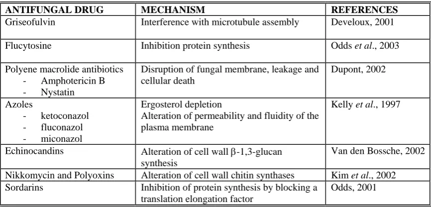
[image:18.595.80.513.542.750.2]Because C. albicans is a eukaryotic organism and shares biological components and processes with human cells, differences between these two types of cells are used for developing antifungal drugs with selective toxicity to C. albicans cells. Several targets have been used for antifungal drugs because they are different or not present in human cells: cell wall, ergosterol synthesis, microtubule assembly, DNA and RNA synthesis, protein synthesis (Odds et al., 2003). Antifungal drugs currently available, their targets and mechanisms of action are detailed in table I.1.
Table I.1- Antifungal agents currently available
ANTIFUNGAL DRUG MECHANISM REFERENCES
Griseofulvin Interference with microtubule assembly Develoux, 2001
Flucytosine Inhibition protein synthesis Odds et al., 2003
Polyene macrolide antibiotics - Amphotericin B - Nystatin
Disruption of fungal membrane, leakage and cellular death
Dupont, 2002
Azoles
- ketoconazol - fluconazol - miconazol
Ergosterol depletion
Alteration of permeability and fluidity of the plasma membrane
Kelly et al., 1997
Echinocandins Alteration of cell wall β-1,3-glucan synthesis
Van den Bossche, 2002
Nikkomycin and Polyoxins Alteration of cell wall chitin synthases Kim et al., 2002 Sordarins Inhibition of protein synthesis by blocking a
translation elongation factor
Given the clinical importance of candidiasis, efforts continue to identify new drugs targets and therefore new anti-fungal drugs, to improve diagnostic assays and to provide alternative therapeutic options (Casadevall et al., 2004).
I.2.4- Incidence of candidiasis
Candidiasis is regarded as one of the fourth most common cause of nosocomial and bloodstream infections (BSIs) in the USA, and published data indicate a similar pattern worldwide (Haynes, 2001; Chauhan et al., 2006). From an analysis of multi-institutional surveys of Candida BSIs performed in Europe, including the large prospective survey by the European Confederation of Medical Mycology, rates of candidaemia ranging from 0.20 to 0.38 per 1000 admissions were reported with a global mortality of 44% of the reported cases (Tortorano et al., 2004). The distribution of the different isolated Candida spp. is presented in figure. I.1.
C. albicans , 57% C. parapsilosis,
19% C. glabrata, 8%
C. tropicalis, 7% C. krusei, 4%
[image:19.595.159.433.432.548.2]Other Candida spp., 5%
Figure I.1- Candida BSIs distribution in a multi-institutional study in Europe. Data obtained from prospective survey by the European Confederation of Medical Mycology (Adapted from Tortorano et al., 2004).
I.3- VIRULENCE FACTORS
stablish disease” (Furman and Ahearn, 1983), “factors that interact directly with mammalian host cells” (Odds et al., 2001) and “a component of a pathogen that damages the host” (Casadevall and Pirofski, 2001). In this sense, C. albicans has to mediate the contact with the host cell, evade the immune system, survive and divide in the host environment and spread to new tissues to start the infection process. The virulence factors described for C. albicans include:
- adhesins
- hydrolytic enzymes - morphogenesis - phenotypic switching
I.3.1- Adhesion
Contact and adherence of C. albicans to host cells are the earlier steps during infection. In this process, different molecules present in the host cell surface can be recognized by fungal adhesins. While the receptors of the host cells are variable (can be constituted by proteins, lipids or sugars), adhesins in C. albicans are mainly mannoproteins.
Specific cell wall proteins play a key role in adhesion. All the primary amino acid sequences of the described adhesins share features of highly glycosylated yeast cell wall proteins with an N-terminal signal peptide and C-terminal features that mediate glycosylphosphatidylinositol (GPI) anchor (Sundstrom, 2002). The most important group of C. albicans adhesins is formed by Als (Agglutinin-like sequence) proteins (for a review see Hoyer, 2001). However, some other kinds of adhesins have been found in
C. albicans: Hwp1p, (hyphal wall protein) a specific mannoprotein of mycelial forms (Staab et al., 1999, Sundstrom, 1999; 2002), and Int1p (integrin-like protein) involved in adhesion and virulence (Gale et al., 1998, 2001). Other kinds of proteins have also been found to be involved in adhesion, like Mnt1p (mannosyltransferase) a type II membrane protein involved in N- and O-glycosylation (Thomson et al., 2000; Buurman
et al., 1998).
ALS family is a gene family in C. albicans encoding eight proteins. It is called
The adhesive function of Als proteins has been suggested by expressing C. albicans ALS genes in the normally nonadherent Saccharomyces cerevisiae that acquires an adhesive phenotype. Genes of the C. albicansALS family encode proteins with three characteristic domains: (i) the N-terminal region contains a signal peptide and is relatively conserved among Als proteins, although it has been suggested that N-terminal sequence variability confers substrate specificity (Sheppard et al. 2004); (ii) a central portion consisting of a variable number of tandem repeats with approximately 36 amino acids each; (iii) the C-terminal domain possesses a serine-threonine rich region and a GPI anchor sequence (Zhao et al., 2003; Hoyer et al., 2001).
Different subcategories of Als proteins have been described depending on their physicochemical properties. Als1p, Als3p and Als5p are considered the Als-group A, Als6p and Als7p are part of the Als-group B, and finally, Als2p, Als4p and Als9p appear to constitute the Als-group C.
I.3.2- Secreted hydrolytic enzymes
Extracellular hydrolytic enzymes are secreted for digesting complex molecules for nutrition. However, pathogenic microorganisms hydrolyse proteins and lipids of host cells to facilitate adhesion and invasion, or damaging cells and molecules of the host defense system (Cunningham and Agard, 2004; Naglik et al., 2004; Stehr et al., 2004).
I.3.2.1- Secreted aspartyl proteinases (Saps)
All secreted C. albicans proteinases belong to the aspartyl proteinases family, and these enzymes are essential for growth when proteins are the sole nitrogen source. Proteolytic activity has also been found in other Candida species like C. dubliniensis, C. tropicalis and C. parapsilopsis; since, the proteolytic activity of other non-pathogenic species is lower, this suggests that virulence is correlated with the level of Sap production (Schaller et al., 2005).
processed when transported via the secretory pathway. Eight of these proteinases (Sap1p to Sap8p) are secreted into the extracellular space while Sap9p and Sap10p are membrane-anchored GPI proteins (Naglik et al., 2004). The presence of 10 SAP genes suggests that different Saps are expressed in C. albicans in reaction to specific environmental conditions and may have a variety of functions during the infectious process. Many experimental studies have shown that Sap1-3ps play an important role in adherence while Sap4-6ps are responsible for the pathogenesis of invasive candidiasis as they are secreted after phagocytosis by macrophages (Hube et al., 1997; Sanglard et al., 1997; Hube and Naglik, 2001).
I.3.2.2- Phospholipases (PL)
Phospholipases are considered virulence factors in different fungi as C. albicans
(Ibrahim et al., 1995), Aspergillus fumigatus or Cryptococcus neoformans. However, among all the Candida species only C. albicans possesses PL activity (Lane and Garcia, 1991), although T. glabrata, C. parapsilopsis, C. tropicalis, C. lusitaniae and I. orientalis are able to secrete PL (Ghannoum, 2000; Kantarcioglu and Yucel, 2002).
Phospholipases are enzymes that hydrolyse ester linkages in glycerophospholipids. Different PL subclasses have been described: PL A, PL B, PL C and PL D depending on the mode of action and the target within the phospholipid molecule. To date, only proteins regarding to PL B subgroup have been found to be secreted, while all the others are supossed to be intracellular PLs. The functions of PLs during infection are not yet known, but may be involved in host cell penetration and interaction with host signal transduction pathways (Schaller et al., 2005).
I.3.2.3- Lipases (Lip)
I.3.3- Morphogenesis
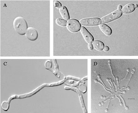
C. albicans can grow in a variety of morphological forms (Fig. I.2) depending on the environmental conditions (Ernst, 2000; Odds, 1988; Sonneborn et al., 1999a): - yeast or blastospore: oval-shaped unicellular budding yeast.
- pseudohyphae: cells grow by unipolar budding, buds develop into elongated cells, which remains attached to mother cell, stop growth and resume budding.
- true hyphae: cells grow by continuous apical extension followed by septation and the result are different mononuclear cells with cytoplasmic communication through porus in the septum.
- chlamydospore: formed by thick-walled spherical cells and developed on pseudohyphal support cells. They are considered resistant cellular forms.
[image:23.595.185.410.443.627.2]The ability to change its morphology is considered to be necessary for virulence in C. albicans. As filamentous forms are invasive, they are supposed to promote tissue penetration during early stages of infection, whereas the yeast form might be more suited for dissemination in the bloodstream (Sudbery et al., 2004).
Figure I.2- Different morphogenic states of C. albicans. (A) yeast, (B) pseudohyphae, (C) hyphae, and (D) chlamydospore.
I.3.3.1- Outside cues
The signals that promote morphological changes in C. albicans often impose stresses (Brown and Gow, 1999). Changes in growth temperature from 30ºC to 37ºC and neutral pH culture induce morphogenetic changes in C. albicans cells (Lee et al., 1975; Saporito-Irwin et al., 1995; Shepherd et al., 1980).
Also, nitrogen and carbon starvation cause hyphal development. In this sense, serum (composed mainly of proteins that are inaccessible nutrient source for the C. albicans cell until they are hydrolised) and N-acetylglucosamine (a poor source of carbon and nitrogen) (Gow and Gooday., 1982; Mattia et al., 1982) are favourable conditions to obtain the filamentous forms. Supplementing these media with amino acids and glucose causes hyphae to revert to yeast cells.
Other in vitro factors facilitating hyphal development are incubation of C. albicans at low cells density (Shepherd et al., 1980), presence of polyamines and hemin (Casanova et al., 1997; Herrero et al., 1999), and different levels of human hormones, suggesting that hyphal induction could be modulated by other host factors in vivo.
I.3.3.2- Signalling pathways
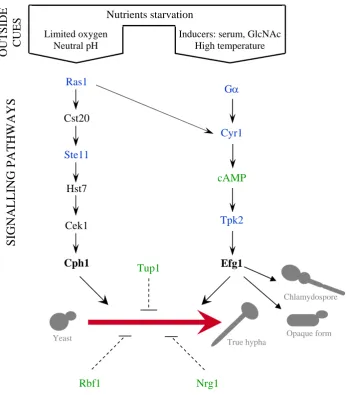
The signal transduction pathways that transduce environmental signals into morphological switching have been extensively studied. These studies have shown that most of the genes involved in hyphal morphogenesis in C. albicans are homologues to those required for polarized growth and pseudohyphal growth in S. cerevisiae. Although no sensors of environmental signals have been identified until this moment, several transduction pathways have been shown to play an important role in C. albicans
morphogenesis (Fig. I.3).
Rbf1 Nrg1
Cph1 Efg1
Tpk2
Cek1 Hst7
Ste11
Cst20
Ras1
cAMP
Cyr1 Gα
Tup1
Limited oxygen Neutral pH
Inducers: serum, GlcNAc High temperature
OUT
SIDE
CUES
SIGNALLING PATHWAYS
Nutrients starvation
Yeast True hypha Opaque form
[image:25.595.127.475.73.469.2]Chlamydospore
Figure I.3. Candida albicans morphogenesis networks. Environmental cues activate separate signalling pathways. Two major pathways, defined by the transcriptional factors Cph1p and Efg1p (in bold), induce hyphal growth. Components shown in black have been established in their position by epistasis analyses. Components shown in green are known to influence morphogenesis in their position unless no epistasis analyses have proved it so far. Components shown in blue represent proteins which have not been confirmed to affect morphogenesis experimentally. Blunt arrows mean negative regulation. Dash lines show components affecting morphogenesis through unknown pathways. (Adapted from Brown and Gow, 1999; Ernst, 2000)
I.3.3.2.1- The cAMP-dependent protein kinase A pathway
Hyphal development is stimulated by exogenous cAMP, and changes in intracellular cAMP levels have been associated with morphogenesis in C. albicans
hyphal morphogenesis as it controls pseudohyphal development in S. cerevisiae (Pan and Heitman, 1999).
cAMP-protein kinaseA (PKA) pathway is controlled in C. albicans by the transcriptional factor Efg1p, homologue to Phd1p in S. cerevisiae. Efg1p belongs to the family of APSES proteins, a conserved class of transcriptional regulators in fungi that contain a basic helix-loop-helix motif (Doedt et al., 2004). The Efg1p protein is a strong regulator of morphogenetic processes, since it influences not only yeast-hypha interconversions (Stoldt et al., 1997; Lo et al., 1997), but also regulates phenotypic switching and chlamydospore formation of this pathogen (Sonneborn et al., 1999a, b). The transcriptional factor Efg1p plays an important role in morphogenesis as disruption of EFG1 causes a complete block of hyphal formation under standard induction conditions (Stoldt et al., 1997) and alterations on biofilms construction (Ramage et al., 2002), whereas Efg1p overexpression produces an increase in C. albicans filamentation. Moreover, Efg1p can act as an activator or a repressor of mophogenesis depending on the environmental cues (Stoldt et al., 1997).
Other components of this signalling pathway, Ras protein (Ras1p), adenyl cyclase (Cyr1p) and protein kinase A (Tpk2p) have been described to be involved in morphogenesis (Ernst, 2000).
I.3.3.2.2- The mitogen-activated protein kinase pathway
Components of the mitogen-activated protein (MAP) kinase pathway in S. cerevisiae are involved in pseudohyphal growth and mating pheromone response (Fig. I.3). Homologues to S. cerevisiae MAP kinase components (Ste20p/Ste7p/Kss1p/Ste12p) have been found in C. albicans
(Cst20p/Hst7p/Cek1p/Cph1p) with regard to structures and functions (Liu et al., 1993; Madhani and Fink, 1997; Alonso-Monge et al., 2003; Eisman et al., 2006).
MAP kinase transduction pathway in C. albicans possesses a lower importance on morphogenesis than the cAMP-PKA pathway, as deficiencies in this pathway affect morphogenesis only under special conditions, but not during induction by serum and other inducers.
of this cascade cause a similar effect to that observed for cph1Δ/cph1Δ (Csank et al., 1998). However, different mutants in MAP kinase components do not display identical virulence phenotypes in systemic infections since cst20Δ/cst20Δ and cek1Δ/cek1Δ
mutants showed reduced virulence whereas hst7Δ/hst7Δ and cph1Δ/cph1Δ did not (Csank et al., 1998; Leberer et al., 1996; Lo et al., 1997).
I.3.3.2.3- Repressing factors
Finally, some other transcription factors have been identified as hyphal repressors unless the mechanisms or pathways in which they function are still unknown. These transcription factors are Tup1p, Nrg1p and Rbf1p (Braun and Johnson, 1997; Braun et al., 2001; Ishii et al., 1997). Cells lacking these proteins grow in filamentous form in all media tested.
I.3.3.3- Co-regulation of virulence and morphogenesis genes
C. albicans exhibits considerable morphogenetic plasticity that is linked to its pathogenicity since most mutants that fail to produce hyphae present reduced virulence. However, it has been pointed out that co-regulation of genes controlling hyphal morphogenesis with genes encoding virulence factors confounds the analysis (Kobayashi and Cutler, 1998; Brown, 2002; Gow et al., 2002; Liu, 2002). The set of virulence factor genes that are co-expressed with hyphal developmental genes are termed as the hyphal regulon, and includes ALS3, HWP1, RBT1, RBT4, SAP4, SAP5 and
SAP6 among others. To ensure that all of the activities that favour virulence are expressed together at the appropriate time, proteins that control cellular form, such as, the hypha-specific G1 cyclin Hgc1p, are co-regulated with adhesins and proteases. (for a review see Kumamoto and Vinces, 2005)
I.3.4- Phenotypic switching
white colonies, while opaque-phase cells appear more elongated and form flatter, darker colonies on solid agar (Bennet and Johnson, 2005). This reversible switching occurs at a frequency of 10-4 to 10-5 per cell generation from white to opaque cells and 5x10⁻⁴ per cell generation from opaque to white phase (Soll, 1997).
Different studies show that C. albicans uses the same signal transduction pathway for regulating dimorphism, mating and phenotypic switching (Bennet and Johnson, 2005; Monge et al., 2006).
Although the role of phenotypic switching in virulence remains to be elucidated, it has been shown that opaque-phase cells are more virulent in cutaneous infections and that white-phase cells are more virulent in systemic infections (Kvaal et al., 1999). DNA microarray experiments with WO-1 strain revealed that it is possible that white-opaque switching in C. albicans plays a role in pathogenesis, as the expression of 400 genes differs between cells in the white and opaque phases (Lan et al., 2002; Tsong et al., 2003). Moreover, specific genes for the white-phase (SAP2, WH11 and EFG1) and opaque-phase (SAP1, SAP3 and CDR3) were found.
I.3.5- Biofilms formation
Pathogenic yeasts have the capability to adhere to different surfaces and devices to gain access to the bloodstream and internal organs of patients (Kojic and Darouiche, 2004).
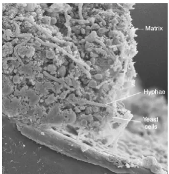
Biofilms are structured microbial communities in which the cells bind to a surface and get embedded in a matrix of extracellular polymeric substances (Ramage et al., 2005). C. albicans biofilms are made up of a mixture of yeast cells, pseudohyphae and hyphae (Nett and Andes, 2006), as seen in Figure I.4. Extracellular matrix is made by cells within the biofilm, and consists of carbohydrate and protein (Douglas, 2003). The function of this extracellular polymeric matrix may be to strengthen the biofilm structure (Nobile and Mitchell, 2006).
C. albicans can form biofilms on almost any medical device. The most commonly infected topical devices are contact lenses and dentures. Systemic devices include vascular and urinary catheters, joint prosthesis, cardiac valves, pace-markers, etc. (Nett and Andes, 2006).
Figure I.4- Scanning electron micrograph of a cross section of a C. albicans biofilm. Different morphologies of C. albicans cells can be observed encased in matrix material. Depth of biofilms can range from 200-500 μm. (Adapted from Nett and Andes, 2006)
C. albicans cells present in biofilms have a characteristic architecture and phenotypic properties that are different from their planktonic counterparts. The most notable difference is a higher resistance to the host immune system and to conventional therapy (Kuhn and Ghannoum, 2004). To better understand biofilms formation and function several studies using in vitro and in vivo models have been performed. These studies showed that there are many factors involved in these processes: expression of transcriptional factors genes required for biofilms formation: EFG1, CPH1, BCR1,
I.4 Candida albicans CELL WALL
Cell wall is the outermost structure present in bacteria, fungi, and plants but it is absent in mammal cells. This is a very important feature for human pathogens such as
C. albicans because its absence in mammal cells turns it into a good target for antifungal therapy.
C. albicans cell wall has several functions:
- Maintenance of cell shape (Ruiz-Herrera, 1992) and adaptation to different morphologies.
- Protection against chemical and physical stresses combining mechanical strength and elasticity and biological aggressions (Klis et al., 2001; Smith et al., 2000).
- As a scaffold for external proteins (Sohn et al., 2006).
- Contribution to pathogenesis, as (i) it is the first structure to come into contact with host cells; (ii) it carries antigenic determinants of the fungus; (iii) it is responsible for the adherence of the pathogen; and (iv) it establishes a cross-talk with the host (Ruiz-Herrera et al., 2006).
I.4.1- Cell wall composition
The cell wall of C. albicans is a complex biochemical entity composed, as in S. cerevisiae, mainly of three components: glucans (β-1,3- and β-1,6-glucan), mannoproteins and chitin (Table I.2) and accounts for approximately 30% of the cell dry weight (Fleet, 1991; Valentin et al., 2000). Besides these major components, some other molecules are present in the cell wall but in minor proportions. This is the case of phospholipomannan, a lipid in C. albicans wall that it has been suggested to be involved in cell adhesion and which reacts with specific antibodies against β -1,2-oligomannosides (Mille et al., 2004)
Table I.2- Distribution of C. albicans cell wall components
MOLECULE % DRY WEIGHT
β-1,3-glucan 40
β-1,6-glucan 20
Mannoproteins 30-40
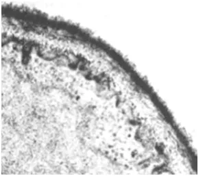
The ultrastructure of C. albicans cell wall has been observed using scanning electron microscopy (Fig. I.5) revealing the existence of different layers in the wall (Tokunaga, 1986; Osumi, 1998).
Figure I.5. Electron micrograph of a C.
albicans cell wall section.
The inner part appears to be electron transparent and consists mainly of β -1,3-glucan, chitin and β-1,6-glucan molecules bound by hydrogen bonds and with a small amount of proteins (Marcilla et al., 1991; Kapteyn et al., 2000). The outer part is mainly made of mannoproteins attached to the polysaccharide network that determine cell surface properties (Klis et al., 2001). All these features are represented in figure I.6.
[image:31.595.88.509.460.731.2]The cell wall is a dynamic structure that requires continuous remodelling of its components and structure in response to isotropic or apical growth, changes in environmental conditions, cell damage, etc. It has been estimated in S. cerevisiae that about 1200 genes cause a cell-wall related phenotype when deleted, supporting the notion that cell wall construction is an integral part of cell physiology (De Groot et al., 2001).
I.4.1.1- β-glucans
They are the most abundant polysaccharides of the fungi and also of the C. albicans cell wall. β-glucans are polymers of D-glucose moieties that can be classified depending on whether they are joined by β-1,3 and/ or β-1,6-glycosidic linkages.
I.4.1.1.1- β-1,3-glucan
β-1,3-glucan chains are made up of around 1500 glucose units per chain. These
polymers present a coiled microfibrillar structure that confers elasticity and tensile strength to the cell wall.
Studies carried out with C. albicans and S. cerevisiae provided evidence that initiation of synthesis of the polymer requires a protein acceptor. β-1,3-glucan chain growth involves a transglycosylation reaction of glucosyl residues from the cytoplasmic donor UDP-glucose to the growing polysaccharide chain (for a review see Ruiz-Herrera
et al., 2004).
In S. cerevisiae β-1,3-glucan synthase subunits have been identified. This enzymatic complex consists of a membrane-bound catalytic subunit (Fks1p and Gsc2p/Fks2p) and a cytosolic GTP-dependent regulatory subunit (Rho1p). FKS1 and
GSC2 are 88% homologs and have similar topology and organization. FKS1 and GSC2
show differential patterns of expression, Fks1p is the major player during vegetative growth on rich medium, with Gsc2p being the protein functioning under more stressful conditions. Fks1p and Fks2p form a redundant essential pair with a double fks1Δfks2Δ
Fks3p function has been related to sporulation and mating (Deutschbauer et al., 2002; Zeitlinger et al., 2003). Rho1p is an essential G protein that in their active GTP-bound state binds and activates its effectors (Fks1p and Gsc2p). In addition to its role as β -1,3-glucan synthase regulator, Rho1p plays an important role in cell wall organization by acting through the PKC cell integrity pathway (Qadota et al., 1996).
In C. albicans three FKS homologs to S. cerevisiae FKS1 have been found:
GSC1/CaFKS1, GSL1 and GSL2 (Mio et al., 1997a). The main activity appears to result from Gsc1p. Interestingly, both GSC1 and RHO1 encode for essential proteins in C. albicans (Mio et al., 1997a; Smith et al., 2002). Moreover, GSL1 and GSL2 are overexpressed during the de novo synthesis of the cell wall as revealed by a transcriptomic study along the protoplasts regeneration process (Castillo et al., 2006).
I.4.1.1.2- β-1,6-glucan
β-1,6-polymer is shorter than β-1,3-glucan, consisting in 350 units of glucose (Kollar et al., 1997). Its structure presents branch points on approximately 7% of the residues in C. albicans (Mio et al., 1997b), resulting in an highly branched structure that acts as a flexible glue by forming covalent cross-links to β-1,3-glucan and chitin and to cell wall mannoproteins (Kollar et al., 1997). Because β-1,6-glucan is the receptor for killer toxin K1, toxin killer resistant-(kre) mutants became an excellent tool for the identification of genes involved in its synthesis.
In S. cerevisiae, at least 10 genes involved in β-1,6-glucan have been identified:
KRE1, KRE5, KRE6, KRE9, KRE11, CNE1, CWH41/GLS1, KNH1, ROT2/GLS2 and
SKN1 (Shahinian and Bussey, 2000). It still remains unclear which synthetic pathway is involved in this process. Genetic and structural studies (Montijn et al., 1999) suggest that β-1,6-glucan synthesis may occur at the cell surface; however, components in the secretory pathway as Kre5p and Cwh41p, located in the ER, and Kre6p and Skn1p transmembrane Golgi proteins are required (Shahinian and Bussey, 2000). It has been shown that numerous genes are involved in the synthesis of this polymer: KRE5, CNE1,
chain by adding linear β-1,6-glucan onto a highly branched acceptor glucan (Boone et al., 1990); KRE9 and KNH1 whose encoded proteins could be related to anchoring or cross-linking the new synthesized β-1,6-glucan in the wall (Brown and Bussey, 1993; Shahinian and Bussey, 2000). Moreover, the GTP dependence of the reaction implies involvement of Rho1p GTP-binding protein in regulating β-1,6-glucan like β -1,3-glucan synthesis.
In C. albicans homologs of several KRE genes have been isolated, providing evidence that the synthesis of β-1,6-glucan occurs by similar mechanisms in both fungi.
I.4.1.2- Chitin
Chitin is a linear polysaccharide made of more than 2000 units of N-acetylglucosamine (GlcNAc) joined by β-1,4-linkages. These chains are associated in an antiparallel fashion through hydrogen bonds to form microfibrils composed of 20-400 chains. This kind of organization makes chitin insoluble, what explains why its linkages to β-1,3-glucan form the basic cell wall scaffold to which mannoproteins are associated.
Chitin is not mainly found in the lateral walls of the cell, but in the chitin ring in the neck of the mother cell, in the primary septum and in the bud scars. Different chitin synthases genes have been found in both S. cerevisae and C. albicans and their regulation is controlled spatially and temporally (Bulawa, 1993; Munro and Gow, 2001)
Synthesis of chitin involves a transglycosylation reaction of GlcNAc residues from the universal substrate UDP-N-acetylglucosamine to the growing chain of polysaccharide and requires a divalent metal, generally Mg2+.
Chitin synthase activity is accumulated in the cytosol of fungi, also in C. albicans, in specialized microvesicles, chitosomes, responsible for the transfer of the enzyme from its site of synthesis to its place of action (Ruiz-Herrera et al., 2004).
In C. albicans four genes encoding chitin synthases (CHS genes) have been described: CHS1, CHS2, CHS3 and CHS8. CHS1 gene expression presents low levels in both yeast and hyphae. Nevertheless, Chsp1p was found to be essential for cell integrity and virulence, apparently being involved in septum formation (Mio et al, 1996; Munro
Chs2p does not affect dimorphism, the levels of chitin in the cell wall or its virulence (Gow et al., 1994; Mio et al., 1996). CHS3 reaches its maximum level of expression during hyphal induction, but Chs3p is responsible for in vivo synthesis of most chitin in both yeast and hyphae (Mio et al., 1996). CHS8 encodes a protein similar to Chs2p.
chs8Δ/chs8Δ mutants display normal growth rates, cellular morphology and chitin content, but their chitin synthase activity is reduced to 25%, and a double homozygous mutant chs2Δ/chs2Δ chs8Δ/chs8Δ possesses less than 3% of the wild-type chitin synthase activity in vitro although has normal growth rate and morphology (Munro et al., 2003).
I.4.1.3- Mannoproteins
Linked to the cell wall polysaccharides are a varied set of glycoproteins. These glycoproteins consist in a protein with glycidic chains covalently attached. It has been shown in yeasts that the carbohydrate bound to the cell wall proteins (CWP) is mainly mannose, and phosphate groups have also been found (Ruiz-Herrera, 1992; Peberdy, 1999). The level of glycosylation is variable, often as high as 50-95% by weight.
The most important roles of cell wall proteins (CWP) in pathogenic fungi, as C. albicans, are:
(i) Enzymatic: some enzymes have been found in the cell wall to play different roles: degradation of impermeable molecules, making the products accessible for nutrition, one example of this is the acid trehalase Atc1p that is required for growth in trehalose (Pedreño et al., 2004); and remodelling of cell wall components, necessary for cell growth, which is carried out by several enzymes with glucanase, chitinase or transglycosidase activities (Chaffin et al., 1998).
(ii) Cell interaction: some cell wall proteins are involved in the contact of C. albicans with host tissues and some of their products such as fibrinogen, complement fragments and extracellular matrix components (Calderone, 1993), but also to abiotic surfaces. The best example for this function is the ALS gene family (see section I.3.1).
(iii)Antigenicity: several wall glycoproteins (not only the protein itself, but their mannan moieties) are antigenic, and are present differentially in the yeast or filamentous form (Sundstrom et al., 1988).
proteins (see section I.3.2.1) are responsible, among other functions, for the elimination of cells and molecules of the host immune system to resist antimicrobial attack by the host (Naglik et al., 2003; Albrecht et al., 2006). Superoxide dismutases (Sods) in C. albicans are located in the cell surface and turns superoxide radicals, produced by the host macrophages, into hydrogen peroxide, defeating one of the host’s defence mechanisms (Fradin et al., 2005).
(v) Structure and morphogenesis: other glycoproteins may be important from a structural point of view, like Ssr1p, a covalently-linked cell wall protein which lack on the cell wall does not produce strong alterations in the cell (Garcera et al., 2003; 2005); proteins with an important role in hypha formation, as Csf4p (Alberti-Segui et al., 2004) or Dfg5p (Casanova et al., 1990); or glycoproteins that have been suggested to be required for the correct assembly of cell wall components, some examples of these are Ecm33p (Pardo et al., 2004) and Pir1p (Martinez et al., 2004).
I.4.1.3.1- Classification of cell wall mannoproteins
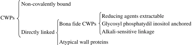
Two methodological approaches have allowed the identification of different cell wall proteins: (i) sequencing and in silico analysis of the whole C. albicans genome, and (ii) proteomic analysis. In C. albicans, these analyses have identified hundreds of putative cell wall proteins (De Groot et al., 2003; Garcera et al., 2003; Eisenhaber et al., 2004) that could be classified depending on the sort of linkage that remains them attached to the cell wall. A scheme of the different kinds of CWPs found in the cell wall is shown in figure I.7.
CWPs
Non-covalently bound
Directly linked
Bona fide CWPs Glycosyl phosphatydil inositol anchored Alkali-sensitive linkage
Reducing agents extractable
[image:36.595.90.494.590.685.2]Atypical wall proteins
I.4.1.3.1.1- Non-covalently linked (NCL-CWPs)
These proteins are bound to the cell wall through weak linkages (Fig. I.7) and they can be extracted by hot water, ionic detergents like SDS (Valentin et al., 1984; Valentin et al., 1987) or chaotropic agents as urea. However, some specific cell wall proteins can be also released using a hot detergent treatment, as is the case of the endoglucanase Bgl2p and the chitinase Cht1p (Capellaro et al., 1998) in S. cerevisiae. Normally, these proteins are not real components of the cell wall but contaminant proteins coming from plasma membrane fragments (Klis, 1994; Eroles et al., 1997; Cappellaro et al., 1998; Klis et al., 2001).
I.4.1.3.1.2- Reducing agents extractable (RAE-CWPs)
This group consists in proteins that are linked to other cell wall proteins through disulphide bridges (Fig. I.7), and consequently, they can be extracted from the cell wall by reducing agents as β-mercaptoethanol (β-ME) or dithiothreitol (DTT) as described by Moukadiri and Zueco (2001a).
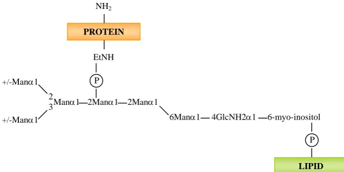
I.4.1.3.1.3- Glycosyl phosphatydil inositol anchored (GPI-CWPs)
GPI proteins appear to be the most abundant ones in C. albicans, representing about 88% of the covalently linked cell wall proteins.
PROTEIN
NH2
EtNH
P
2Manα1 2Manα1
6Manα1 4GlcNH2α1 6-myo-inositol
P
LIPID
Manα1 2 3
+/-Manα1
[image:38.595.124.471.73.247.2]+/-Manα1
Figure I.8- Glycosyl phosphatydil inositol anchor structure of yeast proteins.
EtNH, ethanolamine; (P), phosphate; Man, mannose. (Adapted from Sipos et al., 1995).
Shortly, after protein synthesis in the ER, a preformed GPI anchor is transferred to the C- terminus of the target proteins and replaces the hydrophobic tail (De Groot et al., 2003). The attachment reaction involves a proteolytic cleavage C terminal of the attachment residue and a transamidation between the generated amino acid and the ethanolamine phosphate of the GPI anchor (Tiede et al., 1999; De Groot et al., 2003).
These proteins are bound (through their GPI moiety) via β-1,6-glucan to either
β-1,3-glucan, on 90% of the cases, or to chitin on the remaining 10% (Kapteyn et al.,
2000). The extraction of these proteins can be performed by enzymatic digestion of the polysaccharide network of the cell wall with β-1,6-glucanases (Kapteyn et al., 2000), β -1,3-glucanases (Garcera et al., 2003), chitinases (Marcilla et al., 1991), or chemically by cleaving the phosphate bonds that link GPI-CWPs to β-1,6-glucan chains using hydrofluoric (HF)-pyridine (De Groot et al., 2004).
GPI anchoring is encountered in every eukaryotic cell, including unicellular yeast cells, parasites, and mammalian cells (McConville and Menon, 2000; Bowman et al., 2006). Different studies carried out in S. cerevisiae (Caro et al, 1997; Hamada et al., 1998) have revealed that approximately 70 GPI proteins are present in this organism. An in silico study has identified in C. albicans a total of 234 putative GPI proteins that can be bound to the cell wall or to the plasma membrane (Eisenhaber et al., 2004). In
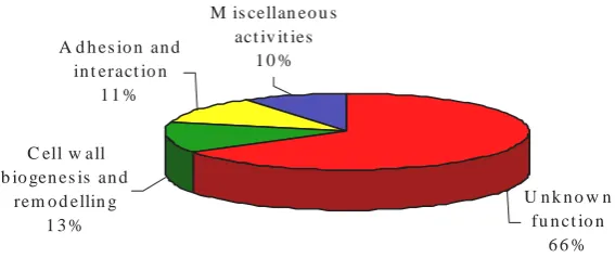
and have published a list of 115 putative GPI proteins corresponding to the C. albicans
genome. Among them, four classes can be described (Fig. I.9): (i) 76 of these putative proteins have unknown function, (ii) 15 proteins with functions related to cell wall biogenesis or remodelling, (iii) 13 related to cell-cell adhesion and other interactions, and (iv) 11 proteins presenting different enzymatic activities.
C ell w all b io gen es is an d
rem o d ellin g 1 3 %
A d h es io n an d in t eract io n
1 1 %
M is cellan eo u s act iv it ies
1 0 %
U n k n o w n fu n ct io n
[image:39.595.157.441.196.315.2]6 6 %
Figure I.9- Function distribution of GPI-CWPs in C. albicans.
(Adapted from Richard and Plaine, 2007).
It is worth to say that among the 76 proteins with unknown function, 41 of them have been shown to be regulated by various transcriptional factors and in different conditions. Some of the transcriptional factors regulating these GPI-protein genes are, Rim101p, Nrg1p, Tup1p, Efg1p, Cph1p and Ssn6p, (Bensen et al., 2004; Murad et al, 2001; Sohn et al., 2003; Setiadi et al., 2006; Garcia-Sanchez et al., 2005).
I.4.1.3.1.4- Alkali-sensitive linkage (ASL-CWPs)
They are a small group of proteins directly attached to the cell wall β-1,3-glucan by covalent linkages sensitive to alkaline treatment (Fig. I.7).
The most remarkable ASL-CWPs are Pir proteins (proteins with internal repeats). These proteins are rich in Ser and Thr residues and are thought to be highly glycosylated. They consist of (i) a putative N-terminal signal peptide, followed by a Kex2p endoprotease cleavage site, (ii) variable number of internal repeats matching the consensus pattern Q[IV]XDGQ[IVP]Q, and (iii) a four C-terminal cysteine-based motif (Toh-e et al., 1993).
Moukadiri and Zueco, 2001a; 2001b), Pir3p (Toh-e et al., 1993; Mrsa et al., 1997) and Pir4p (Moukadiri et al., 1999). Recently, a fifth member of this family, Pir5p, has been identified (M. Ecker, unpublished data). It has been also shown that some of them, Pir2p and Pir4p, can be also released to the culture media (Russo et al., 1992; Moukadiri et al., 2001a) or found attached to other cell wall proteins by disulphide bridges and extracted from the wall using reducing agents (Orlean et al., 1986; Moukadiri et al., 1999; Moukadiri and Zueco, 2001a). The PIR genes show cell-cycle dependent expression, Pir1p, Pir2p and Pir3p are highly expressed when the cells grow isotropically and the new cell wall components have to be assembled which is consistent with their supposed role in β-1,3-glucan cross-linking (De Nobel and Barnett, 1991).
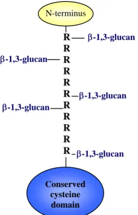
The nature of the linkage that attaches Pir proteins to the cell wall has been recently elucidated. The presence of these proteins linked to β-1,3-glucan that can be released by mild alkali treatment suggested that a glycosidic bound links the β -1,3-glucan and a mannose residue of the O-linked chains of the Pir proteins (Klis et al., 2002). Castillo et al. (2003) have shown by mutational analysis that the repeat sequences are required for binding to β-1,3-glucan. If these repeats, containing the “linking” sequence, are directly involved in binding β-1,3-glucan, then Pir-CWPs may interconnect two or more β-1,3-glucan molecules as represented in figure I.10. Recent studies made with Pir4p have identified the amino acid responsible for the binding of this protein to β-1,3-glucan. Pir4p is attached to the cell wall via glutamine residue 74, within the repeat sequence QIGDGQ74VQ (Ecker et al., 2006). Although which enzyme catalyzes the reaction is still unknown, it has been postulated that an ester linkage between the γ-carboxyl of a glutamate residue, arising from a glutamine, and the sugar hydroxyl is formed (Ecker et al., 2006). This ester bond would be expected to be alkali labile, which is consistent with the knowledge that Pir proteins are extractable from the cell wall using mild alkali treatments.
C. albicans Pir proteins are: (i) the two polypeptides codified by IPF19968 and
IPF15363 are not processed by the endoprotease Kex2p, and (ii) the spacing among cysteines within the cysteines domain is –C-X65-C-X16-C-X12-C-X0-COOH in C. albicans, whereas in S. cerevisiae is –C-X66-C-X16-C-X12-C- X0-COOH. Heterozygous mutants cells in each of the two alleles of PIR1 gene are elongated, form clumps of several cells and are hypersensitive to drugs that affect cell wall assembly. The homozygous mutant has not been obtained yet, suggesting an essential role for PIR1 in
C. albicans (Martinez et al., 2004).
N-terminus
R R R R R R R R R R R
Conserved cysteine
domain
β-1,3-glucan
β-1,3-glucan
β-1,3-glucan β-1,3-glucan
[image:41.595.218.358.256.474.2]β-1,3-glucan
Figure I.10- Internal repeats of Pir proteins may act as crosslinkers between β-1,3-glucan chains. R, internal repeats of the consensus sequence -(Q[I/V]XDGQ[I/V/P]Q)-. (Adapted from Klis et al., 2006).
I.4.1.3.1.5- Atypical wall proteins
Whether these proteins are somehow trapped within the cell wall components or they arrive to the cell wall through a non-classical transport pathway is still unknown.
Putting together all the cell wall components described above, a model of the yeasts cell wall organization and assembly has been developed by the European Consortium Galar Fungail (Fig. I.11).
β-1,3-glucan
β-1,6-glucan β-1,6-glucan
β-1,6-glucan
Chitin Chitin
Gcw
Gcw Gcw
Asl Asl
Ncl Ncl
Rae
Rae
GPI GPI GPI
S
S
S
[image:42.595.124.474.229.479.2]S
Figure I.11- Molecular organization of the cell wall of C. albicans. Gcw, covalently linked GPI-cell wall protein; GPI, glycosyl phosphatidil inositol remmant; Asl, Alkali-sensitive linked cell wall protein; Rae, reducing agents extractable cell wall protein; Ncl, Non-covalently linked protein. Model developed for the Galar Fungail I European Consortium.
I.4.1.3.2- Post-translational modifications of cell wall proteins
Once the cell wall proteins have been synthesized, several post-translational events may occur to make them fully functional and to drive them to the cell surface where they will be anchored to play their specific role.
appropriately modified by addition of a GPI remnant (see section I.4.1.3.1.3) and glycosyl residues (see below). Then the mature proteins are transported to the cell surface in secretion vesicles that merge with the plasma membrane and pour their content outside the cell to favour the attachment of the glycoproteins to the cell wall.
I.4.1.3.2.1- N-glycosylation
N-glycosylation of proteins is present in every eukaryotic cell. In C. albicans it has been shown that this N-linked mannan is important for adhesion to host and surfaces and is also involved in generating the immune response of the host in Candida
infections.
All N-modified glycoproteins acquire the initial oligosaccharide in the ER onto an asparagine residue of the sequence N-X-S-T (where X represents any amino acid except proline), through a direct bond between this asparagines and β -1,4-diacetylchitobiose. Then, cell wall and secreted mannoproteins are extensively mannosylated in the Golgi with a final structure of α-1,6-linked mannose chain of up to 50 mannose residues extending from the N-glycan core and to which are attached shorter chains of α-1,2 residues terminating in α-1,3-linked mannose residues, forming a highly branched structure containing as many as 200 mannose residues (Ballou, 1990; Dean, 1999; reviewed by Cutler, 2001).
N-glycosylation in yeasts has been elucidated using mutant strains in different steps of the process: (i) mnn mutants, mannan synthesis (Ballou, 1990); (ii) alg mutants, asparagines-linked glycosylation deficient (Kukuruzinska et al., 1999), and (iii) och
mutants, outer chain-deficient (Nagasu et al., 1992).
I.4.1.3.2.2- O-glycosylation
O-glycosylation of cell wall proteins is more abundant than N-glycosylation, although the grade of addition of mannose molecules is lower. Despite the small size of the O-linked chains, the number of O-chains per protein can be high and the amount of
O-linked mannose in the cell wall significant (Strahl-Bolsinger et al., 1999).
-mannosyltransferases (PMT family) (Ernst and Prill, 2001). Subsequent steps occur in the Golgi and are catalyzed by mannosyltransferases (Mnt) or mannansynthases (Mnn). The final molecule contains in two residues of mannose being α-1,2-linked and subsequent ones (in C. albicans can be up to five) α-1,3-linked (Jigami and Odani, 1999).
The C. albicans PMT family, consisting in 5 members (Pmt1p, Pmt2p, Pmt4p, Pmt5p, Pmt6p), has been shown to be related not only to O-glycosylation of proteins, but also to morphogenesis and antifungal resistance (Ernst and Prill, 2001; Prill et al., 2005).
I.4.2- Cell wall remodelling
As stated the cell wall is a dynamic structure that needs to be restructured through the activity of different remodelling enzymes during cell cycle and in response to environmental changes or different growth programs.
The cellular localization of these enzymes within the cell wall is compatible with their having important roles as cell wall remodelling proteins.
I.4.2.1- Glucan remodelling enzymes
The product synthesized by β-1,3-glucan synthases is linear. However, the branched structure of the mature β-1,3-glucan network requires shortening of the polymer by nicks and the introduction of polymer branch points. Such reactions are catalized by exo- and endoglucanase activities (Adams, 2004).
In C. albicans some of these enzymes are encoded by PHR1, PHR2, PHR3,
PGA4, PGA5, BGL2, SCW1, SCW11, EXG1 and EXG2 genes (Fonzi, 1999; Eckert et al., 2006; Sarthy et al., 1997; Gonzalez et al., 1997; Stubbs et al., 1999).
I.4.2.2- Chitin remodelling enzymes