DOI 10.1007/s00402-013-1902-7
TrAumA Surgery
Long‑term results of the augmented PFNA: a prospective
multicenter trial
C. Kammerlander · H. Doshi · F. Gebhard · A. Scola · C. Meier · W. Linhart · M. Garcia‑Alonso · J. Nistal · M. Blauth
received: 11 September 2013
© Springer-Verlag Berlin Heidelberg 2013
Methods In 5 european clinics, 62 patients (79 % female, mean age 85.3 years) suffering from an osteoporotic per-trochanteric fracture (AO 31) were treated with the aug-mented PFNA®. The primary objectives were assessment
of activities of daily living, pain and mobility. Furthermore, the X-rays were analyzed for the cortical thickness index, changes of the trabecular structure around the cement and the hip joint space.
Results The mean follow-up time was 15.3 months. We observed callus healing in all cases. The surgical compli-cation rate was 3.2 % with no complicompli-cation related to the cement augmentation. A mean volume of 3.8 ml of cement was injected and no complication was reported due to this procedure. 59.9 % reached their prefracture mobility level until follow-up. The mean hip joint space did not change significantly until follow-up and there were no signs of osteonecrosis in the follow-up X-rays. Furthermore, no blade migration was assessed.
Conclusion This study makes us believe that the stand-ardized augmentation of the PFNA with a perforated blade is a safe method to treat pertrochanteric femoral fractures. It leads to good functional results and is not associated with cartilage or bone necrosis.
Keywords Hip fracture · Augmentation · PmmA · Cement leakage · PFNA · Cut-out · Cement augmentation · Osteoporosis · Cortical thickness index · Cement
distribution · Hip joint space · Osteonecrosis · Long-term follow-up · Parker mobility Score · Pertrochanteric fracture
Introduction
The demographic development leads to an increase of fragility hip fractures and their treatment is still under Abstract
Background Pertrochanteric fractures are increasing and their operative treatment remains under discussion. Fail-ures needing reoperations such as a cut-out are reported to be high and are associated with multiple factors including poor bone quality, poor fracture reduction and improper implant placement. The PFNA® with perforated blade
offers an option for standardized cement augmentation with a PmmA cement to provide more stability to the fracture fixation. It remains unclear if the augmentation of this implant does any harm in a longer time span. This prospective multicenter study shows clinical and radiologi-cal results with this implant with a mean follow-up time of 15 months.
C. Kammerlander (*) · H. Doshi · m. Blauth Department of Trauma Surgery and Sports medicine, medical university of Innsbruck, Anichstrasse 35, 6020 Innsbruck, Austria
e-mail: [email protected]; [email protected]
F. gebhard · A. Scola
Department of Traumatology, Hand-, Plastic-, and reconstructive Surgery, Center of Surgery, Center of musculoskeletal research, university of ulm, ulm, germany
C. meier
Department of Traumatology, Stadtspital Waid, Zurich, Switzerland
W. Linhart
Department of Orthopedics and Trauma Surgery, SLK Kliniken Heilbronn, Heilbronn, germany m. garcia-Alonso · J. Nistal
discussion. Stable A1 fractures are regularly stabilized with a Dynamic Hip Screw [32], whereas there is an increasing trend for intramedullary devices in the treatment of unsta-ble A2 and A3 fractures [1, 2, 26, 30, 39].
Literature shows that screw designs for the cephalic part of the implant come up with failure rates needing revi-sion surgery of up to 16 % [2, 24, 25, 38]. recent stud-ies showed that the PFNA with its blade for the cephalic part of the implant is effective for pertrochanteric fracture fixation [26, 35] with low failure rates of 0.6–3.6 % [20, 26, 35]. It has to be mentioned that these investigations [20, 26, 35] were not limited to the elderly where an underlying osteoporosis could lead to even higher failure rates. Several biomechanical studies have already proven the mechanical superiority of using a blade instead of a screw in osteoporo-tic bone [13] and the fixation construct gets even more sta-ble with the use of an additional augmentation with poly-methylmethacrylate (PmmA) cement in fact independently of the position of the blade within the head-neck element [8–11, 33, 34]. From the clinical side, the use of an aug-mented blade is known to lead to good results in short time [16].
The main concerns about such a device were a potential disturbance of bone metabolism [14, 19, 37] and the induc-tion of cartilage damage although there are several investi-gations rebutting this [4, 41, 43]. The current prospective multicenter trial was conducted to evaluate the long-term clinical outcome as well as the radiological results with the standardized augmentation of the PFNA. To our knowl-edge, this is the first investigation reporting long-term results with this device.
Materials and methods
The study was performed at five orthopedic departments starting in October 2009 and the last follow-up examina-tion was in may 2013. The inclusion criteria were as fol-lows: pertrochanteric fracture (AO/OTA 31A), age 65 and above, low energy trauma and signed informed consent. Patients with a pathological fracture, active malignancy or organ transplant were excluded. The ethical commis-sion approved the study and every single patient signed the informed consent form. A total number of 110 patients have been included. 25 (22.7 %) cases were lost to follow-up due to refusion, concurrent indisposition or weakness which made an additional evaluation impossible. 23 patients died for reasons not related to the surgical procedure. The remaining 62 patients were followed up according to the study protocol. The decision to augment was made by the surgeon without using strict objective measurements.
The surgical technique is described in the first
publica-Figure 1a–d shows a representative case of standardized cement augmentation of the perforated PFNA blade in an 82-year-old lady with an unstable pertrochanteric fracture with a 1-year follow-up X-ray.
Outcome parameters
The WHO Performance Score [28] was used to measure the quality of life before and after the fracture. It consists of five levels in which 0 means full activity without restric-tion and 4 means completely disabled and totally confined to bed or chair.
The Parker mobility Score [29] was used to assess the walking ability before the accident and at the follow-up. The particular capability to walk inside, walk outside and having social contact is evaluated in 4 levels with “no dif-ficulty”, “alone”, “with help from another person” and “not at all”. A maximum of nine points means unlimited walk-ing ability. In addition, the use of a walkwalk-ing aid was docu-mented for every patient before and after the accident.
Pain was assessed using the visual analog scale (VAS) as previously described [6] and widely known. The VAS was found to have good measurement properties for assessing pain in hip fracture patients [5].
The fracture pattern was assessed and classified as AO. The cortical thickness index [7] was assessed in the pre-operative and in the follow-up X-rays. The cortical thick-ness index shows a significant positive correlation with the T-Score of the femoral neck [31] and was therefore used to classify the local bone quality in our study population. A cortical thickness index lower than 0.40 (lateral film) and 0.50 (ap film) has been described as a threshold for osteo-porosis where all measured femora had a lower local bone mineral density than 2.5 standard deviations below the peak bone mass which is the WHO definition of osteopo-rosis [31].
On the postoperative X-rays, we evaluated the quality of fracture reduction as anatomic (no displacement), near-anatomic (<3 mm displacement or 5°–10° varus/valgus and/or anteversion/retroversion) or non-anatomic (>3 mm displacement or >10° varus/valgus and/or anteversion/ret-roversion) [20, 39]. The amount of injected cement was documented, and on the follow-up X-rays, signs of frac-ture healing were assessed. The lateral blade migration was measured as previously described [12, 42].
Intraoperative complications included any unforeseen event during the augmentation such as perforation with the guide wire into the hip joint and cement leakage. Potential postoperative complications were cutting out of the blade from the femoral head, cutting through the blade centrally, any unexpected blade migration, loosening of the blade, implant breakage, infection, additional fracture or bone-healing disturbances and any other general complication within the follow-up period.
Statistical analysis
SPSS 20 (SPSS Inc., Chicago, IL) was used for statisti-cal analysis. All baseline and follow-up parameters were described using standard descriptive statistics. metric scaled data are reported as arithmetic mean and categori-cal data as absolute frequency and percentage distribution. Depending on the distribution form, a t test for independent
Fig. 1 a An 82-year-old female patient with an unstable fracture 31 A2 at her left side after a simple fall in the nursing care home. Intraoperative image intensifier picture after closed reduction and internal fixation with the PFNA and standard-ized cement augmentation of the PFNA blade in ap (b) and lateral (c) views. d Follow-up X-rax after 1 year shows a well-healed fracture and no signs of osteonecrosis of the femoral head
variables or a nonparametric mann–Whitney U test was used. The Kolmogorov–Smirnov test was used to assess the distribution form. A Chi-square test or a Fisher’s exact test was used to analyze categorical data. The probability level was set as p < 0.05.
Results
To test the long-term effects of standardized cement aug-mentation, 62 patients were analyzed. The mean time to follow-up was 15.3 months (365–887 days). The demo-graphics are shown in Table 1. Associated injuries were not noted. The majority of the patients sustained an unstable pertrochanteric fracture (A2/3; 83.9 %). The mean Parker Score was 5.1 before and 4.6 at follow-up. 59.6 % of our patients reached their prefracture mobility level within the follow-up time. The mean WHO Perfor-mance Score was 2 before the fracture occurred and again 2 at follow-up, whereas 70.1 % reached their prefracture WHO Performance Score again. Table 2 shows the main functional outcome parameters split up for the single frac-ture types.
The mean VAS at follow-up was 0.5. All patients were osteoporotic, whereas the mean cortical thickness index (CTI AX) was 0.44 at the time of the fracture and dropped non-significantly (p = 0.86) to 0.42 at follow-up.
The follow-up X-rays showed an anatomic reduction in 59.7 %, whereas in 24.2 %, the reduction was near-ana-tomic and in 1.6 % non-ananear-ana-tomic. An open reduction was necessary in three cases, whereas in 2 cases subtrochanteric
cerclage wires were used to stabilize the reduction and were not removed.
There was one patient with a postoperative haematoma and one patient with a superficial wound infection both requiring a reoperation. One patient had a postoperative pneumonia and two patients a urinary tract infection which required antibiotic treatment. The presence of any of these complications was found to have a significantly negative effect (p = 0.026) on the Parker mobility Score at follow-up.
There was no intraoperative complication reported. In the present study, a mean volume of 3.8 ml (3–6) cement was injected. In all cases, a leakage test was done to detect a perforation into the hip joint and no intraarticular cement was observed.
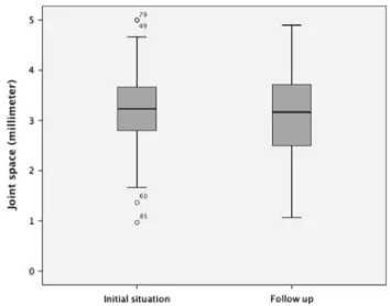
At follow-up, all fractures were healed. No sign of oste-onecrosis of the femoral head or lysis around the cement could be detected. According to the above-mentioned method, the mean joint space showed no significant dif-ference at follow-up (p = 0.44) which is shown in the boxplot in Fig. 3. There was no implant migration (e.g., migration of the blade related to the femoral head) meas-urable. Lateral blade migration averaged 5.2 mm. Lateral blade migration was not found to be associated with frac-ture type (p = 0.86) nor with implant position (p = 0.17), fracture reduction (p = 0.45) or Parker Score at follow-up (p = 0.89). We did not find any unexpected blade migration such as a cut-out, implant loosening or implant breakage within the study period.
Discussion
The PFNA was widely described to be a reliable implant to fix pertrochanteric fractures [18, 20, 26, 35] and the
Table 1 The baseline characteristics of the study population
All (n = 62) %
Age, mean 85.3
Female/male 49/13 79/21
Left/right 36/26 58.1/41.9
AO 31-A1 10 16.1
AO 31-A2 44 71
AO 31-A3 8 12.9
Hospitalization time, mean 12 days
Table 2 The main functional outcome parameters separated for cer-tain fracture types
WHO Perfor-mance Score preoperatively
WHO Perfor-mance Score at follow-up
Parker Score preoperatively
Parker Score at follow-up
A1 2 2.3 4.2 3.2
A2 1.9 2 5.8 4.6
follow-augmented version was previously reported to lead to good short-term functional results [16]. Furthermore, the aug-mentation of the blade gives the fixation construct much more stability due to a larger bone–implant interface [8–11, 37, 40].
59.5 % of our cohort reached their prefracture functional status until the final follow-up. This is an excellent long-term outcome for this age group compared to the literature [17, 20, 26, 35].
Failures requiring reoperation such as a cutting out off the femoral head are still reported to be as high as up to 16 % [2, 25, 29, 36] with screw devices and up to 5.7 % [18] with the PFNA. In this context, it is worth to notice that this affects a high number of people due to the increas-ing incidence of pertrochanteric fractures. In the elderly, where most of these fractures occur, every complication leads to a high perioperative morbidity and mortality due to their little reserves and comorbidities. Studies of the follow-up X-rays showed no unexpected blade migration such as a cut-out or cutting through and no loosening of the blade in this series. These facts suggest that the additional cement augmentation may have prevented the blade from cutting out off the femoral head in this present study.
The surgical complication rate in the presented study was 3.2 % with no complication related to the cement aug-mentation. This is an acceptable rate compared to other reports in the literature [20, 35].
Within the study period in one patient, an additional fall resulted in a subsequent fracture at the tip of the nail.
In this case, the short nail was changed to a long and both the removal of the blade and the reinsertion of a new blade were performed without any complication. unfortunately, the patient died 4 weeks after the second operation due to myocardial infarction. The mean lateral blade migration was 5.2 mm (±4.6) and led in two cases to an irritation of the iliotibial tract with consecutive blade removal (Fig. 4). Both procedures were done without a problem.
The mean cortical thickness index as by Sah et al. [31] was measured in the fracture X-ray and at the final follow-up. It indicated that all patients first suffered from osteopo-rosis and showed no significant change at follow-up.
A mean volume of 3.8 ml of cement was injected and there were no significant associations between the amount of cement and pain at follow-up or functional recovery. Furthermore, the joint space showed no significant change within the study period. Hisatome et al. [15] report a nega-tive influence of PmmA cement to the cartilage if it is placed in a subchondral area where an exothermic reaction while hardening of the cement is proposed to be a possible reason for this finding [14]. Furthermore, a bone necrosis could be imaginable if higher temperatures are reached [14, 19]. Fliri et al. [11] showed that the Traumacem V+® leads
to maximum temperatures of 41 °C at the cement–bone interface when instilled a volume of 3 ml around the blade. The same study also showed that the reached temperature is dependent on the amount of injected cement. For the present study, a lower amount of cement was used (mean 3.8 ml) and it is assumed that this amount does not lead to
negative impact on both bone and cartilage as previously described [4, 43]. Our findings that there were no radiologi-cal signs of osteonecrosis at follow-up and that there was no significant change of the joint space support this state-ment. On the other side, this small volume is enough to biomechanically enhance stability significantly [4, 19, 23, 33, 34, 40]. A possible limitation to the statement about bone necrosis is that previous authors reported an onset after 3 years [3, 22, 27].
The main limitation of this study is the lack of a con-trol group. However, an acceptable long-term outcome of the augmented PFNA could be shown. Furthermore, there were no hints of cartilage damage or bone necrosis within the follow-up period of 15 months. Thus, standardized aug-mentation of the PFNA blade seems to be a safe and feasi-ble method to treat pertrochanteric femoral fractures. From the socio-economic perspective, additional costs have to be taken into account and the number of hip fractures to treat with an augmented PFNA to prevent one failure remains unclear. In this context, it has to be noted that additional cement may not be necessary in stable A1 fractures fixed with a PFNA. To our personal opinion, the standardized augmentation of an unstable pertrochanteric fracture in an osteoporotic bone in the elderly who are known to have several comorbidities and little reserves can provide more safety for the overall treatment. Nevertheless, achieving a good fracture reduction and careful implant placement are essential [21, 26, 35].
A randomized trial comparing geriatric patients with unstable pertrochanteric fractures with a PFNA either with or without augmentation has been initiated and will help identify patients at risk for surgical failures and define the indications for augmentation more precisely.
Conclusion
The standardized augmentation of the PFNA with perfo-rated blade is a safe method to treat pertrochanteric femo-ral fractures. It prevents migration of the implant within the head-neck fragment and leads to good functional results without causing cartilage damage or bone necrosis.
Conflict of interest No benefits in any form have been received or will be received from a commercial party related directly or indirectly to the subject of this article.
References
1. Ahrengart L, Törnkvist H, Fornander P et al (2002) A rand-omized study of the compression hip screw and gamma nail in 426 fractures. Clin Orthop relat res 401:209–222
2. Anglen JO, Weinstein JN (2008) Nail or plate fixation of
intertro-the American Board of Orthopaedic Surgery Database. J Bone Jt Surg Am 90(4):700–707
3. Barnes r, Brown JT, garden rS, Nicoll eA (1976) Subcapital fractures of the femur. A prospective review. J Bone Jt Surg Br 58(1):2–24
4. Boner V, Kuhn P, mendel T, gisep A (2009) Temperature evalua-tion during PmmA screw augmentaevalua-tion in osteoporotic bone—an in vitro study about the risk of thermal necrosis in human femoral heads. J Biomed mater res B Appl Biomater 90(2):842–848 5. Bryant Dm, Sanders DW, Coles CP, Petrisor BA, Jeray KJ,
Laflamme gy (2009) Selection of outcome measures for patients with hip fracture. J Orthop Trauma 23(6):434–441
6. Carlsson Am (1983) Assessment of chronic pain. I. Aspects of the reliability and validity of the visual analogue scale. Pain 16(1):87–101
7. Dorr LD, Faugere mC, mackel Am, gruen TA, Bognar B, mal-luche HH (1993) Structural and cellular assessment of bone qual-ity of proximal femur. Bone 14(3):231–242
8. erhart S, Kammerlander C, el-Attal r, Schmoelz W (2012) Is augmentation a possible salvage procedure after lateral migra-tion of the proximal femur nail antirotamigra-tion? Arch Orthop Trauma Surg 132(11):1577–1581
9. erhart S, Schmoelz W, Blauth m, Lenich A (2011) Biomechani-cal effect of bone cement augmentation on rotational stability and pull-out strength of the proximal femur nail antirotation. Injury 42(11):1322–1327
10. Fensky F, Nuchtern JV, Kolb JP et al (2013) Cement augmenta-tion of the proximal femoral nail antirotaaugmenta-tion for the treatment of osteoporotic pertrochanteric fractures—a biomechanical cadaver study. Injury 44(6):802–807
11. Fliri L, Lenz m, Boger A, Windolf m (2012) ex vivo evaluation of the polymerization temperatures during cement augmentation of proximal femoral nail antirotation blades. J Trauma Acute Care Surg 72(4):1098–1101
12. gardner mJ, Briggs Sm, Kopjar B, Helfet DL, Lorich Dg (2007) radiographic outcomes of intertrochanteric hip fractures treated with the trochanteric fixation nail. Injury 38(10):1189–1196
13. goffin Jm, Pankaj P, Simpson AH, Seil r, gerich Tg (2013) Does bone compaction around the helical blade of a proximal femoral nail anti-rotation (PFNA) decrease the risk of cut-out? A subject-specific computational study. Bone Jt res 2(5):79–83 14. Heini PF, Franz T, Fankhauser C, gasser B, ganz r (2004)
Fem-oroplasty-augmentation of mechanical properties in the osteo-porotic proximal femur: a biomechanical investigation of PmmA reinforcement in cadaver bones. Clin Biomech (Bristol, Avon) 19(5):506–512
15. Hisatome T, yasunaga y, Ikuta y, Fujimoto y (2002) effects on articular cartilage of subchondral replacement with polymeth-ylmethacrylate and calcium phosphate cement. J Biomed mater res 59(3):490–498
16. Kammerlander C, gebhard F, meier C et al (2011) Standardised cement augmentation of the PFNA using a perforated blade: a new technique and preliminary clinical results. A prospective multicentre trial. Injury 42(12):1484–1490
17. Kammerlander C, gosch m, Kammerlander-Knauer u, Luger TJ, Blauth m, roth T (2011) Long-term functional outcome in geriatric hip fracture patients. Arch Orthop Trauma Surg 131(10):1435–1444
18. Lenich A, Vester H, Nerlich m, mayr e, Stockle u, Fuchtmeier B (2010) Clinical comparison of the second and third generation of intramedullary devices for trochanteric fractures of the hip— blade vs screw. Injury 41(12):1292–1296
20. Liu y, Tao r, Liu F et al (2010) mid-term outcomes after intramedullary fixation of peritrochanteric femoral fractures using the new proximal femoral nail antirotation (PFNA). Injury 41(8):810–817
21. Lobo-escolar A, Joven e, Iglesias D, Herrera A (2010) Predictive factors for cutting-out in femoral intramedullary nailing. Injury 41(12):1312–1316
22. Loizou CL, Parker mJ (2009) Avascular necrosis after internal fixation of intracapsular hip fractures; a study of the outcome for 1023 patients. Injury 40(11):1143–1146
23. mattsson P, Alberts A, Dahlberg g, Sohlman m, Hyldahl HC, Larsson S (2005) resorbable cement for the augmenta-tion of internally-fixed unstable trochanteric fractures. A pro-spective, randomised multicentre study. J Bone Jt Surg Br 87(9):1203–1209
24. mattsson P, Larsson S (2003) Stability of internally fixed femo-ral neck fractures augmented with resorbable cement. A pro-spective randomized study using radiostereometry. Scand J Surg 92(3):215–219
25. mattsson P, Larsson S (2004) unstable trochanteric fractures augmented with calcium phosphate cement. A prospective rand-omized study using radiostereometry to measure fracture stabil-ity. Scand J Surg 93(3):223–228
26. mereddy P, Kamath S, ramakrishnan m, malik H, Donnachie N (2009) The AO/ASIF proximal femoral nail antirotation (PFNA): a new design for the treatment of unstable proximal femoral frac-tures. Injury 40(4):428–432
27. murphy AJ, ricketts D, Thomas Wg (1995) Avascular necrosis of the femoral head following pertrochanteric fracture. Injury 26(5):351–352
28. Oken mm, Creech rH, Tormey DC et al (1982) Toxicity and response criteria of the eastern Cooperative Oncology group. Am J Clin Oncol 5(6):649–655
29. Parker mJ, Palmer Cr (1993) A new mobility score for predict-ing mortality after hip fracture. J Bone Jt Surg Br 75(5):797–798 30. Pervez H, Parker mJ, Vowler S (2004) Prediction of fixation
fail-ure after sliding hip screw fixation. Injury 35(10):994–998 31. Sah AP, Thornhill TS, Leboff mS, glowacki J (2007) Correlation
of plain radiographic indices of the hip with quantitative bone mineral density. Osteoporos Int 18(8):1119–1126
32. Saudan m, Lubbeke A, Sadowski C, riand N, Stern r, Hoffmeyer P (2002) Pertrochanteric fractures: is there an advan-tage to an intramedullary nail?: A randomized, prospective study of 206 patients comparing the dynamic hip screw and proximal femoral nail. J Orthop Trauma 16(6):386–393
33. Sermon A, Boner V, Boger A et al (2012) Potential of polymeth-ylmethacrylate cement-augmented helical proximal femoral nail antirotation blades to improve implant stability–a biomechanical investigation in human cadaveric femoral heads. J Trauma Acute Care Surg 72(2):e54–e59
34. Sermon A, Boner V, Schwieger K et al (2012) Biomechanical evaluation of bone-cement augmented Proximal Femoral Nail Antirotation blades in a polyurethane foam model with low den-sity. Clin Biomech (Bristol, Avon) 27(1):71–76
35. Simmermacher rK, Ljungqvist J, Bail H et al (2008) The new proximal femoral nail antirotation (PFNA) in daily practice: results of a multicentre clinical study. Injury 39(8):932–939 36. Simpson AH, Varty K, Dodd CA (1989) Sliding hip screws:
modes of failure. Injury 20(4):227–231
37. Stoffel KK, Leys T, Damen N, Nicholls rL, Kuster mS (2008) A new technique for cement augmentation of the sliding hip screw in proximal femur fractures. Clin Biomech (Bristol, Avon) 23(1):45–51
38. Takigami I, Ohnishi K, Ito y et al (2011) Acetabular perforation after medial migration of the helical blade through the femoral head after treatment of an unstable trochanteric fracture with proximal femoral nail antirotation (PFNA): a case report. J Orthop Trauma 25(9):e86–e89
39. Vidyadhara S, rao SK (2007) One and two femoral neck screws with intramedullary nails for unstable trochanteric frac-tures of femur in the elderly—randomised clinical trial. Injury 38(7):806–814
40. von der Linden P, gisep A, Boner V, Windolf m, Appelt A, Suhm N (2006) Biomechanical evaluation of a new augmenta-tion method for enhanced screw fixaaugmenta-tion in osteoporotic proximal femoral fractures. J Orthop res 24(12):2230–2237
41. von Steyern FV, Kristiansson I, Jonsson K, mannfolk P, Hein-egard D, rydholm A (2007) giant-cell tumour of the knee: the condition of the cartilage after treatment by curettage and cementing. J Bone Jt Surg Br 89(3):361–365
42. Watanabe y, minami g, Takeshita H, Fujii T, Takai S, Hirasawa y (2002) migration of the lag screw within the femoral head: a comparison of the intramedullary hip screw and the gamma Asia-Pacific nail. J Orthop Trauma 16(2):104–107