KEY WORDS: Chlorhexidine, Microgel, N-Isopropylacrylamide, Thermosensitivity. * Author to whom correspondence should be addressed. E-mail:[email protected]
Lat. Am. J. Pharm. 30(8): 1481-6 (2011)
Revised version: June 6, 2011 Accepted: June 9, 2011
The Deposition of Chlorhexidine on Chemically Modified
Thermosensitive PolyNIPA Microgels Assessed by EDXS
in Scanning Electron Microscopy
Witold MUSIAL*
Chair and Department of Pharmaceutical Technology, Faculty of Pharmacy, Wroclaw Medical University, 50-139 Wroclaw, ul. Szewska 38, Poland
SUMMARY. The deposition of chlorhexidine base on thermosensitive N-isopropylacrylamide polymeric
microparticles was assessed in this study using energy dispersive X-ray spectrometry (EDXS) in scanning electron microscopy (SEM) setting. Three different types of polymer were synthesized. The PN1 was a polymer with terminal anionic groups resulting from potassium persulphate initiator. The PN2 was syn-thesized with 2,2’-azobis(2-methylpropionamidine)dihydrochloride, what resulted in cationic amidine ter-minal groups. The PN3 had anionic terter-minals, however increased hydrophobicity was maintained with N-tert-butylacrylate functional groups. The thermosensitivity of the polymer-chlorhexidine complexes was confirmed by the turbidimetric assay. The deposition patterns, observed in/on the polymers give the as-sumption to develop the applications of evaluated polymers as factors influencing chlorhexidine release from the topical formulations.
INTRODUCTION
Chlorhexidine is widely used in dentistry, and in limited range in dermatological applica-tions, as well as disinfectant in medical prophy-lactic procedures, due to its efficient activity against Gram-positive and Gram-negative bacte-ria, as well as against fungi and enveloped viruses 1-3. High topical levels of chlorhexidine may be toxic, however in usually applied con-centrations it is well tolerated by mucosa and the skin. However the local increase of chlorhexidine concentration in the mucosal area or on dysfunctional area of the skin, especially in long term therapy, may lead to unacceptable levels of chlorhexidine also in the central com-partment. Due to this fact new forms of chlorhexidine are developed, with specific poly-meric carriers - synthesized or formed from nat-ural polymers. Reddy et al. proposed gelatin bioresorbable chips to enhance bone gain for periodontal regeneration 4. Interesting form of carrier for prolonged release of chlorhexidine evaluated Meng et al. –the chlorhexidine acetate is intercalated in montmorillonite, a mineral ap-plied as a filler in thermal dressings– the interca-lated complex chlorhexidine-montmorillonite strongly inhibited growth of wide spectrum of pathogens 5. Copolymers of hydroxyethyl
methacrylate and methyl methacrylate were syn-thesized and applied to fabrication of a mem-brane-controlled, reservoir-type controlled-re-lease delivery system to enable intraoral use of chlorhexidine 6. New polymeric carriers were synthesized using urethane dimethacrylate-tri-ethylene glycol dimethacrylate resin system 7. Some authors evaluated special inclusion com-plexes based on porous silica and cyclodextrins 8. Other porous systems were evaluated form methacrylate derivative polymer 9. The research on chlorhexidine carriers for local delivery of this disinfectant to mucosa and skin is still an in-teresting field of study. In our previous work we evaluated systems with chlorhexidine based on methylcellulose and polyacrylic acid 10. Some at-tempts were also made to perform preliminary assessment of chlorhexidine release from N-iso-propylacrylamide beads, however the placement of chlorhexidine in the microgel still was not elucidated 11.
swelled microgels may contain up to 95 % of water, whereas in the deswollen state the water quantity decreases to ca. 20 % of the mass of polymeric matrix. Between numerous ther-mosensitive entities poly(N-isopropylacrylamide) (polyNIPA) is one of the most utilized macro-molecules, characterized by reversible volume phase transition at specified temperature close to 32 ºC volume phase transition temperature (VPTT). The microgel thermosensitive particles may be applied in many medical devices, in-cluding drug forms for topical use. The collaps-ing and expandcollaps-ing of the macromolecule in the aqueous environment gives possibility to release various bioactive molecules in the controlled manner 12. Synthesis of microgel particles, per-formed in the presence of different co-monomers, enables modification of polyNIPA characteristics; consequently anionic, cationic or hydrophobic polyNIPA polymers are obtained 13,14. Newly synthesized colloidal microgels
con-sisting of a crosslinked polymer networks have recently received attention as environmentally responsive systems, including different stimuli, i.e.: temperature, pH, enzymatic activity or con-centration of specified chemical molecules. Due to bibliographic data the microgels have poten-tial in drug release to the skin. They may be of particular importance where the skin barrier is compromised, as in disease state, or in wound management. The controlled delivery of actives to the skin can provide therapeutic levels where required and minimize systemic uptake 15,16. The affinity of some bioactives to the microgels was modified by the temperature of the process of absorption 17, whereas the morphology of the polymeric material was modified by changing of the co-monomers, and reaction conditions 18. Wide spectrum of literature covers release ex-periments and evaluations of various absorption isotherms to identify the proper composition of bioactive substance e.g. chlorhexidine imple-mented or incorporated into polymeric drug car-rier 19-22. Few communications considers the EDXS as a source of information on chlorhexi-dine deposition on polymeric material pre-sumed for controlled drug delivery 23-25, includ-ing one of our paper 10. Fay et al. evaluated mi-crospheres loaded with chlorhexidine and com-posed of polyvinyl alcohol 23, other authors ana-lyzed effect of chlorhexidine on nanoleakage of luting cements in dentistry by the mean of EDXS 24. Interesting application of EDXS in the context of chlorhexidine level in material of high chemical diversity proposed Dynes et al. for quantitative mapping of chlorhexidine in natural river
biofilms, however in described settings the transmission electron microscopy was used 25.
The aim of the present study was to investi-gate the deposition of weakly soluble base chlorhexidine crystals on the surface and inside of the matrix of polyNIPA polymers synthesized in the presence of acidic initiator or basic initia-tor, and compared to the deposition of chlorhexidine on the polyNIPA-co-tert-buty-lacrylate polymer, using EDXS device within SEM setting.
MATERIALS AND METHODS
Materials
N-isopropylacrylamide 97 % (Aldrich), N-tert-butylacrylamide 99 % (Acros organics), N,N’-methylenebisacrylamide 99 % (Aldrich), potassi-um persulphate 98 % (BDH Laboratory Suppli-ers (GPR™), and 2,2’–azobis(2-methylpropi-onamidine) dihydrochloride 97 % (Aldrich) and chlorhexidine base (Aldrich) were obtained from commercial suppliers, and used without further purification. Dialysis bag for purification of microgels of molecular weight cut off, MW-CO: 12000-14000 Da was obtained from Visking Medicell International Ltd. Deionized water ob-tained from the osmotic column TKD 9000 was applied in all the procedures.
Synthesis of Poly(NIPA) microgels
ad-ditionally increased hydrophobicity according to functional tert-butyl acrylate groups implement-ed in the course of synthesis. For the evaluation of the completion of polymerization process the IR spectra assessments were performed. The presence of vinyl groups and other functional groups were analyzed and compared to the sub-strates of the reaction. The spectra of pure chlorhexidine, PN1, PN2, and PN3 were further compared to the freeze-dried complexes: PN1-CHX, PN2-PN1-CHX, PN3-CHX. The obtained poly-mers in dispersions were applied as they were, to be loaded by the chlorhexidine base, for 72 h. The composition of obtained mixtures is pre-sented on the Table 1.
The mixtures were consequently freeze-dried for 24 h, using MINI LYOTRAP LF/LYO/02/1 with vacuum pump model RV5, in high vacuum mode, at 50 % power setting, with vacuum val-ues in the range of 1 × 10⁻¹to 1 × 100mbar (i.e. 1 × 101 to 1 × 10² Pa). The VPTT for obtained mixtures was assessed by turbidimetric analysis of the diluted microgel dispersions without and with the addition of chlorhexidine carried out in the HACH 2100 Turbidimeter, stepwise, with 1 °C increase in every 15 min.
SEM and EDXS evaluation
Surface and morphology of freeze dried sam-ples was assessed by the SEM. The electron mi-croscopes FEI QUANTA 200 3D for uncovered samples and FEI SIRION for samples covered by gold particles were used. Energy Dispersive X-Ray Spectrometry (EDXS), connected to SEM de-vice, was applied for evaluation of the presence of chlorine element in the loaded microgels. The X-rays emitted from the samples under the electron bombardment were collected with a liquid nitrogen-cooled state detector in the SEM equipment - FEI SIRION XL SFG, with INCA EDXS analyzer, X-Sigma Oxford Instruments, and analyzed according to the assessed energy. The number of X-rays detected per channel (10 electron volts/channel) versus assessed energy expressed in KeV were arranged in histograms, and interpreted to evaluate the presence of chlorine element 27.
Sample PN1 (mg ) PN2 (mg ) PN3 (mg ) CHX (mg ) Water (mg )
CHX-PN1 100.0 - - 50.0 2000.0
CHX-PN2 - 100.0 - 50.0 2000.0
CHX-PN3 - - 100.0 50.0 2000.0
Table 1. The composition of evaluated preparations of chlorhexidine with synthesized polymers.
RESULTS AND DISCUSSION
During the SFDP the unsaturated compounds would be saturated and the bands higher than 3000 cm-1 should not be observed, if there are not any other strong absorbancies. The peaks observed in the monomer at 3104.62 cm–1 and 3030.65 cm–1, assigned to the stretching vibra-tions of unsaturated C=C bond (reference 3080 cm–1 and 3020 cm–1) vanished from the spec-trum after reaction. Also the band of 1620.88 cm–1, assigned as the absorption of stretching vi-brations of the C=C bond, and peak at 1409.84 cm–1 of the in-plane deformation vibrations of the C-H bond disappeared from the spectrum of the product, comparing to the monomer. Also the peak near 1300 cm–1 accompanying the peak 1409.84 cm–1vanished from spectrum after saturation. Both out-of-plane deformation vibra-tion of the C-H bond at the 986.91 cm–1 and 918.46 cm–1, as well, were not observed in the polymer specter after finishing the reaction. Also the frequency of carbonyl group accompanied by the vinyl, recognized at the frequency around 960 cm–1disappeared, as well as the fre-quencies 847.56 cm–1, 808.53 cm–1, and 664.19 cm–1. This was evaluated for the PN1, PN2 and PN3, to confirm the course of the reaction. The PN1, PN2, PN3 polymers were loaded with chlorhexidine, and freeze-dried. The obtained PN1-CHX, PN2-CHX, and PN3-CHX preparations were analyzed with the IR spectrophotometry. In Table 2 most relevant observations are pre-sented. The imine stretching vibration of the group C=N-H usually occurs in the region 3400-3300 cm–1 28. Detailed evaluation of infrared as-sessments and results was presented in our for-mer work 26.
band of C=N stretching vibration at 1664 cm–1in the complex is observed at 1636 cm–1, what is an important prerequisite to claim the ionic character of the interaction 29-31. Also the stretch-ing vibration characteristic for the guanidines =N–C=N– shifted from 1598 cm–1 in chlorhexi-dine IR spectrumr to 1534 cm–1 in chlorhexi-dine-PN1 IR specter 31. Similar relations were observed for the PN2-CHX and PN3-CHX com-plexes. This information confirms the ionic na-ture of the interactions between the assessed polymers and chlorhexidine; this range of pNI-PA infrared specter was evaluated by other au-thors to elucidate the conformation changes in the molecule, when collapsing during phase transition, induced by the temperature factors 32. The obtained values of VPTT were in the range between 31 °C and 42 °C for the PN1 and PN2, whereas the VPTT of PN3 was wide, and the collapsing of the microparticles was ob-served up to the temperature of ca. 36 °C, where the turbidity did not increase anymore, indicating the stable minimal hydrodynamic di-ameter of the particles of PN3 over the tempera-ture of 36 °C.
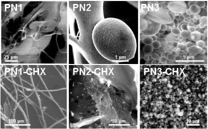
During the synthesis performed by the SFDP method, and consequent loading of the product with chlorhexidine, preparations presenting dif-ferent micromorphology were obtained. In the morphological study by SEM, the synthesized PN1 was identified as continuous structure with some droplets of microspheres, coming from the SFDP process, however in general, the struc-tures would be assessed as continuous hydrogel with some increments, without specific spherical structures (Fig. 1, PN1).
Oppositely, in the case of PN2 and PN3 the material was not continuous, consisting of nu-merous spheres in dimensions between 0.25 µm to 2.5 µm, with some of higher diameter. De-tailed morphology of this kind of material was evaluated in former paper 10. The PN1 material was rather planar, of thickness in the range 1-10 µm and compact, with some empty areas of di-ameter in the range between 10 and 100 µm. With the increase of the optical magnification the small spherical structures were revealed. The loaded freeze-dried polymer PN1-CHX is
Figure 1. The obtained microgels before loading with the chlorhexidine: PN1, PN2, PN3 (first row), and mi-crogels loaded with the chlorhexidine: PN1-CHX, PN2-CHX, PN3-CHX (second row).
Assignation group Reference range CHX PN1-CHX PN2-CHX PN3-CHX
N-H str. vibr. of C=N-H 3400-3300 3468 - -
-C=N str. vibr. of -C=N-H 1690-1640 1664 1636 1635 1631
[image:4.595.285.498.185.317.2]Guanidines str. vibr. =N-C=N- 1685-1580 1598 1534 1532 1535
Table 2. The changes in the IR spectrum after loading process of the polymers, with chlorhexidine.
(Fig. 1, second row, PN3-CHX). This phe-nomenon could be applied to control the re-lease of chlorhexidine from the formulation. As it was mentioned before, the chlorhexidine was presumably embedded into the polymeric ma-trix, both in the case of microparticles, and pla-nar hydrogel structures.
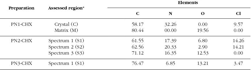
Using the EDXS assessments, the deposition of the chlorhexidine particles was confirmed. However the chlorhexidine was deposited rather on the surface of PN1 and PN2, not inside of the polymeric matrix. In the case of PN3, the chlorhexidine was embedded into the polymeric structure of the microparticles. In the measure-ments, the presence of chlorhexidine was definitively confirmed. As it can be evaluated from the image on the Figure 2 (PN1-CHX), all the needle-like shaped, narrow crystals of chlorhexidine were observed in aggregates on the surface of the polymeric material. The chlorhexidine was distributed on the surface of the material.
[image:5.595.97.311.87.153.2]The EDXS measurements revealed that chlorhexidine was rather on the surface of the polymeric matrix, whereas the actual polymeric material was not loaded by the chlorhexidine: compare the regions Crystal (C) and Matrix (M) and the respective results of chlorine element assessments in this regions: PN1-CHX-Crystal and PN1-CHX-Matrix. We assume that the PN2 Figure 2. The deposition of chlorhexidine on the ob-tained polymers: PN1 (image PN1-CHX), PN2 (image PN2-CHX) and PN3 (image PN3-CHX) - the areas, where EDXS assessments were performed are
marked: C, M, S1-S3, according to the Table 3. Figure 3. The EDXS assessments in various regions of obtained and loaded microgels PN1, PN2, and PN3. The respective details are given in the Table 3.
polymer absorbed the CHX both in the form of visible crystal (Fig. 2, PN2-CHX), and in the form of subtle dispersion - all regions assessed by the EDXS presented significant level of chlo-rine element comparing with the baseline - see PN1-CHX-Spectrum 1 to 3 on the Fig. 3, and re-spective microimages (Fig. 2). The PN3, in mag-nification, presented some increments in circular microparticle visualized on the Fig. 2, section PN3-CHX. According to the EDXS measure-ments, chlorine element was identified within the particle: PN3-CHX-Spectrum 1 (Table 3, Fig. 3). The obtained physical inclusions of chlorhexidine in the polymeric matrix have the potential for the application in topically applied devices for controlled drug release, and will be further examined in the terms of drug release. Respective spectra from EDXS device are at-tached in the Figure 3.
The evaluation of deposition of bioactive,i.e. chlorhexidine base, on or in the polymeric ma-trix is possible by the mean of EDXS. This method gives insight into placement of chlorhexidine microcrystals, when loaded to the polymeric matrix. Due to the simultaneous ob-servation of microscopic SEM images and EDXS assessments the inclusion of chlorhexidine was identified in the case of three various polymers.
Elements Preparation Assessed region*
C N O Cl
PN1-CHX Crystal (C) 58.17 32.26 0.00 9.57
Matrix (M) 80.44 00.00 19.56 0.00
PN2-CHX Spectrum 1 (S1) 61.55 17.39 6.80 14.26
Spectrum 2 (S2) 62.56 20.33 2.90 14.21
Spectrum 3 (S3) 71.12 16.35 12.53 0.00
PN3-CHX Spectrum 1 (S1) 76.47 6.85 13.21 3.47
[image:5.595.325.539.89.204.2] [image:5.595.101.538.606.728.2]The highest levels of chlorine element, which reflected the presence of chlorhexidine in the sample, was determined in polymer PN2 charac-terized by cationic amidine terminal groups – in-terestingly, due to EDXS observations, the chlorhexidine was present both in the form of crystal aggregates and dispersed in the polymer-ic matrix. In the case of highly hydrophobpolymer-ic PN3 polymer, the specific aggregates with chlorhexidine closed within the polymeric mi-crosphere were observed. The EDXS assess-ments may be applied for evaluation of patterns of chlorhexidine absorption to newly synthe-sized thermosensitive polymers.
CONCLUSIONS
Deposition of chlorhexidine base on/in the polymeric matrix may be performed by the mean of EDXS. The placement of chlorhexidine microcrystals, when loaded to the polymeric matrix may be assessed this way. Simultaneous observation of microscopic SEM images and EDXS data gives information on the patterns of deposition of chlorhexidine on the polymeric particle. The highest levels of chlorhexidine were determined in polymer PN2 with cationic amidine terminal groups. The highly hydropho-bic PN3 polymer, implemented the specific ag-gregates with chlorhexidine closed within the polymeric microsphere. The EDXS assessments may be applied for evaluation of patterns of chlorhexidine absorption on newly synthesized N-isopropylacrylamide derivatives.
Acknowledgements. This research was supported by a Marie Curie Transfer of Knowledge Fellowship of the Euro-pean Community 6th Frame Program under contract no. MTKD-CT-2005-029540-POLYSURF. Author would like to thank to Mrs. Tonica Boncina from University of Maribor, Faculty of Mechanical Engineering, University Center for Electron Microscopy for the assistance in SEM measurements.
REFERENCES
1. Lee, I., R.K. Agarwal, B.Y. Lee, N.O. Fishman & C.A. Umscheid (2010) Infect. Control. Hosp. Epidemiol. 31: 1219-29.
2. Al-Tannir, M.A. & H.S. Goodman (1994) Spec. Car. Dentist. 14: 116-22.
3. Noorani, A., N. Rabey, S.R. Walsh & R.J. Davies (2010) Brit. J. Surg. 97: 1614-20. 4. Reddy, M.S., M.K. Jeffcoat, N.C. Geurs, K.G.
Palcanis, T.W. Weatherford, B.M. Traxler et al. (2003) J. Periodontol. 74: 411-9.
5. Meng, N., N.L. Zhou, S.Q. Zhang & J.Shen, (2009) Int. J. Pharm. 382: 45-9.
6. Mirth, D.B., A. Bartkiewicz, R.J. Shern & W.A. Little (1989) Dent. Res. 68: 1285-8.
7. Anusavice, K.J., N.-Z. Zhang & C. Shen (2006) J. Dent. Res. 85: 950-4.
8. Raso, E.M.G., M.E. Cortes, K.I. Teixeira, M.B. Franco, N.D.S. Mohallem & R.D. Sinisterra (2010) J. Incl. Phenom. Macrocycl. Chem. 67: 159-68.
9. Gong, K., M. Braden, M.P. Patel, I.U. Rehman, Z. Zhang & J.A. Darr (2007)J. Pharm. Sci. 96: 2048-56.
10. Musial, W., V. Kokol and B. Voncina (2010) Chem. Pap. 64: 346-53.
11. Musial, W., V. Kokol & B. Voncina (2009) Polim. Med. 39: 3-15.
12. Castro Lopez, V., J. Hadgraft & M.J. Snowden (2005) Int. J. Pharm. 292: 137-47.
13. Gan, D & L.A. Lyon (2002) Macromolecules 35: 9634-9.
14. Wei, H., X.Z. Zhang, W.Q. Chen, S.X. Cheng & R.X. Zhuo (2007) J. Biomed. Mater. Res. A. 83: 980-9
15. Mason, T.G. & M.Y. Lin (2005) Phys. Rev. E. 71: 040801.
16. Liu, F. & M.W. Urban (2008) Macromolecules 41: 6531-9.
17. Pankey, D.A. & L.D. Sabath (2004) Clin. Infect. Dis. 38: 864-70.
18. Wang, H.-D., L.-Y. Chu, X.-Q .Yu, R. Xie, M. Yang, D. Xu et al. (2007) Ind. Eng. Chem. Res. 46: 1511-8
19. Sulea, D., M.V. Ghica, M. Micutz, M.G. Albu, L. Brazdaru, T. Staicu et al. (2010) Rev. Roum. Chim. 55: 543-51.
20. Ceschel, G.C., V. Bergamante, V. Calabrese, S. Biserni, C. Ronchi & A. Fini (2006) Drug. Dev. Ind. Pharm. 32: 53-61.
21. Amin, W.M., M.A. Alawi, R.M. Darwish, M.H. Al-Ali, N.A. Salim & S.K. Al-Tarawneh (2009) Mater. Res. Innov. 13: 448-54.
22. Lin, S., L. Levin, El. Weiss, M. Peled & Z. Fuss (2006) Quintessence Int. 37: 391-4.
23. Fay, F., I. Linossier, G. Legendre & K. Vallee-Rehel (2008) Macromol. Symp. 272: 45-51. 24. Hiraishi, N., C.K.Y. Yiu, N.M. King & F.R. Tay
(2010) J. Biomed. Mater. Res. 94B: 134-40. 25. Dynes, J.J., J.R. Lawrence, D.R. Korber, G.D.W.
Swerhone, G.G. Leppard & A.P. Hitchcock (2006) Sci. Tot. Env. 369: 369-83.
26. Musial, W., B. Vincent, A. Szumny, & B. Vonci-na (2010) Chem. Pap. 64: 602-12.
27. Goldstein, J.I., D.E. Newbury, P. Echlin, D.C. Joy, A.D. Romig, C.E. Lyman, et al. (1992) “Scanning Electron Microscopy and X-ray Mi-croanalysis”, First Eedition, New York, pp. 420-8.
28. Maeda, Y., T. Nakamura & I. Ikeda (2001) Macromolecules. 34: 1391-9.
29. Amatatsu, Y.A., Y. Hamada & M. Tsuboi (1987) J. Mol. Spectroscop. 123: 276-85.
30. Sun, B., Y. Lin, P. Wu & H.W. Siesler (2008) Macromolecules. 41: 1512-20.
31. Cheng, H., L. Shen & C. Wu (2006) Macro-molecules. 39: 2325-9.
32. Katsumoto, Y., T. Tanaka, K. Ihara, M. Koya-ma & Y. Ozaki (2006) J. Phys. Chem. B. 111: 12730-7.