Presentación de caso
Oral rehabilitation after removal of a central cemento-ossifying fibroma
Rehabilitación bucal después de la retirada de un
fibroma cemento-osificante
central
Clóvis Lamartine de Moraes Melo Neto1* https://orcid.org/0000-0003-1477-2055
Liogi Iwaki Filho1 https://orcid.org/0000-0001-9117-6826
Karina Rosso1 https://orcid.org/0000-0001-8182-6792
Rodrigo Lorenzi Poluha1 https://orcid.org/0000-0001-7180-6448
Bruna Milhomens de Sousa1 https://orcid.org/0000-0002-7276-7484
Sérgio Sábio1 https://orcid.org/0000-0002-5261-0228
1State University of Maringá (UEM), Department of Dentistry, Maringá, Paraná, Brazil.
*Autor para correspondencia: [email protected]
ABSTRACT
Introduction: The cemento-ossifying fibroma is a benign bone neoplasm that affects mainly
the female sex during the third or fourth decades of life. This lesion has a slow growth and the treatment is the surgical removal of the tumor, because radiotherapy is not indicated.
Objective: to describe and discuss a case of central cemento-ossifying fibroma.
Case report: A 41-year-old female patient sought dental treatment due to tooth mobility in
Conclusion: Despite being a benign tumor, the central cemento-ossifying fibroma caused
functional and aesthetic damage to the patient and required a complex rehabilitation treatment. After approximately 5 years of tumor removal, there was no recurrence of the lesion or signs of peri-implant or periodontal diseases, evidencing the success of the treatments.
Keywords: fibroma; ossifying; neoplasms; cancer; tumor
RESUMEN
Introducción: El fibroma cemento-osificante es una neoplasia ósea benigna que afecta
principalmente al sexo femenino durante la tercera o cuarta décadas de la vida. Esta lesión tiene un crecimiento lento y el tratamiento es la extirpación quirúrgica del tumor, porque la radioterapia no está indicada.
Objetivo: Describir y analizar un caso de un fibroma cemento-osificante central.
Presentación caso: Paciente de 41 años de edad buscó tratamiento dental debido a la
movilidad dental en la región anterior de la mandíbula. En esta región de la mandíbula se observó un aumento de volumen con una consistencia firme a la palpación, cubierta por la mucosa normal y desplazamiento de los dientes. Radiográficamente, se observó la presencia de una lesión extensa en esta región. Se realizó una biopsia incisional, que indicó el diagnóstico final del fibroma cemento-osificante central. Después el tumor fue
completamente extirpado. Se realizó un injerto óseo autógeno y, posteriormente, se instalaron cuatro implantes osteointegrados para rehabilitar al paciente. Se instalaron prótesis soportadas por implantes (arco inferior) y una dentadura (arco superior) para restablecer la estética y la función de la cavidad bucal de la paciente.
Conclusiones: A pesar de ser un tumor benigno, el fibroma cemento-osificante central
causó daños funcionales y estéticos al paciente y requirió un complejo tratamiento de rehabilitación. Después de 5 años de la extirpación del tumor, no hubo recurrencia de la lesión. Además, no hubo signos de enfermedades periimplantarias y/o periodontales, lo que demuestra el éxito del tratamiento.
Palabras clave: fibroma; osificante; neoplasias; cáncer, tumor.
INTRODUCTION
Cemento-ossifying fibroma (COF) is a benign slow-growing lesion that mainly affects the posterior region of the mandible in 70 % of cases.(1-4) This tumor is more frequently found in
the female gender than in the male gender. In addition, the COF is most commonly seen in
the third and fourth decades of life, (1-4) with a predilection for the black population.(2)
The COF has been described as a well-delimited lesion and occasionally encapsulated, that contains fibrous tissue and varying quantities of mineralized material similar to bone and/or cementum,(1-4) which generally arises from cells in the periodontal ligament.(1,2) The degree
of mineralization is variable, therefore the COF may be radiolucent or radiolucent and radiopaque appearance, depending on the amount of calcification of the lesion.(1,2)
The differential diagnosis for COF includes cemento osseous dysplasia, odontogenic cyst, periapical granuloma, traumatic bone cyst, unilocular ameloblastoma, calcifying epithelial odontogenic tumour and central giant cell granuloma.(1,3) The treatment choice is the
curettage and/or enucleation.(1,3,5) According to Kattiet al.(1) an inadequate surgical treatment
may cause recurrence of the lesion and therefore, an adequate diagnosis and treatment plan are necessary for clinical success.1 Radiotherapy is contraindicated due to the radioresistence
of this lesion and the side effects of the radiation.(5)
Oral rehabilitation after removal of a tumor may be necessary in many cases, especially when the patient loses many oral tissues.(6) The objective of this case report was to describe
and discuss a case of central cemento-ossifying fibroma.
CASE REPORT
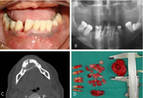
In the panoramic radiograph, a predominantly radiolucent image was visualized, with limits clearly defined, and with a sclerotic margin, thus showing the reason for the displaced teeth (Fig. 1, B). The first procedure for the case was to perform an incisional biopsy and send the biological material extracted for microscopic examination.
Microscopically, a benign fibrous-osseous lesion was observed, with fibroblast proliferation, intermingled with mineralized material, at times in the form of trabeculae that mimetized immature bone, and at other times, in the form of cementoid globules (basophilic and frequently acellular). The osteoid layer thickness varied, and so did the predominance of the peripheral osteoblasts. Fragments of normal tissue completed the microscopic condition of central cemento-ossifying fibroma.
After performing the computed tomography exam (Fig. 1, C), an intraoral access was performed for extracting the mandibular teeth in direct contact with the lesion. A submental extraoral incision was performed for enucleation of the pathology, preserving the mandibular base and lingual cortical. After removal of the lesion (Fig. 1, D), the local reconstruction was performed with 10 mL of tibial bone graft, that was inserted into the surgical cavity. A titanium screen was used as a support for the soft tissue and bone graft. These procedures were performed in the hospital.
Fig. 1 – A: Clinical Front View. B: Panoramic radiography showed that there was a lesion. C:
After 60 days the titanium screen was removed, due to its intraoral exposure. After the emergency phase has been resolved, treatment options were suggested to the patient. The upper arch could be rehabilitated with branemark protocol, overdenture or another CD. For the lower arch, the options were rehabilitation with implant-supported prostheses or a removable partial denture (RPD). Due to the patient's financial condition, she opted for a conventional CD (upper arch) and implant-supported prostheses (lower arch). Considering the large number of surgeries performed and the psychological aspect involved, it was decided to perform the extraction of the retained molar after oral rehabilitation, since it would not interfere with the prosthetic rehabilitation.
Due to the financial conditions of the patient, the rehabilitation treatment was delayed. After 1 years and seven months, clinical and radiographic evaluation showed normality of the mandible. The installations of 4 external hexagon type implants (3.75 x 11.5 mm) (Titanium Fix, AS Technology Ltda., Brazil) with 4.1 platforms were performed in the lower jaw. The patient was referred to the dental prosthesis clinic of the UEM. After 2 year and six months the patient returned (Fig. 2, A and B). A new maxillary CD was being fabricated up to the stage of mounting the teeth.
For the three anterior implants, three Micro Unit abutments (Titanium Fix, AS Technology
Ltda.) were selected, with angulation of 30 degrees each and all with a metal strap 3 mm in
height. For the single implant in the posterior mandibular region, the Esteticone straight abutment (Titanium Fix, AS Technology Ltda.) with a 1 mm strap was selected. The four abutments were installed on the implants and radiographed (to evaluate adaptation) and their respective torques (32 Ncm) were applied.
A plastic tray was tried-in, cut and the transfers of the open tray were adapted on the abutments (Fig. 2, C). Using addition silicone (Elite, Zhermack, S.p.A., Italy) the impression was made, following the manufacturer's recommendations(Fig. 2, D).
The brand and shade (62 - Dental Vipi Ltda., Brazil) of the mandibular acrylic teeth were the same as those of the CD. The VITA Scale (Vita Zahnfabrik, Germany) with reference to shade A-2 was chosen for the porcelain crown.
after a time to the surgery clinic for extraction of the molar retained in the mandible. After approximately 5 years of tumor removal, there was no recurrence of the lesion. In addition, there were no signs of peri-implant and/or periodontal diseases, evidencing the success of the treatment.
Fig. 2 - A: Panoramic Radiograph with implants installed. B: With the old complete denture
the smile was unsatisfactory. C: Abutments and transfers. Subsequently the transfers were united with the acrylic resin (Duralay, Reliance Dental Mfg., USA). D: Addition silicone
mold.
Fig. 3 – A: Implant-supported prostheses. B: View of the mandibular arch with the
DISCUSSION
This lesion affects mainly the female gender,(1-5) with a female-to-male ratio of 5:1,(5) during
the third or fourth decades of life,(1-5) which corroborates with the present case in which the
patient was a 41-year-old woman. In this case report the tumor was found in the anterior region of the mandible. It disagrees with the literature which states that this lesion is more prevalent in the mandibular premolar and molar region.(1-5)
According to Katti et al., the COF can often be neglected by the patient until its growth
results in facial deformity.(1) Pain and paresthesia are rarely associated with this lesion.(1,2,3,4)
In addition, the involved teeth normally conserve their vitality with absence of root resorption.(1,3,5) The root movementand cortical expansion can occur.(1,3,5) The present case
corroborates these reports in the literature.
Irritating factors can start this process (COF), such as trauma and extractions.(1,5) The patient
has a previous history poor oral hygiene, periodontal disease,(7) and dental extractions. This
can explain the development of the COF, in this case. According to the literature, it is
possible that undifferentiated mesenchymal cells of the periodontal ligament had differentiated to produce a calcified material similar to bone or cementum, or a combination of these elements within a predominantly fibrous tissue.(1,5)
In cases of very extensive lesions, some authors recommend a more conservative approach (enucleation/curettage) in order to reduce the need for bone grafting and improve postoperative.(1,4) The recurrence rate of this tumor can reach ≅ 30 %(1,8) Therefore the
lesion was completely removed, including the subjacent periosteum and teeth in direct contact with the lesion. During the treatment, the patient underwent periodontal maintenance and oral hygiene instruction regularly to control the plaque index.
In cases when COF is very large, this lesion associated with surgery may lead to fracture of the mandible.(4) In this case reported this did not occur, however the mandible was
fragilized. Silveira et al.(9) recommend that after extensive surgery, a bone graft and
implants should be performed for reconstruction of the region.(9) This case report
corroborates this situation. The donor area was the tibia, since, according to Hussain,(10) it is
an excellent alternative for bone grafts.(10)
To minimize this, all the information about the surgeries required were provided, and the patient’s doubts were clarified to reduce anxiety and increase her confidence in the dental surgeons.(13,14)
There is a strong body of evidence that poor oral hygiene and history of periodontal disease are risk factors for the development of peri-implant diseases (mucositis and peri-implantitis). Moreover, mucositis is believed to be the precursor of peri-implantitis and these pathologies can lead to implant loss.(15) This patient has a history of periodontal disease, thus, some
measures to prevent these diseases have been taken. Therefore, the choice was to rehabilitate the mandibular arch with screw-retained implant-supported prostheses due to the capacity
of reversibility of this type of dental prosthesis,(16) to improve control of peri-implant health.
Moreover, screw-retained implant-supported prostheses do not have the inconvenience of excess cement, which can affect peri-implant health.(16)
The rehabilitation with RPD probably would not have a good prognosis in this situation, due to the tissue losses, the location, the inclination and amount of teeth remaining of the patient. According to Campbell et al.,(17) the evaluation of the dentition state, tooth position,
adapting structures within the RPD, timely recall, and maintenance are necessary for successful treatment.(17) In addition, the treatment with RPD can cause overload on natural
teeth, periodontal damage and increased incidence of caries.(18)
All the implants received abutments. The anterior abutments facilitated seating of the dental prosthesis, because the Microunit abutments (Titanium Fix, AS Technology Ltda.) are
rotational. With removal of part of the alveolar process, there was the formation of a “bone ramp” that consequently led to the 3 anterior implants becoming “vestibularized”, so that angled abutments of 30 degrees were used. Although angulation increases stress on abutments and implants, this factor does not impair the longevity of the prosthesis and implants because, since according to Cavallaro and Greenstein,(19) this increase is within the
physiological limits.(19)
The installation of the metal framework was passive and there was no gap formation. Passiveness is important to avoid the accumulation of stress at the bone and implant interface, screw loosening, fracture and failure of the implant.(20) After insertion of the
prostheses, an occlusal adjustment was made so that all the teeth would have contact, and for the prevention of an overload in the implants, avoiding biomechanical complications.(21)
To reduce the accumulation of biofilm, which is the main cause of mucositis, (15) the patient
Technologies, USA). It was recommended to brush the new CD with neutral soap.
Despite being a benign tumor, the central cemento-ossifying fibroma caused functional and aesthetic damages to the patient and required a complex rehabilitation treatment. Therefore, regular dental visits are of fundamental importance to discover these types of pathologies while they are still in the initial stages. After approximately 5 years of tumor removal, there was no recurrence of the lesion. In addition, there were no signs of peri-implant and/or periodontal diseases, evidencing the success of the treatment.
BIBLIOGRAPHIC REFERENCES
1. Katti G, Khan MM, Chaubey SS, Amena M. Cemento-ossifying fibroma of the jaw. BMJ Case Rep. 2016;2016:bcr2015214327.
2. Woo SB. Central Cemento-Ossifying Fibroma: Primary Odontogenic or Osseous Neoplasm? J Oral Maxillofac Surg. 2015;73(12 Suppl):S87-93.
3. Dominguete MH, Dominguette AA, Matos BH, Dominguete PR, León JE, Oliveira LR. Extensive presentation of central ossifying fibroma treated with conservative surgical excision. Case Rep Dent. 2014;2014:204258.
4. Ong AH, Siar CH. Cemento-ossifying fibroma with mandibular fracture. Case report in a young patient. Aust Dent J. 1998;43(4):229-33.
5. Naik RM, Guruprasad Y, Sujatha D, Gurudath S, Pai A, Suresh K. Giant cemento-ossifying fibroma of the mandible. J Nat Sci Biol Med. 2014;5(1):190-4.
6. Minichetti JC, D'Amore JC, Schwarz E. Complete oral rehabilitation of a postresection ameloblastoma patient: a clinical case report. J Oral Implantol. 2011;37(6):735-44.
7. Michaud DS, Fu Z, Shi J, Chung M. Periodontal Disease, Tooth Loss, and Cancer Risk. Epidemiol Rev. 2017;39(1):49-58.
8. Knutsen BM, Larheim TA, Johannessen S, Hillestad J, Solheim T, Koppang HS. Recurrent conventional cemento-ossifying fibroma of the mandible. Dentomaxillofac Radiol. 2002;31(1):65-8.
10. Hussain S. Evaluation of alveolar grafting with tibial graft in adolescent patients. Indian J Dent Res. 2013;24(6):659-63.
11. Mochizuki Y, Matsushima E, Omura K. Perioperative assessment of psychological state and quality of life of head and neck cancer patients undergoing surgery. Int J Oral Maxillofac Surg. 2009;38(2):151-9.
12. Tahani B, Razavi SM, Emami H, Alamchi F. Assessment of the quality of life of the patients with treated oral cancer in Iran. Oral Maxillofac Surg. 2017;21(4):429-37.
13. Appukuttan DA. Strategies to manage patients with dental anxiety and dental phobia: literature review. Clin Cosmet Investig Dent. 2016;8:35-50.
14. Riley JL, Gordan VV, Hudak-Boss SE, Fellows JL, Rindal DB, Gilbert GH, et al. Concordance between patient satisfaction and the dentist's view: findings from The National Dental Practice-Based Research Network. J Am Dent Assoc. 2014;145(4):355-62.
15. Heitz-Mayfield LJ. Peri-implant diseases: diagnosis and risk indicators. Clin Oral Implants Res. 2008;35:214–22.
16. Wittneben JG, Joda T, Weber HP, Brägger U. Screw retained vs. cement retained implant-supported fixed dental prosthesis. Periodontol 2000. 2017;73(1):141-51.
17. Campbell SD, Cooper L, Craddock H, Hyde TP, Nattress B, Pavitt SH, et al. Removable partial dentures: The clinical need for innovation. J Prosthet Dent. 2017;118(3):273-80. 18. Zlatarić DK, Celebić A, Valentić-Peruzović M. The effect of removable partial dentures on periodontal health of abutment and non-abutment teeth. J Periodontol. 2002;73(2):137-44 19. Cavallaro J Jr, Greenstein G. Angled implant abutments: a practical application of available knowledge. J Am Dent Assoc. 2011;142(2):150-8.
20. de Araújo GM, de França D G, Silva Neto J P, Barbosa G A. Passivity of conventional and CAD/CAM fabricated implant frameworks. Braz Dent J. 2015;26(3):277-83.
21. Hsu YT, Fu JH, Al-Hezaimi K, Wang HL. Biomechanical Implant Treatment Complications: A Systematic Review of Clinical Studies of Implants with at Least 1 Year of Functional Loading. Int J Oral Maxillofac Implants. 2012;27(4):894-904.
Conflicts of interest