Behaviour of pathogenic gram negative bacteria inoculated in milk and model cheese treated with high hydrostatic pressure
156
0
0
Texto completo
(2) Departament de Ciència Animal i dels Aliments Facultat de Veterinària Universitat Autònoma de Barcelona. BEHAVIOUR OF PATHOGENIC GRAM NEGATIVE BACTERIA INOCULATED IN MILK AND MODEL CHEESE TREATED WITH HIGH HYDROSTATIC PRESSURE. MEMÒRIA PRESENTADA PER OPTAR AL GRAU DE DOCTORA EN CIÈNCIA I TECNOLOGIA DELS ALIMENTS. SÍLVIA DE LAMO CASTELLVÍ Bellaterra (Cerdanyola del Vallès), 2006. Centre Especial de Recerca Planta de Tecnologia dels Aliments (CERPTA).
(3) MARTA CAPELLAS PUIG, Professora Titular de Tecnologia dels Aliments, i ARTUR XAVIER ROIG SAGUÉS, Professor Titular de Tecnologia dels Aliments de la Universitat Autònoma de Barcelona,. FAN CONSTAR: que la llicenciada en Química i en Ciència i Tecnologia dels Aliments Sílvia De Lamo Castellví ha dut a terme, sota la seva direcció, a l’Àrea de Tecnologia dels Aliments de la Facultat de Veterinària de la Universitat Autònoma de Barcelona, el treball titulat “Behaviour of pathogenic Gram negative bacteria inoculated in milk and model cheese treated with high hydrostatic pressure”, que presenta per optar al grau de Doctora.. I perquè així consti, signem el present document a Bellaterra (Cerdanyola del Vallès) a 7 de febrer de 2006.. Marta Capellas Puig. Artur Xavier Roig Sagués.
(4) I don’t believe there would be any science at all without intuition Rita Levi Montalcini, 1986 Nobel Prize in Physiology and Medicine.
(5) Acknowledgements Al Ventura per donar-me l’oportunitat de realitzar aquest treball i entendre els meus desitjos. A l’Artur per tot el teu treball en la direcció d’aquesta tesi i per la teva paciència amb les meves divagacions. A la Marta pel teu treball en la direcció d’aquesta tesi i més enllà, per estar sempre que t’he necessitat i per tenir la paraula i la idea justa en el moment adequat. A la Manoli pels teus magnífics consells i per la teva bona disposició tant en els moments bons com en els difícils. Al Josep, per escoltar-me i ajudar-me. També per l’experiència workshop, inoblidable. Al Ramón per inspirar-me amb la teva recerca i per la teva bona disposició. A la Dolors per ser una molt bona jefa del laboratori i perdonar els meus nombrosos tubs bruts. A la Núria, la Dora i el Manuel, per ser de les millors persones que he conegut i crear aquest màgic vincle. Al Tomás per compartir aventures i desventures en la realització d’aquest projecte i a vegades les tensions pròpies que comporta aquesta feina. Al Wilfido per ser un bon company i amic, per saber escoltar i per alegrar el dia a dia de la recerca. A la Pilar i la Patricia, per ser unes bones conselleres i iniciar la meva immersió en la cultura mexicana i peruana. Als meus companys de despatx Ibrahima, Betty, Diana, Patricia i Osvaldo per compartir el mini espai del que disposem i més coses. A la resta de companys de les dues Unitats que m’han facilitat la feina en molts moments i que m’han fet riure quan calia. A la Inga, la Berta i la Laia per la seva magnífica contribució en la realització d’aquest treball. To Dr. Frank for giving me the chance to open my eyes to a new research world reality..
(6) To Dr. Toledo for letting me use one of his high pressure machines. You made my dream come true. To Carl for helping me all the time. A Danitza, por ser mi mayor apoyo durante mi aventura americana, por ayudarme a salir adelante cuando todo parecía desvanecerse y por creer siempre en mi. To Linda, for being my main support in the lab but specially, for being my friend. To Wut, for being the best officemate that I have ever had. I will never forget our salsa lessons in our lunch breaks. Als meus pares i germans pel vostre afecte, comprensió i ajuda. Al David per la teva infinita paciència i per entendre i compartir aquesta vida boja que portem els que ens dediquem a recerca..
(7) This study was financially supported by: Instituto Nacional de Investigación y Tecnología Agraria y Alimentaria. Project: CAL-00-005-C2-1. Departament d’Universitats, Recerca i Societat de la Informació de la Generalitat de Catalunya, which provided Sílvia De Lamo Castellví a fellowship (2002FI-00079) to carry out this research..
(8) Contents 1. Introduction ...................................................................................................................1 1.1 General principles of high pressure...............................................................................4 1.2 Effects of pressure on microorganism...........................................................................5 1.2.1 Effect of pressure on cell membranes...................................................................6 1.2.2 Effect of pressure on cell wall ..............................................................................7 1.2.3 Effect on cell morphology ....................................................................................8 1.2.4 Effect on biochemical reactions ...........................................................................9 1.2.5 Effect on genetic mechanisms ..............................................................................9 1.3 Effects and interactions of treatment variables..............................................................10 1.3.1 Type of microorganism ........................................................................................10 1.3.2 Pressure level and time .........................................................................................12 1.3.3 Temperature..........................................................................................................12 1.3.4 pH .........................................................................................................................13 1.3.5 Composition of medium .......................................................................................14 1.3.6 Antimicrobial compounds ....................................................................................15 1.4 Injured population .........................................................................................................16 1.5 Kinetics of microbial inactivation ...............................................................................19 1.6 Milk and dairy products as a vehicle of foodborne pathogens ......................................21 1.7 Gram negative bacteria used for this research...............................................................23 1.7.1 Yersinia enterocolitica..........................................................................................23 1.7.2 Salmonella spp......................................................................................................24 1.7.3 Escherichia coli ....................................................................................................25 1.8 Effect of high hydrostatic pressure on microorganisms in milk....................................26 1.9 Effect of high hydrostatic pressure on microorganisms in cheese ................................30 2. Aim of this dissertation and Sampling protocol .........................................................33 3. Materials and Methods .................................................................................................39 3.1 Bacterial strains .............................................................................................................41 3.2 Inocula preparation ........................................................................................................42 3.3 Starter culture preparation .............................................................................................42 3.4 Sample preparation ........................................................................................................42 3.4.1 Skimmed milk ......................................................................................................42 3.4.2 Model cheese manufacture ...................................................................................43 3.5 High pressure treatment.................................................................................................45 3.6 Microbiological analyses...............................................................................................45 3.6.1 Skimmed milk ......................................................................................................45 3.6.2 Model cheese ........................................................................................................45 3.7 Expression of results......................................................................................................46 3.8 pH measurement ............................................................................................................46 3.9 Statistical analyses.........................................................................................................47 3.10 Kinetic study of microbial inactivation .......................................................................47.
(9) 4. Papers included in the dissertation ..............................................................................51 4.1 Survival and growth of Yersinia enterocolitica strains inoculated in skimmed milk treated with high hydrostatic pressure. International Journal of Food Microbiology (2005), 102:337-342 ...................................................................................................... 53 4.2 Behavior of Yersinia enterocolitica strains inoculated in model cheese treated with high hydrostatic pressure. Journal of Food Protection (2005), 68:528-533................... 61 4.3 Fate of Escherichia coli strains inoculated in model cheese produced with or without starter and treated by high hydrostatic pressure. Submitted to be published in Journal of Food Protection .......................................................................................69 4.4 Response of two Salmonella enterica strains inoculated in model cheese treated with high hydrostatic pressure. Submitted to be published in International Journal of Food Microbiology ................................................................95 5. Results and Discussion ..................................................................................................125 5.1 Skimmed milk ...............................................................................................................127 5.1.1 High Pressure effect at day 0................................................................................127 5.1.2 Behaviour of Y. enterocolitica after high hydrostatic pressure treatments..........127 5.1.3 Kinetic study of microbial inactivation ................................................................128 5.2 Model cheese .................................................................................................................129 5.2.1 Behaviour of initial inoculum during model cheese production .........................129 5.2.2 High Pressure effect at day 0 in pathogenic bacteria............................................130 5.2.3 Behaviour of pathogenic bacteria after high hydrostatic pressure treatment in model cheese produced with starter culture .........................................................133 5.2.4 Behaviour of pathogenic bacteria after high hydrostatic pressure treatment in model cheese produced without starter culture ....................................................134 5.2.5 Behaviour of starter culture in model cheese inoculated with Y. enterocolitica serotype O:3 (long term experiments) or with S. enterica strains (short term experiments) .........................................................................................................138 6. Conclusions ....................................................................................................................141 7. References ......................................................................................................................145.
(10) List of Tables Table 1.. Volume changes accompanying the formation of major interactions and bonds in biosystems...........................................................................................4. Table 2.. Temperature changes of selected substances due to compression heating........5. Table 3.. Outbreaks associated with milk and dairy products contaminated with pathogenic bacteria ............................................................................................21. Table 4.. Heat treatments more common used in dairy industry .....................................22. Table 5.. Bacterial strains .................................................................................................41. Table 6.. Details of high hydrostatic pressure equipments and the conditions of the treatments applied..............................................................................................48. Table 7.. Culture media and incubation conditions used for cheese samples analyses. ...49. Table 8.. Cell counts at 0 and 15 days of Y. enterocolitica inoculated in milk and pressurized at 500 MPa at 20ºC for 10 min.......................................................128. Table 9.. Values of pH in blank cheese samples(non inoculated and untreated) produced with and without starter and stored at 8 or 12ºC for 15 or 60 days ....................................................................................................132. Table 10. Cell counts at 0 and 15 or 60 days of pathogenic bacteria inoculated in cheese produced with starter culture and pressurized at 300, 400 or 500 MPa at 20ºC for 10 min ............................................................................................135 Table 11. Results of the enrichment process (24 h at 37ºC or 24 h at 32ºC) applied to selected cheese samples produced with starter culture (those which showed cell counts below the detection level [10 cfu/g] during the storage time).........136 Table 12. Cell counts at 0 and 15 days of bacteria inoculated in cheese produced without starter culture and pressurized at 300, 400 or 500 MPa at 20ºC for 10 min ..........................................................................................................137.
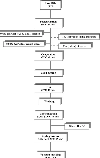
(11) List of Figures Figure 1. Structural and functional changes in microorganisms at different pressures ....6 Figure 2. Microbial stress, injury, adaptation and resistance to processing .....................19 Figure 3. Milk experiments...............................................................................................36 Figure 4. Model cheese experiments with Y. enterocolitica strains..................................37 Figure 5. Model cheese experiments with E. coli and S. enterica strains.........................38 Figure 6. Cheese making process......................................................................................44 Figure 7. Cell counts in TALm at day 0 of pathogenic bacteria inoculated in model cheese produced (a) with and (b) without starter culture and treated at 300 and 400 or 500 MPa for 10 min at 20ºC............................................................131 Figure 8. Behaviour of starter culture in cheese samples inoculated with Y. enterocolitica (serotype O:3) and treated at 300 and 400 MPa for 10 min at 20ºC and ripened for 60 days at 8ºC..................................................................139.
(12) 1. Introduction. 1.
(13) Introduction. 1. Introduction Consumers in the 21st century are demanding high quality foods that are free from additives, fresh tasting, microbiologically safe and with an extended shelf-life. One food technology that has the potential to meet these demands is high pressure processing. High pressure processing, also known as high hydrostatic pressure (HHP), uses pressures up to 900 MPa to inactivate many of the microorganisms found in foods (Patterson, 2005). This technology has been proposed as a viable alternative to conventional heat treatment for preserving food. In contrast to thermal processing, the application of high hydrostatic pressure to food causes negligible impairment of nutritional value, taste, colour or flavour (Smelt, 1998). It is well known that the effectiveness of any food preservation technique has to be evaluated by its ability to eradicate any pathogenic microorganism to assure product safety and to inactivate spoilage microorganisms to improve the shelf life of the food (McClements et al., 2001). High hydrostatic pressure induces a number of changes to morphology, biochemical reactions and genetic mechanisms, and to cell membrane and wall of microorganisms (Hoover et al., 1989) and is generally effective to inactivate most vegetative bacteria, yeasts, and molds between 300 and 700 MPa (Smelt, 1998). Nevertheless, it is important to remark that there are different factors that affect the resistance of bacteria to high hydrostatic pressure treatments: temperature, magnitude and duration of pressure treatment, the stage of growth and the composition of medium (McClements et al., 2001). Moreover, in food, two effects always determine microbiological safety and stability: the effect of the food matrix during treatment (Patterson and Kilpatrick, 1998), and its effect after treatment, during the repair phase of the microorganisms (Smelt, 1998).. 3.
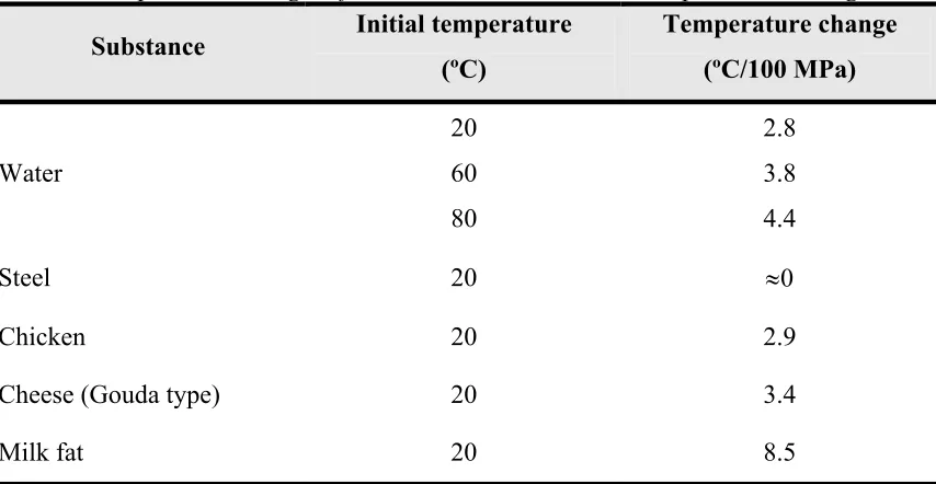
(14) Introduction. 1.1 General principles of high pressure The thermodynamic term describing the effects of pressure on chemical or biochemical systems is ∆V, the variation of the molar volume between two states (such as the final and the initial states) at constant temperature (Balny and Masson, 1993). High hydrostatic pressure is governed by two principles: the principle of Le Chatelier and the isostatic principle. Firstly, the principle of Le Chatelier states that any phenomenon (phase transition, chemical reactivity, change in molecular configuration or chemical reaction) accompanied by a decrease in volume will be enhanced by pressure (Table 1). An antagonistic effect of temperature is expected from the fact that temperature increase results in a volume increase. Depending on the nature of the product, the initial product temperature and the applied pressure, the adiabatic temperature increase may vary from 3 to 9ºC/100 MPa (Table 2) (de Heij et al., 2003). On the other hand, the reaction rate increases with increasing temperature according to Arrhenius' law. Table 1. Volume changes accompanying the formation of major interactions and bonds in biosystems. Interaction and bonds ∆V≠ of –10 ml/mol for the formation of covalent bonds, and nearly zero ∆V values for exchanges in the covalent bonds. a. Covalent bonds. b. Electrostatic interactions. ∆V of – 10 ml/mol for hydratation of a charged group and a positive value of 1020 ml/mol for the formation of an electrostatic interaction. Hydrophobic interactions. A positive ∆V of 10-20 ml/mol CH2groups entering the hydrophobic contact. Stacking of aromatic rings. Nearly zero values of ∆V. Hydrogen bonds. Nearly zero values of ∆V. Differences in the volumes of the activated (V≠) and initial states. Differences in the volumes of the final and the initial states Source: Mozhaev et al., 1994. a. b. 4.
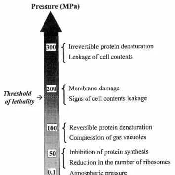
(15) Introduction Table 2. Temperature changes of selected substances due to compression heating Initial temperature Temperature change Substance (ºC) (ºC/100 MPa) 20. 2.8. 60. 3.8. 80. 4.4. Steel. 20. ≈0. Chicken. 20. 2.9. Cheese (Gouda type). 20. 3.4. Milk fat. 20. 8.5. Water. Source: de Heij et al., 2003. Secondly, the isostatic principle (isostatic pressure) that shows that the pressure is instantaneously and uniformly transmitted independently of the size and the geometry of the food (Smelt, 1998).. 1.2 Effects of pressure on microorganisms Microorganisms are inactivated when they are exposed to factors that substantially alter their cellular structure or physiological functions. Structural damage includes DNA strand breakage, cell membrane rupture or mechanical damage to cell envelope. Cell functions are altered when key enzymes are inactivated or membrane selectivity is disabled. A preservation technology may cause cell death through multiple mechanisms. High pressure is well known to produce alterations (Figure 1) in biochemical reactions, genetic mechanisms, structural changes in the morphology, cell wall and membranes of microorganisms (Hoover et al., 1989; Cheftel, 1995). However, the mechanisms of microbial inactivation are still not fully understood (Patterson, 2005).. 5.
(16) Introduction. Figure 1. Structural and functional changes in microorganisms at different pressures. Source: Lado and Yousef (2002).. 1.2.1 Effect of pressure on cell membranes The cell membrane is generally considered to be a primary site of pressure damage in microorganisms (Hoover et al., 1989). The cell membrane is composed by a bilayer of phospholipids with embedded functional proteins that play an important role in transporting ions and other substances across the membrane. The effect of pressure on the membranes might be due to the fact that lipids are particularly sensitive to pressure, being an order of magnitude more compressible than proteins (Fernandes, 2005). It has been observed that lipid bilayers undergo phase transitions under pressure (San Martin et al., 2002). Membrane fluidity it is a multifaceted phenomenon that has contributions from molecular packing (order) and molecular motion (viscosity). The changes in fatty acyl composition may alter either or both of these aspects of fluidity and may exert its influence through control of the ion pumps in the membrane that are essential for maintaining pH homeostasis (Russell, 2002). The composition of lipid membrane is known to be altered in response to variations of pH, external osmolality and low temperature to retain membrane fluidity and functionality (Scheyhing et al., 2004). Kato and Hayashi (1999) suggested that the fluidity of lipid bilayers decreases with an increase in the pressure applied, resulting in functional disorder of membrane associated enzymes, accompanied by a reversible phase transition of the biomembrane, and finally, the lipid bilayer is irreversible fragmented, accompanied by enzyme denaturation. Casadei et al. (2002) reported that membrane fluidity affects the pressure resistance of. 6.
(17) Introduction exponential and stationary-phase cells in a similar way, but it is the dominant factor in exponential-phase cells whereas in stationary-phase cells, its effects are superimposed on a larger effect of the physiological stationary-phase cell response that is itself temperature dependent. Moreover, loss of membrane-bound ATPase activity and loss of the ability to maintain a transmembrane pH gradient have been shown to occur during inactivation of Lactobacillus plantarum by high pressure (Wouters et al., 1998). Physical damage to cell membrane has been demonstrated as leakage of ATP or UVabsorbing material -in the case of bacteria (Smelt et al., 1994)-, and solubilization and leakage of intracellular substances such as amino acids -in the case of yeasts (Kato and Hayashi, 1999)-, subjected to pressure occurs. A loss of osmotic responsiveness or increased uptake of the fluorescent dyes such as ethidium bromide and propidium iodide that do not normally penetrate the membranes of healthy cells (Benito et al., 1999; Ulmer et al., 2000; Pagán and Mackey, 2000) has also been observed. Benito et al. (1999) found that when Escherichia coli O157:H7 strains were exposed to the same pressure for different times, the pressure-sensitive strains took up stain sooner than the more resistant strain. The differences in resistance may be related to susceptibility to membrane damage and possibly to a property associated with the protein component. Mañas and Mackey (2004) reported that exponential-phase cells are inactivated under high pressure by irreversible damage to the cell membrane. In contrast, stationary-phase cells have a more robust cytoplasmic membrane that can better withstand pressure treatments. The retention of an intact membrane appears to allow the stationary-phase cell to repair gross changes in other cellular structures and to remain viable at pressures that are lethal to exponential-phase cells.. 1.2.2 Effect of pressure on cell wall Hoover at el. (1989) reported that pressures of 20-40 MPa can cause larger cells to lyse from mechanical disruption of a stressed cell wall. Kato and Hayashi (1999) suggested that the cell wall structure is altered by high pressure treatment, which leads to solubilization of intra-cellular substances. Malone et al. (2002) found that the pressure treatment at 100 MPa promoted cell wall hydrolase activity in Lactococcus lactis subsp.. 7.
(18) Introduction cremoris MG1363 while treatment at pressures in excess of 300 MPa inactivated the enzyme activity.. 1.2.3 Effect on cell morphology Intracellular damage has been studied for several researchers by observation under a light microscope. Hoover et al. (1989) reported a variety of morphological changes in cells after applying pressure treatments that included longer individual cells, separation of the cell wall from the cytoplasmic membrane, thickened cell walls with no membrane structure, clear zones of spongy or reticular structures in the cytoplasm, and a decrease in number of ribosomes. Ritz et al. (2001, 2002), using scanning electron microscopy, reported the occurrence of bud scars on the surface of Listeria monocytogenes cells after 10 min of treatment at 400 MPa in citrate buffer. Intracellular regions of low density were observed in L. monocytogenes and L. lactis subsp. cremoris MG1363 using transmission electron microscopy (Mackey et al., 1994; Malone et al., 2002). These authors suggested that the low-density regions are caused by transient membrane invaginations under pressure that are subsequently reversed upon pressure release, leaving the low-density regions adjacent to the cell membrane. However, these lowdensity regions were not observed in high pressure treated Salmonella enterica serovar Thompson (Mackey et al., 1994), suggesting that only the Gram positive cell wall may be associated with this phenomenon. Mañas and Mackey (2004) reported that membranes of exponential-phase cells showed physical disruption after pressurization, with the formation of vesicles, areas of engrossment, and also invaginations toward the cytoplasm. These authors suggested that the vesicles observed in pressurized exponential-phase cells could be from outer membrane materials, whereas engrossment areas, which occupied part of the cytoplasm, could be from the cytoplasmic membrane.. 8.
(19) Introduction 1.2.4 Effect on biochemical reactions The application of pressure enhances reactions that lead to a volume decrease and generally retards reactions involving a volume increase. Most biochemical reactions result in a change in volume. Consequently, biological processes are influenced by pressure application (Hoover, 1989). Pressure acts as a modulator of biochemical processes ranging from inhibition of bacterial growth or viability of viral particles to activation-inactivation of enzymatic reactions. Moreover, high pressure affects the structure and function of enzymes in a complex way by altering intra and intermolecular interactions involved in protein stability (Dallet and Legoy, 1996). Simpson and Gilmour (1997) found that L. monocytogenes intracellular enzymes could be affected by high pressure treatments. The electrophoretic mobility of those enzymes was modified after pressure treatment corresponding to some variations of their conformational forms. In some instances, the activity of those enzymes appeared to decrease as pressure increased.. 1.2.5 Effect on genetic mechanisms Nucleic acids are relatively resistant to high pressures and as the structure of the DNA helix is largely the result of hydrogen bond formation, it is also stable under pressure (Patterson, 2005). However, an extreme condensation of the nuclear material was observed in L. monocytogenes and S. typhimurium (Mackey et al., 1994). The hypothesis is that at elevated pressures, DNA comes in contact with endonucleases, which cleave DNA. This condensation has been found in many other instances and it is reversible and, presumably, also an enzyme responsible for renaturation is involved. If this enzyme is deactivated by high hydrostatic pressure, the cell is no longer able to multiply (Smelt, 1998). One of the mechanisms that bacteria have developed to survive in unfavourable conditions is the ability to respond to stress situations. This stress response is mediated by a changed pattern of gene expression that typically results in an increased tolerance of the bacteria for the stress factor that triggered the response and usually also for a 9.
(20) Introduction number of other stress factors (Aertsen et al., 2004). Upon temperature stress and other environmental stresses, gene expression adjusts to adapt to such environmental changes through regulations by several DNA-binding proteins (Ishii et al., 2005). These authors studied pressure effects on E. coli using DNA microarray analysis and found that heat and cold stress responses were induced simultaneously by the elevated pressure.. 1.3 Effects and interactions of treatment variables The degree of inactivation depends on different factors: type of microorganism, amount of pressure applied, temperature and time of the treatment, pH of the dispersion medium, composition of the medium and the presence of antimicrobial compounds.. 1.3.1 Type of microorganism Generally, yeast and moulds are very sensitive to high pressure, Gram positive microorganisms are the most resistant and Gram negative are moderately sensitive. Yeast viability during hydrostatic pressure treatment decreases with increasing pressure and this effect is more pronounced when cells are submitted to pressures above 100 MPa (Fernandes, 2005). Vegetative bacteria forms are inactivated by pressures between 400 and 600 MPa while bacterial spores are extremely resistant and survive pressures in excess of 1,000 MPa. Smelt (1998) suggested that bacteria with a more fluid membrane are more resistant to high pressure. Gram positive bacteria are less sensitive than Gram negative probably due to the thicker cell wall (the rigidity of the teichoic acids in the peptidoglycan layer) of the former (Russell, 2002; Lado and Yousef, 2002). Bacteria of small size and coccoid in shape are generally more resistant to high hydrostatic pressure treatments than the large rod-shaped ones (Ludwing and Schreck, 1997). Several authors have suggested that the structure and thickness of the bacterial spores coats could be the reason for their high resistance. The formation of pores in spore coat during treatment at 50-300 MPa may indicate that high hydrostatic pressure induces spore germination. No germination was observed at high pressures, likely because the environmental 10.
(21) Introduction conditions were potentially lethal to germinating spores (Smelt, 1998). Spore inactivation by pressure is due to the induction of spore germination by pressure, with the germinated spores then being killed rapidly by pressure (Sojka and Ludwing, 1994; Nakayama et al., 1996; Van Opstal, 2004). However, there are many exceptions of these general rules. Patterson et al. (1995) studied a clinical isolate of E. coli O157:H7 that possesses a pressure resistance comparable to spores. Arroyo et al. (1999) found that the resistance to high pressure was greater in Saccharomyces cerevisiae than in bacteria. This finding indicates that other factors than the shape and size of the cell are involved in cell resistance and the nature of the cell membrane influences cell resistance to alternative preservation processes. Paidhungat et al. (2002) reported that although the coats are important in spore resistance to some agents, they are not important in the resistance of spores to high pressure. Moreover, although one or more spore coat proteins are involved somehow in spore germination the spore coats are not important in pressure-induced spore germination. The conditions and stage of growth of microorganisms play an important role in determining their sensitivity to high pressure. McClements et al. (2001) demonstrated that L. monocytogenes strains in stationary phase showed higher inactivation rate when grown at 30ºC than grown at 8ºC. However, for Bacillus cereus, stationary-phase cells grown at 30ºC were more resistant than those grown at 8ºC. Actually, bacterial cells in exponential-phase of growth are less pressure resistant than those in stationary phase of growth (Smelt, 1998). In addition, a considerable variation in pressure resistance within strains of the same species has been demonstrated in Gram positive and Gram negative bacteria (Styles et al., 1991; Patterson et al., 1995; Alpas et al., 1999). Benito et al. (1999) suggested that the differences in resistance between E. coli O157:H7 strains were related to their susceptibility to membrane damage. The development of high levels of barotolerance in microorganisms has important implications for the practical application of pressure technology in food preservation. Hauben et al. (1997) found that E. coli cells subjected to successive pressurization cycles (18) developed barotolerance. For the parent strain, a pressure treatment of 220 MPa produced a 10% of surviving cells, whereas at this same pressure, 50% of the 11.
(22) Introduction barotolerant mutant survived. Fernandes (2005) reported that S. cerevisiae cells submitted to a mild sublethal pressure do not acquire resistance to a subsequent rise to severely high hydrostatic pressure. Baroresistance after pressure treatment is only acquired if the yeast cells are incubated at room pressure for a short period of time before the severe high hydrostatic pressure stress. The protective effect is seen after 15 min of incubation at atmospheric pressure and persists for one hour in the presence of severe pressure.. 1.3.2 Pressure level and time Generally, it is assumed that increases in the magnitude of pressure or length of exposure correspond to increasingly more adverse effects, and even death, to the microorganisms (Hoover, 1989). However, Kalchayanand et al. (1998a,b) reported that pressurization for a longer time at low pressure range (to minimize adverse effects on food texture and color) was not a great advantage for microbial inactivation, even if antibacterial compounds were included.. 1.3.3 Temperature Temperature during pressure treatment can have a significant effect on microbial survival. Increased inactivation is usually observed at temperatures above or below 20ºC (Patterson, 2005). Bacterial cells are relatively less sensitive to hydrostatic pressure at 20-35ºC but become more sensitive to pressurization above 35ºC due to phase transition of membrane lipids (Kalchayanand et al., 1998b). Moreover, membrane repair of pressure induced pores could be harder to accomplish when intermolecular forces are weakened by warming (Russell, 2002). Several authors have reported that the effect of temperature on the viability of all strains studied was more pronounced at higher pressures (Hauben et al., 1997). Alpas et al. (2000) found that increasing the pressurization temperature from 25 to 35 and 45ºC and then to 50ºC had a significant effect on the viability loss of all the strains (Staphylococcus aureus, L. monocytogenes, E. coli O:157:H7, S. enteritidis and S. thypimurium) tested.. 12.
(23) Introduction The effects of temperature are of great practical interest, because combined pressure and temperature processing may cause equivalent microbial inactivation ratios while operating at lower pressure levels and/or for shorter periods of time (Cheftel, 1995). It has been demonstrated that significant reductions in spore survival can be obtained by application of a cyclic process alternating between low and high pressures at moderate temperatures between 40 to 70°C (Wuytack et al., 1998). Cléry-Barraud et al. (2004) showed that the combination of heat and pressure resulted in complete destruction of B. anthracis spores, with a D value of approximately 4 min after pressurization at 500 MPa and 75°C, compared to 160 min at 500 MPa and 20°C and 348 min at atmospheric pressure and 75°C. Refrigeration temperatures can also enhance pressure inactivation. Gervilla et al. (1997a) reported that pressure treatments at 2ºC were more effective inactivating L. innocua inoculated in ewe’s milk than at room temperature (25ºC), but less effective than at 50ºC. The lower resistance of microorganisms at lower temperature could be also attributable to alteration of the cell membrane with resultant phase transition from liquid into gel (Eze, 1990). Hayakawa et al. (1998) showed that at sub-zero temperatures, the high pressure conditions generated (60-140 MPa) were able to inactivate microorganisms effectively: yeasts (S. cerevisiae and Zygosaccharomyces rouxii), bacteria (Lactobacillus brevis and E. coli), and fungi (Aspergillus niger and A. oryzae). However, S. aureus was only partly inactivated under the same conditions.. 1.3.4 pH The application of pressure can alter the pH of a medium, as well as, progressively, narrow the pH range for growth (Hoover et al., 1989; Stippl et al., 2004). Vegetative bacteria are quite sensitive to pressure and low pH and are more sensitive to suboptimal pH after pressure treatment (Smelt, 1998). Alpas et al. (2000) found that pressurization at 345 MPa of peptone water inoculated with strains of four foodborne pathogens in the presence of either citric or lactic acid increased their viability loss by an additional 1.2–3.9 log (cfu/ml) at pH 4.5 for both acids. Pagán et al. (2001) observed that pressure-damaged E. coli O157 cells were more acid sensitive than native cells suggesting that pressure sensitization may involve loss of cell protective or repair 13.
(24) Introduction functions. Whereas, yeasts and moulds are quite resistant to low pH and a pH lower than 4.0 hardly sensitises these microorganisms against pressure (Smelt, 1998).. 1.3.5 Composition of medium Several authors have reported that certain foods could protect microorganisms from pressure inactivation with factors such as low water activity, sodium chloride, sucrose and readily available supply of nutrients. Carbohydrates are generally more protective than salts. In general, low water activity protects cells against pressure but microorganisms injured by pressure are generally more sensitive to low water activity (Smelt, 1998). Salmonella spp. were more sensitive to pressure treatment in phosphate buffer saline (PBS) than in richer media such as chicken baby food (Metrick et al., 1989). UHT milk and raw milk inoculated with L. monocytogenes appeared to provide a protective effect and lessened cell death as compared to pressurization in PBS (Styles et al., 1991). Patterson et al. (1995) found that L. monocytogenes and E. coli were more resistant to pressure treatment in ultra heat treated (UHT) milk than in poultry meat or PBS and E. coli O157:H7 inactivation was significantly greater in poultry meat than in milk. In contrast, S. aureus was more resistant in milk than in poultry meat (Patterson and Kilpatrick, 1998). Gervilla et al. (1997a) found a baroprotective effect of ewe’s milk (6% fat) on L. innocua. Black et al. (2005) detected that milk had a clearly protective effect in the inactivation of Pseudomonas fluorescens at 250 MPa for 5 min at 20ºC. The reduction in milk was only 4.51 log (cfu/ml) whereas in PBS the cell population was reduced to undetectable levels from an initial number of 1.6 x 10⁸ cfu/ml. The inactivation of E. coli by high hydrostatic pressure treatment at up to 550 MPa and 20ºC was studied in potassium phosphate buffer containing high concentrations of sucrose. E. coli strain MG1655 was pressure-sensitive in the absence of sucrose, but became highly pressure resistant in the presence of 10% to 50% (w/v) sucrose (Van Opstal et al., 2003). However, Ellenberg and Hoover (1999) reported no notable protective effect when Aeromonas hydrophila and Yersinia enterocolitica were pressurized in pork as compared to buffer or tryptic soy broth (TSB), or when they were pressurized in TSB as compared to buffer. 14.
(25) Introduction 1.3.6 Antimicrobial compounds Several authors have reported a synergistic effect between high pressure and antimicrobial compounds like nisin (Masschalck et al., 2001; Kalchayannand et al., 2004), lysozyme (López-Pedemonte et al., 2003), lacticin 3147 (Ryan et al., 1996) and lactoperoxidase system (García-Graells et al., 2003). Nisin is an antibacterial peptide produced by L. lactis subsp. lactis and it is active principally against Gram positive bacteria, but Gram negative bacteria, damaged cells in particular, may be sensitive to nisin as well. The primary site of action of nisin against vegetative cells is considered to be the cytoplasmatic membrane, acting as a depolarizing agent in a voltage-depenent fashion (Ray, 1992). Ponce et al. (1998) found that high hydrostatic pressure inactivation of E. coli and L. innocua inoculated in liquid whole egg was improved significantly with nisin addition at concentrations of 1.25 and 5 mg/l. The combination of nisin and pressure promoted a large reduction in mechanically recovered poultry meat cell counts, specially for psychrotrophe populations (Yuste et al., 1998). The combination of high pressure and nisin has been also used to increase the inactivation of B. cereus in model cheese (López-Pedemonte et al., 2003). When the treatment (60 MPa at 30ºC for 210 min to germinate spores followed by 400 MPa at 30ºC for 15 min to eliminate the vegetative cells) was done adding 1.56 mg/l of nisin in the model cheeses the highest inactivation of the spores was obtained (2.4 log [cfu/g]). Lysozyme is a natural component of egg white and their bactericidal properties were primarily ascribed to its N-acetylmuramoylhydrolase enzymic activity, resulting in peptidoglycan hydrolysis and cell lysis. However, new findings support the existence of a nonenzymic and/or nonlytic mode of action (Masschalck et al., 2002). Gram negative bacteria are normally insensitive to lysozyme by their outer membrane that acts as a physical barrier preventing the access of the enzyme (Masschalck and Michiels, 2003), but, several studies have showed that high hydrostatic pressure treatments can produce membrane permeabilization. Lacticin 3147 is a broad spectrum bacteriocin produced by L. lactis DPC3147, and is inhibitory to a wide range of Gram-positive organisms, including Listeria, Clostridium, 15.
(26) Introduction Enterococcus, Staphylococcus and Streptococcus (Ryan et al., 1996). Lacticin 3147 elicits a bactericidal effect on sensitive cells first by interacting with the cell membrane. This causes the membrane to become porous for K+ and inorganic phosphate, which leak out of the cells. In an attempt to reaccumulate these ions, ATP dependent uptake systems lead to hydrolysis of internal ATP. Since ATP is required for the maintenance of essential cellular functions, such as the pH gradient at the cell membrane, cellular functions are disrupted and the cell eventually loses energy and dies. When cells are exposed to pressure in addition to lacticin induced membrane damage, the level of cell damage results in a greater kill than either treatment alone (Morgan et al., 2000). These researchers showed that the combination of 250 MPa (2.2 log [cfu/ml] of reduction) and lacticin 3147 (1 log [cfu/ml] of reduction) resulted in more than 6 log (cfu/ml) of viability loss of S. aureus ATCC6538 inoculated in 10% reconstituted skimmed milk. Lactoperoxidase is a native milk enzyme that catalyzes the oxidation of thiocyanate by peroxide into short-lived reactive oxidation products, such as the hypothiocyanite anion, that, in turn, rapidly oxidizes many biomolecules. Most relevant for microbial inactivation is probably the oxidation of enzymes and other proteins in the bacterial cell membrane that have exposed sulfhydryl groups. The first direct effect of lactoperoxidase action on the cell is membrane damage resulting in loss of pH gradient, K+ leakage, and inhibition of transport of solutes, such as amino acids and glucose. This enzyme has a broad working spectrum being effective for Gram positive and Gram negative bacteria (García-Graells et al., 2000). These authors detected an strong synergistic interaction of high pressure treatments and lactoperoxidase system for L. innocua strains. Inactivation over 7 log (cfu/ml) was achieved for all strains with a mild treatment (400 MPa, 15 min, 20°C), which in the absence of the lactoperoxidase system caused only 2 to 5 log (cfu/ml) of inactivation depending on the strain.. 1.4 Injured population Bacterial cells exposed to different physical and chemical treatments suffer injury that could be reversible in food materials during storage (Figure 2). Injury has been observed for many bacterial cells (Bozoglu et al., 2004). Several authors have detected injured cells after high pressure treatments (Metrick et al., 1989; Styles et al., 1991; 16.
(27) Introduction Patterson et al., 1995; Simpson and Gilmour, 1997; McClements et al., 2001; Chen and Hoover, 2003), suggesting that, given favourable conditions such as prolonged storage in a suitable substrate, these cells may be able to repair. Further studies have been done to confirm this behaviour. Carlez et al. (1994) found that, although there were not counts of Pseudomonas spp. after minced meat was pressurized (400 MPa, 20 min), growth was detected at day 6 of storage at 3ºC. García-Risco et al. (1998) showed that no psychrotrophic bacteria (<10 cfu/ml) were detected immediately after the pressure treatment (400 MPa, 30 min) of raw whole and skimmed milk, but after storage at 7ºC for 15 days the psychrotrophic counts reached levels similar to those of the mesophilic bacteria (5.30 and 5.68 log [cfu/ml]). Black et al. (2005) detected recovery and subsequent growth of P. fluorescens in milk during refrigerated storage from days 1 to 10 in samples treated at 250 MPa for 5 min at 20ºC. This potential repairing is problematic and has particular significance for psychrotrophic foodborne pathogens that can survive and grow at refrigeration temperatures (McClements et al., 2001). On the other hand, high hydrostatic pressure induced injury can be advantageous in high acid foods, where lower pressure can be used to produce injured cells that could not repair in acidic medium (Bozoglu et al., 2004). Some authors suggest that high hydrostatic pressure treatments could restrict the pH range that bacteria can tolerate, possibly due to the inhibition of ATPase-dependent transfer protons and cations -or directly their denaturation- or the dislocation of bound ATPase in the membrane, and therefore recovery of sublethally injured cells would be reduced (Pagán et al., 2001; Wouters et al., 1998). Traditionally, injured cells produced by high pressure treatments have been detected using classic culture techniques (comparing viable counts on non-selective and selective media). Nevertheless, sublethally injured cells generally do not grow on or in selective media. This is because these media contain agents that may inhibit the repair of damaged cells, and so they are not detected. Alternative options have been proposed to these methods. Several researchers have been adding chemical products (sodium chloride, sodium dodecyl sulphate, a mixture of citric acid and Na2HPO4, or glucose) in nutritive culture media to act as selective agents and inhibit growth of injured cells (Patterson et al., 1995; Wuytack et al., 2002; O’Reilly et al., 2000). Patterson et al. (1995) detected differences between E. coli counts obtained in trypticase soy agar enriched with 6% of yeast extract (TSAYE) and TSAYE supplemented with 2% of 17.
(28) Introduction sodium chloride after 15 min of treatment at 600 MPa for UHT milk, poultry meat and PBS. Kang and Fung (1999) proposed the thin agar layer (TAL) method to recover injured microorganisms. This method consists of selective medium overlaid with nonselective medium. During the first few hours of incubation of TAL plates, injured cells recover and start to grow on the non-selective medium top layer, whereas the agents of the selective medium gradually diffuse to the top layer. Then, the target microorganism performs most reactions that it typically does on selective medium, and growth of most other microorganisms is inhibited by the now lower concentration of selective agents (Kang and Fung, 1999, 2000). This method has been effective to recover inoculated pathogens injured by heat, acid, and chemicals treatments (Kang and Fung, 1999, 2000; Wu et al., 2001; Hajmeer et al., 2001; Yuste and Fung, 2002) and new research is trying to find if this method is useful to recover injured microorganisms produced by high hydrostatic pressure treatments or mainly allows differentiation of the inoculated pathogens from background microbiota (Yuste et al., 2003; Yuste et al., 2004). Bozoglu et al. (2004) reported that there are three states of cells just after pressure treatment: active cells (those that can form visible colonies on selective and nonselective agar), primary injury (those that can form visible colonies on non-selective agar but not on selective agar, but can form colonies on selective agar during prolonged storage) and secondary injury (those that can not form visible colonies on either nonselective or selective agar but can form colonies first on non-selective agar and later on selective agar during prolonged storage). These two types of injuries were observed in high pressure treated cells depending on the species and the level of pressure applied. Moreover, some researchers have detected, using flow cytometry and different fluorescent dyes, the presence of viable subpopulations that did not form colonies and were therefore not detected on agar media (Arroyo et al., 1999). Ritz et al. (2001) reported that high hydrostatic pressure treatments produce heterogeneous cell populations likely due to variations in levels of injury, metabolic activity and viability among the cells and due to this heterogeneity the reversible damage and the cellular repair that may occur under favourable conditions should not be ruled out. Ananta et al. (2004) reported that loss of capacity of forming colonies on agar as a consequence of pressure treatments beyond 500 MPa did not necessarily correlate with the absence of metabolic activity, in particular esterase activity.. 18.
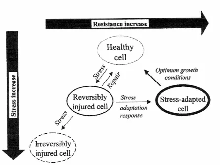
(29) Introduction. Figure 2. Microbial stress, injury, adaptation and resistance to processing. Source: Lado and Yousef (2002).. 1.5 Kinetics of microbial inactivation The traditional approach to inactivation kinetics is based on the assumption that microorganisms or their spores die exponentially following first-order kinetics (Mussa and Ramaswamy, 1997; Gervilla et al., 1999). It also assumes that cells or spores have identical pressure resistance. Mussa et al. (1998) inoculated raw milk (containing the natural microbiota) with L. monocytogenes Scott A. Samples were subjected to different pressure treatments (150 to 400 MPa) for selected holding times of up to 120 min to evaluate kinetics of destruction of this microorganism. These authors found that all the pressures applied followed a first order rate of death in the destruction of L. monocytogenes as well as microbiota present in raw milk. Dogan and Erkmen (2004) determined high hydrostatic pressure kinetics of aerobic bacteria and L. monocytogenes inactivation in brain heart infusion broth, milk and in peach and orange juice. For this purpose, different pressures (200-700 MPa) at 25ºC for different holding times (25 to 100 min) were applied. The decimal reduction times (D values) that these researchers obtained were about 3.04 and 2.43 min for aerobic bacteria and L. monocytogenes, respectively, in milk at 600 MPa, 2.13 and 1.52 min, respectively, in peach juice, and 1.24 and 0.87 min, respectively, in orange juice.. 19.
(30) Introduction However, deviations in log-linear models have been detected in form of tails (biphasics) and shoulders (Heldman and Newsome, 2003). Several authors have detected this behaviour. Kinetics of microbial inactivation of L. monocytogenes was not linear in time and Patterson et al. (1995) stated that it was not possible to calculate conventional D values as a method to determine relative pressure sensitivities of different organisms. In general, inactivation curves did not follow first-order kinetics but tended to be exponential, with an initial decrease in numbers, during the first 15 min of treatment followed by a tail, suggesting that a small fraction of the population was more pressure resistant. Gervilla (2001) reported that, in some of the microorganisms analysed, the kinetics changed from first order to second order after the first 15-20 min of high hydrostatic pressure treatment. A small group of cells could survive after a considerably long treatment that was expected to be completely lethal. This behaviour suggested that cells had the ability to adapt to the medium in high pressure conditions. However, Metrick et al. (1989) worked with Salmonella spp and pressurized the supposed resistant cells a second time. Comparing the resistance to pressure between the original culture and the remaining population, they found no significant changes. Tay et al. (2003) investigated the inactivation kinetics of L. monocytogenes Scott A (pressuresensitive strain) and OSY-8578 (pressure-resistant strain) at 350 and 800 MPa. Firstorder kinetics was not suitable to describe the inactivation, and extended pressure treatment did not eliminate the tailing phenomenon. Chen and Hoover (2003) reported a strong tailing in the survival curves of Y. enterocolitica obtained at four pressure levels (300, 350, 400 and 450 MPa) in PBS and whole milk. They indicated that the best models for predicting pressure inactivation of this organism were the non-linear regression and the Weibull models and suggested that in each food is necessary to develop new models to predict the inactivation kinetics of microorganisms. Moreover, these authors (2004) studied also the survival curves of L. monocytogenes Scott A inactivated by high hydrostatic pressure at seven pressure levels (300, 350, 400, 450, 500, 550 and 600 MPa) in UHT whole milk. In these case, the shapes of the survival curves were concave upward or downward depending on the treatment pressure levels and the Weibull model produced also a better fit than the linear model. Guan et al. (2005) studied the inactivation of S. typhimurium DT 104 in UHT whole milk by high hydrostatic pressure applying different pressures (350, 400, 450, 500, 550, and 600 MPa) at room temperature and using four modelling methods (linear, Weibull, modified Gompertz and log-logistic models) to fit the results. A strong tailing was observed in all 20.
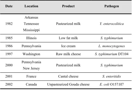
(31) Introduction survival curves and log-logistic model was the model that produced the best fit to survival data. 1.6 Milk and dairy products as a vehicle of foodborne pathogens Milk is a nutritious medium that presents a favourable environment (near neutral pH) for the multiplication of microorganisms and supports a wide range of spoilage and pathogenic bacteria. Traditionally, raw or unpasteurized milk has been a major vehicle for transmission of pathogens. Dairy products have long been recognized as being susceptible to postprocessing contamination (Donnelly, 1990). Food poisoning outbreaks and other illnesses involving milk and milk products have been reported since the beginning of the dairy industry (Table 3). Nowadays, special attention is focused on milk and dairy products contaminated with pathogenic bacteria as L. monocytogenes, Y. enterocolitica, Campylobacter jejuni, A. hydrophila and enteropathogenic E. coli (Vasavada, 1988; Donnelly, 1990; Klausner and Donnelly, 1991; Jayarao and Henning, 2001). Table 3. Some outbreaks associated with milk and dairy products contaminated with pathogenic bacteria Date. Location. Product. Pathogen. Pasteurized milk. Y. enterocolitica. Arkansas 1982. Tennessee Mississippi. 1985. Illinois. Low fat milk. S. typhimurium. 1986. Pennsylvania. Ice cream. L. monocytogenes. 1997. Washington. Raw milk cheese. S. typhimurium DT104. Pasteurized milk. S. typhimurium. 2000. Pennsylvania New Jersey. 2001. France. Cantal cheese. S. enteritidis. 2002. Canada. Unpasteurized Gouda cheese. E. coli O157:H7. Sources: Vasavada, 1988; Villar et al., 1999; Haeghebaert et al., 2003; Olsen et al., 2004; Honish et al., 2005.. 21.
(32) Introduction Heat treatment is the oldest and most widely used technological process applied to milk (Table 4). Table 4. Heat treatments more common used in dairy industry Temperature Time Treatment (ºC) (s or min) Termization. 63-65. 15-30 s. High Temperature Short Time (HTST). 72-75. 15-20 s. Low Temperature Long Time (LTLT). 63-65. 30 min. 125-138. 2-4 s. Ultra High Temperature (UHT). 135-140. 2-4 s. In-bottle. 115-120. 20-30 min. Pasteurization. Ultra Pasteurization Sterilization. Source: Gervilla, 2001 The principal objective of pasteurization is the elimination of pathogens and the reduction of the natural microbiota that can be present in milk. But it has been reported that pasteurization may not destroy all foodborne pathogens in milk due to pathogens such as L. monocytogenes that can survive and thrive in post-pasteurization processing environments, thus leading to recontamination of dairy products (Oliver et al., 2005). Traditionally, the heat treatment usually applied to cheese milk is pasteurization. However, some researchers have found differences between cheeses made from raw and pasteurized milk; cheeses made from raw milk tend to develop a stronger flavour and generally ripen faster than cheeses made from pasteurized, due to inactivation of enzymes and destruction of the heat sensitive microbiota by pasteurization treatment (Grappin and Beuvier, 1997).. 22.
(33) Introduction 1.7 Gram negative bacteria used for this research 1.7.1 Yersinia enterocolitica Y. enterocolitica is a facultative anaerobic, Gram negative, non-spore forming, short rod-shaped bacterium, currently classified as part of the family Enterobacteriaceae (ICMSF, 1998). It has been isolated from animals belonging to various taxonomic groups, from mammals to insects. This microorganism comprises over 60 serotypes but only a few are pathogenic for humans (Kapperud, 1991). This organism has been associated with human gastro-intestinal disease called yersiniosis, but may also cause further infections including skin, eye, muscle and wound abscesses, endocarditis and pharyngitis, and postinfection immunologic consequences in the form of arthritis and its complications, erythema nodosum, and thyroid disorders (Schiemann, 1987). The majority of Yersinia infections are food-borne. The spread of the disease is mainly dependent on the degree of hygiene standard at every step of the technological process of food processing, storage and distribution (Stojek, 1999). Y. enterocolitica has been isolated from humans in many countries, but it seems to be most frequently found in cooler climates (Kapperud, 1991). In Europe, Canada and Japan, the majority of cases are due to Y. enterocolitica serogroup O:3 and to O:9 on a lesser extent, and are caused by consumption of pork products. Pigs are regarded as major reservoirs of pathogenic Y. enterocolitica since it is often carried in the oral cavity or intestinal tract of healthy exemplars (Tauxe et al., 1987; Doyle, 1990; Andersen et al., 1991). In contrast, in the United States, the prevalent serotype between 1976 and 1982 had been O:8. However, after 1988, several US outbreaks of Y. enterocolitica O:3 suggested a serogroup shift (Ackers et al., 2000). In Spain, O:3 is the most frequent serotype identified in human yersiniosis cases (Anonymous, 2001). The organism has been frequently isolated from raw milk and even from pasteurized milk (Larkin et al., 1991). Milk has been implicated in several outbreaks of yersiniosis. The cases with more people affected have been reported in the United States. In September and October 1976, an outbreak of illness linked to consumption of chocolate milk contaminated with Y. enterocolitica resulted in the hospitalization of 36 children. Investigation of this outbreak revealed that Yersinia was introduced during hand mixing. 23.
(34) Introduction of unpasteurized chocolate syrup with pasteurized milk in an open vat (Black et al., 1978). In July 1981, gastrointestinal disorders of varying severity were observed in 239 campers and staff members at a summer camp in New York State. Y. enterocolitica serogroup O:8 was isolated from 54% of persons examined. Isolates belonging to the same serogroup and biogroup of human isolates were recovered from dissolved powdered milk and a milk dispenser, amongst others (Morse et al., 1984). In June and July 1982, a large interstate outbreak of yersiniosis occurred in Tennessee, Arkansas and Mississippi. Pasteurized milk was epidemiologically implicated as the vehicle of the infection (Tacket et al., 1984). Although yersiniosis outbreaks from cheese have not been reported, some studies have showed the ability of Y. enterocolitica to grow in Brie cheese (Little and Knochel, 1994) and survive in Turkish feta cheese (Erkmen, 1996). The psychrotrophic nature of this organism is of particular significance in milk and milk products that are normally stored at low temperatures. In raw milk, Y. enterocolitica strains could survive in presence of high numbers of competing microorganisms and could maintain the virulence plasmid during extended storage at refrigeration temperatures (Larkin et al., 1991). In pasteurized milk, contamination has been mainly attributed to the inadequate pasteurization or post process contamination (Klausner and Donnelly, 1991; Kushal and Anand, 1999).. 1.7.2 Salmonella spp. Salmonella is ubiquitous in nature and the serotypes that can cause human infections occur in all types of animals, including a significant but unknown number of domestic animals. Infections exhibit a number of clinical presentations, but gastrointestinal disorders are the most common clinical manifestation. The severity and duration of symptoms depend on the type of Salmonella present, the amount of food eaten and the susceptibility of the person involved (El-Gazzar and Marth, 1992). The two most prevalent serovars of Salmonella currently isolated from foodborne outbreaks in USA and Europe are S. enterica serovar enteritidis and S. enterica serovar typhimurium (Mattick et al., 2001; Anonymous, 2003). Moreover, S. enterica serovar enteritidis is one of the Salmonella serotypes most commonly associated with morbidity and mortality in humans (Ahmed et al., 2000). 24.
(35) Introduction. Raw and pasteurized milk and different types of cheeses have been involved in several outbreaks (CDC, 1985; Table 3). Some studies have showed that when milk becomes contaminated with Salmonella spp. after pasteurization, the pathogen could survive the cheese making process and persist for several months in the cheese (Leyer and Johnson, 1992). In ripened Cheddar cheese, Salmonella counts were detected for up to 7 months at 7ºC. In cold-packed cheeses, cells were found depending on the pH value and preservative used (El-Gazzar and Marth, 1992). Moreover, fat and proteins in cheese can protect foodborne pathogens from gastric acidity, reducing the number of organisms necessary to cause clinical infections (Altekruse et al., 1998). These trends increase the need of detecting low numbers of Salmonella cells in cheeses .. 1.7.3 Escherichia coli E. coli is a Gram negative, non-spreforming, rod-shaped organism. Different groups have been recognized, namely, enteropathogenic, enterotoxigenic, enteroinvasive, enteroaggregative and enterohaemorrhagic (Vasavada, 1988; Nataro and Kaper, 1998; Gonzalez Garcia, 2002). E. coli O157:H7 has emerged as a foodborne pathogen of major concern for the food industry due to its ability to cause severe illness, in particular haemorrhagic colitis, haemolytic uraemic syndrome and thrombotic thrombocytopenic purpura. Dairy cattle is considered a main reservoir of E. coli O157:H7 for human infection (Doyle, 1991) being fecal contamination of milk an important vehicle for its transmission (Borczyk et al., 1987; Gonzalez Garcia, 2002). This pathogen has been isolated from raw milk (Reitsma and Henning, 1996), and multiple outbreaks of E. coli O157:H7 linked to ingestion of raw milk and dairy products have been reported (Altekruse et al., 1998; Honish et al., 2005). In many countries, the fact that raw milk is used for cheese manufacture increases the risk that pathogenic bacteria would contaminate the product (De Buyser et al., 2001). Several authors have studied the ability of E. coli O157:H7 to grow and survive in different types of cheese. In fresh cheese, E. coli O157:H7 grew 2 log (cfu/g) during cheese 25.
(36) Introduction manufacture (Arocha et al., 1992), but total inactivation was obtained during the heat treatment. Reitsma and Henning (1996) found that this microorganism was able to grow during Cheddar cheese manufacture even with an initial inoculum in milk of 1 cfu/ml. E. coli O157:H7 also survived the manufacture of Camembert and Feta cheeses (Ramsaran et al., 1998) and reached counts higher than those present in milk after 75 and 65 days of storage, respectively. Moreover, Maher et al. (2001) found that this pathogen was able to survive all stages of smear-ripened cheese production up to 70 days post-manufacture.. 1.8 Effect of high hydrostatic pressure on microorganisms in milk The interest in high hydrostatic pressure for treatment of milk has increased mainly due to the possibility of reducing the microbial number without significant effects on flavour or nutritional components. In fact, several studies have currently examined pressure inactivation of microorganisms that are either naturally present in milk or introduced into the milk artificially. Styles et al. (1991) studied the effect of high pressure treatments in raw and UHT milk inoculated with 6 log (cfu/ml) of L. monocytogenes Scott A. The total inactivation of this microorganism was obtained after treating samples at 340 MPa for 80 min, in the case of UHT milk, and 60 min, in the case of raw milk. López-Fandiño et al. (1996) evaluated the effects of high pressure treatments (from 100 to 400 MPa) applied for different periods (10 to 60 min), on the biochemical and microbiological characteristics of raw milk. Psychrotrophic bacteria were reduced with pressure more quickly than total bacterial counts. After applying 300 MPa for 30 min and 10 min at 400 MPa, no psychrotrophic bacteria were detected by the plate count method (<10 cfu/ml). These authors suggested that this failure in growth initiation could be due to the lower incubation temperature and these microorganisms could have been regenerated under more favourable conditions. Gervilla et al. (1997b) inoculated ovine milk (6% fat) with E. coli and P. fluorescens at a rate of 106 and 107 cfu/ml, respectively and treated the samples with different combinations of pressure (300, 400, 450 and 500 MPa), temperatures (2, 10, 25 and 26.
(37) Introduction 50ºC) and times (5, 10 and 15 min). Inactivation (over 6 log [cfu/ml]) of both strains was observed at 50ºC for all the pressures and times and the lowest reduction was observed at 10ºC for E. coli and at 25ºC for P. fluorescens. Mussa and Ramaswamy (1997) used different combinations of pressure (200-400 MPa) and times (5 to 20 min) to inactivate microorganisms present in raw milk. These authors found that milk subjected to a microbial inactivation of 4 times de D value at 350 MPa had a shelf-life of 25 days at 0ºC, 18 days at 5ºC or 12 days at 10ºC. Patterson and Kilpatrick (1998) used different combinations of pressure, temperatures and times to eliminate the population of E. coli O157:H7 and S. aureus inoculated in UHT milk and poultry. In UHT milk, population of E. coli was reduced 5 log (cfu/ml) when was treated at 400 MPa for 15 min at 50ºC and for S. aureus, the reduccion was approximately 6 log (cfu/ml) when it was treated at 500 MPa for 15 min at 50ºC. Mussa et al. (1998) applied different pressures (150-400 MPa) for selected holding times of up to 120 min to raw milk inoculated with 106 cfu/ml of L. monocytogenes Scott A to study the kinetics of destruction of this microorganism and the natural flora present in the milk. At 400 MPa, the instantaneous pressure kill (destruction achieved due to application of a pressure pulse with no hold-time) was higher for L. monocytogenes than the normal flora (2.10 log [cfu/ml]). The effects of high pressure on the viability and acidifying and peptidolytic activities of L. lactis ssp. lactis, L. casei ssp. casei and L. plantarum strains were studied by Casal and Gómez (1999). Cells, inoculated in 10% reconstituted bovine skim milk, were treated at different pressures (100 to 400 MPa) for 20 min at 20°C. The Lactococci were more sensitive than the Lactobacilli to pressures of 100 to 350 MPa. The pressuretreated cells exhibited lower acidification rates, even with treatments that did not affect cell viability and increased the hydrolytic activity of these microorganisms on the carboxyl-terminal fragment from b-casein (C-peptide), which contributes to bitterness in cheese. Gervilla et al. (1999) inoculated ovine milk with S. aureus and L. helveticus at a concentration of 107 cfu/ml and treated the samples using different combinations of pressures (200, 300, 400, 450 and 500 MPa), temperatures (2, 10, 15, 25 and 50ºC) and times (5, 10 and 15 min). These authors found that S. aureus was extremely resistant to pressure and cell reductions above 7 log (cfu/ml) were only achieved after applying 500 MPa at 50ºC for 15 min. For L. helveticus the pressure treatments were more effective 27.
(38) Introduction at low (2 and 10ºC) and moderately high (50ºC) temperatures than at room temperature (25ºC). Linton et al. (2001) studied the pressure resistance of a range of pathogenic E. coli in skimmed milk applying different pressures (200-700 MPa) for 15 min at 20ºC. A pressure treatment of 500 MPa for 40 min gave only a 4 log (cfu/ml) reduction of the two most resistant strains (NCTC 11601 and NCTC 9706) but after a treatment of 600 MPa for 30 min, no survivors of either strain could be detected (> 7 log [cfu/ml]). Molina-Höppner (2002) reported that high pressure treatments of L. lactis spp. cremoris MG1363 inoculated in milk buffer, initially affected metabolic activity and subsequently damaged membrane integrity. After a treatment for 5 min at 300 MPa the metabolic activity was 10-12% of the activity of untreated microorganisms and after 12 min of treatment the cells did not show any metabolic activity. During the treatment at 300 MPa cell death was closely followed by the loss of metabolic activity, but cultures retained about 25% of metabolic activity, even after 3 log (cfu/ml) reduction in cell counts. Bozoglu et al. (2004) studied the effect of high pressure treatments on two Gram positive (L. monocytogenes and S. aureus) and two Gram negative (E. coli O157:H7 and S. enteritidis) bacteria inoculated in milk and the formation of injured cells. The pathogens were pressurized at 350, 450 and 550 MPa for 10 min and stored at 4, 22 and 30ºC over a period of four weeks. Except for L. monocytogenes where there was survival at 350 MPa (colony formation on both selective and non-selective agars), pathogens were either inactivated or injured at all pressures studied. Gao et al. (in press) employed response surface methodology (RSM) to create a quadratic equation to predict the optimum process parameters for a 6 log (cfu/ml) reduction of L. monocytogenes inoculated in milk buffer. The conditions found to obtain this reduction with this microorganism were a pressure treatment at 448 MPa for 11 min at 41°C. Moreover, several researchers have studied the potential of combining high hydrostatic pressure treatments with antimicrobial compounds to reduce the pressures required for microbial inactivation in milk. Morgan et al. (2000) evaluated the combination of high pressure (150-600 MPa, 30 min, 25ºC) and lacticin 3147 (10,000 AU/ml) to improve the inactivation of S. aureus ATCC6538, inoculated in 10% reconstituted skimmed milk, 28.
(39) Introduction and L. innocua DPC1770, inoculated in 20% reconstituted demineralised whey powder. The results showed a more than additive effect when both treatments were used in combination for both bacterial species. In the case of S. aureus, up to 200 MPa little or no effect on viability was observed. Above this pressure, a rapid decline in viability occurred, with approximately a 6 log (cfu/ml) kill being detected at 300 MPa. L. innocua demonstrated results similar to those observed for the other strain up to 200 MPa. At 275 MPa, a 3 log (cfu/ml) reduction in viable cell numbers was observed and, at 325 MPa, viable cells were not detectable and the reduction in population was greater than 6 log (cfu/ml). García-Graells et al. (2003) studied the inactivation of different bacteria (E. coli MG1655 and LMM1010, S. typhimurium, P. fluorescens, S. aureus, Enterococcus faecalis, L. innocua and L. plantarum) by high pressure in skimmed milk supplemented with the lactoperoxidase-hydrogen peroxide-thiocyanate (LP) system at naturally concentration. In absence of pressure treatment, the effect of LP system varied with the bacterial strain from bactericidal (P. fluorescens) to bacteriostatic or inhibitory (E. coli MG1655, L. innocua, S.aureus, L. plantarum and E. faecalis), or no effect (S. typhimurium and E. coli LMM1010). The presence of the LP system affected inactivation by high pressure in a cell density dependent manner. At low concentration (106 cfu/ml), the LP system strongly increased high pressure inactivation except E. coli LMM1010 (pressure resistant strain) and at high concentration (109 cfu/ml) only inactivation of L. innocua, E. faecalis and L. plantarum were enchanced at relatively mild pressures (400-450 MPa). The behaviour of Gram negative (E. coli and P. fluorescens) and Gram positive bacteria (L. innocua and L. viridescens) associated with milk, in response to application of high pressure (250–500 MPa, 5 min, 20ºC) combined with nisin (0, 250, or 500 IU/ml) was evaluated by Black et al. (2005). These authors found that the combination of high pressure and nisin not only showed a synergistic effect to enhance the level of inactivation for bacteria in milk but also an effective effect in sensitising E. coli and P. fluorescens to this bacteriocin.. 29.
Figure
+7


Documento similar
and (iv) the in vitro digestive stability and bioaccessibility of bioactive compounds in control and HHP-treated prickly pear fruits. On one hand, pulps were studied due to
Samples of “Turrialba cheese” (Latin-type fresh cheese) were obtained from different locations in Costa Rica to determine the levels of contamination with
Improvement of the Rett Syndrome phenotype in a Mecp2 mouse model upon treatment with Levodopa and a Dopa Decarboxylase Inhibitor?. The lack of Mecp2 protein results
To test the direct effect of bacteria on the r-SDF, sperm samples from 6 different bulls classified in the group of individuals without bacteria were
With regard to redness, the recorded values at the beginning of storage were lower than those reported for fresh seabream fillets (Andrés-Bello et al., 2015; Oliveira et al.,
Parsi [39] compared an online care model for follow-up treatment of patients with psoriasis with a conventional in-office model and did not find significant differences in
monocytogenes due to the application of high-pressure processing (HPP) after ripening and storage of fuet-type (FT) and snack-type (ST) sausages spontaneously fermented (no starter)
Effect of high hydrostatic pressure and thermal processing on the nutritional quality and enzyme activity of fruit smoothie... Effects of combined treatment of
