Molecular and serological characterization of
occult hepatitis B infection in blood donors from Mexico
Beatriz María García-Montalvo,* Laura Patricia Ventura-Zapata** Centro Médico Nacional Ignacio García Téllez, Instituto Mexicano del Seguro Social, Mérida, Yucatán, Mexico.
ABSTRACT
Occult hepatitis B virus (HBV) infection (OBI) is characterized by presence of HBV DNA in blood or liver tis-sue without detectable HBV surface antigen (HBsAg), with or without antibodies to hepatitis B core anti-gen (anti-HBc) or antibodies against HBsAg (anti-HBs). A molecular and serological characterization was done of OBI in blood donors from Yucatan, Mexico. HBV DNA was found in 24 (6.4%) of the 372 evaluated samples. Anti-HBs was present in 15/24 samples (62.5%), and no significant difference was observed bet-ween HBV DNA positivity and anti-HBs levels. HBV genotype H was detected in 66.7% of samples, followed by genotypes D (20.8%) and F (8.3%). Amino acid substitutions were identified in the core region of nine samples, and most of these changes were located in immunodominant epitopes. No precore stop codon 28 mutant (W28Stop) was identified among the analyzed HBV isolates. In conclusion, genotype H is the main circulating HBV strain among OBI blood donors from Yucatan, Mexico. Mutations in the core region may contribute to viral persistence.
Key words. Blood donors. Occult hepatitis B. Genotype. Mutations. Hepatitis B core protein.
Correspondence and reprint request: Dra. Beatriz María García-Montalvo Calle 19 No. 430 x 50, Fracc. Jardines de Mérida
97135 Mérida, Yucatán, Mexico. Tel.: +52 (999) 943-61-36. Fax: +52 (999) 922-27-84 E-mail: [email protected]
Manuscript received: October 12, 2010. Manuscript accepted: November 21, 2010.
INTRODUCTION
Serological evidence of acute or chronic hepatitis B is commonly generated by hepatitis B surface anti-gen (HBsAg) assays. This sensitive serological test for HBsAg detection is widely applied but transmis-sion of hepatitis B virus (HBV) infections by trans-fusion continues to occur.1,2 Occult HBV infection
(OBI) is characterized by presence of HBV DNA in blood or liver tissue with no detectable HBsAg, and with or without antibodies to hepatitis B core anti-gen HBc) or antibodies against HBsAg (anti-HBs).3,4 Clinical observations suggest that OBI
carriers may be a HBV transmission source via blood transfusion or orthotopic liver transplanta-tion. It is also considered a possible risk factor for developing hepatocarcinoma.4 Carrier rates for
HB-sAg vary geographically and Mexico, like other La-tin American countries, is considered a low endemi-city area (< 2% of population are carriers).5
However, high anti-HBc prevalence (91.1%) has been reported in populations on Mexico’s southern border.6 Very little data has been collected on OBI
prevalence among blood donors in Mexico.7 The
pre-sent study objective was to molecularly and serolo-gically characterize OBI in a population of blood donors from Mexico.
METHODS
Sample selection
were selected for further analysis. These were sto-red at -20 °C until analysis. Donors provided writ-ten informed consent at the time of blood donation and this study was approved by the Institution Ethics Committee.
Serological and Biochemical assays
All anti-HBc-positive plasmas were further tested for anti-HBs by MEIA AxSYM AUSAB (Abbott La-boratories, Abbott Park, IL, USA). DNA-positive samples were also tested for hepatitis B e antigen (HBeAg) and antibodies to HBeAg (anti-HBe) using commercial kits (ARCHITECT, Abbott Laboratories, Wiesbaden, Germany). Alanine aminotransferase (ALT) levels were identified with an automated Di-mension RXL (Dade Behring Inc., Newark, USA) fo-llowing manufacturer instructions.
HBV DNA detection
DNA was extracted from plasma samples using a commercial kit (QIAamp DNA Mini Kit, QIAGEN GmbH, Hilden, Germany) following manufacturer instructions. HBV DNA identification was done for all 372 plasma samples by PCR amplification of a 560-bp fragment of the C gene, followed by nested amplification of a 438-bp fragment. The outer pri-mers were: sense (5'-TTCAAGCCTCCAAGCTGT-GCCTTGG-3', nt 1863 to 1887) and antisense (5'-TCTGCGACGCGGCGATTGAGA-3', nt 2402 to 2422). The inner primers were: sense (5'-CCTTGGG-TGGCTTTGGGGCA-3', nt 1882 to 1901) and anti-sense (5'-AGGATAGGGGCATTTGGTGGTCTATA-3', nt 2294 to 2319).8 First-round amplification was
done in a volume of 50 µL, containing New England BioLabs 1 x PCR buffer, 0.5 µM of each primer, 0.2 mM dNTP mix; 2.5 U Taq polymerase (New En-gland BioLabs) and 10 µL DNA. The reaction was run in a MyCycler Thermal Cycler (Bio-Rad) using the following specifications: initial denaturation at 94 °C for 5 min; followed by 40 cycles at 94 °C for 1 min; 63 °C for 1 min; and 72 °C for 2 min; and final extension at 72 °C for 10 min. The second PCR round was done under the same conditions except that annealing was done at 55 °C with 10 µL of the first-round product. PCR-amplified products (10 µL) were separated by electrophoresis on 2% agarose gel, stained with ethidium bromide and viewed un-der ultraviolet light. Recombinant HBV DNA sam-ples were used as positive controls for PCR amplification and for establishing the lower detec-tion limit of the assay as described previously.7
De-tection limit for the nested PCR assay was approxi-mately 30 copies per mL. Plasma samples from blood donors negative for all HBV serological mar-kers and reagents without DNA were used as nega-tive controls. Neganega-tive and posinega-tive controls were added to each amplification round. To prevent cross-contamination, DNA extraction and PCR reaction mixture preparation were done in rooms separate from that in which the amplified samples were handled. Furthermore, all instruments used in the procedures were ultraviolet-irradiated before use. A sample was considered positive when repea-tedly found positive after amplification of newly ex-tracted material.
DNA sequencing
After 2% agarose gel electrophoresis, the second-round PCR products were purified using a commer-cial kit (QIAquick Gel Extraction Kit, QIAGEN GmbH, Hilden, Germany). Direct sequencing of the purified product was done with the dideoxy chain termination method using the same sense and anti-sense inner primers used in the PCR, with a BigDye Terminator Cycle Sequencing Kit and a 3730xl DNA Analyzer (Applied Biosystems, Foster City, CA, USA).
Sequence analysis
Statistical analysis
Statistical differences were evaluated by applying a Chi-square test and a Fisher’s exact test. P values less than 0.05 were considered significant. All analy-ses were done using the SPSS software package (Version 14, SPSS Inc. Chicago, Illinois, USA).
RESULTS
Serological characterization
Of the 372 blood donors positive for anti-HBc and negative for HBsAg, 202 (54.3%) were also anti-HBs-positive. Twenty-four (6.4%) of the 372 evaluated anti-HBc positive samples were also DNA-positive and thus categorized as OBI carriers. All the HBV DNA-positive samples were identified as such only af-ter a second PCR amplification round. No HBV DNA was detected in any of the negative controls. In the positive controls, a visible 560-bp DNA band was de-tected after the first amplification round and a 438-bp DNA band after the second round. All donors were clinically asymptomatic and none of the viremic do-nors had previously received HBV immunization. Additional serological markers such as anti-HBe and
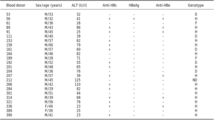
HBeAg were evaluated using HBV DNA-positive sam-ples. No significant difference (P = 0.53) was found between HBV DNA positivity and anti-HBs levels. OBI-positive donor serological, biochemical and mole-cular data are summarized in Table 1.
Genotype identification
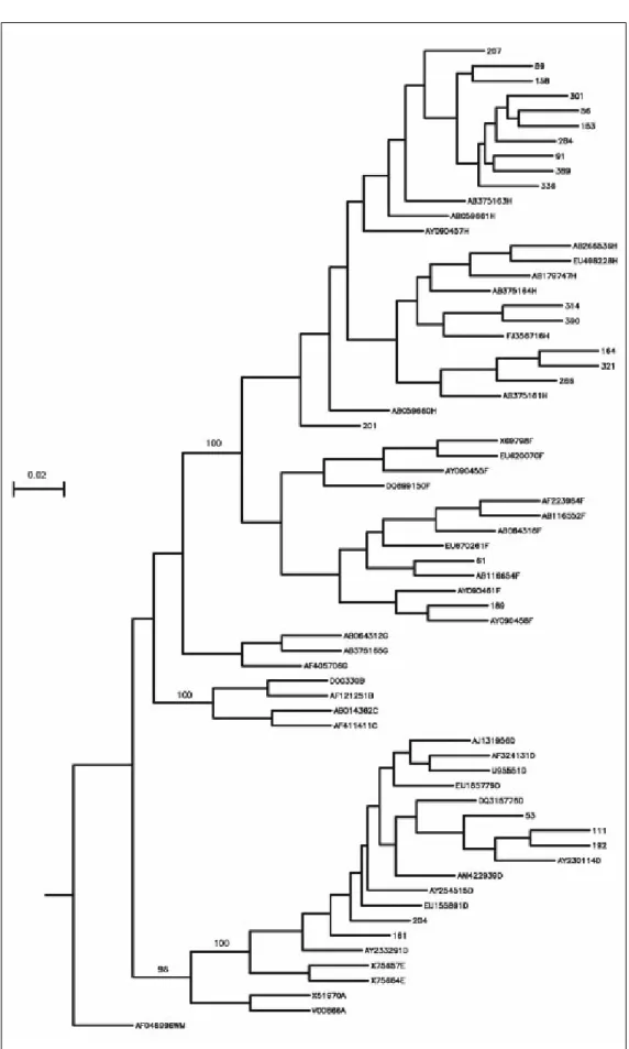
Phylogenetic analysis comparing donor sequen-ces with those of various HBV genotype strains re-vealed that sixteen (66.7%) isolates belonged to genotype H, 5 (20.8%) to genotype D and 2 (8.3%) to genotype F (Figure 1). Genotype could not be de-termined in one (4.2%) donor, and therefore the results include only 23 donors.
C gene mutations
In an effort to identify mutations in the precore and core regions, an analysis was done of the 23 sequences with an identified genotype by comparing each geno-mic fragment sequence with a consensus sequence constructed from representative strains of each isolate’s respective genotype. The GenBank accession numbers for the H, D and F genotypes used to build the consensus sequence were the same as those used in
Table 1. Serological, biochemical and molecular data of 24 donors with OBI.
Blood donor Sex/age (years) ALT (U/I) Anti-HBs HBeAg Anti-HBe Genotype
53 M/53 32 - - - D
56 M/32 41 + + + H
61 M/36 26 - - - F
89 M/43 86 + - + H
91 M/45 25 + - + H
111 M/40 39 - - - D
153 M/57 62 + - - H
158 M/60 79 + - - H
161 M/57 60 + - - D
164 M/46 82 - - - H
189 M/28 71 - - - F
192 M/52 55 + - - D
201 M/48 65 + - - H
204 M/36 76 - - - D
207 M/57 39 + - + H
212 M/45 125 - - - ND
266 M/42 110 + - - H
284 M/29 82 + - - H
301 M/51 44 - - - H
314 M/39 68 + - + H
321 M/56 76 + - - H
336 F/49 23 + - + H
389 F/39 25 - - - H
390 M/41 23 + - - H
ND: ND: ND: ND:
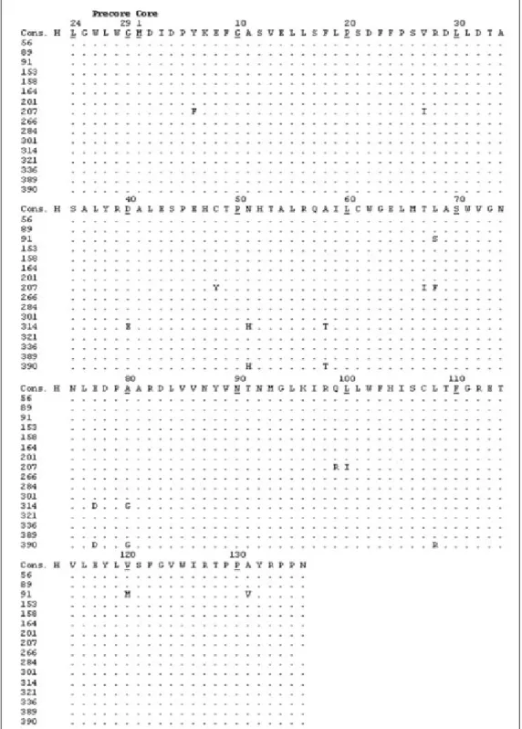
Figure 2. Deduced ami-no acid sequences encoded by a portion of the hepati-tis B virus (HBV) C gene from blood donors with oc-cult hepatitis B infection. Each donor’s sequence was aligned with reference se-quences of HBV genotype H (consensus sequence). Numbers indicate amino acid position within the protein and each dot co-rresponds to a non-variable amino acid.
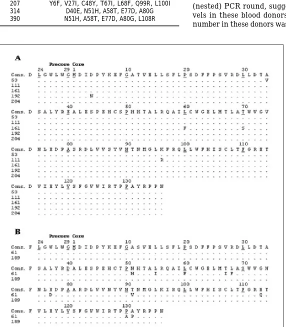
the phylogenetic analysis (Figure 1). Amino acid chan-ges were detected in HBV isolates from nine donors (Table 2, Figures 2 and 3), while the remaining four-teen HBV isolates had wild type amino acid sequences. Of the nine samples with amino acid mutations in the core protein, six (91, 161, 192, 207, 314, and 390) were
Table 2. Amino acid substitutions in the C region.
Blood donor Amino acid change
53 A34V
61 N51H, L55I, L60F; T67I, L68F, E77D, T91V, E113Q, P130A, A131P 91 L68S, V120M, A131V
111 K96R
161 L60F, T70S
192 D4N
207 Y6F, V27I, C48Y, T67I, L68F, Q99R, L100I 314 D40E, N51H, A58T, E77D, A80G 390 N51H, A58T, E77D, A80G, L108R
Figure 3. Deduced ami-no acid sequences encoded by a portion of the hepati-tis B virus (HBV) C gene from blood donors with oc-cult hepatitis B infection. Each donor’s sequence was aligned with reference se-quences for the same geno-type. Consensus sequences: A) HBV genotype D; and B) HBV genotype F. Numbers indicate amino acid position within the protein and each dot corresponds to a non-variable amino acid.
codon 28 mutant (W28Stop) can interfere with secre-ted HBeAg production, but none was observed among the 23 analyzed HBV isolates (Figures 2 and 3). Nei-ther insertions nor deletions were recorded within the C gene.
DISCUSSION
Blood donor screening for HBV infection in Mexi-co involves only HBsAg. However, this marker may not be detected for a number of reasons, such as HBV infection during the window period, OBI and presence of HBsAg escape mutants.9 Studies in
other countries have shown that HBsAg seronegati-ve donors can transmit HBV to these units’ recep-tors.1,10,11 In Mexico, very little research has been
done on OBI occurrence,7,12,13 and no data is
availa-ble on post-transfusion HBV incidence.
Previous reports have shown that OBI is charac-terized by a very low viral load.3,14 In the present
Prevalence of OBI individuals (with or without detectable anti-HBs) varies depending on the stu-died population. For example, the 7.4% observed in the present study is notably higher than reported for Venezuela in HBsAg-negative donors with HBV DNA who are also anti-HBc and anti-HBs positive (1.42, 4.8 and 4.3%),15-17 and higher than reported
for Germany (1.59%).18 The 62.5% of donors with
OBI as well as anti-HBs in the present study is com-parable to the 42.86% reported in Greece19 and the
63.63% reported for Venezuela.17 No association was
identified here between HBV DNA positivity and anti-HBs levels, which coincides with other repor-ts.17 This is a vital aspect since transmission via
blood containing anti-HBc and anti-HBs has been reported recently.2 The present finding of high HBV
DNA prevalence among HBsAg-negative blood do-nors from Mexico is particularly significant because Mexico is considered a low HBV prevalence region. Roman, et al.13 have suggested that the high
anti-HBc prevalence versus the low HBsAg prevalence observed in Mexico may be an artifact of the limited sensitivity immunoassays used for HBsAg detection in the country and/or the presence of as yet uniden-tified immunogenetic characteristics within the Mexican population.
Very little research has been done on HBV geno-type distribution in Mexico and most reports have been done on HBsAg-positive individuals.20-23 The
HBV genotype distribution pattern observed here is similar to that reported by Sánchez et al.21 in a
group of patients with chronic and acute hepatitis; the exception is genotype A, which was not observed in the present study. As reported previously for HBV in Mexico,13,21-23 the H genotype was found to
be dominant in the present study. To our knowled-ge, no previous reports exist of HBV genotypes in OBI blood donors from Mexico.
The hepatitis B core protein (HBc) is highly im-munogenetic whereas HBV envelope proteins have comparatively low immunogeneticity.24 This means
that HBc is an important target for immune-media-ted viral clearance via inducement of B cell, T hel-per cell and cytotoxic T limphocyte (CTL) responses.25 Important B cell epitopes of the HBc
protein are located near amino acid sequences 74-89 and 126-135,25,26 while important HBc protein
im-mune recognition sites for T helper epitopes are at sequences 1-20, 28-47, 50-69, 72-105 and 108-165, and those for CTL epitopes are at 18-27, 88-96 and 141-151.25-27 In the present results, mutations in
im-munodominant epitopes were identified in 9/23 (39.1%) of the analyzed OBI donors.
Changes in amino acids were observed in positions 40, 51, 58, 60, 77, 80 and 91, which coincides with re-ports for OBI donors in Venezuela.17 Amino acid
substitution also occurred at position 27 in one of the studied OBI donors. In a study of patients administe-red interferon therapy, Naoumov, et al.28 reported
that HBeAg-positive patients did not respond initially or subsequently developed amino acid changes in the CTL epitope (residues 21 and/or 27), whereas these mutations did not occur in patients who responded. Mutant HBV strains resistant to antiviral therapy have also been reported in chronic asymptomatic HBV carriers who have not received treatment.29
Substitution of amino acids at position 27 may in-fluence immune modulation, which may in turn con-tribute to development of viral persistence.30
Amino acid substitution was also observed here in positions 77 and 80, which is known to reduce HBe and HBc antigenicity.31 Substitution of
gluta-mine for arginine at position 99 within the CTL epitope was identified in the present study in a do-nor negative for HBeAg but positive for anti-HBe, a phenomenon reported elsewhere in a chronic liver disease patient who was also HBeAg-negative and anti-HBe-positive.32 In addition, six OBI donors
ex-hibited amino acid changes in the hepatitis B core antigen hot-spot mutational domain (residues 80-120) reported to be associated with severe forms of liver disease.32,33 Amino acid changes in positions 27,
40, 48, 60, 67, 77, 80, 100, 108, 113 and 130 obser-ved here have also been reported in hepatocarcino-ma, exacerbated chronic hepatitis B and liver fibrosis patients.33-36
Mutations in the HBV core are also reported in HBeAg-negative and anti-HBe positive chronic liver patients with wild type precore sequences, sugges-ting that absence of HBeAg is never solely due to a high proportion of precore mutants.30,32 Core region
mutations have frequently been identified in pa-tients with chronic HBV infections and these muta-tions are even more common in HBeAg-negative patients,30 which coincides with the present results.
Amino acid changes in HBc protein immune recogni-tion sites are probably influenced by the host immu-ne system and core mutation frequency may be associated with hepatitis severity.26,32,33
CONCLUSION
ABBREVIATIONS
• HBV: Hepatitis B virus
• HBsAg: Hepatitis B surface antigen • OBI: Occult hepatitis B virus infection
• HBc: Antibodies to hepatitis B core anti-gen
• anti-HBs: Antibodies against HBsAg • MEIA: Microparticle enzyme immunoassay • HBeAg: Hepatitis B e antigen
• anti-HBe: Antibodies to HBeAg • ALT: Alanine aminotransferase • PCR: Polymerase chain reaction • HBc: Hepatitis B core protein • CTL: Cytotoxic T lymphocyte
ACKNOWLEDGEMENTS
The authors thank the blood bank staff of the Ig-nacio Garcia Tellez National Medical Center, Meri-da, Yucatan, Mexico for their assistance. This research was financed by the FOMIX CONACYT-Gobierno del Estado de Yucatán (grant No. YUC-2006-C05-66115).
REFERENCES
1 . Norder H, Hammas B, Larsen J, Skaug K, Magnius LO. Detection of HBV DNA by PCR in serum from an HBsAg negative blood donor implicated in cases of post-transfusion hepatitis B. Arch Virol Suppl 1992; 4: 116-8.
2. Levicnik-Stezinar S, Rahne-Potokar U, Candotti D, Lelie N, Allain JP. Anti-HBs positive occult hepatitis B virus carrier blood infectious in two transfusion recipients. J Hepatol
2008; 48: 1022-5.
3. Allain JP. Occult hepatitis B virus infection: implications in transfusion. Vox Sang 2004; 86: 83-91.
4. Raimondo G, Allain JP, Brunetto MR, Buendia MA, Chen DS, Colombo M, Craxì A, et al. Statements from the Taormina expert meeting on occult hepatitis B virus infection. J He-patol 2008; 49: 652-7.
5. Tanaka J. Hepatitis B epidemiology in Latin America. Vacci-ne 2000; 18(Suppl. 1): S17-9.
6. Alvarez-Muñoz T, Bustamante-Calvillo E, Martínez-García C, Moreno-Altamirando L, Guiscafre-Gallardo H, Guiscafre JP, Muñoz O. Seroepidemiology of the hepatitis B and delta in the southeast of Chiapas, Mexico. Arch Invest Med (Mex) 1989; 20: 189-95.
7. García-Montalvo BM, Farfán-Ale JA, Acosta-Viana KY, Puerto-Manzano FI. Hepatitis B virus DNA in blood donors with anti-HBc as a possible indicator of active hepatitis B virus infection in Yucatan, Mexico. Transfus Med 2005; 15: 371-8.
8. Gutiérrez C, León G, Liprandi F, Pujol FH. Low impact of silent hepatitis B virus infection on the incidence of post-transfusion hepatitis in Venezuela. Rev Panam Salud Pu-blica 2001; 10: 382-7.
9. Comanor L, Holland P. Hepatitis B virus blood screening: unfinished agendas. Vox Sang 2006; 91: 1-12.
10. Weber B, Muhlbacher A, Melchior W. Detection of an acute asymptomatic HBsAg negative hepatitis B virus infection in a blood donor by HBV DNA testing. J Clin Virol 2005; 32: 67-70.
11. Almeida RP, Cardoso DD. Detection of HBV DNA by nested-PCR in an HBsAg and anti-HBc negative blood bank donor. J Clin Virol 2006; 36: 231-4.
12. Torres-Baranda R, Bastidas-Ramírez BE, Maldonado-Gonzá-lez M, Sánchez-Orozco LV, Vázquez-Vals E, Rodríguez-No-riega E, Panduro A. Occult hepatitis B in Mexican patients with HIV, an analysis using nested polymerase chain reac-tion. Ann Hepatol 2006; 5: 34-40.
13. Roman S, Tanaka Y, Khan A, Kurbanov F, Kato H, Mizokami M, Panduro A. Occult hepatitis B in the genotype H-infec-ted Nahuas and Huichol native Mexican population. J Med Virol 2010; 82: 1527-36.
14. Gonçales FL Jr, Pereira JS, Da Silva C, Thomaz GR, Pavan MH, Fais VC, Magna LA, et al. Hepatitis B virus DNA in sera of blood donors and of patients infected with hepatitis C virus and human immunodeficiency virus. Clin Diagn Lab Immunol 2003; 10: 718-20.
15. Gutierrez C, Leon G, Loureiro CL, Uzcategui N, Liprandi F, Pujol FH. Hepatitis B virus DNA in blood samples positive for antibodies to core antigen and negative for surface antigen. Clin Diagn Lab Immunol 1999; 6: 768-70.
16. León G, López JL, Maio A, García L, Quiroz AM. Investiga-tion of HBV-DNA using the polymerase chain reacInvestiga-tion (PCR) in HBsAg-negative, anti-HBc-positive Venezuelan do-nors. Sangre (Barc) 1999; 44: 342-6.
17. Gutiérrez C, Devesa M, Loureiro CL, León G, Liprandi F, Pujol FH. Molecular and serological evaluation of surface antigen negative hepatitis B virus infection in blood do-nors from Venezuela. J Med Virol 2004; 73: 200-7. 18. Hening H, Puchta I, Luhm J, Schlenke P, Goerg S, Kirchner
H. Frequency and load of hepatitis B virus DNA in first-time blood donors with antibodies to hepatitis B core anti-gen. Blood 2002; 100: 2637-41.
19. Katsoulidou A, Paraskevis D, Magiorkinis E, Moschidis Z, Haida C, Hatzitheodorou E, Varaklioti A, et al. Molecular characterization of occult hepatitis B cases in Greek blood donors. J Med Virol 2009; 81: 815-25.
20. Sánchez LV, Maldonado M, Bastidas-Ramírez BE, Norder H, Panduro A. Genotypes and S-gene variability of Mexican hepatitis B virus strains. J Med Virol 2002; 68: 24-32. 21. Sánchez LV, Tanaka Y, Maldonado M, Mizokami M, Panduro
A. Difference of hepatitis B virus genotype distribution in two groups of Mexican patients with different risk fac-tors. High prevalence of genotype H and G. Intervirology
2007; 50: 9-15.
22. Alvarado-Esquivel C, Sablon E, Conde-González CJ, Juárez-Figueroa L, Ruiz-Maya L, Aguilar-Benavides S. Molecular analysis of hepatitis B virus isolates in Mexico: predomi-nant circulation of hepatitis B virus genotype H. World J Gastroenterol 2006; 12: 6540-5.
23. Ruiz-Tachiquín ME, Valdez-Salazar HA, Juárez-Barreto V, Dehesa-Violante M, Torres J, Muñoz-Hernández O, Alva-rez-Muñoz MT. Molecular analysis of hepatitis B virus “a” determinant in asymptomatic and symptomatic Mexican carriers. Virol J 2007; 4: 6.
24. Chisari FV. Rous-Whipple Award Lecture. Viruses, immuni-ty, and cancer: lessons from hepatitis B. Am J Pathol
2000; 156: 1117-32.
25. Pumpens P, Grens E. HBV core particles as a carrier for B cell/T cell epitopes. Intervirology 2001; 44: 98-114. 26. Bozkaya H, Ayola B, Lok AS. High rate of mutations in the
of chronic hepatitis B virus infection. Hepatology 1996; 24: 32-7.
27. Khakoo SI, Ling R, Scott I, Dodi AI, Harrison TJ, Dusheiko GM, Madrigal JA. Cytotoxic T lymphocyte responses and CTL epitope escape mutation in HBsAg, anti-HBe positive individuals. Gut 2000; 47: 137-43.
28. Naoumov NV, Thomas MG, Mason AL, Chokshi S, Bodicky CJ, Farzaneh F, Williams R, et al. Genomic variations in the hepatitis B core gene: a possible factor influencing response to interferon alpha treatment. Gastroenterolo-gy 1995; 108: 505-14.
29. Kobayashi S, Ide T, Sata M. Detection of YMDD motif mu-tations in some lamivudine-untreated asymptomatic hepa-titis B virus carriers. J Hepatol 2001; 34: 584-6.
30. Shanmugam S, Velu V, Nandakumar S, Madhavan V, Shan-mugasundaram U, Shankar EM, Murugavel KG, et al. Low frequency of precore mutants in anti-hepatitis B e antigen positive subjects with chronic hepatitis B virus infection in Chennai, Southern India. J Microbiol Biotechnol 2008; 18: 1722-8.
31. Günther S, Paulij W, Meisel H, Will H. Analysis of hepatitis B virus populations in an interferon-alpha-treated patient reveals predominant mutations in the C-gene and chan-ging e-antigenicity. Virology 1998; 244: 146-60.
32. Ehata T, Omata M, Yokosuka O, Hosoda K, Ohto M. Varia-tions in codons 84-101 in the core nucleotide sequence correlate with hepatocellular injury in chronic hepatitis B virus infection. J Clin Invest 1992; 89: 332-8.
33. Ehata T, Omata M, Chuang WL, Yokosuka O, Ito Y, Hosoda K, Ohto M. Mutations in core nucleotide sequence of hepa-titis B virus correlate with fulminant and severe hepahepa-titis. J Clin Invest 1993; 91: 1206-13.
34. Kim H, Jee Y, Mun HS, Song BC, Park JH, Hyun JW, Hwang ES, et al. Comparison of full genome sequences between two hepatitis B strains with or without preC mutation (A1896) from a single Korean hapatocellular carcinoma pa-tient. J Microbiol Biotechnol 2007; 17: 701-4.
35. Pollicino T, Raffa G, Costantino L, Lisa A, Campello C, Squadrito G, Levrero M, et al. Molecular and functional analysis of occult hepatitis B virus isolates from patients with hepatocellular carcinoma. Hepatology 2007; 45: 277-85.