Biotechnological Applications of Artificial Microtissues
157
0
0
Texto completo
(2) TABLE OF CONTENTS Summary .................................................................................................... 5 Sumario ...................................................................................................... 6 General Introduction................................................................................. 7 Engineering Artificial Microtissues ................................................................................ 8 Gene Transfer and Gene Regulation of Artificial Microtissues ....................................... 12 Applications of Artificial Microtissues ........................................................................... 17. Contributions of the work ......................................................................... 41 Chapter 1: Physiology of Artificial Microtissues. 44. Abstract ....................................................................................................... 45 Introduction ................................................................................................. 45 Material and Methods CMOS-based high-density microelectrode array (HD-MEA) .......................................... 47 Isolation of neonatal rat cardiomyocytes and culture on HD-MEAs ................................ 49 Design, production and transduction of lentiviral particles.............................................. 49 Microtissue production ................................................................................................... 51 Western Blot .................................................................................................................. 51 Immunofluorescence microscopy ................................................................................... 52 Drugs ............................................................................................................................. 53 Data analysis .................................................................................................................. 53 Statistical analysis .......................................................................................................... 54. Results HD-MEA-based electrophysiological analysis of neonatal rat cardiomyocytes ............... 54 Modulation of neonatal rat cardiomyocyte electrophysiology by genetic manipulation ... 59 Electrophysiological regulation of cardiac-like microtissue ............................................ 66 Pacemaker-like activity of myocardial microtissues........................................................ 69.
(3) Discussion ................................................................................................... 71 Acknowledgments ....................................................................................... 75 References ................................................................................................................... 75 Chapter 2: (Trans) Differentiation of Artificial Microtissues. 82. Abstract ....................................................................................................... 83 Introduction ................................................................................................. 83 Material and Methods Cell culture and microtissue production .......................................................................... 86 Vector design and lentiviral transduction ........................................................................ 86 Lentivirus transduction and transdifferentiation of adipose-like spheroids into osteo-like microtissues ................................................................................................... 87 FACS analysis................................................................................................................ 88 Western blot analysis...................................................................................................... 88 Quantification and visualization of lipid accumulation and calcium incorporation90 Immunofluorescence analysis ......................................................................................... 90 Microscopy .................................................................................................................... 90 Quantitative RT-PCR ..................................................................................................... 91 Quantification of the mineralization rate......................................................................... 92 Transmission electron microscopy.................................................................................. 92. Results Differentiation of human primary preadipocytes into adipocytes in 3D microtissue cultures ........................................................................................................ 92 Transdifferentiation of human adipose-like spheroids to osteo-like microtissues............. 95 Gene expression profiling of transdifferentiating human adipose-like microtissues to osteo-like microtissues ........................................................................... 99 Morphological conversion from human adipose-like to osteo-like microtissues .............. 101 Calcium mineralization rate during the transdifferentiation process of adipose-like to osteo-like microtissues............................................................................ 103 ΔFosB induces preadipopcyte factor 1 in differentiated adipocytes ................................. 108.
(4) Discussion ................................................................................................... 109 Acknowledgments ....................................................................................... 113 References .................................................................................................. 113 Chapter 3: Protein Productivity of Artificial Microtissues. 118. Abstract ....................................................................................................... 119 Introduction ................................................................................................. 119 Material and Methods Cell culture..................................................................................................................... 120 Microtissue production ................................................................................................... 121 Lentivirus-based transduction technology....................................................................... 123 Transduction of monolayers and microtissues................................................................. 125 Immunofluorescence-based analysis ............................................................................... 125 Hematoxylin-Eosin staining............................................................................................ 126 Microscopy .................................................................................................................... 126 Quantification of secreted α-amylase (SAMY) and secreted human placental alkaline phosphatase (SEAP) ........................................................................... 126 Glucose and lactate profiling .......................................................................................... 127 Quantitative RT-PCR ..................................................................................................... 127. Results Specific productivity of SEAP-producing CHO-K1 grown as monolayers or microtissues ............................................................................................................... 127 Microtissue-based production of lentiviral particles ........................................................ 132 Specific productivity of transduced cell lines and primary cells cultivated as monolayers or microtissues with lentiviral particles........................................................ 133 Penetration of lentiviral particles into microtissues ......................................................... 134 Vascularization of protein-producing microtissues.......................................................... 135. Discussion ................................................................................................... 137 Acknowledgments ....................................................................................... 139 References ................................................................................................... 139.
(5) Conclusions and Outlook .......................................................................... 146 Acknowledgements .................................................................................... 151 Agradecimientos ........................................................................................ 153 Curriculum vitae ....................................................................................... 155.
(6) Summary. 5. Summary Advances in tissue engineering and cell-based therapies will require the development of a range of specific technologies. These include systems for the cultivation of primary cells in three dimensions (3D) to form artificial tissues, efficient gene transduction technologies, and human-compatible gene regulation systems for adjustable molecular interventions all of which should enable the rational reprogramming of mammalian cells to achieve desired cell phenotypes, functionality, and to optimize cellular productivity. The aim of this thesis is to (i) study and modulate the (electro) physiological properties of artificial myocardial microtissues by BMP-2 regulation to control and to regenerate cardiac functionality, (ii) study, induce and characterize differentiation and transdifferentiation processes of adipose- and bone-microtissues by overexpression of BMP-2 and ΔFosB genes in order to treat bone-related diseases such as osteoporosis, and (iii) evaluate artificial microtissue productivity to optimize mammalian production processes. Finally, the purpose is also to automate artificial microtissue production in order to satisfy requirements for industrial scale applications. Scaffold-free artificial microtissues represent an alternative to existing tissue engineering strategies for cell-based therapies and tissue regeneration, as well as for cellbased biopharmaceutical applications. Within a research context, artificial microtissues could be used as a cell culture model to study/screen novel molecules/genes/proteins that are involved in different cellular processes (e.g. differentiation, proliferation, apoptosis, protein synthesis and secretion), while in a development context, artificial microtissues could be used as a tool to evaluate such target molecules/genes/proteins for their therapeutic action. Finally, once a therapeutic molecule/protein is identified, artificial microtissues may provide a more suitable or optimized environment for therapeutic protein production..
(7) Sumario. 6. Sumario Avances en ingeniería de tejidos y en terapias celulares requerirán el desarrollo de un nuevo abanico de tecnologías. Estas incluirán sistemas de cultivo de líneas primarias en tres dimensiones (3D) con el fin de construir tejidos artificiales, sistemas de transducción eficientes y sistemas de regulación genética compatibles en humanos para ajustar intervenciones moleculares. Todo ello permitirá reprogramar las células animales con el objetivo de obtener fenotipos deseados y de optimizar la productividad celular. El objetivo de esta tesis es (i) estudiar y manipular las propiedades (electro) fisiológicas de los tejidos artificiales de miocardio mediante la regulación de BMP-2 para controlar y regenerar la funcionalidad cardiaca, (ii) estudiar, inducir y caracterizar los procesos de diferenciación y transdiferenciación de microtejidos adiposos y oseos mediante la sobre expresión de los genes BMP-2 y ΔFosB para tratar enfermedades relacionadas con el tejido oseo como la osteoporosis, y (iii) evaluar la productividad de los microtejidos artificiales para optimizar los procesos de producción con células animales. Por último, el objetivo es también automatizar la producción de microtejidos artificials para satisfacer las necesidades de la industria. Microtejidos libres de scaffold no solo presentan una alternativa a la ingeniería de tejidos para terapias celulares y regeneración de tejidos, sino también tienen un gran potencial en estudios celulares para aplicaciones en la industria bio-farmacéutica. Mientras en el área de investigación, los microtejidos artificiales presentan un modelo para el estudio de nuevos genes y proteínas implicadas en diferentes procesos celulares (diferenciación, proliferación, apoptosis, síntesis de proteínas, etc.), en el area de desarrollo, los microtejidos artificiales representan una herramienta para el análisis de dichos genes y proteínas por su acción terapéutica. Finalmente, una vez la proteína terapéutica ha sido identificada, los microtejidos artificiales podrían proveer un ambiente más beneficioso para la producción de proteínas terapéuticas..
(8) General Introduction. 7. General Introduction.
(9) General Introduction. 8. Engineering Artificial Microtissues Tissue engineering is an interdisciplinary field that applies the principles of engineering and life sciences towards the development of biological substitutes that restore, maintain, or improve tissue function 1, 2. 2D vs. 3D Cell Culture In vitro cultivation of mammalian cells is predominantly carried out by growing cells on adhesive monolayer cell culture surfaces (i.e. in two-dimensions, 2D). However, the in situ environment of a cell in a living organism has a three-dimensional (3D) architecture. The cells not only adhere to each other, but are also embedded in an extracellular matrix (ECM) containing proteins such as collagens, integrins, laminin and fibronectin 3, which affect cell shape 4, polarity 5, tension 6, proliferation 7, differentiation 8. and help communication between the cells 9 (Figure 1). Local disruption of the ECM by. pharmacologic or genetic means results in selective programmed cell death (apoptosis) amongst adjacent cells. 10, 11. . Cell functionality depends on such cell shape, cell-cell. communication, cell-ECM interactions and on a signal gradient. Critically, this molecular gradient plays a key role in biological differentiation, determination of cell fate, organ development and signal transduction. Such gradients cannot be replicated in two dimensions. As a tissue consists of living cells organized within this complex structural and functional framework of 3D ECMs, it is no surprise that cells grown in flat monolayers miss many biological subtleties 12, 13. Cell communication and the cellular environment determine gene expression and cellular biological behavior starting in the earliest stages of embryonic development. 14. .. For example, embryonic cells (non-malignant tissue) implanted into an unusual location transform to malignancy and to cancer, whereas implantation of the same cells into the correct location leads to normal embryogenesis..
(10) General Introduction. 9. Change of physical. Redistribution of. tension. transmembrane receptors. Reconfiguration of. Remodeling the ECM. cytoskeleton Alternate signaling pathways. Protein secretion. Differential gene expression. Migration. Apoptosis. Adhesion. Protein synthesis. Differentiation. Homeostasis. Secretion. Polarity Proliferation. Figure 1: Role of three-dimensional shape, tension, external factors and extracellular matrix (ECM) on migration, adhesion, differentiation, secretion, proliferation, polarization, homeostasis, protein synthesis and apoptosis. Adapted from 13.. Due to the inability of 2D cell culture technologies to produce tissue-like phenotypes, biologists are progressively turning to 3D cell culture technologies. 12, 15. .. There are already many ongoing applications of 3D cell culture systems; in (i) tissue modeling or remodeling embryonic development. 18. 16, 17. , (ii) embryoid body characterization and studies of. , (iii) studies of cell-cell interactions, particularly with respect. to drug resistance of multicellular tumors 11, 19, 20, (iv) gene/drug-function studies for cellbased therapies and studies of cell phenotype and differentiation stages, (v) therapeutic orientated studies, particularly for cancer research (necrosis, drug resistance, angiogenesis, metastasis, and proliferation) 21-24..
(11) General Introduction. 10. 3D Cell Culture Systems There are three principal tissue engineering strategies for treating diseased or injured tissues in patients. Implantation of: (i) biomaterials without additional cells which are used to convey biological signals to surrounding tissues to recruit cells and to promote inherent regeneration; (ii) cells together with a biomaterial scaffold that acts as a framework for developing tissues; and (iii) cells without any biomaterial which spontaneously form tissues. Scaffold-based 3D cell culture systems are currently the standard technology for (micro)tissue generation. The scaffold serve as a substrate on which cell populations can attach, migrate, proliferate, differentiate, and be used as a drug carrier and delivery device to induce specific cellular functions 25, 26. Critical variables in scaffold design and function includes the biomaterials themselves, including bulk materials, surface chemistry, mechanical properties, initial environment in the area of the scaffold, and late scaffold environment, which is often determined by degradation characteristics 27. A host of different materials have been used in tissue engineering applications: (i) synthetic organic materials (polymers, hydrogels), (ii) synthetic inorganic materials (such as hydroxyapatite, ceramics, metals), (iii) organic material of natural origin (such as collagen, alginates, matrigel), and (iv) inorganic materials of natural origin (such as coralline, hydroxyapatite) 28. However, such matrices contain biological information and elicit biological responses, which may differ from the response found in the natural microenvironment. 29. . Despite considerable advances. 30. , current approaches to engineer. cell-surface interactions do not entirely mimic the complexity of signals through which surrounding tissue regulates cell behavior such as induction of angiogenesis 13. Scaffold-free 3D cell culture systems are an alternative to the use of biomaterials and are based upon cell self-assembly. Self-assembly can be characterized as the autonomous organization of cells into structures without human intervention. Such processes are common throughout nature occurring for example during histogenesis and organogenesis, and relying upon cell-cell and cell-ECM interactions to enable the organisms and its developing parts to gradually acquire their final shape. 31. . As tissue. engineers aim to not only create desirable organs but also to better understand of cell-cell and cell-matrix adhesion, cell self-aggregates represent ideal models for fulfilling these.
(12) General Introduction. 11. 32. requirements. . Therefore, strategies employing natural reaggregation processes to. assemble monodispersed cells in a tissue-mimicking manner represent a valuable extension of current scaffold-based initiatives. Scaffold-free reaggregation may occur following (i) cultivation in shake flasks, gyratory shakers, roller bottles or non-adhesive surfaces. 33, 34. (ii) centrifugation-based compression. 35. , (iii) maintenance in cell culture. inserts 36, or (iv) gravity-enforced assembly in hanging drops 37. Hanging-drop Technology Originally pioneered for the production of embryoid bodies and blastocysts to study differentiation potential of stem cells 38, gravity-enforced assembly of microtissues in hanging drops has been found to be compatible with a variety of cell types. 39. .. Importantly, hanging drop technology enables self-tissue organization resulting in 3D biological structures of a prescribed size without compromising intracellular crosstalk or creating biocompatibility-associated issues. 37, 39. . Hanging drop technology has been. extensively used in areas such as cancer therapies due to the ability of multicellular tumor spheroids (MCTS) to reproduce tumor microenvironment more accurately than 2D cultures 11, 21, 22, 24, 40-42. The microenvironment of the tumor is deeply involved in cancer initiation, progression, and especially in chemotherapeutic sensitivity (multicellular resistance). 43. . Microtissues formed in hanging drops have been also used for gene-. function analysis of differentiation phenomena and development. 44. and for the designing. of functional microlivers, microcartilage and microhearts 45, 46 (Figure 2).. Plate Medium Cells. Figure 2. Gravity enforced self-assembly of microtissues in hanging drops. Droplets of a single cell suspension are placed onto a surface and cultivated upside down. After 1-4 days, depending on the cell type, cells form a cellular reaggregate..
(13) General Introduction. 12. Overall, gravity-enforced microtissue production has demonstrated its potential to (i) control tissue size in order to avoid oxygen and nutrients limitations of the in vitro culture. 37. , (ii) develop an ECM. 47. , (iii) maintain tissue-specific functionality. provide complex feeder structures for sensory neurons to differentiate endothelial cells to vascularize host tissues. 50. 45, 49. 48. 37. , (iv). and for. , (v) supports seamless integration of implants into. and (vi) offer tissue-like environments to improve drug/gene function. correlations in current discovery programs 51, 52.. Gene Transfer and Gene Regulation of Artificial Microtissues The major goal of gene therapy is to introduce a functional gene into a target cell to restore protein production, which either does not occur or is deficient due to a genetic disorder or metabolic disease 53, 54. Over the past years a number of gene transfer vehicles have been developed, which include both nonvirus- and virus-based gene delivery systems 55-57. Viral delivery systems are based on recombinant viruses delivering genetic information to the host cells by utilizing the viral infection process. Lentiviruses Lentiviruses are complex members of the Retroviridae family. The name is derived from lentus (slow) because of the prototypic slow progression of neurological diseases in sheep caused by the maedi visna virus. The most known lentivirus is the human immunodeficiency virus (HIV). Retroviruses are enveloped particles, 80 to 100 nm in diameter, which contain linear single-stranded RNA with positive polarity 58. They infect target cells by fusion with cell membrane and convert viral RNA into a DNA intermediate through the viral-encoded reverse transcriptase enzyme and, through the integrase enzyme, they integrate this DNA molecule into the host genome where it resides as a provirus. 59. . Compared to other retroviruses, lentiviruses contain several. accessory genes (tat, rev, nef, vpr, vif, and vpu), some of which are known to play a regulatory role (tat and rev) and some others to have important functions during the viral life cycle and viral pathogenesis (Figure 4) 60. However, the outstanding characteristic of lentiviruses are their ability to infect not only mitotic cells, but also non-dividing,.
(14) General Introduction terminally differentiated cells. 13 61, 62. because the preintegration complex can traverse the. intact membrane of the nucleus in the target cell 63.. Figure 4: Schematic representation of the wild-type HIV provirus. The virus genome contain the 5’ long terminal repeat [5’LTR, formed by the unique 3 prime (U3), a repeat (R) and the unique 5 prime (U5)], the primer-binding site (PBS), the extended packaging signal (ψ+) contained in the gag coding region (coding for the main structural proteins), the pro and pol coding regions (encoding the enzymes required for the viral lifecycle), the accessory genes vif, vpr, vpu and nef (which play important roles in the infectivity and pathogenesis of the virus), the env gene (coding for the envelope proteins), the regulatory genes tat and rev, the REV response element (RRE), the central polypurine track (PPT), and the 3’ long terminal repeat (3’LTR; like 5’LTR formed by U3, R and U5).. In general, the genome of viruses encodes genes and cis-acting elements. While the coding sequences contain the genetic information of the structural and regulatory proteins required for the life cycle of the virus (infection, virus entry, replication and propagation) the cis-acting elements are essential for the packaging of the viral genome and integration in the host genome. To generate replication-deficient viral vectors, all protein-encoding sequences are removed from the virus and replaced by a therapeutic gene (or gene of interest), leaving the cis-acting sequences intact. 53, 64-66. . The non-. replicative viral particles, containing the genetic information of the therapeutic gene, are produced by transfecting the producer cells with the viral vector together with other vectors that provide the viral proteins in trans. Many different viral vectors are currently available for gene therapy 53, 67..
(15) General Introduction. 14. Lentiviral Vectors Lentiviral vector systems typically include the transfer vector, which contains the transgene cassette, flanked by cis-acting elements required for encapsidation, reverse transcription and integration. These include (i) 5’LTR and 3’LTR, (ii) a relevant part of the gag gene containing the packaging signal (ψ+), which is necessary for virus packaging, (iii) the env-derived sequences encompassing the Rev response element (RRE), allowing the transport of full length or singly spliced mRNA from the nucleus to the cytoplasm, and (iv) the polypurine tracts (cPPT and PPT) which support nuclear import of the proviral DNA in the transduced cell. The 3’LTR contains a large deletion in the U3 region (ΔU3) which results in transcriptional inactivation of LTR and reduces the risk of recombination with the wild-type virus 53, 68. These engineered vectors have been referred to as self-inactivating vectors (SIN) and have markedly increased the safety of lentivirus-derived vectors. In addition to the transfer plasmid the helper and the envelope plasmids supply all the trans-acting elements necessary for the information about structural proteins, enzymes, and the envelope protein (VSV-G) which are all required for the production of non-replicating lentiviral particles 66 (Figure 5).. Figure 5: Schematic representation of the HIV-derived lentiviral vectors. In the transfer plasmid viral LTR regions flank the promoter and the transgene. Packaging the viral RNA genome is ensured by the presence of the packaging signal (ψ+) comprised of the 5’ untranslated region of the 5’ sequence of the gag ORF. In addition the vector contains two additional cis-acting sequences, the RRE and the PPTs. The helper plasmid supplies the structural matrix and capsid proteins encoded in gag ORF and the integrase and the reverse transcriptase encoded in pro/pol ORF. Other accessory proteins are provided by the helper plasmid (tat,.
(16) General Introduction. 15. rev, vif, vpr, vpu) and activate RNA transcription or increase viral tropism. Finally, the envelope plasmid encodes the vesicular stomatitis virus G protein (VSV-G), which pseudotyped the lentiviral particles.. Overall, lentiviral vectors integrate into the host genome and promote long-term transgene expression without eliciting a host immune response. Moreover lentiviral vectors can transduce non-dividing cells with high gene transfer efficiency and can be pseudotyped with different surface proteins, thus enlarging the virus tropism and the utilization of these viral vectors for gene therapy purposes 54, 64, 69, 70. Lentiviral Regulable Gene Delivery Regulation systems allow the adjustable expression of heterologous genes in response to extracellular stimuli (temperature), small molecules (antibiotics, hormones, isopropyl β-D-1-thiogalactopyranoside [IPTG]) or metals (copper, cadmium). 71. . A. satisfactory artificial gene regulation system must fulfill the following criteria: (i) the system does not interfere with endogenous regulatory networks and it is activated exclusively by exogenous nontoxic drug (specificity), (ii) high inducibility, that is a low base line and a high induction ratio, (iii) bioavailability of the inducer agents in all cells and tissues of animals and humans, (iv) high reversibility, (v) low potential for eliciting host immune responses (immunogenicity), (vi) high modularity of the regulating components (flexibility), (vii) correlation between the response of the system and the concentration of the inducer (dose-dependence). 71. . Regulated gene expression has. influenced a wide variety of basic and applied biological research strategies. Therefore, lentiviral vectors engineered to regulate transgene expression 72-74 provide a powerful tool for tissue engineering, gene therapy, biopharmaceutical production and functional genomics strategies 75-81. Many mammalian gene regulation systems have been developed in order to increase the window of applications 82-84. Streptrogramin-Responsive Expression System (Pip System) Bacterial antibiotic resistance regulons (collection of genes under control of the same antibiotic) represents an ideal source for mammalian gene regulation concepts. They show an extremely high binding affinity between the antibiotic and the.
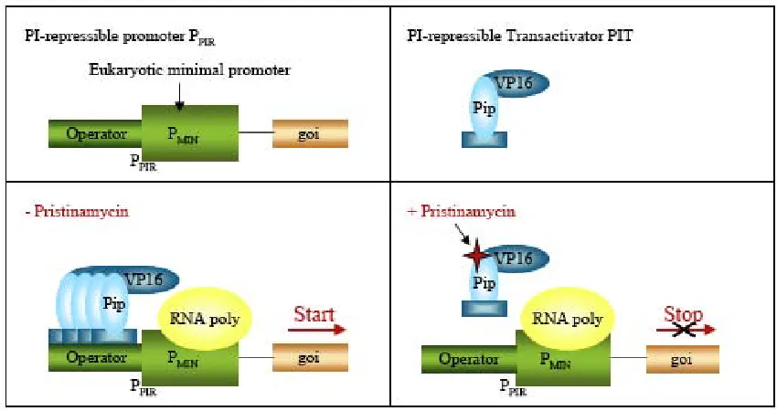
(17) General Introduction. 16. corresponding repressor and they are often compatible for human therapies since the inducer antibiotic is clinically licensed and has minimal to no side-effects within the dose responsive range. Streptomyces have developed many resistance mechanisms to tolerate their own antibiotic products 85. S. pristinaespiralis was shown to contain a pristinamycin resistance determinant (ptr) whose expression is induced by the streptogramin pristinamycin I (PI). Like other streptogramins, pristinamycin is a mixture of two structurally dissimilar molecules, the streptogramin A component pristinamycin II (PII) and the streptogramin B component pristinamycin I (PI). The regulation of the ptr promoter (PPTR) requires a DNA sequence motif (GTACRSYGTAY), which forms the binding site for Pip (pristinamycin-induced protein). Pip was purified and its gene was cloned from S.coelicolor. PI was demonstrated to reverse biding of Pip to PPTR. The streptograminrepressible gene regulation system (PipOFF) is based on the PI-dependent transactivator (PIT: Pip fused to VP16, transactivator domain) which binds and activates PI-repressible promoter derivatives (PPIR) in the absence of PI, whereas in the presence, PIT binding is prevented and PPIR activity is repressed 86, 87 (Figure 6).. Figure 6: The Pip OFF System 83..
(18) General Introduction. 17. Acetaldehyde-Inducible Expression System (Air System) The fungus A.nidulans harbors a specific regulon responsible for coordinating the utilization of ethanol as its sole carbon source. Alcohol is oxidized via acetaldehyde into acetate, which enters mainstream metabolism in its activated form, acetyl-CoA. In the presence of acetaldehyde, a Zn2Cys6-type AlcR transactivator bind to a specific operator module and induces transcription of its own cistron as well as those encoding the alcohol (alcA) and aldehyde dehydrogenases (aldA). The alcA promoter (PalcA) harbors seven AlcR-specific operators (OalcA) each of which interacts with AlcR at high affinity 88. PAIR (acetaldehyde-adjustable promoter) was designed by placing an A.nidulans PalcA-derived heptameric operator module 5’ of a minimal version of the human cytomegalovirus immediate early promoter (PhCMVmin). AlcR activated PAIR in an acetaldehyde-inducible manner. Acetaldehyde enters into the gas phase and dissolves into the media and is a FDA-approved compound used as a food additive 72, 89.. Applications of Artificial Microtissues Strategies based on the assembly of single/monodispersed cells by natural reaggregation have already provided insight into cell-cell interactions, tissue function and regulatory networks. Gravity-mediated self-assembly by the hanging drop technique has been used for the aggregation of several cell types in which cell-specific phenotype and functionality has been maintained. This technology has subsequently been widely used to study the differentiation potential of stem cells and to model tumor behavior in vitro by providing a surrounding microenvironment capable of promoting tumor initiation and progression. Scaffold-free microtissues represent an alternative to existing tissue engineering strategies and they might have a strong potential in cell-based studies. In Physiological-based Studies To this end, a cell embedded within a three-dimensional configuration more closely reproduces its in vivo physiology than when it is cultured by means of standard culture technologies (two-dimensional monolayer cultures) 12, 15..
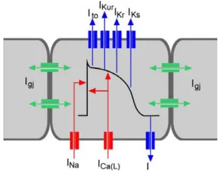
(19) General Introduction. 18. (Electro)Physiological Studies To date, electrophysiological studies have been performed by fundamentally two different techniques; (i) intracellular recordings, which impales a cell with a fine electrode, also called “patch-clamp” technique. 90, 91. and (ii) extracellular recordings, in. which the electrode is in tight contact with the cell/tissue. 92-97. . Although the “patch. clamp” technique yields very accurate information on the electrophysiological properties of the cell, is an invasive method that reduces cell viability and limits the number of possible cell recordings. This can be circumvented by extracellular recordings, where the cells are cultured directly on to an array of transducers, typically known as microelectrode arrays (MEAs), thus recording ion flow across the membrane of a cell/tissue during excitation. Besides the detection of the origin of excitation, the direction and velocity of the excitation spread, the contractility rate can also be measured 98, 99. . MEAs have been recently shown to be able to characterize further properties of the. extracellularly recorded field potential, such as action potential upstroke velocity and duration 100. The standard model used to understand cardiac action potentials are ventricular cardiomyocytes. Cardiac myocyte excitability results from action potentials. When one cell is electrically stimulated, typically by an electric current from an adjacent cell, an increase in the cell permeability allow the Na+-gated channels to initiate a rapid opening producing a fast depolarization due the rapid entrance of Na+ ions. The next phase occurs immediately after the maximum peak of Na+ flow and is recognized as a partial repolarization of the membrane (Ito). The following phase, plateau phase, is unique to ventricular and purkinje fiber myocytes. An electrical plateau is generated due to the opening of Ca2+ channels (L-type calcium channels) allowing inward Ca2+ flow, and outward potassium flow through several types of potassium channels. Some Ca2+ ions originate from the extracellular fluid and others from the endoplasmatic reticulum. The total amount of current during the plateau phase of the cardiac action potential is small and can be maintained for 0.3 sec. Ca2+ ions subsequently bind to troponin, allowing the actin and myosin filaments to slide against each other, producing tension (strength of contraction). In the last phase the repolarization of the membrane is mediated by outward potassium currents, in which K+ channels allow dissipation of K+ ions down their.
(20) General Introduction. 19. concentration. There are two main repolarizing potassium currents IKr and IKs involved in the final repolarization. Concurrently Na+ and Ca2+ channels start closing, re-establishing the resting/basal potential (-90mV) 101 (Figure 9).. Figure 9: Schematic representation of a cardiac action potential with voltage on the Inward sodium current (1Na) mediates the rapid depolarization phase. Transient outward potassium (K+) current (Ito) mediate rapid repolarization phase. The L-type inward calcium current contributes to the long plateau duration. Many outward K+ currents are responsible for repolarization of the cardiac action potential, including rapidly activating delayed rectifying IKr' slowly activating delayed rectifying IKs' and inward rectifying IK1. Thus the coordinated opening and closing of caridac ion channels is responsible for cardiac excitability. Adapted from 102.. Artificial Myocardial Microtissues Primary cardiomyocyte cultures from fetal, neonatal, or adult vertebrate hearts present a reliable source for pioneering cardiac tissue engineering. Even though cardiomyocytes maintained in monolayer cultures have enabled detailed insight for major structure and gene-function studies. 103. , they are less responsive to growth and. differentiation factors compared to cardiomyocytes assembled into 3D cultures. 104. .. Moreover, while the latest generation of biocompatible scaffolds combine shapesupporting capacity with functional and bioactive properties. 105, 106. , some have been.
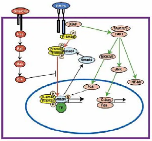
(21) General Introduction. 20. associated with post implantation side-effects resulting from toxic degradation products, inflammatory reaction, and poor resorption. 107. . In contrast, myocardial microtissues. produced in hanging drops show (i) inter-microtissue superstructures, (ii) retention of cardiomyocyte-specific cell qualities, (iii) ECM development, (iv) high-lentiviral transduction rates, and (v) strong potential for regenerative medicine, with the successful integration of myocardial microtissues implanted into the pericardial cavity of adult rats maintaining cardiomyocyte specific phenotype. 46, 50. . However, myocardial microtissue. functionality hasn’t been yet evaluated. In order to study cardiac electrophysiology we tested the BMP-2 gene, known to be related in cardiac functionality. BMP Signaling Pathway (BMP-2) Bone morphogenetic proteins (BMPs) are members of the transforming growth factor-β (TGF-β) superfamily. The BMP signaling pathway, like the TGF-β signaling pathway, modifies target gene transcription by receptor-mediated intracellular signaling 108. . There are two types of BMP receptors; type I (BMPR1A and BMPR1B) and type II. (BMPR2). In the common BMP pathway, type I BMP receptors activate SMAD1/5/8 by phosphorylation. 109. . A dimer of phosphorylated R-SMADs then forms a complex with. SMAD4. This heterodimic complex translocates into the nucleus and binds to transcription factors regulating the expression of target genes. 110, 111. . In the uncommon. BMP pathway, activated BMP receptors interact with a X-linked inhibitor of apoptosis (XIAP), which activates the MAPK (mitogen activated protein kinase) pathway. XIAP links the BMP receptor signal to TAK1, and TAK1 binding proteins (TAB1/2/3) can trigger Jun N-terminal kinase (JNK) and NF-kB. In the common BMP pathway, SMAD function can be inhibited through blocking its translocation to the nucleus by Erk, in response to GF/CK signaling through Ras/Raf/Mek (Figure 7)..
(22) General Introduction. 21. Figure 7. The BMP signaling transduction pathway. Adapted from 108.. BMPs were originally identified as growth and differentiation factors for the skeletal development but are now considered multifunctional cytokines, which play important roles in the development of many organs, including lung, kidney, gut, skin, teeth, and heart. 112. . BMP-2, a member of this family of proteins, is expressed during. mouse embryonic heart development cells into neuronal phenotypes bony sites in adult animal. 115. 114. 113. . It also directs the development of neural crest. and is able to induce de novo formation of bone at no. . Preclinical and clinical studies have shown that BMP-2 can. be applied in various therapeutic interventions such as bone defects, non-union fractures, spinal fusion, osteoporosis and root canal surgery. 116. . In addition, BMP-2 has been. recently associated with cardiac functionality via MEF-2A (myocyte enhancer factor 2A) through the PI3K (Phosphatidylinositol 3-Kinase) pathway. 117. , suggesting BMP-2/MEF-. 2A pathway as an alternative for the heart failure treatment (Figure 8)..
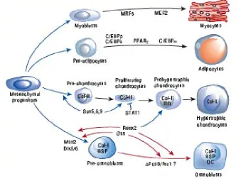
(23) General Introduction. 22. BMP-2. PI 3 kinase Smad 1/5. MEF-2A ? Cardiac differentiation. Cardiac contractility. Figure 8: Scheme of BMP-2-induced MEF-2A-dependent BMP-2 expression and cardiac differentiation. BMP-2 stimulates BMP-specific Smads to regulate cardiac differentiation. BMP-2 increases PI 3-kinase activity, which regulates MEF-2A-dependent transcription of BMP-2 suggesting a plausible mechanism of autoregulation of BMP-2 expression. Also, increased MEF-2A expression regulates MHC expression, a marker for cardiac differentiation. The dotted red arrow indicates a plausible interaction between BMPspecific Smad and MEF-2A (yet to be identified). MEF-2A is suggested to control cardiac contractility. Adapted from Ghosh-Choudhury et.al 118.. In Differentiation- and Transdifferentiation-based Studies Differentiation processes are determined by cellular environment via cell-to-cell, cell-to-cellular matrix, and ECM interactions. Critically, this matrix is reduced and modified when cells are cultivated in conventional two-dimensional cultures 12, 15. Mesenchymal Stem Cells Differentiation and Transdifferentiation Mesenchymal Stem Cells (MSCs) differentiate and transdifferentiate in response to changes in the microenvironment and to signals in the ECM. 119. . Mesenchymal stem. cells (MSCs) are multipotent stem cells that can differentiate into myocytes, adipocytes, condrocytes, or osteoblasts 120 (Figure 10). Differentiated cells (adult cells) are those that express the phenotypic properties characteristic of the functionally mature cell in vivo and beyond which the cell cannot progress. Such cells can change their phenotypes into another. differentiated. cell. type,. in. a. process. named. transdifferentiation.. Transdifferentiation belongs to a wider class of cell type conversions referred to as.
(24) General Introduction ‘metaplasias’. 121. 23. . There is still controversy about whether transdifferentiation processes. are a direct switch from an already differentiated cell to another differentiated cell or if dedifferentiation takes also place 122.. Figure 10: Overview of the mesenchymal stem cell differentiation. Adapted from 123.. However, mesenchymal-derived differentiated cells have been shown to directly transdifferentiate from myoblasts to adipocytes. 124, 125. and from osteoblast to adipocytes. 126. . Adipocytes and osteoblasts represent two morphologically and functionally distinct. cell types. While adipose tissue is a soft tissue able of storing large amounts of fat, which serve as an energy reserve, bone tissue is a stiff tissue formed by a mineralized ECM, which serves as a tissue support. Adipocyte differentiates from preadipocyte in a process named adipogenesis. Adipogenesis is regulated by an elaborate network of transcription factors. The two principal adipogenic factors are PPARγ and C/EBPα 127, 128 (Figure 11)..
(25) General Introduction. 24. Figure 11: Adipogenesis. Exposure of preadipocytes to a cocktail of adipogenic inducers comprised of insulin, glucocorticoids, agents that elevate cAMP (isobutylmethylxanthine), and fetal bovine serum activates expression of several transcription factors that converge on PPARγ. PPARγ then induces C/EBPα expression, and together, these factors oversee terminal adipogenesis. Adapted from 127.. On the other hand, osteoblasts differ from their precursors fibroblasts (preosteoblasts) through a process named osteogenesis, in which the expression of the Cbfa1/Runx2 transcription factor is essential. Cbfa1/Runx2 regulates the expression of all known marker genes expressed during osteogenesis, such as osteocalcin, and is required for bone ECM production 123, 129, 130. The primary function of osteoblasts is to synthesize and secrete ECM which results in the deposition of new bone (Figure 12).. Figure 12: Osteogenesis. Some of the known molecules affecting the differentiation of pre-osteoblasts into osteoblasts as well as some of the inhibitory proteins such as Hoxa2 and Leptin. Adapted from. 130. ..
(26) General Introduction. 25. 3D Cell Culture Systems of Adipose Tissue Adipose tissue has recently become a focus area for tissue engineering, encouraged by the large number of reconstructive, cosmetic and correctional indications that could be addressed with clinically translatable adipose tissue engineering strategies 131. . Adipose tissue is the largest tissue in the body which is often carried in excess and. can therefore be harvested without creating contour deformities. Until now, 3D scaffolds have been predominantly used to replace adipose volume but not function complete adipose tissue engineering meant to restore both volume and function. 132. , but. 133. . 3D. scaffolds systems have been demonstrated to direct cell differentiation and organization into multicellular communities that approximate the in vivo architecture and function, in particular the specific secretory functions of adiopocytes 1. Therefore, 3D cell cultures systems of adipose tissue are a promising tool to study/understand the cellular and the molecular basis of adipocyte differentiation and metabolism in physiological and pathological states in order to prevent and to treat obesity and its complications 134, 135. 3D Cell Culture Systems of Bone Tissue Bone tissue repair is one of the major concerns of regenerative medicine. The current need for tissue replacements has necessitated the development of a new science termed 'bone tissue engineering'. Bone development requires the concentrated action of several microenvironmental signals such as cytoquines/growth factors, ECM molecules and cell/cell interactions. 136. . During osteogenesis, cells differentiate into pre-osteoblasts. and then undergo cellular condensation, a process that precedes osteoblast differentiation and matrix mineralization. 137, 138. . Cellular condensation is mimicked in high-density. cultures, such as scaffold-free reaggregation where up-regulation of the ECM components and ECM mineralization takes place. 34. . 3D porous scaffolds formulated. from biocompatible and osteoconductive materials that are not immunoreative are intended to mimic the native in vivo microenvironment. This demands construction of bioactive scaffolds that are also capable of supporting vascularization as well as cell proliferation and osteogenic differentiation. 3D cell cultures for bone regeneration provide a surgical tool for bone tissue engineering directed to enhance bone repair and wounding for orthopedic and cranio-maxillofacial clinical applications 139..
(27) General Introduction. 26. Transdifferentiation of adipose tissue to bone tissue Obesity (fat tissue disease) and osteoporosis (bone tissue disease), two disorders of body composition, share several features including a genetic predisposition as well as a hormonal or environmental disorder. Bone remodeling is centrally regulated through the hypothalamus and sympathetic nervous system, a pathway that also regulates the metabolic fate and distribution of adipose tissue. With aging, the composition of bone marrow shifts to favor the presence of adipocytes with a corresponding decline in osteoblast function, resulting in osteoporosis. The relationship between bone and fat formation within the bone marrow microenvironment is suggested to be one of the causes of osteoporosis, and is accordingly an active area of investigation. 140,. 141. .. Transdifferentiation provides an alternative method for osteoporosis and cell basedtherapies in order to replace sick or damaged cells or tissue using patient’s own cells 119, 142. . In order to induce transdifferention from adipse tissue to bone tissue we evaluate the. effect of BMP-2 (see above) and ΔFosB. AP-1 Transcription Factors Family (ΔFosB) The AP-1 (activator protein 1) family of transcription factors consist of dimeric complexes of Fos (c-Fos, FosB, ΔFosB, Fra-1 and Fra-2) and Jun-related (cJun, JunB and JunD) proteins. 143. . Some members of the ATF and CREB families of proteins are also. part of the AP-1 complexes 144. These are basic leucine zipper proteins that modulate the transcription of a variety of genes through interactions with specific sequences on the target gene promoters. Members of the AP-1 family participate in a large variety of biological processes including cell differentiation, proliferation, apoptosis and oncogenic transformation 145. AP-1 components, mainly members of the Fos family, are involved in the regulation of bone cell proliferation and differentiation, and the promoters of several genes related to bone formation contain AP-1 consensus sequences. In addition several regulators of bone formation (TGF-β, parathyroid hormone and 1,25-dihydroxy vitamin D) induce AP-1 expression. 146, 147. . The various components of the AP-1 complex are. differentially expressed during osteoblast maturation in vitro. 148. . During osteoblast. proliferation, the levels of all Fos and Jun proteins are high. Subsequently, during the.
(28) General Introduction. 27. period of ECM production and mineralization, their levels decrease, and Fra-2 and JunD become the principal components of the AP-1 complex in fully differentiated osteoblasts. Fra-1 overexpression results in increased bone formation and osteosclerosis developement of the entire skeleton 149. However, Fra-1 deficient mice display no skeletal 150. abnormalities. Furthermore mice overexpressing c-Fos develop osteosarcomas whereas c-fos knockout mice lack osteoclasts and develop osteopetrosis. ,. 151, 152. . In. contrast no bone abnormalities have been described in fosB knockout mice, or in mice where FosB. 153. , Fra-2, c-Jun or JunB alone were overexpressed. 147. . Transgenic mice. expressing ΔFosB show not only an osteosclerotic phenotype in osteoblasts, but also a pronounced decrease in adipogenesis by altering the DNA-binding of C/EBPβ 154, 155. ΔFosB is a C-terminally truncated splice variant of FosB that lacks the prolinerich transactivation domain but has maintained the ability to bind DNA and heterodimerize with other AP-1 factors. 156-158. . It can be induced in a region-specific. manner in the brain in response to several types of chronic perturbation, including drug abuse, antipsychotic drugs, antidepressant drugs, seizures and lesions. 159. . Transgenic. expression of ΔFosB, widely expressed in many tissues in addition to bone, stimulates bone formation and increases bone mass in mice, but it is not known how this protein, which lacks a transactivation domain, increases bone formation 123. In Productivity-based Studies Three-dimensional (3D) cell cultures differ form the two-dimensional (2D) ones not only in specific tissue functionality, but also in their metabolic activity. In contrast to 2D cell cultures, cells assembled within microtissues typically showed lower proliferation rates 24. Proliferation-controlled cell cultures showed to increase cell specific productivity 75. , suggesting that cells growing in a three-dimensional system could optimize the. performance of production cell lines. Heterelogous Protein Production Biopharmaceutical manufacturing is the science of producing large amounts of high-quality. recombinant. protein. pharmaceuticals. 160. .. During. recent. years,. biopharmaceutical products manufactured by processes that use mammalian cell cultures.
(29) General Introduction have gained increasing importance. 28 161. . The basis of production processes with. mammalian cells was commended in 1949 by Enders who infected cultured HeLa cells with poliomyelitis virus to create a vaccine 162. In the late 1970’s, Kholer and Millstein’s work using fused lymphocyte-myeloma mouse cell line was another milestone in the development of animal cell culture 163. Besides the production of antibodies and vaccines, the 1980’s saw significant advances in genetic engineering leading to mammalian cell culture processes in which major pharmaceutical recombinant proteins were produced at large scale, including tissue plasminogen activator (tPA, Activase; produced for the first time in CHO cells in 1987 by Genentech), erythropoietin (EPO; produced for the first time in a mammalian cell line in 1989 by Amgen) and Factor VIII (produced for the first time in a mammalian cell line in 1992 by Baxter Healthcare/Genetics Institute). Improving cell productivity in bioprocesses has long a major effort within the biotechnological industry to reduce costs of manufacturing and increase product quality 164. . High cell density is particularly desirable as product concentration is directly. proportional to viable cell concentration over the time. For maximizing product concentration it is necessary to use cell lines with the ability to proliferate indefinitely to maintain the cells in their productive stage by supplying them with the essential nutrients. However, an uncontrolled proliferation of the cells in a batch mode results in nutrient and oxygen depletion, accumulation of lactate, ammonia and other toxic subproducts, which can lead to the deterioration of the target product. It is therefore useful to maintain cells in a static productive state. Also the development of defined medium (serum-free media) for mammalian cell culture has been relevant for the quality and biosafety of the desired product. Different strategies for increasing specific productivity of mammalian production cell lines have been performed. Increased productivity by reducing specific growth rates was observed for the first time in hybridoma cells. 165, 166. . Jenkin’s and coworkers. demonstrated that the high requirements for nucleotides and protein during DNA synthesis and mitosis might limit availability of substrates for recombinant RNA and protein synthesis. Their statement is based on Miller’s kinetic analysis, which demonstrated that cells with small specific growth rate spend proportionally less time in mitosis. Several approaches, such as low temperature cultivation, chemicals addition and.
(30) General Introduction. 29. genetic engineered cells, have achieved a G1 phase arrest during the cultivation period to improve cell productivity. 75, 167-172. . This correlation is supported with in nature whereby. many cells of a mature mammal display no further growth after terminal differentiation yet continue to produce and often secret proteins during the lifetime of the organism. The productivity of recombinant mammalian cell lines is often limited by the rapidity with which cells entry into the decline phase (apoptosis), the period of culture after maximum cell density is reached, and where cell viability starts to decrease. Heterologous protein production by itself is a stimulus of apoptosis. Therefore obviation of apoptosis is essential to prolong the production phase but also important during the adaptation to serum-free media conditions. 173. . Apoptosis is triggered by several stimuli,. such as serum deprivation, nutrient limitation, oxygen limitation and mechanical stress 174. . Indeed, spatial organization of the cells within a tissue and factors associated with the. tissue microenvironment are implicated in programmed cell death in lower organisms 175. Viability of Artificial Microtissues (vascularization) Cells within the microtissue suffer nutrient and oxygen limitation. Endothelial cells are the central organizational unit of the vascular structure 176, which is responsible of supplying oxygen and nutrients to the tissues. Monodispersed human umbilical vein endothelial cells (HUVEC), co-cultured onto microtissue forming human primary fibroblasts produced in hanging drops, show high angiogenic potential. 49. . By controlling microtissue size, and without administration. of any growth factor, 6-day old HUVEC coated human aortic fibroblast (HAF) microtissues establish a dense microvessel network (Figure 3). A. Initiation of a feeder microtissue (HAF). B. Addition of a second cell type (HUVEC). C. D. Development of a cell layer. Development of capillarylike structures.
(31) General Introduction. 30. Figure 3: Endothelial-mediated vascularization of microtissues formed in hanging drops. (A) Single cell suspension is aliquoted into 60-well plates and cultivated upside down. After 2-6 days, depending on the cell type and the media composition, cells form a cellular aggregate (microtissues). (B) Generation of a multilayer microtissues by adding a second cell type into the hanging drop. (C) The added cells from an addition cell layer around the feeder spheroid. (D) Development of a capillary network through the microtissues after 6 days of co-cultivation.. Size-controlled microtissues exhibit size-dependent expression of vascular endothelial growth factor (VEGF), which indicated the oxygen levels within the microtissue. Capillary-like filled microtissues reduce VEGF expression thus increasing oxygenation within the tissue and demonstrating functional angiogenesis. 49. . Moreover,. macrotissues, assembled from HUVEC-coated fibroblasts microtissues, develope a vascular system, which functionally connects to the chicken embryo’s vasculature after implantation. This collectively demonstrates that scaffold-free vascularized microtissues could be used for the scale-up and production of artificial tissue implants for future tissue engineering strategies. 45,. 50. . Moreover, vascularized microtissues optimized the. microtissue yield representing an advantage in further biotechnological applications.. References 1. 2. 3. 4. 5. 6. 7. 8.. Langer, R. and Vacanti, J.P. Tissue engineering. Science (New York, N.Y 260, 920, 1993. Lysaght, M.J. and Reyes, J. The growth of tissue engineering. Tissue engineering 7, 485, 2001. Juliano, R.L. Signal transduction by cell adhesion receptors and the cytoskeleton: functions of integrins, cadherins, selectins, and immunoglobulin-superfamily members. Annual review of pharmacology and toxicology 42, 283, 2002. Goldmann, W.H. Mechanical aspects of cell shape regulation and signaling. Cell Biol Int 26, 313, 2002. Boudreau, N.J. Organized living: from cell surfaces to basement membranes. Sci STKE 2003, pe34, 2003. Tarone, G., Hirsch, E., Brancaccio, M., De Acetis, M., Barberis, L., Balzac, F., Retta, S.F., Botta, C., Altruda, F. and Silengo, L. Integrin function and regulation in development. Int J Dev Biol 44, 725, 2000. Sechler, J.L. and Schwarzbauer, J.E. Control of cell cycle progression by fibronectin matrix architecture. J Biol Chem 273, 25533, 1998. Bokel, C. and Brown, N.H. Integrins in development: moving on, responding to, and sticking to the extracellular matrix. Dev Cell 3, 311, 2002..
(32) General Introduction 9. 10. 11.. 12. 13. 14. 15. 16.. 17. 18. 19. 20. 21. 22. 23. 24. 25. 26. 27. 28.. 31. Schenk, S. and Quaranta, V. Tales from the crypt[ic] sites of the extracellular matrix. Trends Cell Biol 13, 366, 2003. Boudreau, N., Sympson, C.J., Werb, Z. and Bissell, M.J. Suppression of ICE and apoptosis in mammary epithelial cells by extracellular matrix. Science (New York, N.Y 267, 891, 1995. Weaver, V.M., Lelievre, S., Lakins, J.N., Chrenek, M.A., Jones, J.C., Giancotti, F., Werb, Z. and Bissell, M.J. beta4 integrin-dependent formation of polarized three-dimensional architecture confers resistance to apoptosis in normal and malignant mammary epithelium. Cancer Cell 2, 205, 2002. Abbott, A. Cell culture: biology's new dimension. Nature 424, 870, 2003. Vogel, V. and Baneyx, G. The tissue engineeting puzzle: a molecular perspective. Annu Rev Biomed Eng 5, 441, 2003. Mathis, L. and Nicolas, J.F. Cellular patterning of the vertebrate embryo. Trends Genet 18, 627, 2002. Zhang, S. Beyond the Petri dish. Nature biotechnology 22, 151, 2004. Khalil, M., Shariat-Panahi, A., Tootle, R., Ryder, T., McCloskey, P., Roberts, E., Hodgson, H. and Selden, C. Human hepatocyte cell lines proliferating as cohesive spheroid colonies in alginate markedly upregulate both synthetic and detoxificatory liver function. J Hepatol 34, 68, 2001. Schmeichel, K.L. and Bissell, M.J. Modeling tissue-specific signaling and organ function in three dimensions. Journal of cell science 116, 2377, 2003. Duguay, D., Foty, R.A. and Steinberg, M.S. Cadherin-mediated cell adhesion and tissue segregation: qualitative and quantitative determinants. Developmental biology 253, 309, 2003. Desoize, B. and Jardillier, J. Multicellular resistance: a paradigm for clinical resistance? Crit Rev Oncol Hematol 36, 193, 2000. Yamada, K.M., Pankov, R. and Cukierman, E. Dimensions and dynamics in integrin function. Braz J Med Biol Res 36, 959, 2003. Ingber, D.E. Cancer as a disease of epithelial-mesenchymal interactions and extracellular matrix regulation. Differentiation 70, 547, 2002. Jacks, T. and Weinberg, R.A. Taking the study of cancer cell survival to a new dimension. Cell 111, 923, 2002. Zahir, N. and Weaver, V.M. Death in the third dimension: apoptosis regulation and tissue architecture. Curr Opin Genet Dev 14, 71, 2004. Bissell, M.J., Radisky, D.C., Rizki, A., Weaver, V.M. and Petersen, O.W. The organizing principle: microenvironmental influences in the normal and malignant breast. Differentiation 70, 537, 2002. Elisseeff, J., McIntosh, W., Fu, K., Blunk, B.T. and Langer, R. Controlled-release of IGF-I and TGF-beta1 in a photopolymerizing hydrogel for cartilage tissue engineering. J Orthop Res 19, 1098, 2001. Richardson, T.P., Peters, M.C., Ennett, A.B. and Mooney, D.J. Polymeric system for dual growth factor delivery. Nature biotechnology 19, 1029, 2001. Griffith, L.G. and Naughton, G. Tissue engineering--current challenges and expanding opportunities. Science (New York, N.Y 295, 1009, 2002. Lavik, E. and Langer, R. Tissue engineering: current state and perspectives. Appl Microbiol Biotechnol 65, 1, 2004..
(33) General Introduction 29. 30. 31. 32. 33. 34. 35.. 36. 37. 38. 39. 40. 41. 42.. 43. 44.. 32. Hunziker, E.B. Articular cartilage repair: are the intrinsic biological constraints undermining this process insuperable? Osteoarthritis Cartilage 7, 15, 1999. Hench, L.L. and Polak, J.M. Third-generation biomedical materials. Science (New York, N.Y 295, 1014, 2002. Whitesides, G.M. and Grzybowski, B. Self-assembly at all scales. Science (New York, N.Y 295, 2418, 2002. Jakab, K., Neagu, A., Mironov, V., Markwald, R.R. and Forgacs, G. Engineering biological structures of prescribed shape using self-assembling multicellular systems. Proc Natl Acad Sci U S A 101, 2864, 2004. Furukawa, K.S., Ushida, T., Sakai, Y., Suzuki, M., Tanaka, J. and Tateishi, T. Formation of human fibroblast aggregates (spheroids) by rotational culture. Cell transplantation 10, 441, 2001. Kale, S., Biermann, S., Edwards, C., Tarnowski, C., Morris, M. and Long, M.W. Three-dimensional cellular development is essential for ex vivo formation of human bone. Nature biotechnology 18, 954, 2000. Muraglia, A., Corsi, A., Riminucci, M., Mastrogiacomo, M., Cancedda, R., Bianco, P. and Quarto, R. Formation of a chondro-osseous rudiment in micromass cultures of human bone-marrow stromal cells. Journal of cell science 116, 2949, 2003. Watzka, S.B., Lucien, J., Shimada, M., Edwards, V., Yeger, H., Hannigan, G. and Coles, J.G. Selection of viable cardiomyocytes for cell transplantation using three-dimensional tissue culture. Transplantation 70, 1310, 2000. Kelm, J.M., Timmins, N.E., Brown, C.J., Fussenegger, M. and Nielsen, L.K. Method for generation of homogeneous multicellular tumor spheroids applicable to a wide variety of cell types. Biotechnol Bioeng 83, 173, 2003. Wobus, A.M., Wolf, E. and Beier, H.M. Embryonic stem cells and nuclear transfer strategies. Present state and future prospects. Cells, tissues, organs 166, 1, 2000. Kelm, J.M. and Fussenegger, M. Microscale tissue engineering using gravityenforced cell assembly. Trends Biotechnol 22, 195, 2004. Radisky, D., Muschler, J. and Bissell, M.J. Order and disorder: the role of extracellular matrix in epithelial cancer. Cancer Invest 20, 139, 2002. Santini, M.T., Rainaldi, G. and Indovina, P.L. Apoptosis, cell adhesion and the extracellular matrix in the three-dimensional growth of multicellular tumor spheroids. Crit Rev Oncol Hematol 36, 75, 2000. Weaver, V.M., Petersen, O.W., Wang, F., Larabell, C.A., Briand, P., Damsky, C. and Bissell, M.J. Reversion of the malignant phenotype of human breast cells in three-dimensional culture and in vivo by integrin blocking antibodies. J Cell Biol 137, 231, 1997. Timmins, N.E., Dietmair, S. and Nielsen, L.K. Hanging-drop multicellular spheroids as a model of tumour angiogenesis. Angiogenesis 7, 97, 2004. Itskovitz-Eldor, J., Schuldiner, M., Karsenti, D., Eden, A., Yanuka, O., Amit, M., Soreq, H. and Benvenisty, N. Differentiation of human embryonic stem cells into embryoid bodies compromising the three embryonic germ layers. Molecular medicine (Cambridge, Mass 6, 88, 2000..
(34) General Introduction 45. 46. 47. 48. 49. 50.. 51. 52.. 53. 54. 55. 56. 57. 58. 59. 60. 61. 62.. 33. Kelm, J.M., Djonov, V., Ittner, L.M., Fluri, D., Born, W., Hoerstrup, S.P. and Fussenegger, M. Design of custom-shaped vascularized tissues using microtissue spheroids as minimal building units. Tissue engineering 12, 2151, 2006. Kelm, J.M., Ehler, E., Nielsen, L.K., Schlatter, S., Perriard, J.C. and Fussenegger, M. Design of artificial myocardial microtissues. Tissue engineering 10, 201, 2004. Anderer, U. and Libera, J. In vitro engineering of human autogenous cartilage. J Bone Miner Res 17, 1420, 2002. Kelm, J.M., Ittner, L.M., Born, W., Djonov, V. and Fussenegger, M. Selfassembly of sensory neurons into ganglia-like microtissues. Journal of biotechnology 121, 86, 2006. Kelm, J.M., Diaz Sanchez-Bustamante, C., Ehler, E., Hoerstrup, S.P., Djonov, V., Ittner, L. and Fussenegger, M. VEGF profiling and angiogenesis in human microtissues. Journal of biotechnology 118, 213, 2005. Kelm, J.M., Djonov, V., Hoerstrup, S.P., Guenter, C.I., Ittner, L.M., Greve, F., Hierlemann, A., Sanchez-Bustamante, C.D., Perriard, J.C., Ehler, E. and Fussenegger, M. Tissue-transplant fusion and vascularization of myocardial microtissues and macrotissues implanted into chicken embryos and rats. Tissue engineering 12, 2541, 2006. Kunz-Schughart, L.A., Freyer, J.P., Hofstaedter, F. and Ebner, R. The use of 3-D cultures for high-throughput screening: the multicellular spheroid model. J Biomol Screen 9, 273, 2004. Layer, P.G., Weikert, T. and Willbold, E. Chicken retinospheroids as developmental and pharmacological in vitro models: acetylcholinesterase is regulated by its own and by butyrylcholinesterase activity. Cell and tissue research 268, 409, 1992. Kootstra, N.A. and Verma, I.M. Gene therapy with viral vectors. Annual review of pharmacology and toxicology 43, 413, 2003. Walther, W. and Stein, U. Viral vectors for gene transfer: a review of their use in the treatment of human diseases. Drugs 60, 249, 2000. Gomez-Vargas, A. and Hortelano, G. Nonviral gene therapy approaches to hemophilia. Seminars in thrombosis and hemostasis 30, 197, 2004. Pfeifer, A. and Verma, I.M. Gene therapy: promises and problems. Annual review of genomics and human genetics 2, 177, 2001. Wells, D.J. Gene therapy progress and prospects: electroporation and other physical methods. Gene therapy 11, 1363, 2004. Fields, B.N., Knipe, D.M., Howley, P.M. and Al, E. Fields Virology. Philadelphia: Lippincott-Raven Publishers, 1996. Tang, H., Kuhen, K.L. and Wong-Staal, F. Lentivirus replication and regulation. Annual review of genetics 33, 133, 1999. Coffin, J.M., Hughes, S.H. and Varmus, H.E. Retroviruses. New York: Cold Spring Harbor Laboratory Press, 1997. Naldini, L. and Verma, I.M. Lentiviral vectors. Advances in virus research 55, 599, 2000. Trono, D. Lentiviral vectors: turning a deadly foe into a therapeutic agent. Gene therapy 7, 20, 2000..
(35) General Introduction 63. 64. 65. 66. 67. 68. 69. 70. 71. 72. 73. 74.. 75. 76.. 77. 78.. 34. Amado, R.G. and Chen, I.S. Lentiviral vectors--the promise of gene therapy within reach? Science (New York, N.Y 285, 674, 1999. Buchschacher, G.L., Jr. and Wong-Staal, F. Development of lentiviral vectors for gene therapy for human diseases. Blood 95, 2499, 2000. Hansen, A.C. and Pedersen, F.S. Safety features of retroviral vectors. Current opinion in molecular therapeutics 4, 324, 2002. Hu, W.S. and Pathak, V.K. Design of retroviral vectors and helper cells for gene therapy. Pharmacological reviews 52, 493, 2000. Kay, M.A., Glorioso, J.C. and Naldini, L. Viral vectors for gene therapy: the art of turning infectious agents into vehicles of therapeutics. Nature medicine 7, 33, 2001. Miyoshi, H., Blomer, U., Takahashi, M., Gage, F.H. and Verma, I.M. Development of a self-inactivating lentivirus vector. Journal of virology 72, 8150, 1998. Lever, A.M., Strappe, P.M. and Zhao, J. Lentiviral vectors. Journal of biomedical science 11, 439, 2004. Quinonez, R. and Sutton, R.E. Lentiviral vectors for gene delivery into cells. DNA and cell biology 21, 937, 2002. Fussenegger, M. The impact of mammalian gene regulation concepts on functional genomic research, metabolic engineering, and advanced gene therapies. Biotechnology progress 17, 1, 2001. Hartenbach, S. and Fussenegger, M. Autoregulated, bidirectional and multicistronic gas-inducible mammalian as well as lentiviral expression vectors. Journal of biotechnology 120, 83, 2005. Mitta, B., Weber, C.C. and Fussenegger, M. In vivo transduction of HIV-1derived lentiviral particles engineered for macrolide-adjustable transgene expression. The journal of gene medicine 7, 1400, 2005. Mitta, B., Weber, C.C., Rimann, M. and Fussenegger, M. Design and in vivo characterization of self-inactivating human and non-human lentiviral expression vectors engineered for streptogramin-adjustable transgene expression. Nucleic acids research 32, e106, 2004. Fussenegger, M., Schlatter, S., Datwyler, D., Mazur, X. and Bailey, J.E. Controlled proliferation by multigene metabolic engineering enhances the productivity of Chinese hamster ovary cells. Nature biotechnology 16, 468, 1998. Fux, C., Langer, D. and Fussenegger, M. Dual-regulated myoD- and msx1-based interventions in C2C12-derived cells enable precise myogenic/osteogenic/adipogenic lineage control. The journal of gene medicine 6, 1159, 2004. Fux, C., Mitta, B., Kramer, B.P. and Fussenegger, M. Dual-regulated expression of C/EBP-alpha and BMP-2 enables differential differentiation of C2C12 cells into adipocytes and osteoblasts. Nucleic acids research 32, e1, 2004. Koponen, J.K., Kankkonen, H., Kannasto, J., Wirth, T., Hillen, W., Bujard, H. and Yla-Herttuala, S. Doxycycline-regulated lentiviral vector system with a novel reverse transactivator rtTA2S-M2 shows a tight control of gene expression in vitro and in vivo. Gene therapy 10, 459, 2003..
(36) General Introduction 79.. 80. 81. 82. 83. 84. 85. 86. 87.. 88. 89. 90. 91. 92. 93. 94.. 35. Regulier, E., Trottier, Y., Perrin, V., Aebischer, P. and Deglon, N. Early and reversible neuropathology induced by tetracycline-regulated lentiviral overexpression of mutant huntingtin in rat striatum. Human molecular genetics 12, 2827, 2003. Vigna, E., Cavalieri, S., Ailles, L., Geuna, M., Loew, R., Bujard, H. and Naldini, L. Robust and efficient regulation of transgene expression in vivo by improved tetracycline-dependent lentiviral vectors. Mol Ther 5, 252, 2002. Vogel, R., Amar, L., Thi, A.D., Saillour, P. and Mallet, J. A single lentivirus vector mediates doxycycline-regulated expression of transgenes in the brain. Human gene therapy 15, 157, 2004. Weber, W. and Fussenegger, M. Artificial mammalian gene regulation networksnovel approaches for gene therapy and bioengineering. Journal of biotechnology 98, 161, 2002. Weber, W. and Fussenegger, M. Approaches for trigger-inducible viral transgene regulation in gene-based tissue engineering. Current opinion in biotechnology 15, 383, 2004. Weber, W. and Fussenegger, M. Pharmacologic transgene control systems for gene therapy. The journal of gene medicine 8, 535, 2006. Cundliffe, E. How antibiotic-producing organisms avoid suicide. Annual review of microbiology 43, 207, 1989. Fussenegger, M., Morris, R.P., Fux, C., Rimann, M., von Stockar, B., Thompson, C.J. and Bailey, J.E. Streptogramin-based gene regulation systems for mammalian cells. Nature biotechnology 18, 1203, 2000. Weber, W., Kramer, B.P., Fux, C., Keller, B. and Fussenegger, M. Novel promoter/transactivator configurations for macrolide- and streptograminresponsive transgene expression in mammalian cells. The journal of gene medicine 4, 676, 2002. Flipphi, M., Kocialkowska, J. and Felenbok, B. Characteristics of physiological inducers of the ethanol utilization (alc) pathway in Aspergillus nidulans. The Biochemical journal 364, 25, 2002. Weber, W., Rimann, M., Spielmann, M., Keller, B., Daoud-El Baba, M., Aubel, D., Weber, C.C. and Fussenegger, M. Gas-inducible transgene expression in mammalian cells and mice. Nature biotechnology 22, 1440, 2004. Neher, E. and Sakmann, B. Single-channel currents recorded from membrane of denervated frog muscle fibres. Nature 260, 799, 1976. Cole, K.S. Some physical aspects of bioelectric phenomena. Proceedings of the National Academy of Sciences of the United States of America 35, 558, 1949. DeBusschere, B.D. and Kovacs, G.T. Portable cell-based biosensor system using integrated CMOS cell-cartridges. Biosens Bioelectron 16, 543, 2001. Fromherz, P. Electrical interfacing of nerve cells and semiconductor chips. Chemphyschem 3, 276, 2002. Jimbo, Y. and Robinson, H.P. Propagation of spontaneous synchronized activity in cortical slice cultures recorded by planar electrode arrays. Bioelectrochemistry (Amsterdam, Netherlands) 51, 107, 2000..
(37) General Introduction 95. 96.. 97.. 98.. 99. 100. 101. 102. 103. 104. 105. 106. 107. 108. 109. 110. 111. 112.. 36. Morefield, S.I., Keefer, E.W., Chapman, K.D. and Gross, G.W. Drug evaluations using neuronal networks cultured on microelectrode arrays. Biosens Bioelectron 15, 383, 2000. Olsson, R.H., 3rd, Buhl, D.L., Sirota, A.M., Buzsaki, G. and Wise, K.D. Bandtunable and multiplexed integrated circuits for simultaneous recording and stimulation with microelectrode arrays. IEEE transactions on bio-medical engineering 52, 1303, 2005. Pancrazio, J.J., Gray, S.A., Shubin, Y.S., Kulagina, N., Cuttino, D.S., Shaffer, K.M., Eisemann, K., Curran, A., Zim, B., Gross, G.W. and O'Shaughnessy, T.J. A portable microelectrode array recording system incorporating cultured neuronal networks for neurotoxin detection. Biosens Bioelectron 18, 1339, 2003. Feld, Y., Melamed-Frank, M., Kehat, I., Tal, D., Marom, S. and Gepstein, L. Electrophysiological modulation of cardiomyocytic tissue by transfected fibroblasts expressing potassium channels: a novel strategy to manipulate excitability. Circulation 105, 522, 2002. Wang, L. and Duff, H.J. Developmental changes in transient outward current in mouse ventricle. Circulation research 81, 120, 1997. Halbach, M., Egert, U., Hescheler, J. and Banach, K. Estimation of action potential changes from field potential recordings in multicellular mouse cardiac myocyte cultures. Cell Physiol Biochem 13, 271, 2003. Keating, M.T. and Sanguinetti, M.C. Molecular and cellular mechanisms of cardiac arrhythmias. Cell 104, 569, 2001. Jongsma, H.J. Sudden cardiac death: a matter of faulty ion channels? Curr Biol 8, R568, 1998. Severs, N.J. The cardiac muscle cell. Bioessays 22, 188, 2000. Armstrong, M.T., Lee, D.Y. and Armstrong, P.B. Regulation of proliferation of the fetal myocardium. Dev Dyn 219, 226, 2000. Hubbell, J.A. Bioactive biomaterials. Current opinion in biotechnology 10, 123, 1999. Polonchuk, L., Elbel, J., Eckert, L., Blum, J., Wintermantel, E. and Eppenberger, H.M. Titanium dioxide ceramics control the differentiated phenotype of cardiac muscle cells in culture. Biomaterials 21, 539, 2000. Yang, S., Leong, K.F., Du, Z. and Chua, C.K. The design of scaffolds for use in tissue engineering. Part I. Traditional factors. Tissue engineering 7, 679, 2001. Zhang, J. and Li, L. BMP signaling and stem cell regulation. Developmental biology 284, 1, 2005. Zwijsen, A., Verschueren, K. and Huylebroeck, D. New intracellular components of bone morphogenetic protein/Smad signaling cascades. FEBS letters 546, 133, 2003. Massague, J., Blain, S.W. and Lo, R.S. TGFbeta signaling in growth control, cancer, and heritable disorders. Cell 103, 295, 2000. Shi, Y. and Massague, J. Mechanisms of TGF-beta signaling from cell membrane to the nucleus. Cell 113, 685, 2003. Hogan, B.L. Bone morphogenetic proteins in development. Curr Opin Genet Dev 6, 432, 1996..
Figure

+7


Documento similar
Second, hierarchical clustering of pancreatic cancer cell lines with primary tumours enabled the translation of drug sensitivity data from cell lines to the tumour subtypes found
(B) Oxygen Consumption Rate -OCR- was measured as an evaluation of mitochondrial respiratory profile changes in control cells (C), non-transduced (MSUD) or lentiviral-transduced
In low density cultures, spheres are clonally derived colonies that are composed of a minority of stem cells and a majority of progenitor cells, the founding cell of the colony being
Accordingly, analysis of a large cohort of primary and metastatic melanoma cell lines (Supplementary Table 1) confirmed increased LOXL3 expression at both LOXL3
17,18 Contrary to graphene, the band gap in ML-MDS separating the valence and conduction bands is naturally large and due to the absence of inversion symmetry in ML-MDS the
When cells were cultivated in an imbalanced minimal salts medium with a high C/N ratio and octanoic acid as the sole carbon source, the expression of phaC1 during the
Given a set of service de- scriptions, already classified under some classification taxonomy, and a new service description, we propose a heuristic for automated
(E) Western blot analysis of PEDF e protein levels in CM and HIF1a in whole-cell extracts from M438 primary melanocytes, SBcl2 and WM164 melanoma cell lines incubated in normoxia
