TítuloMarine natural products from the Yucatan peninsula
24
0
0
Texto completo
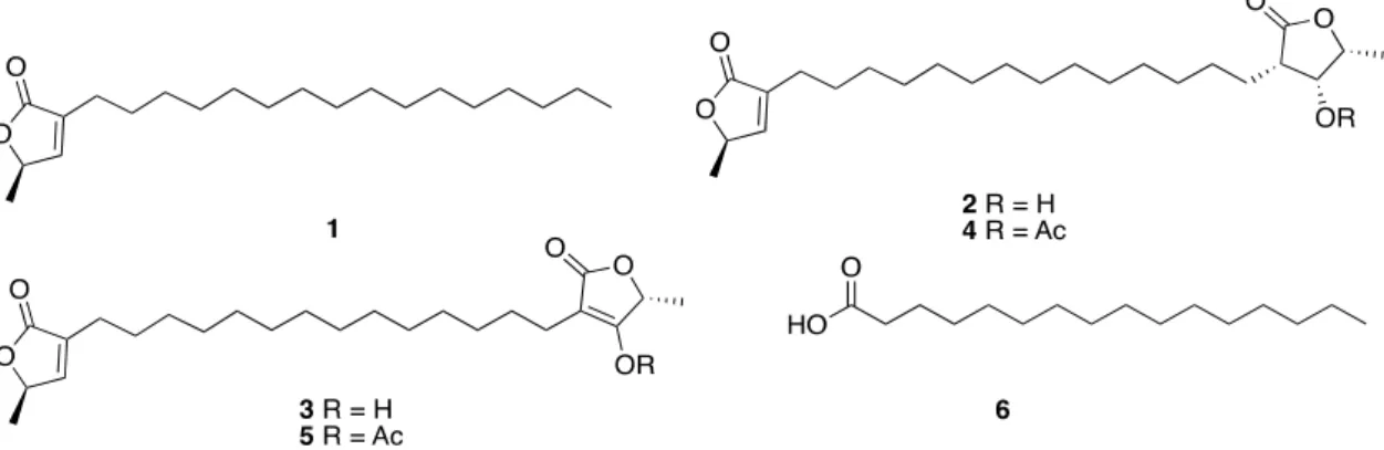
(2) Mar. Drugs 2020, 18, 59. 2 of 24. On the other hand, a priority task for the conservation and management of coastal areas, such as the Yucatan Peninsula, and for the discovery of new sources of novel natural products, is the study of their biodiversity. The first effort aimed at determining the state of health of the coast of the Yucatan Peninsula was reported in 2010. Pech-Pool and Ardisson Herrera described the identification of more than 400 thousand organisms from marine and coastal environments and lagoons, belonging to 529 species, which were distributed in 13 phyla, 26 classes, 28 orders, 113 families, and 358 genera. Of the registered species, 45% (237) corresponded to the Arthropod (Crustacea); 22% (118) to Mollusca; 14% (72) to the Nematoda; and, 13% (68) to the Annelida phyla. The remaining 6% (33) belonged to the Echinodermata, Nemertea, Platyhelminthes, Sipuncula, Porifera, Chaetognatha, Chordata, and Cnidaria phyla [6]. More specifically, taxonomic identification of coral and sponges was also described. Thus, from a total of 31 registered coral species, 15 corresponded to order Scleractinia, being Poritidae and Faviidae the most important families that were represented by four species each; other 15 species were octocorals, seven of them belonged to Plexauridae family and, finally, the remaining species was a hydrocoral [7]. On the other hand, most of the registered sponges belonged to three classes: Calcarea, Hexactinellida and Demospongiae, the last being the most predominant and with the greatest diversity. The 50 species registered were distributed in 10 orders, two subclasses, 25 families, and 35 genera [8]. The results of studies that were focused on marine biodiversity in specific benthic communities were also reported. For example, the biodiversity analysis of the Alacranes Reef, one of the largest platform-type reefs in Mexico, covering an approximate area of 333.7 km2 , showed that this benthic community mainly consists of macroalgae (50.1%), seagrass (16.2%), algal mat (13.6%), scleractinia corals (11.1%), octocorals (7.6%), sponges (0.6%), and other vagile and sessile organisms (0.5%) and hydrocorals (0.3%) [9]. A very recent report published in 2019 describes 31 ascidian species from the Yucatan Peninsula that were grouped into 13 families and 19 genera, being two species, Clavelina sp. and Pyura sp., described for the first time [10]. With the present review, we will cover the current knowledge of bioprospecting and the exploration of the natural product diversity of marine organisms that were collected along the coasts of the Yucatan Peninsula up to mid-2019. 2. Marine Natural Products from the Yucatan Peninsula Although the number of secondary metabolites that were isolated from marine organisms collected along the coasts of the Yucatan Peninsula is not very high, they display a great diversity of structures and biological activities. They can be grouped into the following categories as polyketides (aliphatic polyketides, glycolipids, and aromatic acids), terpenoids (diterpenes and sesterterpenes, steroids, and triterpenoids saponins), nitrogen compounds (indole derivatives, nucleosides, nitrogenous bases, and conotoxins), and biopolymers, based upon the putative biogenetic origins. The following sections show a detailed list of the isolated natural products from the reported species, along with their biological activities, as well as the taxonomic identification of marine organisms from which they were obtained. 2.1. Polyketides 2.1.1. Aliphatic Polyketides The interest on the study of fatty acids derivatives from marine organisms, specifically ω-3 polyunsaturated fatty acids, was sparked by the approval of Lovaza® by the FDA as a mixture of ethyl esters of eicosapentaenoic acid and docosahexaenoic acid used as therapeutic agent for reducing serum triglycerides [1]. The Italian researcher group led by Cimino from Naples published in 1999 the isolation and structure elucidation of three new fatty acids derivatives, the butenolide lipids 1–3 from the gorgonian Pterogorgia anceps, which were collected at Puerto Morelos, Quintana Roo state. The new fatty acids derivatives were identified as (R)-3-hexadecyl-5-methylfuran-2 (5H)-one (1), (R)-3-(14-((3S,4R,5R)-4-hydroxy-5-methyl-2-oxotetrahydrofuran-3-yl)tetradecyl)-5-methylfuran-2(5H).
(3) Mar. Drugs 2020, 18, 59. 3 of 24. -one (2), and (R)-4-hydroxy-5-methyl-3-(14-((R)-5-methyl-2-oxo-2,5-dihydrofuran-3-yl)tetradecyl)furan -2(5H)-one (3). The proposed stereochemistry was confirmed by acetylation of 2 and 3 to give the Mar. Drugs 2020, 18, x FOR PEER REVIEW 3 of 24 acetate derivatives 4 and respectively [11]. Palmitic acid (6) was also isolated from the sponge Mar. Drugs 2020, 18, x FOR PEER5,REVIEW 3 of 24 Haliclona tubifera (now H. (Reniera) tubifera) that was collected the coasts of the Yucatan [12] the sponge Haliclona tubifera (now H. (Reniera) tubifera) that wason collected on the coasts of thestate Yucatan the sponge Haliclona tubifera (now H. (Reniera) tubifera) that was collected on the coasts of the Yucatan (Figure 1). state [12] (Figure 1). state [12] (Figure 1).. Figure 1. Structures of the aliphatic polyketides 1–3, along with their synthetic acetate derivatives 4 Figure 1. of the aliphatic polyketides 1–3, along with synthetic acetate derivatives 4 1. Structures aliphatic Pterogorgia polyketidesanceps along with their their synthetic acetate derivatives and 5, isolated from thethe gorgonian and palmitic acid (6) isolated from the sponge and 5, isolated from the gorgonian Pterogorgia anceps and palmitic acid (6) isolated from the sponge isolated the gorgonian Pterogorgia anceps palmitic isolated the sponge Haliclona tubifera (now H. (Reniera) tubifera). Haliclona Haliclona tubifera tubifera (now (now H. H. (Reniera) (Reniera) tubifera). tubifera).. 2.1.2. Glycolipids Glycolipids 2.1.2. 2.1.2. Glycolipids Marine glycolipids glycolipids are are amphiphilic amphiphilic compounds compounds that that are are divided divided into into two two main main groups: groups: Marine Marine glycolipids are amphiphilic compounds that are divided into two main groups: glycoglycerolipids (GGLs) (GGLs) and glycoglycerolipids and glycosphingolipids glycosphingolipids (GSLs). (GSLs). Glycoglycerolipids Glycoglycerolipidsare arecomposed composedbybya glycoglycerolipids (GGLs) and glycosphingolipids (GSLs). Glycoglycerolipids are composed by a glycerol unit glycosylated at one primary alcoholic function. Sulfoquinovosyldiacylglycerols a glycerol unit glycosylated at one primary alcoholic function. Sulfoquinovosyldiacylglycerols glycerol glycosylated at one types primary alcoholic function. Sulfoquinovosyldiacylglycerols constituteunit one most common of glycoglycerolipids (GGLs) found in found marine in organisms constitute oneofofthethe most common types of glycoglycerolipids (GGLs) marine constitute one of the most common types of glycoglycerolipids (GGLs) foundofin marine organisms and some of them are present in large amounts in photosynthetic membranes cyanobacteria, algae organisms and some of them are present in large amounts in photosynthetic membranes of and some of themplants. are present in large amounts in photosynthetic membranes of cyanobacteria, algae and higher Antiviral activity against HIV-1 was reported for some cyanobacteria, algae and higher plants. Antiviral activity against HIV-1 was reported for some and higher plants. Antiviral activity against HIV-1 was [13]. reported for some sulfoquinovosyldiacylglycerols that were were isolated from cyanobacteria cyanobacteria Freile–Pelegrin and sulfoquinovosyldiacylglycerols that isolated from [13]. Freile–Pelegrin and sulfoquinovosyldiacylglycerols that were isolated from cyanobacteria [13]. Freile–Pelegrin and collaborators reported in 2010, from the brown algae Lobophora variegata, collected at Puerto Morelos collaborators reported in 2010, from the brown algae Lobophora variegata, collected at Puerto collaborators reported in 2010, from theand brown algae Lobophora variegata, collected at Puerto Morelos in Quintana Roo state, isolation, structure elucidation of a new 1-OMorelos in Quintana Roothe state, the isolation, and structure elucidation of a glycoglycerolipid, new glycoglycerolipid, in Quintana Roo state, the isolation, and structure elucidation of a new glycoglycerolipid, 1-O000 -sulfo-α-d-quinovopyranosyl)-glycerol palmitoyl-2-O-oleoyl-3-O-(6′′′-sulfo-αD-quinovopyranosyl)-glycerol (7), along with two 1-O-palmitoyl-2-O-oleoyl-3-O-(6 (7), along withknown two palmitoyl-2-O-oleoyl-3-O-(6′′′-sulfo-αD-quinovopyranosyl)-glycerol (7), along with two known 000 glycolipids: 1-O-palmitoyl-2-O-myristoyl-3-O-(6′′′-sulfo-αD -quinovopyranosyl)-glycerol (8), and known glycolipids: 1-O-palmitoyl-2-O-myristoyl-3-O-(6 -sulfo-α-d-quinovopyranosyl)-glycerol glycolipids: 1-O-palmitoyl-2-O-myristoyl-3-O-(6′′′-sulfo-α-D-quinovopyranosyl)-glycerol (8), three and 1,2-di-O-palmitoyl-3-O-(6′′′-sulfo-α-D-quinovopyranosyl)-glycerol (9). The mixture the (8), and 1,2-di-O-palmitoyl-3-O-(6000 -sulfo-α-D-quinovopyranosyl)-glycerol (9). Theofmixture of 1,2-di-O-palmitoyl-3-O-(6′′′-sulfo-α-D-quinovopyranosyl)-glycerol (9). The mixture of Entamoeba the three sulfoquinovosyldiacylglicerols 7–9 showed high in vitro antiprotozoal activity against the three sulfoquinovosyldiacylglicerols 7–9 showed high in vitro antiprotozoal activity against sulfoquinovosyldiacylglicerols 7–9 showed high in vitro against antiprotozoal activity against Entamoeba histolytica (IC 50 value of 3.9 μg mL−1) and moderate activity Trichomonas trophozoites Entamoeba histolytica (IC50 value of−1 3.9 µg mL−1 ) and moderate activity against vaginalis Trichomonas vaginalis histolytica (IC 50 value of 3.9 μg mL ) and moderate activity against Trichomonas vaginalis trophozoites −1), with good−1 (IC50 value 8.0 μg mL selective indexselective (SI > 10). However, theyHowever, were lessthey effective than trophozoites (IC value 8.0 µg mL ), with good index (SI > 10). were less 50 (IC 50 value 8.0 μg mL−1), with good selective index (SI > 10). However, they were less effective than −1 metronidazole being used as control (IC 50 = 0.13 and 0.04 μg mL−1/0.759 and 0.230 nM, respectively) effective than metronidazole being used as control (IC50 = 0.13 and 0.04 µg mL /0.759 and 0.230 nM, metronidazole being used as control 50 = 0.13 and 0.04 μg mL−1/0.759 and 0.230 nM, respectively) [14]. The relative of the(IC glycerol in 7–9 was not (Figure 2). (Figure 2). respectively) [14].configuration The relative configuration ofunit the glycerol unit inspecified 7–9 was not specified [14]. The relative configuration of the glycerol unit in 7–9 was not specified (Figure 2).. Figure Structures of Figure 2. 2. Structures of marine marine glycolipids glycolipids isolated isolated from from the the brown brown algae algae Lobophora Lobophora variegata. variegata. Figure 2. Structures of marine glycolipids isolated from the brown algae Lobophora variegata..
(4) Mar. Drugs 2020, 18, 59. Mar. Drugs 2020, 18, x FOR PEER REVIEW. 4 of 24. 4 of 24. 2.1.3. Aromatic Acids 2.1.3. Aromatic Acids Several aromatic acids, such as p-hydroxybenzaldehyde (10), vanillin (11), benzoic acid (12), Several aromatic acids, such as p-hydroxybenzaldehyde (10), vanillin (11), benzoic acid (12), pp-hydroxybenzoic acid (13), and phenylacetic acid (14), were isolated from the sponge Haliclona tubifera hydroxybenzoic acid (13), and phenylacetic acid (14), were isolated from the sponge Haliclona tubifera (now H. (Reniera) tubifera) [12] (Figure 3). (now H. (Reniera) tubifera) [12] (Figure 3).. Figure 3. Structures Structures of aromatic isolated fromsponge the sponge Haliclona tubifera H. Figure 3. of aromatic acidsacids isolated from the Haliclona tubifera (now H. (now (Reniera) (Reniera) tubifera). tubifera).. 2.2. Terpenoids 2.2. Terpenoids 2.2.1. Diterpenes and Sesterterpenes 2.2.1. Diterpenes and Sesterterpenes Terpenoid biogenesis is one of the dominant pathways of most marine natural products, mainly biogenesis is one of the dominant pathways most marine natural products, mainly thoseTerpenoid that were isolated from cnidarians followed by sponges.ofThe wide variety of biological activities thosewere that were from cnidarians followed bytheir sponges. The wide biological activities that foundisolated in marine terpenes, together with ecological rolevariety in the of marine environment, that were found in marine terpenes, together with their ecological role in the marine environment, makes them a very interesting target of study apart from being potential drugs [15]. Pech-Puch et al. makes them a very target of study characterization apart from beingof potential drugs [15].from Pech-Puch et al. reported in 2019 theinteresting isolation and structural seven terpenoids the sponge reportedtubulifera in 2019 the isolation and structural of seven from the sponge Spongia (now S. (Spongia) tubulifera)characterization that were collected at Rioterpenoids Indio, Quintana Roo state. Spongia tubulifera (now S. (Spongia) tubulifera) that were collected at Rio Indio, Quintana Roo Two of them resulted in being new natural products, 3β-hydroxyspongia-13(16),14-dien-2-onestate. (15) Two19-dehydroxy-spongian of them resulted in being new natural products, 3β-hydroxyspongia-13(16),14-dien-2-one (15) and diterpene 17 (16), while the remaining five corresponded to previously and 19-dehydroxy-spongian diterpene 17 (16), while9-nor-3-hydroxyspongia-3,13(16)14-trien-2-one the remaining five corresponded to previously reported terpenes, three spongia furanoditerpenes: reported terpenes, three spongia furanoditerpenes: 9-nor-3-hydroxyspongia-3,13(16)14-trien-2-one (17), 3β, 19 dihydroxyspongia-13(16),14-dien-2-one (epispongiadiol) (18), and spongian diterpene 17 (17), 3β, dihydroxyspongia-13(16),14-dien-2-one (epispongiadiol) (18),(21). and spongian diterpene 17 (19); the 19 furanoditerpene ambliol C (20) and the sesterterpene scalarin The pharmacological (19); the furanoditerpene ambliol C (20) and the sesterterpene scalarin (21). The pharmacological analysis of the isolated compounds displayed a very mild cytotoxic activity for 15, 18, and 20, while analysis of thenoisolated compounds displayed a very mild cytotoxic activity for Klebsiella 15, 18, and 20, while they showed antimicrobial (Acinetobacter baumannii, Pseudomonas aeruginosa, pneumoniae, theyStaphylococcus showed no antimicrobial (Acinetobacter baumannii, Pseudomonas aeruginosa, and aureus) or antiviral (HAdV5 and HAdV5-GFP) activities [16] Klebsiella (Figure 4).pneumoniae, and Staphylococcus aureus) or antiviral (HAdV5 and HAdV5-GFP) activities [16] 4). From the organic extracts of the brown seaweed Dictyota ciliolata, collected at(Figure the Caribbean coast From the organic extracts of the brown seaweed Dictyota ciliolata, collected at the Caribbean coast of Quintana Roo state, Caamal-Fuentes et al. isolated the diterpenes pachydictyol A (22) and dictyol B of Quintana Roo state, Caamal-Fuentes et al. isolated the diterpenes pachydictyol A (22) and dictyol acetate (23) in 2014. Cytotoxic and antiproliferative activities of the isolated compounds were evaluated B acetate in 2014. antiproliferative activities of theofisolated compounds were on a panel(23) of cancer cell Cytotoxic lines (oral and carcinoma (KB), epithelial carcinoma the larynx (Hep-2), breast evaluated on a panel of cancer linesadenocarcinoma (oral carcinoma (KB), epithelial carcinoma the larynxkidney (Hepadenocarcinoma (MCF-7), andcell cervix (SiHa)) and a human cell of embryonic 2), breast adenocarcinoma (MCF-7), and cervix adenocarcinoma (SiHa)) and a human cell embryonic cell line HEK-293 as the control). Compound 22 exhibited inhibitory activity against all of the tested kidneycell cell lines, line HEK-293 the control). Compound 22 exhibited activitycarcinoma against all of of the the cancer whereasas diterpene 23 showed cytotoxic activity inhibitory against epithelial −1 tested cancer cell whereas diterpene 23 showed cytotoxic activity against epithelial carcinoma larynx-HEP-2 (CClines, 50 = 19.6 µg mL /0.056 µM) and antiproliferative activity against breast-MCF-7 of the larynx-HEP-2 (CC 50 = 19.6 μg mL−1/0.056 μM) and antiproliferative activity against breast-MCF−1 (IC50 = 38.3 µg mL /0.11 µM) and cervix-SiHa (IC50 = 34.4 µg mL−1 /0.099 µM) [17] (Figure 4). 7 (IC50 = 38.3 μg mL−1/0.11 μM) and cervix-SiHa (IC50 = 34.4 μg mL−1/0.099 μM) [17] (Figure 4)..
(5) Mar. Drugs 2020, 18, 59 Mar. Drugs 2020, 18, x FOR PEER REVIEW. 5 of 24 5 of 24. Figure 4. Structures of diterpenes 15–20 and sesterterpene 21 isolated from the sponge Spongia tubulifera Figure 4. Structures of diterpenes 15–20 and sesterterpene 21 isolated from the sponge Spongia (now S. (Spongia) tubulifera) and diterpenes 22 and 23 isolated from the algae Dictyota ciliolata. tubulifera (now S. (Spongia) tubulifera) and diterpenes 22 and 23 isolated from the algae Dictyota ciliolata. 2.2.2. Steroids. High diversity unusual structures of steroid derivatives with multiple potential biological 2.2.2. Steroids properties have been isolated from marine organisms. Bohlin et al. reported several marine High diversity unusual structures of steroid derivatives with multiple potential biological steroids as acetates the from sponge Teichaxinella (now Axinella corrugata) collected properties have beenfrom isolated marine organisms. morchella Bohlin et al. reported several marine steroids at as a acetates depth of 15the m sponge at Puerto Morelos in Quintana Roo state in 1981. new of sterols, from Teichaxinella morchella (now Axinella corrugata) collectedTwo at a depth 15 (22E,24S)-3E-acetoxymethyl-24-methyl-27-nor-A-nor-5α-cholest-22-ene (A-nor-patinosterol) (24) m at Puerto Morelos in Quintana Roo state in 1981. Two new sterols, (22E,24S)-3E-acetoxymethyl-24andmethyl-27-nor-A-nor-5α-cholest-22-ene (22E,24R)-3E-acetoxymethyl-23, (A-nor-patinosterol) 24-dimethyl-A-nor-5α-cholest-22-ene (A-nor-dinosterol) (24) and (22E,24R)-3E-acetoxymethyl(25), along with six known sterols,(A-nor-dinosterol) (22E)-3E-acetoxymethyl-A-nor-5α-cholest-22-ene) (26), 23, 24-dimethyl-A-nor-5α-cholest-22-ene (25), along with six known sterols, (22E)3E-acetoxymethyl-A-nor-5α-cholestane (27), (22E,24S)-3E-acetoxymethyl-24-ethyl-A-nor-5α-cholest-22 3E-acetoxymethyl-A-nor-5α-cholest-22-ene) (26), 3E-acetoxymethyl-A-nor-5α-cholestane (27), -ene(22E,24S)-3E-acetoxymethyl-24-ethyl-A-nor-5α-cholest-22-ene (28), (22E,24R)-3E-acetoxymethyl-24-ethyl-A-nor-5α-cholest-22-ene (29), (22E,24S)-3E-acetoxymetyl (28), (22E,24R)-3E-acetoxymethyl-24ethyl-A-nor-5α-cholest-22-ene (29), (22E,24S)-3E-acetoxymetyl-24-methyl-A-nor-5α-cholest-22-ene -24-methyl-A-nor-5α-cholest-22-ene (30), and (22E,24R)-3E-acetoxymethyl-24-methyl-A-nor-5α-colest(30),(31) and (31) were isolated. 22-ene were(22E,24R)-3E-acetoxymethyl-24-methyl-A-nor-5α-colest-22-ene isolated. Furthermore, four known steroids were also detected, (24S)-3E-acetoxymethyl Furthermore, four known steroids were also detected, (24S)-3E-acetoxymethyl-24-methyl-A-nor-5α-24-methyl-A-nor-5α-cholestane (32), (24R)-3E-acetoxymethyl-24-methyl-A-nor-5α-cholestane (33), cholestane (32), (24R)-3E-acetoxymethyl-24-methyl-A-nor-5α-cholestane (33), (24R)-3E(24R)-3E-acetoxymethyl-24-ethyl-A-nor-5α-cholestane (34), and (24S)-3E-acetoxymethyl-24-ethyl-A-nor acetoxymethyl-24-ethyl-A-nor-5α-cholestane (34), and (24S)-3E-acetoxymethyl-24-ethyl-A-nor-5α-5α-cholestane (35) [18]. The relative configuration at C-3 and C-20 of 24–35 was not specified (Figure 5), (35) [18]. relative configuration at C-3 and C-20 of 24–35 was not specified (Figure 5), andcholestane no biological dataThe were reported for these compounds. and no biological data were reported for these compounds. From two brown algae, Padina sanctae-crucis and Turbinaria tricostata, which were collected at the From two brown algae, Padina sanctae-crucis and Turbinaria tricostata, which were collected at the Caribbean coast of Quintana Roo state, were reported in 2014 from the isolation of fucosterol (36) and Caribbean coast of Quintana Roo state, were reported in 2014 from the isolation of fucosterol (36) and 24E-hydroperoxy-24-vinylcholesterol (37). Cytotoxic (CC50 ) and antiproliferative (IC50 ) activity assays 24E-hydroperoxy-24-vinylcholesterol (37). Cytotoxic (CC50) and antiproliferative (IC50) activity assays on a panel of human cancer cell lines (KB, Hep-2, MCF-7, and SiHa) and a human cell embryonic on a panel of human cancer cell lines (KB, Hep-2, MCF-7, and SiHa) and a human cell embryonic kidney cell line HEK-293 as the control, showed that 36 is cytotoxic against Hep-2 and SiHa cell lines kidney cell line HEK-293 as−1the control, showed that 36 is cytotoxic against Hep-2 and SiHa cell lines (CC(CC 14.8 and 18.6 µg mL /0.036 and 0.045 µM, respectively), with a high selectivity index towards 50 of 50 of 14.8 and 18.6 μg mL−1/0.036 and 0.045 μM, respectively), with a high selectivity index towards Hep-2 (SI(SI = 10) and and SiHa SiHa(IC (IC5050ofof43.3 43.3and and 34.0 µg/mL/0.10 Hep-2 = 10) andantiproliferative antiproliferativeactivity activityagainst against MCF-7 MCF-7 and 34.0 μg/mL/0.10 andand 0.083 µM, respectively). Fucosterol (36) was also isolated from the brown algae Dictyota ciliolata. 0.083 μM, respectively). Fucosterol (36) was also isolated from the brown algae Dictyota ciliolata. −1 /7.0 nM), but also Steroid 37 displayed not only the highest cytotoxic activity (CC of 3.1 µg mL −1 Steroid 37 displayed not only the highest cytotoxic activity (CC5050of 3.1 μg mL /7.0 nM), but also a a high on KB KB cell cell lines. lines. Additionally, Additionally,37 37exhibited exhibiteda amoderate moderatecytotoxic cytotoxic highselectivity selectivity index index (SI (SI = = 16.2) 16.2) on −1 /0.024, −1/0.024, activity towards thethe Hep-2, MCF-7, andand SiHa cell cell lineslines (CC50 of5010.5, 12.1,12.1, and and 18.9 18.9 µg mL 0.027, activity towards Hep-2, MCF-7, SiHa (CC of 10.5, μg mL and 0.042 µM, respectively) with a lower selectivity index (SI of 4.7, 4.1, and 12.6, respectively) [17] (Figure 5)..
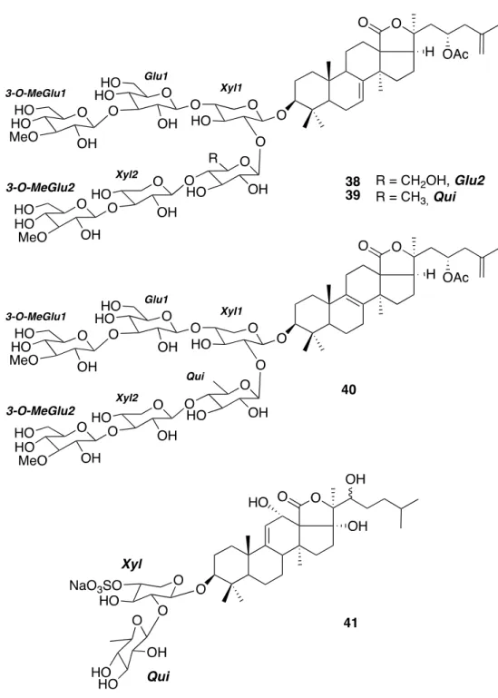
(6) Mar. Drugs 2020, 18, x FOR PEER REVIEW. 6 of 24. 0.027, and2020, 0.042 Mar. Drugs 18, μM, 59 respectively) with a lower selectivity index (SI of 4.7, 4.1, and 12.6, respectively) 6 of 24 [17] (Figure 5).. Figure 5. Steroid structures of the A-nor-5α-cholestanes 24–35 isolated from the sponge Figure 5. Steroid structures of the A-nor-5α-cholestanes 24–35 isolated from the sponge Teichaxinella Teichaxinella morchella (now Axinella corrugata) and the cholesterol derivatives 36 and 37 isolated morchella (now Axinella corrugata) and the cholesterol derivatives 36 and 37 isolated from the brown from the brown algae Padina sanctae-crucis and Turbinaria tricostata. algae Padina sanctae-crucis and Turbinaria tricostata.. 2.2.3. Triterpenoid Saponins 2.2.3. Triterpenoid Saponins Sea cucumbers constitute a rich source of triterpenoid saponins, with some of them exerting Sea cucumbers constitute a rich of triterpenoid saponins, withcollected some ofon them pharmacological effects [19]. From thesource sea cucumber Astichopus multifidus, the exerting Yucatan pharmacological effects [19]. From the sea cucumber Astichopus multifidus, collected on the Yucatan Peninsula coasts, Mena-Rejón and collaborators reported the isolation and the structural elucidation Peninsula coasts, Mena-Rejón and collaborators isolation and elucidation of three oligoglycoside triterpenes in 2016. Tworeported of them,the stichloroside B2 the (38)structural and astichoposide C of three oligoglycoside triterpenes in 2016. Two of them, stichloroside B 2 (38) and astichoposide C (39), were known, while the third one, named as astichoposide D (40), turned out to be a new natural (39), wereAntiproliferative known, while the third one, named as two astichoposide D MCF-7 (40), turned out to be a new product. activity assays against cancer lines, (ATCC HTB-22) and anatural highly product. Antiproliferative activity assays against two cancer lines, MCF-7 (ATCC HTB-22) and a invasive triple-negative breast cancer MDA-MB-231 (ATCC HTB-26), displayed that 38 had the highest highly invasive triple-negative breast cancer (ATCC HTB-26), displayed that 38 had antiproliferative activity against MCF-7 cellsMDA-MB-231 (6.45 µM), while 39 had the highest antiproliferative the highest antiproliferative activity against MCF-7 cells (6.45 μM), while 39 had the highest activity against the MDA-MB-231 cells (3.80 µM) [20]. The research group of Mena-Rejón also reported antiproliferative activity against the MDA-MB-231 cells (3.80 μM) [20]. The research group of Menain 2013 the isolation of the known triterpenoid saponin holothurin B2 (41) from the sea cucumber Rejón also floridana reported(now in 2013 the isolationfloridana) of the known triterpenoid saponin holothurin B2 (41) from Holothuria H. (Halodeima) [21] (Figure 6). the sea cucumber Holothuria floridana (now H. (Halodeima) floridana) [21] (Figure 6)..
(7) Mar. Drugs 2020, 18, 59. Mar. Drugs 2020, 18, x FOR PEER REVIEW. 7 of 24. 7 of 24. Figure6. 6. Structures the triterpenoid isolated the seathecucumbers: the Figure Structures of the of triterpenoid saponins saponins isolated from the seafrom cucumbers: hexaglycosides hexaglycosides 38–40 from Astichopus multifidus and the diglycoside 41 from Holothuria floridana 38-40 from Astichopus multifidus and the diglycoside 41 from Holothuria floridana (now H. (Halodeima) (now H. (Halodeima) floridana). floridana).. 2.3. Nitrogen Compounds 2.3. Nitrogen Compounds 2.3.1. Indole Derivatives 2.3.1. Indole Derivatives Olguin-Uribe et al. isolated two indoles, indole-3-carbaldehyde (42) and its brominated derivate, Olguin-Uribe et al. isolated two indole-3-carbaldehyde (42) the andtunicate its brominated derivate, 6-bromoindole-3-carbaldehyde (43),indoles, in 1997 from two different sources, Stomozoa murrayi 6-bromoindole-3-carbaldehyde (43), in 1997 from two different sources, the tunicate Stomozoa murrayi (currently known as Stomozoa roseola) and the bacterium Acinetobacter sp. associated to its surface [22]. (currently known Stomozoa thembacterium sp. associated to very its surface [22]. The tunicate was as collected at aroseola) depth and of 3–5 in Puerto Acinetobacter Morelos, Quintana Roo state, close to the The tunicate was collected at a depth of 3–5 m in Puerto Morelos, Quintana Roo state, very close to Institute of Marine Sciences and Limnology research station of the National Autonomous University the Institute of Marine Sciences and Limnology research station of the National Autonomous of Mexico (UNAM). These compounds were evaluated in several biological assays. The brominated University of Mexico (UNAM). These compounds were evaluated in several biological assays. The brominated indole 43 displays antimicrobial activity by inhibiting the growth of four marine bacterial.
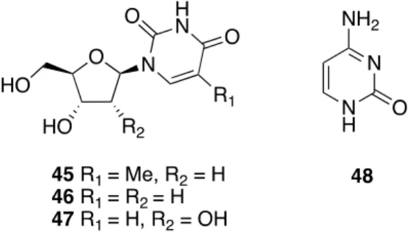
(8) Mar. Drugs 2020, 18, x FOR PEER REVIEW. 8 of 24. strains SM-S2, SM-Z, Bacillus marinus, and Vibrio campbellii, while its debrominated analog 42 shows Mar. Drugs 2020, 18, 59 8 of 24 no inhibitory activity. On REVIEW the other hand, both of the compounds exhibit antifouling activity Mar. Drugs 2020, 18, x FOR PEER 8 of by 24 completely inhibiting the settlement of Balanus amphitrite (now Amphibalanus amphitrite) at the highest strains SM-S2, SM-Z, Bacillus marinus, and Vibrio campbellii, while debrominated 42 shows indole 43 displays antimicrobial activity by inhibiting theμM growth of its four marine SM-S2, concentration tested at 100 μg mL−1 (0.13 and 0.084 respectively) and, bacterial even, analog thestrains most active no inhibitory activity. On the other hand,byboth the compounds antifouling activity by SM-Z, Bacillus marinus, and Vibrio campbellii, while analog 42 shows inhibitory compound 43 can inhibit larval settlement 80%of atits 10debrominated μg mL−1/0.044 exhibit μM. Finally, theseno compounds completely inhibiting the settlement of Balanus amphitrite (now Amphibalanus amphitrite) at the highest activity. no On antipredatory the other hand,(deterrent) both of theactivity compounds exhibit activity bywhich completely showed against the antifouling Serranus cabrilla fish, were inhibiting collected −1 (0.13 and 0.084 μM respectively) and, even, the most active concentration tested at 100 μg mL(now thethe settlement of Balanus amphitrite Amphibalanus the highest concentration tested in Mediterranean Sea, or significant antialgal activityamphitrite) against theatdiatom Nitzchia acicularis (Figure −1 −1/0.044 μM. Finally, these compounds compound 43 can inhibit larval settlement by 80% at 10 μg mL at 100 µg mL (0.13 and 0.084 µM respectively) and, even, the most active compound 43 can inhibit 7). −1 showed no antipredatory activity against the Serranus cabrilla fish, which were collected larvalPech-Puch settlement at(deterrent) 10 µg /0.044 Finally, these compounds showed antipredatory etby al.80% reported themL isolation of µM. another indole, serotonin (44), from theno salivary glands in the Mediterranean Sea,in orthe significant antialgal activity against the diatomin Nitzchia acicularis (Figure (deterrent) activity against Serranus cabrilla fish, which wereneurotoxic collected the Mediterranean Sea, of Octopus maya collected Sisal, Yucatan state in 2016 [23]. The activity previously found 7). or its significant antialgal activity against the diatom Nitzchia in extract was attributed to that compound (Figure 7). acicularis (Figure 7). Pech-Puch et al. reported the isolation of another indole, serotonin (44), from the salivary glands of Octopus maya collected in Sisal, Yucatan state in 2016 [23]. The neurotoxic activity previously found in its extract was attributed to that compound (Figure 7).. Figure 7.7.Structures Structures of indole derivatives 4243 and 43the from the tunicate murrayi (now Figure of indole derivatives 42 and from tunicate StomozoaStomozoa murrayi (now Stomozoa Stomozoa roseola) and the bacterium Acinetobacter sp. and 44 from the mollusk Octopus maya. roseola) and the bacterium Acinetobacter sp. and 44 from the mollusk Octopus maya.. Pech-Puch et al. reported the isolation of another indole, serotonin (44), from the salivary glands 2.3.2.Figure Nucleosides and Nitrogenous Bases 42 and 43 from the tunicate Stomozoa murrayi (now Stomozoa 7. Structures of indole derivatives of Octopus maya collected in Sisal, Yucatan state in 2016 [23]. The neurotoxic activity previously found roseola) and the bacterium Acinetobacter sp. and 44 from the mollusk Octopus maya. importance of the to study nucleosides comes in itsThe extract was attributed that of compound (Figure 7). from the fact that the arabino-nucleosides spongothymidine and spongouridine, isolated from a marine sponge, were the first marine natural 2.3.2. and Nitrogenous Bases 2.3.2. Nucleosides Nucleosides andtheir Nitrogenous products that showed potentialBases as drugs, because they constituted the basis of the development The importance of the study of comes from that the thecytarabine arabino-nucleosides of theThe firstimportance synthetic nucleosides approved as therapeutic thefact anticancer (ara-C) and of the study of nucleosides nucleosides comes drugs: from the the fact that arabino-nucleosides spongothymidine and spongouridine, isolated from a marine sponge, were the first marine the antiviral vidarabine (ara-A) [1]. spongothymidine and spongouridine, isolated from a marine sponge, were the first marine natural natural products showed potential as because constituted the the development Threethat nucleosides, thymidine 2′-desoxyuridine (46), and uridine (47), of were from products that showed their their potential(45), as drugs, drugs, because they they constituted the basis basis of theisolated development of the first synthetic nucleosides approved as therapeutic drugs: the anticancer cytarabine (ara-C) and the sponge Halichondria magniconulosa (now (Halichondria) at (ara-C) 0.5–1 m of of the first synthetic nucleosides approved as H. therapeutic drugs:magniconulosa) the anticancercollected cytarabine and the antiviral vidarabine (ara-A) [1]. depth in Chabihau, Yucatan state, and reported by the research group of Mena-Rejón in 2018 [24]. the antiviral vidarabine (ara-A) [1]. Three nucleosides, thymidine (45), (46), and uridine (47), were isolated from The same Medina-Gómez et al. reported the isolation of cytosine (48)(47), from the sponge Haliclona Threeyear, nucleosides, thymidine (45), 202′-desoxyuridine -desoxyuridine (46), and uridine were isolated from the the sponge Halichondria magniconulosa (now H. (Halichondria) magniconulosa) collected at 0.5–1 m of tubifera (now H. (Reniera) tubifera) [12] (Figure 8). No biological data were reported for 45–48. sponge Halichondria magniconulosa (now H. (Halichondria) magniconulosa) collected at 0.5–1 m of depth depth in Chabihau, Yucatan state, and reported by the research group of Mena-Rejón in 2018 [24]. in Chabihau, Yucatan state, and reported by the research group of Mena-Rejón in 2018 [24]. The same The year, Medina-Gómez et al. reported the isolation of cytosine the sponge Haliclona year,same Medina-Gómez et al. reported the isolation of cytosine (48) from(48) thefrom sponge Haliclona tubifera tubifera H. (Reniera) [12] (Figure 8). No biological data were reported for 45–48. (now H.(now (Reniera) tubifera)tubifera) [12] (Figure 8). No biological data were reported for 45–48.. Figure 8. Structures of nucleosides 45–47 and nitrogenous base 48 isolated from the sponges Halichondria magniconulosa (now H. (Halichondria) magniconulosa) and Haliclona tubifera (now H. (Reniera) tubifera), respectively. Figure 8. 8. Structures Structuresofof nucleosides nucleosides 45–47 45–47 and and nitrogenous nitrogenous base base 48 48 isolated isolated from from the the sponges sponges Figure Halichondria magniconulosa (now H. (Halichondria) magniconulosa) and Haliclona tubifera (now H. (Reniera) Halichondria magniconulosa (now H. (Halichondria) magniconulosa) and Haliclona tubifera (now H. tubifera), respectively. (Reniera) tubifera), respectively..
(9) Mar. Drugs 2020, 18, 59. 9 of 24. 2.3.3. Conotoxins The venomous fish-hunting cone snails that belong to the Conus genus are composed of a collection of toxin peptides that serve to immobilize prey by targeting different physiological mechanisms in their neuromuscular system. In this way, ω-conotoxin MVIIA, isolated from Conus magus, is commercialized as the synthetic Prialt® (ziconatide), which constituted the first FDA-approved drug that was directly derived from a marine natural product as a pain control drug [1]. On the basis of this background, the research group lead by Aguilar from the Institute of Neurobiology-UNAM at Queretaro in Mexico, reported the isolation of new conotoxins from three different snails, mainly belonging to the Conus genus, which were collected along the coasts of the Mexican Caribbean. Thus, the study of the extract of the venomous duct of Conus delessertii (now Conasprella delessertii), mollusk collected in Quintana Roo state, allowed Aguilar and collaborators to report four new peptides: conotoxin de13a (49) and conotoxin de7a (50), reported in 2005, conotoxin de7b (51), reported in 2009, and conotoxin de13b (52), reported in 2013. Conotoxin de13a (49) contains 32 amino acids (3486.76 Da) and it was defined as a new class of conotoxins. This peptide is characterized by the presence of high content of post-translational modified amino acids, such as 5-hydroxylysine, and the residues of cysteine arranged in a pattern (C-C-C-CC-C-C-C), which were not previously described in conotoxins [25]. On the other hand, conotoxin de7a (50) contains 28 amino acids (3170.0 Da), with some of them being post-transductionally modified, and a residue (-γCCS-) previously found only in two other conotoxins [26]. Conotoxin de7b (51), bearing 28 amino acids, including six cysteine residues, is characterized by existing in different post-transductional modified isomorphs (with molecular masses varying from 3078.6 to 3154.6 Da), some of them containing γ-carboxy-glutamate and/or 4-hydroxyproline at positions 4, 7, and/or 14 [27]. Finally, conotoxin de13b (52) has the same arrangement of cysteine residues as conotoxin de13a (49) [28] (Figure 9). From the extract of the venom duct of a second mollusk, Conus spurius, Aguilar and collaborators reported the isolation of twelve new conotoxin derivatives 53–64, from two different places: Quintana Roo and Campeche states. Conotoxin sr5a (53), which was reported in 2006, is a hydrophobic peptide belonging to the T-1 conotoxin family with a molecular mass of 1616.60 Da and a pair of disulfide bridges. In a biological test in mice, this conotoxin caused a depressed behavioral activity [29]. One year later, two new α-conotoxins of 18 amino acids, SrIA (54) and SrIB (55), with a molecular mass of 2202.9 and 2158.8 Da, respectively, were reported. Conotoxis 54 and 55 were evaluated as antagonists to nicotinic acetylcholine receptors in order to search for new therapeutic alternatives against brain diseases (schizophrenia, nocturnal frontal lobe epilepsy and Alzheimer’s disease). The results suggested not only that these conotoxins can operate as nicotinic acetylcholine receptor inhibitors, but also that they bind to nicotinic acetylcholine receptors with a very high affinity, increasing their intrinsic cholinergic response, and making them excellent model tools for studying toxin-receptor interaction [30]. The fourth new peptide, conotoxin sr11a (56), with a molecular weight of 3650.77 Da, was reported in 2007, being the first I-conotoxin that was isolated from the Western Atlantic. This peptide produces a stiffening of body, limbs, and tail when intracranially injected into mice [31]. Conotoxin sr7a (57), containing 32 amino acids (3330.74 Da) and reported in 2007, displays several in vivo effects, such as hyperactivity in mice and paralysis in freshwater snails (Pomacea paludosa), while it was inactive in intramuscular trials with the limpet Patella opea and the freshwater fish Lebistes reticulatus [32]. In contrast, conorfamide-Sr2 (CNF-Sr2, 58), as reported in 2008, with a molecular mass of 1468.70 Da and without cysteine residues, exhibits paralytic activity in the limpet Patella opea and produces hyperactivity in the freshwater snail Pomacea paludosa and mice [33]. From specimens of Conus spurius, collected in Isla Arena, Campeche state, were isolated and identified by reverse transcription polymerase chain reaction, seven conotoxins. Four of them belong to the T-1 conotoxin family, (18V) sr5a (59), (18T) sr5a (60), “extended” (61), and “hydrophilic” (62), which were reported in 2009 [34], and they are very similar to the conotoxin sr5a (53). The other three, reported in 2010, were the known conotoxin sr11a (56) already reported in 2007 [31] and the new conotoxins, sr11b (63) and sr11c (64) [35] (Figure 9)..
(10) Mar. Drugs 2020, 18, 59. 10 of 24. Mar. Drugs 2020, 18, x FOR PEER REVIEW. 10 of 24. Finally, Aguilar and collaborators reported in 2009 the isolation of a new peptide, pal9a (65) Finally, Aguilar and collaborators reported in 2009 the isolation of a new peptide, pal9a (65) (3678.84 Da) with 34 amino acids, including six cysteine residues, from a third mollusk, Polystira albida, (3678.84 Da) with 34 amino acids, including six cysteine residues, from a third mollusk, Polystira collected in Campeche state. This is the first P-conotoxin-like turritoxin isolated from a member of the albida, collected in Campeche state. This is the first P-conotoxin-like turritoxin isolated from a member familyofTurridae from the Western Atlantic [36] (Figure 9). the family Turridae from the Western Atlantic [36] (Figure 9). 49. DCOTSCOTTCANGWECCKGYOCVNKACSGCTH* O = 4-hydroxyproline W = 6-bromotryptophan K = 5-hydroxylysine * = amidated C-terminus 50. ACKOKNNLCAITγMAγCCSGFCLIYRCS* O = hydroxyproline γ = γ-carboxyglutamate * = amidated C-terminus 51. DCI(P/O)GG(E/ γ)NCDVFR(O/P)YRCCSGYCILLLCA O = 4 hydroxyproline γ = γ-carboxyglutamate 3 disulfide bridges between C-C-CC-C-C 52. DCPTSCPTTCANGWECCKGYPCVRQHCSGCNH* W = 6-bromotryptophan K = 5-hydroxylysine * = amidated C-terminus 53. IINWCCLIFYQCC 54. RTCCSROTCRMγYPγLCG* O = hydroxyproline γ = γ-carboxyglutamate 2 disulfide bridges between CC-C-C * = amidated C-terminus 55. RTCCSROTCRMEYPγLCG* O = hydroxyproline γ = γ-carboxyglutamate 2 disulfide bridges between CC-C-C * = amidated C-terminus 56. CRTEGMSC γ γNQQCCWRSCCRGECEAPCRFGP* γ = γ-carboxyglutamate 4 disulfide bridges between C-C-CC-CC-C-C * = amidated C-terminus 57. CLQFGSTCFLGDDDICCSGECFYSGGTFGICS* 3 disulfide bridges between C-C-CC-C-C * = amidated C-terminus. 58. GPMγDPLγIIRI* γ = γ-carboxyglutamate * = amidated C-terminus. 59. IINWCCLVFYQCC. 61. INWCCLIFYQCCL. 60. IINWCCLTFYQCC. 62. IMAGCCPRFYQCCYP* * = amidated C-terminus. 63. CDSDGTSCTSNMECCGYGCCSGTCQTPCRFGP* 4 disulfide bridges between C-C-CC-CC-C-C * = amidated C-terminus 64. CSDEGASCEKKSDCCFLSCCWSVCDRPCRLVP* 4 disulfide bridges between C-C-CC-CC-C-C * = amidated C-terminus 65. NVCDGDACPDGVCRSGCTCDFNVAQRKDTCFYPQ* 3 disulfide bridges between C-C-C-C-C-C * = amidated C-terminus. Figure 9. Structures of conotoxins 49–65isolated isolated from from cone to the Conus genus and and Figure 9. Structures of conotoxins 49–65 conesnails snailsbelonging belonging to the Conus genus Polystira albida. Polystira albida..
(11) Mar. Drugs 2020, 18, 59 x FOR PEER REVIEW Mar. Mar. Drugs 2020, 18, x FOR PEER REVIEW. 11 11 of of 24 11 of 24. 2.4. Biopolymers 2.4. 2.4. Biopolymers Biopolymers Freile-Pelegrín and collaborators reported the characterization of L-carrageenan (66) in 2018, Freile-Pelegrín andcollaborators collaboratorsreported reported the characterization of L-carrageenan (66) in being 2018, Freile-Pelegrín thered characterization of l-carrageenan in 2018, being obtained fromand the direct extraction of the algae Solieria filiformis collected(66) at Telchac in the being obtained from the direct extraction of the red algae Solieria filiformis collected at Telchac in the obtained from the extraction ofshows the red algae Solieriaactivity filiformisagainst collected at Telchac in the Yucatan Yucatan state. Thisdirect polysaccharide high antiviral Herpes simplex virus with Yucatan state. This polysaccharide shows high antiviral activity against Herpes simplex virus with state. This polysaccharide shows antiviral activity −1 an EC50 value of 6.3 μg mL−1 /0.019 high μM [37] (Figure 10). against Herpes simplex virus with an EC50 −1 an EC 50 value of 6.3 μg mL /0.019 μM [37] (Figure 10). value of 6.3 µg mL /0.019 µM [37] (Figure 10).. Figure 10. Structure of L-carrageenan isolated from the red algae Solieria filiformis. Structure of of L-carrageenan L-carrageenan isolated from the red algae Solieria filiformis. Figure 10. Structure. 3. Bioprospecting Overview 3. Bioprospecting Overview Overview 3. Bioprospecting A total total of 95 scientific documents were recorded and analyzed inreview, this review, including 82 A documents were recorded and and analyzed in thisin including 82 articles, A totalofof9595scientific scientific documents were recorded analyzed this review, including 82 articles, four postgraduate dissertation theses, and nine meeting abstract communications. They four postgraduate dissertation theses, andtheses, nine meeting abstract communications. They describe the articles, four postgraduate dissertation and nine meeting abstract communications. They describerelated the to reports related to research surveys on pharmacological surveys of extracts, chemical reports research on pharmacological of extracts, chemical composition, and isolation describe the reports related to research on pharmacological surveys of extracts, chemical composition, and isolation of marine natural products. Amarine total of 145 speciesare of marine organisms are of marine natural products. A totalnatural of 145 products. species ofA enclosed, belonging composition, and isolation of marine total of organisms 145 species of marine organisms are enclosed, belonging to 12 phyla (Tables 1 and 2), being the most representative Rhodophyta (27%), to 12 phyla (Tables 1toand being the most representative Rhodophyta (27%), Chlorophyta (22%), enclosed, belonging 12 2), phyla (Tables 1 and 2), being the most representative Rhodophyta (27%), Chlorophyta(17%), (22%),and Phaeophyta (17%), and Cnidaria (14%) (Figure 11). Phaeophyta Cnidaria (14%) (Figure 11). Chlorophyta (22%), Phaeophyta (17%), and Cnidaria (14%) (Figure 11).. Figure 11. Distribution of the reported marine organisms by phylum. Figure 11. Distribution of the reported marine organisms by phylum. Figure 11. Distribution of the reported marine organisms by phylum..
(12) Mar. Drugs 2020, 18, 59. 12 of 24. Table 1. Reported marine species of the Yucatan Peninsula in which natural products were isolated. Phylum. Species. Compounds Isolated and Biogenetic Origin. References. Proteobacteria. Acinetobacter sp.. 42, 43 (Nitrogen compounds). [22]. Chordata (Ascidian). Stomozoa murrayi (now Stomozoa roseola). 42, 43 (Nitrogen compounds). [22]. Cnidaria (Coral). Pterogorgia anceps. 1–3 (Polyketides). [11]. Astichopus multifidus. 38–40 (Terpenoids). [20]. Holothuria floridana (now H. (Halodeima) floridana). 41 (Terpenoid). [21]. Conus delessertii (now Conasprella delessertii). 49–52 (Nitrogen compounds). [25–28]. Conus spurius. 53–64 (Nitrogen compounds). [29–35]. Octopus maya. 44 (Nitrogen compound). [23]. Polystira albida. 65 (Nitrogen compound). [36]. Halichondria magniconulosa (now H. (Halichondria) magniconulosa). 45–47 (Nitrogen compounds). [24]. Haliclona tubifera (now H. (Reniera) tubifera). 6, 10–14 (Polyketides) and 48 (Nitrogen compound). [12]. Spongia tubulifera (now S. (Spongia) tubulifera). 15–21 (Terpenoids). [16]. Teichaxinella morchella (now Axinella corrugata). 24–35 (Terpenoids). [18]. Dictyota ciliolata. 22, 23, 36 (Terpenoids). [17]. Lobophora variegata. 7–9 (Polyketides). [14]. Padina sanctae-crucis. 36, 37 (Terpenoids). [17]. Turbinaria tricostata. 36, 37 (Terpenoids). [17]. Solieria filiformis. 66 (Biopolymer). [37]. Echinodermata (Sea cucumbers). Mollusca (Mollusks). Porifera (Sponges). Phaeophyta (Brown algae). Rhodophyta (Red algae).
(13) Mar. Drugs 2020, 18, 59. 13 of 24. Table 2. Reported marine species from Yucatan Peninsula related to bioprospecting without determining the chemical composition. Domain: Bacteria Kingdom: Bacteria Phylum. Genus or Species. References. Actinobacteria. Streptomyces Saccharomonospora Dietzia Nocardiopsis Pseudonocardia Verrucosispora Brachybacterium Jiangella Salinispora. [38]. Domain: Eukarya Kingdom: Animalia Arthropoda (Crustaceans). Bathynomus giganteus Limulus Polyphemus. [39] [40]. Chordata (Ascidians). Trididemnum solidum. [41]. Cnidaria (Anemones). Cnidaria (Corals). Cnidaria (Jellyfish). Echinodermata (Sea cucumbers). Mollusca (Mollusks). Actiinidae Gen. sp. nov *. Anthopleura texaensis Bartholomea annulata Bunodeopsis antilliensis Bunodeopsis globulifera (now Viatrix globulifera) Condylactis gigantea Lebrunia danae (now L. neglecta) Stichodactyla helianthus Telmatactis bernoni*. [42] [43–46] [47] [48,49] [50] [47,51] [47,52] [43]. Millepora alcicornis Millepora complanata Porites astreoides Pseudodiploria strigosa Siderastrea siderea. [53–57] [53,55,58–62] [63] [63] [63]. Aurelia aurita Carybdea marsupialis Cassiopea xamachana (now Cassiopea andromeda) Linuche unguiculata Pelagia noctiluca. [64,65] [47,66,67] [47,68,69] [47] [70]. Holothuria mexicana (now H. (Halodeima) mexicana). [71]. Isostichopus badionotus. [72–74]. Conus austini (now C. cancellatus) Conus spurius Gemmula periscelida Octopus maya Polystira albida Domain: Eukarya Kingdom: Plantae. [75] [75,76] [77] [78] [75,77].
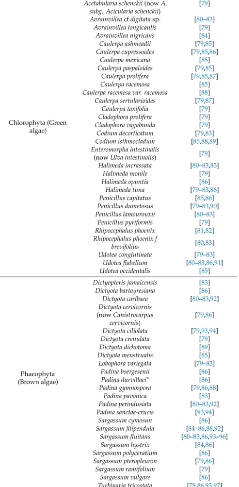
(14) Mar. Drugs 2020, 18, 59. 14 of 24. Table 2. Cont.. Chlorophyta (Green algae). Phaeophyta (Brown algae). Acetabularia schenckii (now A. subg. Acicularia schenckii) Avrainvillea cf digitata sp. Avrainvillea longicaulis Avrainvillea nigricans Caulerpa ashmeadii Caulerpa cupressoides Caulerpa mexicana Caulerpa paspaloides Caulerpa prolifera Caulerpa racemosa Caulerpa racemosa var. racemosa Caulerpa sertularioides Caulerpa taxifolia Cladophora prolifera Cladophora vagabunda Codium decorticatum Codium isthmocladum Enteromorpha intestinalis (now Ulva intestinalis) Halimeda incrassata Halimeda monile Halimeda opuntia Halimeda tuna Penicillus capitatus Penicillus dumetosus Penicillus lamourouxii Penicillus pyriformis Rhipocephalus phoenix Rhipocephalus phoenix f brevifolius Udotea conglutinata Udotea flabellum Udotea occidentalis Dictyopteris jamaicensis Dictyota bartayresiana Dictyota caribaea Dictyota cervicornis (now Canistrocarpus cervicornis) Dictyota ciliolata Dictyota crenulata Dictyota dichotoma Dictyota menstrualis Lobophora variegata Padina boergesenii Padina durvillaei* Padina gymnospora Padina pavonica Padina perindusiata Padina sanctae-crucis Sargassum cymosun Sargassum filipendula Sargassum fluitans Sargassum hystrix Sargassum polyceratium Sargassum pteropleuron Sargassum ramifolium Sargassum vulgare Turbinaria tricostata Turbinaria turbinata. [79] [80–83] [79] [84] [79,85] [79,85,86] [85] [79,85] [79,85,87] [85] [88] [79,87] [79] [79] [79] [79,83] [85,88,89] [79] [80–83,85] [79] [86] [79–83,86] [85,86] [79–83,90] [80–83] [79] [81,82] [80,83] [79–83] [80–83,86,91] [85] [83] [86] [80–83,92] [79,86] [79,93,94] [79] [89] [85] [79–83] [86] [86] [79,86,88] [83] [80–83,92] [93,94] [86] [84–86,88,92] [80–83,86,93–96] [84,86] [86] [79,86] [79] [86] [79,86,93,97] [80–83,86,92].
(15) Mar. Drugs 2020, 18, 59. 15 of 24. Table 2. Cont.. Rhodophyta (Red algae). Tracheophyta (Seagrasses). Acanthophora spicifera Agardhiella sp. Agardhiella subulata Bryothamnion triquetrum (now Alsidium triquetrum) Ceramium nitens Champia salicornioides Chondria atropurpurea Chondria baileyana Chondrophycus papillosus (now Palisada perforata) Chondrophycus poiteaui (now Yuzurua poteaui) Digenea simplex Eucheuma isiforme (now Eucheumatopsis isiformis) Gracilaria blodgettii Gracilaria bursa-pastoris Gracilaria caudata (now Crassiphycus caudatus) Gracilaria cervicornis Gracilaria cornea (now Crassiphycus corneus) Gracilaria crassissima (now Crassiphycus crassissimus) Gracilaria cylindrica Gracilaria damaecornis* Gracilaria sp. Gracilaria tikvahiae Gracilariopsis tenuifrons Halymenia floresia (now H. floresii) Heterosiphonia gibbesii Hydropuntia cornea (now Crassiphycus corneus) Hypnea musciformis Hypnea spinella Jania capillacea Laurencia intricata Laurencia microcladia Laurencia obtusa Laurencia papillosa (now Palisada perforata) Laurencia poiteaui (now Palisada poiteaui) Liagora ceranoides Nemalion helmintoides* Rhodymenia pseudopalmata Solieria filiformis Spyridia filamentosa Syringodium filiforme Thalassia testudinum. [79] [80–83] [89] [79–83,85,86,89] [79–83,85] [79,80,82,83] [79] [79] [79] [79] [79,85,86] [79–83,85,88,98] [99] [79] [79–83,85] [80–83,99] [79,85,88,100] [99,100] [79] [80–83] [80–83] [79] [79,101] [79,80,82–85,89,102, 103] [79–83] [80–83,95] [104] [79] [80–83] [79,86,87] [80–83] [79,84–86] [86] [85,86] [79] [79] [95,105,106] [95,107,108] [87] [87] [87]. * Organisms not found in World Register of Marine Species (WORMS database) [109].. The mollusk Conus spurius and two algae, Halymenia floresia (now H. floresii) and Sargassum fluitans, with nine reports each, were the most reported species. The coral Millepora complanata and eight algae, Halimeda tuna, Penicillus dumetosus, Udotea flabellum, Bryothamnion triquetrum (now.
(16) Tracheophyta (Seagrasses). Syringodium filiforme Thalassia testudinum. [87] [87]. * Organisms not found in World Register of Marine Species (WORMS database) [109].. The 2020, mollusk Mar. Drugs 18, 59 Conus. spurius and two algae, Halymenia floresia (now H. floresii) and Sargassum 16 of 24 fluitans, with nine reports each, were the most reported species. The coral Millepora complanata and eight algae, Halimeda tuna, Penicillus dumetosus, Udotea flabellum, Bryothamnion triquetrum (now Alsidium Alsidium triquetrum), triquetrum), Ceramium Ceramium nitens, nitens, Eucheuma Eucheuma isiforme isiforme (now (now Eucheumatopsis Eucheumatopsis isiformis), isiformis), Gracilaria Gracilaria caudata (now Crassiphycus caudatus), Lobophora variegata, and Turbinaria turbinata, with 6–8 reports each, caudata (now Crassiphycus caudatus), Lobophora variegata, and Turbinaria turbinata, with 6–8 reports following the listthe as list it isas shown in Figure 12. 12. each, following it is shown in Figure. Figure 12. 12. Number of publications of reported reported marine marine species species organized organized by by phylum. phylum. Only those species that have three three or or more more reports reports are are displayed. displayed.. From point of of view, From the the territorial territorial distribution distribution point view, the the highest highest number number of of reports reports corresponds corresponds to to marine organisms that were collected at the coast of the Yucatan state (38%), followed by the coasts of marine organisms that were collected at the coast of the Yucatan state (38%), followed by the coasts Quintana RooRoo statestate (36%) and,and, finally, the coasts of Campeche state (4%). 22% of 22% the reports of Quintana (36%) finally, the coasts of Campeche state However, (4%). However, of the Mar. Drugs 2020, 18, x FOR PEER REVIEW 17 of 24 did not specify statethe where marine were collected (Figure 13). reports did not the specify statethe where the organisms marine organisms were collected (Figure 13).. Figure 13. Geographic distribution of collections sources in percentage by state. Figure 13. Geographic distribution of collections sources in percentage by state.. Figure 14 displays the number of reports per year. As far as we know, the first report was Figure 14 displays the number of reports per year. As far as we know, the first report was published in 1981 and, since then, the number of publications related to the search for natural marine published in 1981 and, since then, the number of publications related to the search for natural marine products of the Yucatan Peninsula has been increasing. However, this increase was not constant, being products of the Yucatan Peninsula has been increasing. However, this increase was not constant, the years 2007, 2013, 2014, and 2016, with seven publications each, when more reports were published. being the years 2007, 2013, 2014, and 2016, with seven publications each, when more reports were published..
(17) Figure 14 displays the number of reports per year. As far as we know, the first report was published in 1981 and, since then, the number of publications related to the search for natural marine products of the Yucatan Peninsula has been increasing. However, this increase was not constant, being the years 2007, 2013, 2014, and 2016, with seven publications each, when more reports17were Mar. Drugs 2020, 18, 59 of 24 published.. Figure 14. Number of of publications publications per per year. year. Figure 14. Number. 4. Conclusions 4. Conclusions The present review represents the first comprehensive report of natural products that have The present review represents the first comprehensive report of natural products that have been been isolated from marine organisms collected along the coasts of the Yucatan Peninsula, covering isolated from marine organisms collected along the coasts of the Yucatan Peninsula, covering literature up to mid-2019. As result of 38 years of investigations of marine organisms that were literature up to mid-2019. As result of 38 years of investigations of marine organisms that were collected in the Yucatan Peninsula, 66 marine natural products were isolated from 18 species collected in the Yucatan Peninsula, 66 marine natural products were isolated from 18 species belonging to eight different phyla (Proteobacteria (Acinetobacter sp.), Chordata (ascidian: Stomozoa belonging to eight different phyla (Proteobacteria (Acinetobacter sp.), Chordata (ascidian: Stomozoa murrayi (now Stomozoa roseola), Cnidaria (Pterogorgia anceps), Echinodermata (Astichopus multifidus murrayi (now Stomozoa roseola), Cnidaria (Pterogorgia anceps), Echinodermata (Astichopus multifidus and Holothuria floridana (now H. (Halodeima) floridana)), Mollusca (Conus delessertii (nowConasprella and Holothuria floridana (now H. (Halodeima) floridana)), Mollusca (Conus delessertii (nowConasprella delessertii), Conus spurius, Octopus maya, and Polystira albida), Porifera (Halichondria magniconulosa (now delessertii), Conus spurius, Octopus maya, and Polystira albida), Porifera (Halichondria magniconulosa H. (Halichondria) magniconulosa), Haliclona tubifera (now H. (Reniera) tubifera), Spongia tubulifera (now S. (now H. (Halichondria) magniconulosa), Haliclona tubifera (now H. (Reniera) tubifera), Spongia tubulifera (Spongia) tubulifera) and Teichaxinella morcella (now Axinella corrugata)), Rhodophyta (Solieria filiformis), (now S. (Spongia) tubulifera) and Teichaxinella morcella (now Axinella corrugata)), Rhodophyta (Solieria Mar. Drugs 2020, 18, x(Dictyota FOR PEERciliolata, REVIEW Lobophora variegata, Padina sanctae-crucis, and Turbinaria tricostata) 18 of 24 and Phaeophyta filiformis), and Phaeophyta (Dictyota ciliolata, Lobophora variegata, Padina sanctae-crucis, and Turbinaria (Table 1). Out of the 66 marine natural products identified, 26 correspond to structures that were not tricostata) 1). Outreported. of the 66 marine natural products identified, 26 correspondtotothree structures that were not (Table previously These 26 new chemical entities correspond aliphatic previously reported. These 26 new chemical entities correspond to three aliphatic polyketides (1–3), polyketides (1–3), one glycolipid (24, 25), one triterpenoid one glycolipid (7), two diterpenes (7), (15,two 16), diterpenes two steroids(15, (24,16), 25),two onesteroids triterpenoid saponin (40), and 17 saponin (40), and 17 conotoxins (49–65). conotoxins (49–65). Figure 15 15 displays displays the the overall overall biogenetic biogenetic distribution distribution of of the the reported reported compounds. compounds. The Figure The terpenoid terpenoid biogenesis is again the most prominent pathway (40.9%), enclosing the diterpenes-sesterterpenes biogenesis is again the most prominent pathway (40.9%), enclosing the diterpenes-sesterterpenes (nine compounds), compounds), steroids steroids (14 (14 compounds), compounds), and and triterpenoids triterpenoids saponins saponins (four (four compounds). compounds). On On the the (nine other hand, conotoxins, with 17 compounds (25.8%) constitute the largest group of the reported other hand, conotoxins, with 17 compounds (25.8%) constitute the largest group of the reported natural products. products. natural. Figure 15. Biosynthetic classes of the reported marine natural products. Figure 15. Biosynthetic classes of the reported marine natural products.. The biological studies of the isolated compounds are focused on cytotoxic or antiproliferative The biological studies of the isolated compounds are focused on cytotoxic or antiproliferative activities (diterpenes 15, 18, 20, 22, and 23; steroids 36 and 37, and triterpenoids saponins 38 and 39), activities (diterpenes 15, 18, 20, 22, and 23; steroids 36 and 37, and triterpenoids saponins 38 and 39), the antimicrobial, antifouling, antipredatory (deterrent), and antialgal activity (indole derivative 42 and the antimicrobial, antifouling, antipredatory (deterrent), and antialgal activity (indole derivative 42 and 43), the antiprotozoal activity (glycolipids 7–9), neurotoxic activity (indole derivative 44), behavioral activity in animal models (conotoxins 53, 56–58), and finally in the interesting pharmacological activities against brain diseases of the new conotoxins 54 and 55, and the high antiviral activity of the known biopolymer L-carrageenan (66). As a concluding remark, this review shows the potential of the Yucatan Peninsula as an.
(18) Mar. Drugs 2020, 18, 59. 18 of 24. 43), the antiprotozoal activity (glycolipids 7–9), neurotoxic activity (indole derivative 44), behavioral activity in animal models (conotoxins 53, 56–58), and finally in the interesting pharmacological activities against brain diseases of the new conotoxins 54 and 55, and the high antiviral activity of the known biopolymer L-carrageenan (66). As a concluding remark, this review shows the potential of the Yucatan Peninsula as an interesting source of new marine natural products, not only because of its unique and rich diversity of marine organisms, but also due to the small number of works that have been published so far, which indicates that this area of research has been poorly investigated. For these reasons, the marine biodiversity of the Yucatan Peninsula can be considered as a poor exploited source of new bioactive marine natural products, which could be the base of the development of new drugs. Author Contributions: Conceptualization, C.J., D.P.-P. and J.R.; Funding acquisition and Resources, J.R. and C.J.; Writing—original draft, D.P.-P.; M.P.-P., O.A.L.-R. Writing—review & editing, D.P.-P.; J.R. and C.J. All authors have read and agree to the published version of the manuscript. Funding: This work was supported by grants RTI2018-093634-B-C22 (AEI/FEDER, EU) from the State Agency for Research (AEI) of Spain, co-funded by the FEDER Programme from the European Union, and GRC2018/039 and ED431E 2018/03 of CICA-INIBIC strategic group from Xunta de Galicia. D.P.P. received a fellowship from the program National Council of Science and Technology (CONACYT) of Mexico and the Secretariat of Research, Innovation and Higher Education (SIIES) of Yucatan (Mexico). O.A.L.R. received a financial support from MostMicro unit. Project LISBOA-01-0145-FEDER-007660 of Portugal. Conflicts of Interest: The authors declare no conflict of interest.. References 1. 2. 3.. 4. 5. 6.. 7.. 8.. 9.. 10. 11. 12.. Jiménez, C. Marine Natural Products in Medicinal Chemistry. ACS Med. Chem. Lett. 2018, 9, 959–961. [CrossRef] Altmann, K.H. Drugs from the Oceans: Marine Natural Products as Leads for Drug Discovery. Chimia 2017, 71, 646–652. [CrossRef] Mayer, A.M.S.; Rodríguez, A.D.; Taglialatela-Scafati, O.; Fusetani, N. Marine Pharmacology in 2012–2013: Marine Compounds with Antibacterial, Antidiabetic, Antifungal, Anti-Inflammatory, Antiprotozoal, Antituberculosis, and Antiviral Activities; Affecting the Immune and Nervous Systems, and Other Miscellaneous Mechanisms of Action. Mar. Drugs 2017, 15, 273. Barrera, A. La Península de Yucatán Como Provincia Biótica. Rev. Soc. Mex. Hist. Nat. 1962, 23, 71–105. Hernández-Bolio, G.I.; Ruiz-Vargas, J.A.; Peña-Rodríguez, L.M. Natural Products from the Yucatecan Flora: Structural Diversity and Biological Activity. J. Nat. Prod. 2019, 82, 647–656. [CrossRef] [PubMed] Pech Pool, D.; Ardisson Herrera, P.L. Diversidad en el Bentos Marino-Costero. In Biodiversidad y Desarrollo Humano en Yucatán; Duran, R., Méndez, M., Eds.; CICY; PPD-FMAM; CONABIO; SEDUMA: Mérida, Yucatán, Mexico, 2010; pp. 144–146. ISBN 978-607-7823-08-7. Garza Pérez, J.R.; Simões, N.; Chiappa Carrara, X.; Cucio, C.; Mascaró Miquelajáuregui, M.; Oseguera Cruz, M.; Lozano Aburto, M.; Acosta González, G. Comunidades Coralinas de las Bajas de Sisal. In Biodiversidad y Desarrollo Humano en Yucatán; Duran, R., Méndez, M., Eds.; CICY; PPD-FMAM; CONABIO; SEDUMA: Mérida, Yucatán, Mexico, 2010; pp. 148–149. ISBN 978-607-7823-08-7. Torruco Gómez, D.; González Solís, A. Las Esponjas y Su Importancia. In Biodiversidad y Desarrollo Humano en Yucatán; Duran, R., Méndez, M., Eds.; CICY; PPD-FMAM; CONABIO; SEDUMA: Mérida, Yucatán, Mexico, 2010; pp. 202–203. ISBN 978-607-7823-08-7. Acosta González, G.; Arias González, J.E. Comunidades Bentónicas Del Arrecife Alacranes. In Biodiversidad y Desarrollo Humano en Yucatán; Duran, R., Méndez, M., Eds.; CICY; PPD-FMAM; CONABIO; SEDUMA: Mérida, Yucatán, Mexico, 2010; p. 147. ISBN 978-607-7823-08-7. Palomino-Alvarez, L.A.; Moreira Rocha, R.; Simões, N. Checklist of Ascidians (Chordata, Tunicata) from the Southern Gulf of Mexico. Zookeys 2019, 832, 1–33. [CrossRef] Guo, Y.W.; Gavagnin, M.; Mollo, E.; Trivellone, E.; Cimino, G. Three New Butenolide Lipids from the Caribbean Gorgonian Pterogorgia anceps. J. Nat. Prod. 1999, 62, 1194–1196. [CrossRef] Medina-Gómez, S.; Mirón-López, G.; Quijano-Quiñones, R. Caracterización Estructural de Compuestos Obtenidos a Partir de Esponja de Mar Empleando Resonancia Magnética Nuclear y Cálculos Teóricos. In 11◦.
(19) Mar. Drugs 2020, 18, 59. 13.. 14. 15. 16.. 17.. 18.. 19. 20.. 21.. 22.. 23.. 24.. 25.. 26.. 27.. 28.. 29.. 30.. 19 of 24. Foro en Ciencias Químicas y Bioquímicas; Posgrado en Ciencias Químicas y Bioquímicas: Mérida, Mexico, 2018; pp. 23–24. Fattorusso, E.; Mangoni, A. Progress in the Chemistry of Organic Natural Products; Herz, W., Kirby, G.W., Moore, R.E., Steglich, W., Tamm, C., Eds.; Springer: Berlin, Germany, 1997; pp. 215–301. ISBN 978-3-7091-7456-2. Cantillo-Ciau, Z.; Moo-Puc, R.; Quijano, L.; Freile-Pelegrín, Y. The Tropical Brown Alga Lobophora variegata: A Source of Antiprotozoal Compounds. Mar. Drugs 2010, 8, 1292–1304. [CrossRef] Carroll, A.R.; Copp, B.R.; Davis, R.A.; Keyzers, R.A.; Prinsep, M.R. Marine Natural Products. Nat. Prod. Rep. 2019, 36, 122–173. [CrossRef] [PubMed] Pech-Puch, D.; Rodríguez, J.; Cautain, B.; Sandoval-Castro, C.A.; Jiménez, C. Cytotoxic Furanoditerpenes from the Sponge Spongia tubulifera Collected in the Mexican Caribbean. Mar. Drugs 2019, 17, 416. [CrossRef] [PubMed] Caamal-Fuentes, E.; Moo-Puc, R.; Freile-Pelegrín, Y.; Robledo, D. Cytotoxic and Antiproliferative Constituents from Dictyota ciliolata, Padina sanctae-crucis and Turbinaria tricostata. Pharm. Biol. 2014, 52, 1244–1248. [CrossRef] Bohlin, L.; Sjöstrand, U.; Djerassi, C.; Sullivan, B. Minor and Trace Sterols in Marine Invertebrates. Part 20. 3E-Hydroxy-methyl-A-nor-Patinosterol and 3E-Hydroxymethyl-A-nor-dinosterol. Two New Sterols with Modified Nucleus and Side-Chain from the Sponge Teichaxinella morchella. J. Chem. Soc. Perkin Trans. 1981. [CrossRef] Khotimchenko, Y. Pharmacological Potential of Sea Cucumbers. Int. J. Mol. Sci. 2018, 19, 1342. [CrossRef] Graniel-Sabido, M.J.; Mirón-López, G.; León-Deniz, L.V.; Moo-Puc, R.E.; Quintal-Novelo, C.J.; Quijano, L.; Mena-Rejón, G.J. Total NMR Assignment of a New Antiproliferative Triterpene Oligoglycoside from the Sea Cucumber Astichopus multifidus. Tetrahedron Lett. 2016, 57, 4375–4378. [CrossRef] Salazar-Mendoza, J.; Padilla-Montaño, N.; León-Deniz, L.V.; Mena-Rejón, G.J.; Quijano, L. Actividad Antifúngica de Metabolitos Aislados de la Pared Corporal de Holoturia Floridana. In Foro en Ciencias Químicas y Bioquímicas. Posgrado Institucional en Ciencias Químicas y Bioquímicas; Universidad Autónoma de Yucatán: Mérida, Mexico, 2013; pp. 19–20. Olguin-Uribe, G.; Abou-Mansour, E.; Boulander, A.; Débard, H.; Francisco, C.; Combaut, G. 6-Bromoindole-3-Carbaldehyde, from an Acinetobacter sp. Bacterium Associated with the Ascidian Stomozoa murrayi. J. Chem. Ecol. 1997, 23, 2507–2521. [CrossRef] Pech-Puch, D.; Cruz-López, H.; Canche-Ek, C.; Campos-Espinosa, G.; García, E.; Mascaro, M.; Rosas, C.; Chávez-Velasco, D.; Rodríguez-Morales, S. Chemical Tools of Octopus maya During Crab Predation Are Also Active on Conspecifics. PLoS ONE 2016, 11, e0148922. [CrossRef] Salazar Mendoza, J.; Mirón López, G.; Mena Rejón, G.J. Estudio Químico de Halichondria Magniconulosa (Porifera: Demospongiae) del Litoral del Estado de Yucatán. In 11◦ Foro en Ciencias Químicas y Bioquímicas; Posgrado Institucional en Ciencias Químicas y Bioquímicas: Mérida, Mexico, 2018; pp. 5–6. Aguilar, M.B.; López-Vera, E.; Ortiz, E.; Becerril, B.; Possani, L.D.; Olivera, B.M.; Heimer de la Cotera, E.P. A Novel Conotoxin from Conus delessertii with Posttranslationally Modified Lysine Residues. Biochemistry 2005, 44, 11130–11136. [CrossRef] [PubMed] Aguilar, M.B.; López-Vera, E.; Imperial, J.S.; Falcón, A.; Olivera, B.M.; Heimer de la Cotera, E.P. Putative γ-Conotoxins in Vermivorous Cone Snails: The Case of Conus delessertii. Peptides 2005, 26, 23–27. [CrossRef] Aguilar, M.B.; Flores-Torres, A.; Batista, C.V.F.; Falcón, A.; López-Vera, E.; Heimer de la Cotera, E.P. Structural Characterization of Five Post-translationally Modified Isomorphs of a Novel Putative δ-Conotoxin from the Vermivorous Snail Conus delessertii from the Mexican Caribbean Sea. Peptides 2009, 30, 458–466. [CrossRef] Aguilar, M.B.; Ortiz, E.; Kaas, Q.; López-Vera, E.; Becerril, B.; Possani, L.D.; Heimer de la Cotera, E.P. Precursor De13.1 from Conus delessertii Defines the Novel G Gene Superfamily. Peptides 2013, 41, 17–20. [CrossRef] Aguilar, M.B.; Lezama-Monfil, L.; Maillo, M.; Pedraza-Lara, H.; López-Vera, E.; Heimer de la Cotera, E.P. A biologically active hydrophobic T-1-Conotoxin from the Venom of Conus spurius. Peptides 2006, 27, 500–505. [CrossRef] [PubMed] López-Vera, E.; Aguilar, M.B.; Schiavon, E.; Marinzi, C.; Ortiz, E.; Restano Cassulini, R.; Batista, C.V.F.; Possani, L.D.; Heimer de la Cotera, E.P.; Peri, F.; et al. Novel α-Conotoxins from Conus spurius and the.
Figure


+7


Documento similar
Super Natural II also provides infor- mation about the pathways associated with synthe- sis and degradation of the natural products, as well as their mechanism of action with respect
1/6 Course Specification Quality assurance of natural products Fifth Year 2020/2021 University: Minia Faculty: Pharmacy Department: Pharmacognosy 1- Administrative Information
Using an initial set of 29,779 natural products that are annotated with their natural source and an experimentally validated virtual screening procedure previously developed in our lab
Market Surveys & Brief for Two Botanical Natural Products 1 Market Surveys & Brief for Two Botanical Natural Products Camu camu (Myrciaria dubia) and Sacha inchi (Plukenetia volubilis)
INFORME NATURAL PRODUCTS EXPO WEST 2012 P ? g i n a | 2 Elaborado por Carla Vaca Espinoza 2012 P ? g i n a | 3 ?ndice 1 Antecedentes y Justificaci?n 2 Ficha t?cnica de la feria 3
Secondary Metabolites Isolated from Chilean Marine Algae: A Review • Arrieche, DioniaSend mail to Arrieche D.; • Carrasco, HéctorbSend mail to Carrasco H.; • Olea, Andrés F.bSend
Because of the chemical diversity and biological activities of benzimidazole and pyrrole ring contained natural products against many diseases, they have an important role in the
A micellar electrokinetic capillary chromatography MECC method was tested to analyze secondary metabolites produced by 20 mold strains commonly found in dry-cured meat products.. In
