FACULTAD DE MEDICINA
UNIVERSIDAD DE CANTABRIA
GRADO EN MEDICINA
TRABAJO FIN DE GRADO
Role of gut microbiota in obesity
Papel de la microbiota intestinal en la obesidad
Autor: Dña. Elena Blanco Martín
Director/es: Dña. Asunción Seoane Seoane
CONTENTS
Abstract ... 1
1. Introduction ... 2
2. Discovering the human microbiome ... 2
3. The healthy microbiome ... 6
4. The intestinal microbial community ... 7
5. Biogeography of gut microbiota ... 8
6. Global variation of human gut metagenomes ... 10
7. Establishment of gut microbiota in different stages of life ... 12
8. Some roles of gut microbiota ... 13
9. Microbiota-associated immunomodulatory metabolites ... 15
10. Short-chain fatty acids (SCFAs) ... 17
10.1 Microbial cross-feeding dynamics in SCFAs production ... 18
10.2 Gut integrity and biological effects of SCFAs ... 20
10.3 SCFAs and the Immune System ... 20
10.4 Role of SCFAs on lipid metabolism ... 22
10.5 SCFAs and appetite regulation... 22
10.6 Regarding the methodology employed in SCFAs experiments ... 23
11. Contribution of microbiota to the development of diseases ... 23
11.1 Autoimmune diseases ... 23
11.1.1 Rheumatoid arthritis (RA) ... 23
11.1.2 Systemic lupus erythematosus (SLE) ... 24
11.1.3 Ankylosing spondylitis (AS) ... 24
11.1.4 Inflammatory bowel disease (IBD) ... 24
11.2 Neurodevelopmental, psychiatric and neurodegenerative diseases ... 24
11.3 Atherosclerosis and thrombosis risk ... 25
11.4 Type 1 diabetes ... 26
11.5 Type 2 diabetes ... 27
12. Obesity ... 28
12.1 Diet and obesity ... 28
12.1.1 Role of diet in gut microbiota composition ... 29
12.2 Host-gut microbiota cross-talk contribution in metabolic syndrome and obesity ... 31
12.2.1 Mechanisms that link gut bacteria and energy metabolism ... 34
12.3 Gut-brain axis in the regulation of energy balance ... 36
12.3.1 Control of energy homeostasis by the endocannabinoid system (ECS) ... 38
13. Modulation of gut microbiota with therapeutic purposes ... 39
13.1 Prebiotics and probiotics used in prevention and treatment of obesity ... 40
13.2 Role of Akkermansia muciniphila and Faecalibacterium prausnitzii ... 41
Bibliography references ... 44
Abbreviations ... 46
Glossary ... 47
Biological classification system of organisms ... 47
1 ABSTRACT
Obesity is considered the epidemic of today, which limits the quality of life and predisposes to suffer numerous health problems. There is an increasingly evidence of the close relationship between the microbiota and the risk of developing obesity, although the exact mechanism remains unclear as many studies give contradictory results probably due to the multiple conditions that in turn influence the development of obesity such as genetic, sociodemographic and environmental factors.
In this review, reference is made to the importance of the molecular cross-talk of the microbiota with the host, linking it with the development of obesity and metabolic inflammation, as well as the evidence of microbiota plasticity according to diets. Special mention is also made of the central role that the commensal bacterium Akkermansia muciniphila has in improving the metabolic profile of the host. Finally, it is pointed out the growing importance of gut microbiota’s manipulation with therapeutic purpose against obesity and related pathologies through interventions like fecal matter transplants, prebiotics or probiotics.
Keywords: gut microbiota, obesity, metabolic syndrome, SCFAs, microbiota-host crosstalk.
La obesidad es considerada una de las epidemias más importantes de hoy en día, que limita la calidad de vida y predispone a padecer numerosos problemas de salud. Cada vez hay más pruebas de la estrecha relación entre la microbiota y el riesgo de desarrollar obesidad, aunque el mecanismo exacto no está claro ya que son muchos los estudios que ofrecen resultados contradictorios probablemente debido a las múltiples condiciones que a su vez influyen en el desarrollo de la obesidad como la genética o los factores sociodemográficos y ambientales.
En esta revisión, se hace referencia a la importancia de la comunicación molecular de la microbiota con el huésped, relacionándola con el desarrollo de la obesidad y la inflamación metabólica así como la evidencia de la plasticidad de la microbiota de acuerdo a las dietas. También se hace mención especial del papel central que desempeña la bacteria comensal Akkermansia muciniphila en la mejora del perfil metabólico del huésped. Finalmente, se señala la importancia creciente que ha cobrado la manipulación de la microbiota intestinal con fines terapéuticos contra la obesidad y las patologías relacionadas, a través de intervenciones como trasplantes de materia fecal, prebióticos o probióticos.
Palabras clave: microbiota intestinal, obesidad, síndrome metabólico, AGCC, comunicación microbiota-huésped.
2 1. INTRODUCTION
Nowadays, obesity is an epidemic that deeply worries around the world and although its etiology is multifactorial (environmental, dietary, lifestyle, genetic and pathological factors), in the recent years it has been giving great importance to the role of the intestinal microbiota in the development of overweight. Short chain fatty acid (SCFA) production, hormones’ stimulation, chronic low-grade inflammation or bile acid metabolism are some of the mechanisms suggested to link the intestinal microbiota with obesity; however, further studies should be done in mice and humans to clarify the cause relationship between microbiota and obesity because there are still many controversies. Moreover, metagenomic studies are essential in order to elucidate concrete functions or mechanisms through which microbiota relates to host metabolic status.
Differences between lean and obese individuals microbiota have led to propose numerous mechanisms that could contribute to host adiposity; as well as experimental activities to check if phenotype is transmissible by fecal matter transplantation (FMT). Anyway, we should not forget that researches have been done mainly in animals with what this means when extending it to humans.
Changes in diet involve changes in microbiota composition and in this way it is thought to be crucial in prevention of diseases related with obesity. One of the links between nutrition, gut microbiota and pathology are thought to be microbial derived metabolites like SCFA, which may protect body against poor metabolic control and inflammatory status associated with Western lifestyles (Morrison and Preston 2016). Researchers are exploring possible therapeutic gut microbiota manipulations not only for obese people but for other pathological diseases like diabetes or metabolic syndrome, with the final aim of finding an effective one that achieves significant changes in the development of the status. Thus, not only microbiota manipulation through diet, but also supplementation with prebiotics or probiotics have been proposed as a treatment for obesity, as well as novel therapeutic approaches like FMT, membrane protein from Akkermansia muciniphila or exogenous peptide tyrosine tyrosine (PYY).
2. DISCOVERING THE HUMAN MICROBIOME
In the 17th century, A. van Leeuwenhoek created powerful lenses with which he observed bacteria from the plaque between his teeth for first time. He called it “animalcules”. Later, in the 20th, Elie Metchnikoff explained that humans are born sterile and later are populated by diverse microbes which increase in quantity along the gastrointestinal (GI) tract (Tropini et al. 2017). For long time, it has been difficult to culture microbiota, since most of bacteria are obligate anaerobes; but technological advances and reduction of sequencing costs have given us a greater knowledge of microbiota composition. In fact, recent development of genetic analysis carried out by culture-independent methods based on 16S rRNA sequencing and metagenomics analysis have begun to catalogue microorganisms in a particular ecosystem (microbiota) and evaluate its genomes (microbiome)(Blum 2017). Through the 16S rRNA gene analysis, evolutionary relationships between organisms can be determined by comparison of their rRNA gene sequences (Namsolleck et al. 2004). However, it is still a challenge to understand the role of each individual member of an ecosystem.
With the aim of revealing microbe’s interactions, the human microbiome project (HMP) and the “Metagenomics of the Human Intestinal tract” (Meta-Hit) Consortium coordinating efforts to carry
3
out deep sequencing of all microbes (eukaryotes, archaea, bacteria and viruses) that inhabit specific body sites (such as the mouth, throat, airways, stomach, intestine, urogenital system and skin) (Fig.1A). This characterization could have diagnostic, therapeutic and preventive implications, once the composition of the microbiome is categorized according to different diseases, such as in inflammatory bowel disease (IBD), type 2 diabetes (T2D) or necrotizing enterocolitis (Fig. 1B) (Rosenbaum et al. 2015). However, only an average of 10-50% of cultures is successful due to the complexity of culturing the microbiota from the anaerobic environment (Patterson et al. 2016).
Fig 1. (A) Different microbiomes in humans. (B) Intestinal microbiome in healthy individuals and patients (Blum 2017).
As stated above, culture-independence sequencing technologies of microbiota have generated lots of data, but without success in providing information about the function of each population. In consequence, the use of germfree animals has been a key point to improve our understanding about gut microbiota and its interaction with host metabolism and immunity. For example, it is evidenced that in germfree mice there are defects in immune system development with defects in gut lymphoid tissue and cell-turnover rates along with smaller Peyer’s patches and mesenteric lymph nodes (Patterson et al. 2016).
Nevertheless, metagenomics, proteomics and metabolomics approaches (or systematic study of chemical fingerprints that specific cellular processes leave behind) attempt to elaborate a crosstalk map among gut microbes, and between them and the host. These techniques are applied on stool, which allow repeating samples without invasive procedures but with the inability to capture variation in localization and function along the gastrointestinal (GI) tract (Tropini et al. 2017). Nowadays, metabolomics are used to identify biomarkers that could indicate presence of a disease or response to drug intervention, to determine biochemical stresses and to characterize microbial metabolism and human health of disease. Indeed, it has been applied to studies of gut microbiota
4
focused on the exploration of disease-related metabolites in order to obtain detailed information on gut metabolic pathways. It was concluded that the microbiota is involved in several biochemical functions associated to physiological or pathological conditions (Table 1) (Vernocchi et al. 2016)
Table 1. Role of gut microbiota metabolites on health and disease.
BENEFICIAL MICROBIAL
ACTIVITIES BENEFITS
HARMFUL MICROBIAL
ACTIVITIES DRAWBACKS
SCFAs and vitamin production, recovery of
energy
Nutrients and energy providing
Lipopolysaccharide supply, inflammation
Obesity and metabolic syndrome
Butyrate production, fermentation of
non-digestible fibers
Cancer prevention Toxins production,
inflammation Cancer promotion
Antimicrobials production (e.g. bacteriocins, H2O2,
acids, etc), intestinal pH regulation, competition of ecological niche Inhibition of pathogens Tissue invasion, inflammation, disruption of the gut barrier/homeostasis
Infectious diseases, leaky gut Anti-inflammatory vs. pro-inflammatory signals development Normal GI immune function Pro-inflammatory vs. anti-inflammatory signals development
IBD, immune disorders
Non-digestible
carbohydrates metabolism Normal gut motility Metabolism imbalance
Irritable bowel syndrome, metabolic disease aggravation Propionate production Gluconeogenesis, cholesterol synthesis, inhibition
Acetate production Cholesterol synthesis, cardiovascular diseases
(Vernocchi et al 2016)
Mainly there are two ways of designing experimental studies to find strategies that will improve human health by gut microbiota modulation:
- The first one, is based on the collection and comparison of data obtained by multi-omic analysis between healthy and non-healthy people (with metabolic disorders), indicating genes, pathways or molecules as possible targets for therapeutic intervention. These multi-omic approaches are used to increase the understanding of how the microbiota may affect human metabolism. In a second step, in vitro or animal models are used to clarify the underlying mechanisms that lead to each status as well as possible therapies for the modulation of gut microbiota. This is how bases for human intervention trials are established (Sonnenburg and Bäckhed, 2016).
5
For instance, to generate a testable hypothesis about how specific microbes can affect in a disease state, multi-omic approaches and specific evaluation of microbiota should be made. Then, the created hypothesis first need to be validated in animals, followed by double blind placebo-controlled interventions in humans (Fig.2) (Duranti et al. 2017).
- The second way is to begin with human studies as a starting point to identify strategies that modulate the intestinal microbiota from components of the diet, since this intervention is considered “safe”. Subsequently, data processing algorithms (like machine learning) can be used to identify aspects of the clinical profile of individuals, including data about microbiota. After validating these predictive elements in independent cohorts, they can be used to improve human health or to guide mechanistic studies in experimental models (Fig. 3) (Sonnenburg and Bäckhed, 2016).
However, the microscope is still an important tool to determine which bacteria are where, or even how global spatial organization changes in health and disease. Early efforts to visualize the gut microbiota within the colon were based on electron microscopy. Optical microscopy can be used to visualize the spatial organization of the gut microbiota and confocal microscopy is necessary to distinguish individual cells. Interestingly, microscope shows how local environment varies in health and disease, not only by staining intestinal tissues with hematoxylin and eosin but also with specific antibodies or fluorescent probes which permit analysis of specific species or structures such us biofilms (Tropini et al. 2017). Besides, flow cytometry and fluorescence in situ hybridization (FISH) can be used for enumeration of fluorescently labelled bacteria cells. In fact, FISH is a very useful method to enumerate bacteria in complex habitat like human gut and it does not require cultivation of target organisms (Namsolleck et al. 2004).
Fig.2. Strategies for modulating the gut microbiota to improve human health. From biological samples to clinical studies (Sonnenburg and Bäckhed, 2016).
Fig.3. Strategies for modulating the gut microbiota to improve human health. From dietary interventions in humans to mechanistic studies in experimental models(Sonnenburg and Bäckhed, 2016).
6
On the other hand, the Insertion Sequencing (INSeq) method allows to detect the genetic factors that led members of the gut microbiota to flourish within this niche. This approach seeks to find out insertion site and relative abundance of large numbers of transposon mutants in a mixed population of isogenic mutants of a sequenced microbial specie. INSeq can provide gene-level characterization of species which can establish gut robustness or perturbation through diet, disease and clinical treatment. For example, using this approach, a strain-specific and diet-specific determinant (arabinoxylan utilization locus) was discovered in Bacteroides cellulosilyticus WH2 that is decisive for the organism’s fitness during high fat (HF)/simple–sugar feeding (Patterson et al. 2016).
It is important to highlight the role of molecular and cell biology in clinical practice and not only in biomedical research. On the one hand we could make reference to the International “Haplotype Map” Project (HapMap), which produced a genome-wide database of human genetic variation for use in genetic association studies of common diseases. This project was launched in 2002 as a catalog of common genetic variants or single nucleotide polymorphisms (SNPs) that are often inherited together in segments of DNA called haplotypes; and on the other hand the Genome-wide association study (GWAS) can be used as an analysis of genetic variation along the human genome with the aim of linking it with an observable feature. In this way it has been possible to associate SNPs with phenotypic characteristics (like hair or eye color, body mass index (BMI), etc), specific human diseases or different individuals’ response to drug treatment (e.g. to lithium). As we can imagine, this can mean an advance in the diagnosis, treatment and prevention of human diseases, even in the assessment of individual risk to develop a disease. However, it needs a careful evaluation before entering in clinical practice (Blum 2017).
3. THE HEALTHY MICROBIOME
To identify the configuration of microbiome in disease, first is necessary to recognize healthy one. For this we rely on two hypotheses: The first one is based on a “core” of microbes present in healthy individuals; in consequence, its absence would point for dysbiosis. The second one is focused in a “functional core”, which sustain that functions are performed by microbiome within a particular habitat but not necessarily by the same organisms across people (Lloyd-Price et al.2016). In contrast to what it can be thought due to the high taxonomic variability, functional variability is not so elevated, which suggests that despite a different taxonomic composition between individuals, the metabolic pathways and the functional result are quite constant (Rosenbaum et al. 2015). In this way, the “functional core” includes:
- House-keeping functions required for individual microbial life, like transcription, translation or energy production.
- Specific functions to microbes’ niches such as adhesion to host cell surfaces or creation of compounds concerned with host-microbe interaction.
- Own specialized functions: For example in the gut, the production of short-chain fatty acids (SCFAs), vitamins or essential amino acids, etc.
Furthermore, an important characteristic of a healthy microbiome is the degree of resilience to internal or external changes, which refers to the stress resistance and its ability to recover to a healthy functional profile. So, this consists on a dynamic equilibrium. (Fig.4) (Lloyd-Price et al. 2016).
7
Healthy gut microbiome has also been associated with high diversity of microbes which leads to temporal stability thanks to functional redundancy, even if the functional potential could be achieved with fewer taxa. For instance, a lack of diversity is observed in obesity, inflammatory bowel disease (IBD), type 1 or type 2 diabetes (T1D or T2D) (Lloyd-Price et al. 2016). We can also find a frequent drastic reduction after antibiotic treatment, with a highly variable recovery dynamics. In healthy individuals after recovery period, relative microbe’s abundance mostly resembles the pretreatment state. This would point about anatomical sites where reservoirs of bacterial cells are located and can proliferate again in the lumen, like for example crypts in colon and appendix (Donaldson et al. 2015).
4. THE INTESTINAL MICROBIAL COMMUNITY
First of all, microbiota means the microbial community of commensal, symbiotic and pathogenic microorganisms (Kim et al. 2016). It is estimated that the human gut microbiota, which has a symbiotic relationship with human host, consists of up to 100 trillion microbes (1014) representing a
total bacterial mass of 1-2kg and bacteria outnumber human by a ratio of 10:1. However, recent estimations suggest that the number of bacteria in the reference man is 3.9x10¹³ microbes and this
slightly exceeds the number of human cells which was estimated around 3.7x10¹³ cells (Postler and
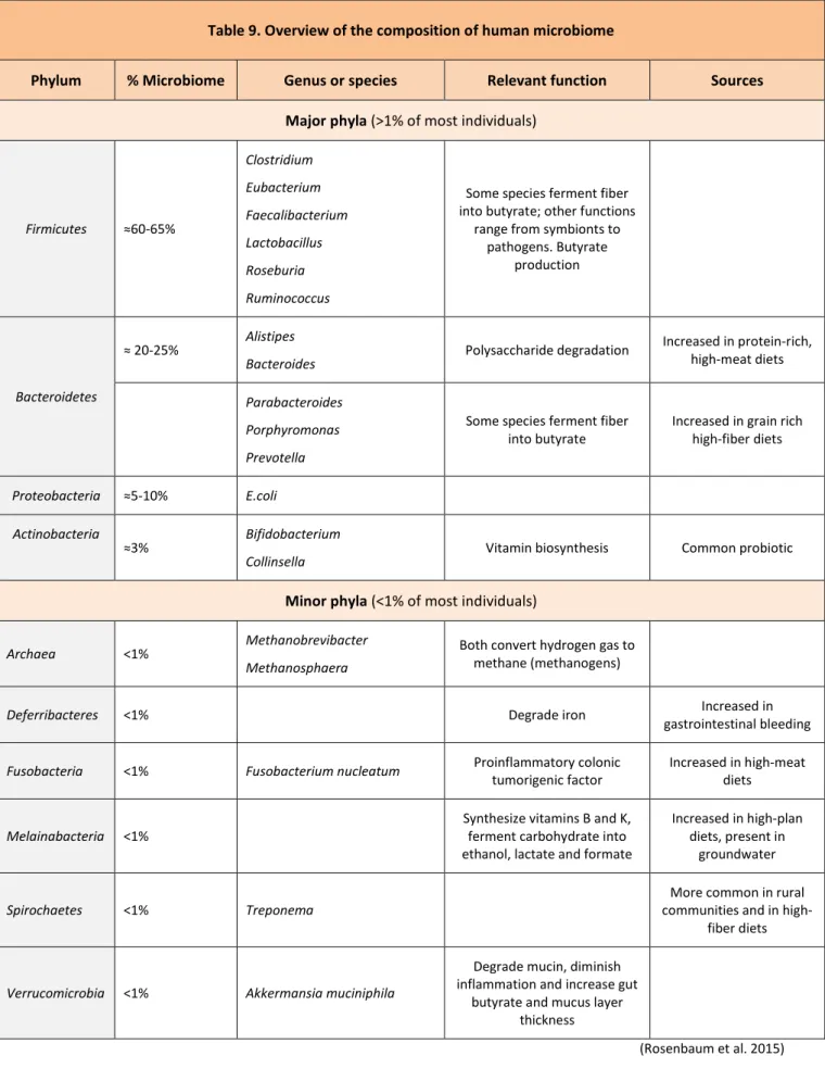
Ghosh, 2017). Therefore, these numbers need to be revisited. Interestingly, metagenomics sequencing of fecal samples has identified more genes than human genome, in fact gut microbiome is containing >150 times more genes than human genome (Patterson et al. 2016). Moreover, each person has at least 160 different species, which contrast with the restricted diversity at the phylum level, mainly dominated by two phyla, the Firmicutes (65%, no true outer membrane, mostly Gram-positive) and the Bacteroidetes (25%, outer membrane, Gram-negative). Proteobacteria (5%) and Actinobacteria (3%) are less important (Postler and Ghosh, 2017).
The amount of bacteria differs in every location of the GI tract with a rostro-caudal increasing gradient, as the consequence of chemical and physical factors alongside with the transit time (Fig.5) (Fetissov, 2016).
Fig.4. Healthy microbiome can be characterized in terms of its dynamics, depicted here in a simplified model as a conceptual energy landscape. The infant microbiome (yellow point) starts out in an unstable state and gradually descends towards one of potentially several healthy adult attractor states. Perturbations (dashed red arrows) can either be resisted (blue point) or can move the microbiome out of the healthy state (white point), after which a resilient microbiome will return to a healthy state or fall into an unhealthy state (red point) (Lloyd-Price et al. 2016).
8
In the gut, the growth of microbial community is influenced not only by bacteria-intrinsic regulation but diet too, and limited by chemical (like digestive juices that cause bacterial lysis and prevent mucosal adhesions) and physical factors (like intestinal peristalsis that cause elimination of bacteria by feces) of the host. These negative and positive influences determine the relative stability of microbiota at each part of GI tract (Fig.6) (Fetissov 2016).
5. BIOGEOGRAPHY OF GUT MICROBIOTA
Microbial variations along the length and cross-section of GI tract depend on chemical gradients, oxygen levels, nutrient availability and immune effectors (Fig.7) (Tropini et al. 2017). For example, the small intestine is more acidic and with high levels of oxygen and antimicrobials than the colon, for that reason, fast-growing facultative anaerobes that tolerate this environment will compete with host bacteria or other bacteria for the simple carbohydrates in this region. Another case is the bactericidal properties of bile acids to certain species, which can shape the composition of small intestine mainly. Indeed, it was evidenced a growth of Firmicutes (while Bacteroidetes decreased) in
Fig.5. Presence of chemical and digestive factors and the transit time along the gastrointestinal tract might underlie the increasing rostro-caudal gradient of bacterial content. In the upper gut, the transit time is apparently, shorter than the time necessary for the bacterial population to reach the stationary growth phase (Fetissov 2016).
Fig.6. Host factors influencing gut bacterial growth. Key host-related factors influencing the balance between stimulation and inhibition of the bacterial cell number in the gut. The role of the immune system is not shown, but it contributes by stabilizing the autochthonic community and neutralizing pathogenic invaders (Fetissov 2016).
9
mice fed with excess of bile acids (Donaldson et al. 2015). Besides, the shorter transit time in the small intestine compared with the colon makes bacterial adherence an important factor for persistent colonization of the small intestine (Donaldson et al. 2015).
Additionally, some host factors lead to cross-sectional microbial variation of the gut. The colon wall folds over itself and creates inter-fold regions that are different from the central lumen (Fig.7). In mouse studies, laser capture microdissection was used to indicate specific microbial communities. Specifically, Firmicutes families Lachnospiraceae and Ruminococcaceae were enriched in the inter-fold regions, whereas Bacteroidetes families, Prevotellaceae, Bacteroidaceae and Rikenellaceae were prevalent in the luminal compartment. Both sites contain lots of mucus as a nutrient source of some bacteria (Donaldson et al. 2015).
As it was stated in the previous section, the adult intestinal microbiota is mainly enriched in bacteria dominated by two phyla: Bacteroidetes and Firmicutes. But other microbial domains have been identified as well, like archaea genera (Methanobrevibacter smithii, which optimize digestion of dietary polysaccharides), viruses (each person has a unique virome consisting primarily of
Fig.7. Microbial habitats in the human lower gastrointestinal tract. The dominant bacterial phyla in the gut are Bacteroidetes, Firmicutes, Actinobacteria, Proteobacteria and Verrucomicrobia. The dominant bacterial families of the small intestine and colon reflect physiological differences along the length of the gut. For instance, a gradient of oxygen, antimicrobial peptides (like bile acids) and pH limits the bacterial density in the small intestinal community, whereas the colon carries high bacterial loads. In the small intestine, the families Lactobacillaceae and Enterobacteriaceae dominate, whereas the colon is characterized by the presence of species from the families Bacteroidaceae, Prevotellaceae, Rikenellaceae, Lachnospiraceae and Ruminococcaceae (colors correspond with the relevant phyla). A cross-section of the colon shows the digesta, which is dominated by Bacteroidaceae, Prevotellaceae and Rikenellaceae, and the inter-fold regions of the lumen, which are dominated by Lachnospiraceae and Ruminococcaceae. Cfu (colony-forming units) (Donaldson et al. 2015)
10
Fig.8. Genus abundance variation box plot for the 30 most abundant genera as determined by read abundance. Genera are colored by the respective phylum (see inset for color key). Inset shows phylum abundance box plot. Genus and phylum level abundances were measured using reference-genome-based mapping with 85% and 65% sequence similarity cutoffs. Unclassified genera under a higher rank are marked by asterisks (Arumugam et al. 2011).
bacteriophages, which serve as means of horizontal gene transfer among distant related bacteria) or fungi (that typically are pathogens, but they can be present also in healthy populations like Candida, Malassezia or Saccharomyces) (Lloyd-Price et al. 2016).
Actually, direct mutualistic relationships between humans and fungi have been found and these trans-kingdom interactions are responsible for an immune and ecological balance of the healthy microbiome for example Lactobacillus control of fungi in the gut. Also, some examples exist about mutualistic relationships between humans and fungi like the probiotic yeast Saccharomyces boulardii (Lloyd-Price et al. 2016). This heterogeneous ecosystem indicates that co-evolution of the host with its gut microbial symbionts (either commensals or mutualists) has generated powerful selective mechanisms (Donaldson et al. 2015).
6. GLOBAL VARIATION OF HUMAN GUT METAGENOMES
Gut metagenome is all the genes in the community of gut microorganisms (Boulangé et al. 2016). A study to compare metagenomics of human gut was carried out by M. Arumugam et al., in which a total of 39 samples were analyzed. The majority of these samples belonged to bacteria and they identified 30 most abundant genera and the respective phylum level (Fig. 7). They conclude that Firmicutes and Bacteroidetes were the dominant phyla and Bacteroides were the most abundant, but also the most variable genus (Arumugam et al. 2011).
As it is showed in the box plot (Fig.8), there is a “long tail effect” corresponding to species in low abundance while there are just a little of predominant species. This ecosystem distribution is the result of the homeostasis by selective pressure from host and microbial competitors. This outcome points to “survival strategies” by which the low abundance microbes share abundant functions (Arumugam et al. 2011).
11
Fig.9. A) Abundances of the main contributors of each enterotype from the Sanger metagenomes. B) Co-occurrence networks of the three enterotypes from the Sanger metagenomes. Unclassified genera under a higher rank are marked by asterisks in B (Arumugam et al. 2011).
During the study, they were able to link the most abundant molecular functions with the most dominant species; but also low abundance genera like Escherichia contribute in over 90% of pilus assembly and assists in plasmids’ transfer for protective functions (like antibiotic resistance) (Arumugam et al. 2011). In conclusion, abundance genera cannot reveal the whole functional complexity of gut microbiota.
They also pointed the main contributors for each of the 3 enterotypes: Bacteroides (enterotype 1), Prevotella (enterotype 2) and Ruminococcus (enterotype 3), and the co-occurrence networks between these three enterotypes from the Sanger metagenomes (Fig.9). Definitely, there is evidence that enterotypes are not as sharply delimited as blood groups, but they are likely to characterize individuals (Arumugam et al. 2011).
It is significant to note that Arumugam et al. study showed no significant correlation between host properties (like nationality, sex, age or BMI) and enterotypes (exc. Enterotype 1 which is enriched in Japanese individuals), and what's more how enterotypes do not seem to differ in functional richness. They only found a negative correlation between age and abundance of Clostridium genus, but did not find any correlation between BMI and Firmicutes:Bacteroidetes ratio (in contrast with the typical discussion of the relationship between obesity and this ratio) (Arumugam et al. 2011). However, although popular press has focused on the idea of discrete enterotypes, individual’s enterotype can be highly variable with an extraordinary within -and between- diversity. In fact, there are plenty of evidences that support continuous gradients of prevalent taxa rather than discrete enterotypes (Knights et al. 2014).
So, the assumption that individual’s enterotype is relatively fixed over time is not true at all; actually it was found that an individual’s enterotype crosses from one putative presumed enterotype to another (Fig.10A) as it was demonstrated by time series of 1 year’s daily gut microbiota samples from a single individual (Knights et al. 2014). Indeed, it was observed that microbiome of the individual time series occupies nearly every area in the ternary plot (Fig.10B)
A
12
which demonstrates that enterotypes are fluid and continuous (Knights et al. 2014). That is why, summarizing microbiota variation into separate clusters would mean they are relatively stable over years, and for the moment, it is not true for healthy subjects (Duranti et al. 2017).
Thus, while members of the gut microbiota can be stable for long time, the community structure and the relative quantity of each member is highly dynamic (Patterson et al. 2016) suggesting the existence of microbial community gradients rather than distinct enterotypes. Hence, the possibility of a microbiome-based classification of human individuals is still subject to debate.
7. ESTABLISHMENT OF GUT MICROBIOTA IN DIFFERENT STAGES OF LIFE
Traditionally, environment in utero has been considered sterile, but DNA based analyses have identified low levels of bacterial species in maternal placenta, amniotic fluid, umbilical cord blood and meconium (Blum 2017, Nogacka et al. 2017). So it has been proposed that an initial colonization process occurs during gestation (Duranti et al. 2017).
After birth there is a fast expansion and successively changing composition of microbiota that becomes relatively stable in adulthood (Blum 2017). The neonatal intestinal tract is rapidly and densely colonized by bacteria from the mother and surrounding environment following the birth (Patterson et al. 2016). Although neonate’s intestinal microbiota has a relative prevalence of Proteobacteria and Actinobacteria (Duranti et al. 2017), its composition depends on several factors (Patterson et al. 2016, Nogacka et al. 2017) such as the delivery method (Natural, which has a larger population of Bacteroides and Bifidobacterium species, versus caesarean section, which are initially colonized with skin bacteria from the mother), the way the baby is fed (breast-fed versus formula-fed babies), the gestational age and an early life antibiotic exposure. Indeed, maternal exposure to antibiotics during pregnancy is associated with decreased bacterial diversity on the first stool of the neonate and reduced abundance of lactobacilli and bifidobacteria in the infant gut; it also affects
Fig.10. Enterotype can be unstable, continuous and driven by sampling frame (A and B). Genus-level enterotype time series superimposed on putative clusters derived from 33 subjects. A) Two selected trajectories of consecutive daily samples are shown for a single male subject. Meta-HIT samples are colored by putative enterotype cluster. The two selected trajectories show the subject’s microbiome profile “walking” from one putative enterotype to another over the course of several days. B) Ternary plot of composition of Bacteroides, Prevotella and other genera daily for a year for a single subject and for published cross-sectional samples. These analyses demonstrate the temporal fluidity of enterotypes and provide clear proof by counterexample that enterotypes are not discrete states that separate individuals (Knights et al. 2014).
13
the vaginal microbiota of the mother which could block the later transfer of microbes to the baby during delivery (Nogacka et al. 2017, Rosenbaum et al. 2015).
Moreover, it is suggested that maternal microbiota during gestation also configure the future immune system of the children; and thus, prenatal antibiotics exposition (just with penicillin or chloramphenicol) may be associated with asthma in childhood. In fact, some data advise that the gut resistome (collection of all genes from the gut microbiome that potentially encode for resistance to antibiotics) begins to develop in the utero with the transmission of antibiotic resistance genes from mother to infant. It has also been reported in several studies that early antibiotics exposure could link with allergic disease in later life (“hygiene hypothesis”) and with an increase in body fat and weigh gain. Furthermore, it is a pending task to demonstrate the impact of early life antibiotic administration in antimicrobial resistance by gut microbiota. It is known from in vitro studies that exposition of microbial communities to constant antibiotics, they acquire multidrug resistance (Nogacka et al. 2017).
Afterward, gut microbiota continues to develop throughout childhood and adolescence and becomes more stable. It is generally assumed that a 3 years old child’s microbiota closely resembles that of an adult (Patterson et al. 2016). Thus, it stays relatively stable throughout adult life until old age when its composition changes again. Nevertheless, it is crucial to warn that gut microbiota can be altered by changes such as infection, antibiotic treatment, pregnancy or long-term change of lifestyle (Kim et al. 2016).
With regard to obesity in offspring, maternal obesity could be a predictor for child overweight. As it has been demonstrated, child from obese mother had different levels of Faecalibacterium spp., Oscillabacter spp., Blautia spp. and Eubacterium spp. compared to child born from a lean mother. Also, high level of Lactobacillus spp. and low level of Bacteroides spp. during the first 3 months of life may cause child obesity. Concerning human breast milk, it is crucial from a nutritional point of view but also for vertical transmission of bacteria. Breast milk of obese mothers has a reduced microbiota diversity compared to lean mothers who showed higher levels of Bifidobacterium spp. and lower Staphylococcus. In fact, the increased presence of Bifidobacterium spp. in early stages of life may provide protection against overweight (Duranti et al. 2017).
8. SOME ROLES OF GUT MICROBIOTA
Gut microbiota has some important functions like a protective role from harmful bacteria or a direct competition for limited nutrients which gives benefits for the host. It also helps the host immune system to recognize foreign elements and convert otherwise indigestible food into absorbable nutrients and energy for the host (Fig.11) (Kim et al. 2016).
Traditionally, microbiome has been described to has a commensal relationship with the host (nor damaging or helpful) but truly it is often highly mutualistic. For instance, bacteria helps the host in digesting complex food (by conversing complex carbohydrates into absorbable substrates) and synthesize some essential vitamins (like B and K) while the host provides the bacteria food and protection. It is interesting to note that, more or less, 10% of metabolites found in mammalian blood are derived from gut microbiota. In a very real sense, humans and their microbiome thus form a composite organism, a so-called holobiont. In the same way, microbiota is able to modulate host energy balance and stores, as will be described later, what could lead to recognize it as an enteroendocrine organ (Postler and Ghosh, 2017).
14
The important responsibility of microbiota in the protection of host against pathogens can be verified after antibiotic treatment which leads to overgrowth of Clostridium difficile and also in germ-free mice which have more risk of infection than conventionally raised mice suggesting that microbiota might play an important role in the protection of the host against pathogens. Here are some ways to clarify the host protection by indigenous microbiota (Kim et al. 2016):
- Mutualistic bacteria can inhibit colonization of pathogens by competing for the same nutrients: E.coli commensal strain is in competition with enterohaemorrhagic E.coli (EHEC). - Some bacterial products can regulate the virulence gene of pathogenic microorganisms:
Bacteroides thetaiotaomicron encodes fucosidases, which generates fucose from host mucin and subsequently, contributes to the expression of EHEC virulence genes, inducing its colonization.
- Commensal bacteria prevent the attachment of pathogens to the surface of intestinal epithelium avoiding infection at initial stage; in relation to this, it was proved how germ free mice have thinner mucus layer leading to less production of antimicrobial molecules than conventionally raised mice.
- Epithelial barrier function can be improved thanks to some metabolites produced by the microbiota: bifidobacteria produce acetate which inhibits translocation of EHEC Shiga toxin from gut lumen to the blood.
Fig.11. Multiple roles of the microbiota in the gut. Symbiotic microbiota inhibit the colonization of pathogens by competing for the same nutrients, regulating virulent gene through by-product and inducing of production of mucus and antimicrobial peptides from epithelial cells. SCFAs produced by gut microbiota are used as an energy source in the epithelial cells and participate in cholesterol homeostasis in the peripheral tissues and in glycogenesis in the liver. In addition, gut microbiota liberate nutrients from otherwise indigestible food (Kim et al.2016).
15
9. MICROBIOTA-ASSOCIATED IMMUNOMODULATORY METABOLITES
Gut microbiota is able to produce or modify metabolites that work as signaling molecules for an “intelligent communication system” in the body. “Pharmabiotics” (bioactive metabolites derived from bacteria), contribute to the correct function of epithelial barrier and immune cells. However, many identified metabolites have not been functionally characterized yet (Postler and Ghosh, 2017). These microbiota-associated metabolites can be classified in those produced by the host but modified by gut bacteria, those that are produced by bacteria from dietary components and metabolites that are synthesized de novo by gut microbes.
Some of the metabolites secreted by the host but modified by gut bacteria are secondary bile acids and taurine (Table 2). Intestinal microbes are able to convert the primary bile acids secreted by the host’s liver such as cholic acid and chenodeoxycholic acid to secondary ones such as deoxycholic and lithocholic acid mostly in the colon, by a multi-step process that involves deconjugation and dehydroxylation. Free taurine is released in to the intestinal lumen during the deconjugation process, promoting the integrity of the mucosal epithelium by increasing autocrine production of IL-18. On the other hand, primary and secondary bile acids are signaling molecules for immune system that inhibit the secretion of pro-inflammatory cytokines by macrophages, Kupffer and dendritic cells (Postler and Ghosh, 2017).
Table 2.
Immunomodulatory metabolites that are produced by the host and biochemically modified by gut bacteria
Metabolite
Molecular mechanism(s) of
action
Origin Effects on immune system
Secondary bile acids Extracellular: activation of GPBAR1 Intracellular: activation of BAR
Derived from host-produced primary bile
acids by intestinal microbiota
Inhibit NF-κβ dependent transcription of pro-inflammatory genes in monocytes, macrophages, dendritic
cells. Inhibit production of pro-inflammatory cytokine osteopontin by
NKT cells
Taurine
Enhancement of NLRP6 inflammasome
activation
Derived from host-produced primary bile
salts by intestinal microbiota
Enhances epithelial barrier function and maintenance by promoting epithelial
production of IL-18
16
The metabolites that are produced by bacteria directly from dietary components are the indole derivatives, polyamines and Short-chain fatty acids (SCFAs) (Table 3). The SCFAs are described in more detail below in next section (see point 10). Indole derivatives are obtained from diet with tryptophan and promote the secretion of IL-22 strengthening the integrity of the intestinal mucosa and inducing antimicrobial peptides secretion from epithelial cells, production of mucins and proliferation of globet cells. In the same way, polyamines also develop intestinal mucosa, being such its importance that if endogenous production is blocked, maintenance and repair of intestinal epithelium will be affected. On the contrary to previous statement, exogenous polyamines can reduce IL-18 release, a cytokine that promotes epithelial repair and barrier function (Postler and Ghosh, 2017). They also have direct influence on innate immune cells; in fact, the polyamine named spermine inhibits the lipopolysaccharide (LPS) induced expression of pro-inflammatory cytokines, leading to anti-inflammatory effect (Postler and Ghosh, 2017).
(Postler and Ghosh, 2017).
Table 3.
Immunomodulatory metabolites that are produced by bacteria from dietary components
Metabolite Molecular mechanism(s) of action
Origin Effects on immune system
Indole
derivatives Activation of AhR and NR1l2
Derived from dietary tryptophan by different intestinal bacteria
AhR activation promotes maintenance of ILC3 cells, which strengthen integrity of
gut mucosa by secreting IL-22. NR1l2 activation also enhances epithelial barrier
function Polyamines (primarily putrescine, spermidine, spermine)
Unclear. Inhibit expression of pro-inflammatory cytokines in
conjunction with AHSG. Also inhibit activation of NLRP6
inflammasome
Derived from arginine by host
and bacteria
Enhance development and maintenance of gut mucosa and resident immune cells.
Inhibit expression of pro-inflammatory cytokines by LPS-stimulated monocytes
and macrophages SCFAs (primarily acetate, propionate, butyrate) Extracellular: activation of FFAR2 (GPR43), FFAR3 (GPR41) and HCAR2 (GPR109A, butyrate only). Intracellular: inhibition of
histone acetyl transferases (butyrate and propionate only).
Can also activate extracellular OR51E2 and intracellular PPAR-γ
(butyrate only), but no relevance for immune system
demonstrated yet Polysaccharides’ fermentation by colonic microbiota Bacteroidetes: acetate and propionate Firmicutes: butyrate
Generally anti-inflammatory, protect against colitis. Promote integrity and function of the gut epithelium. Inhibit production of pro-inflammatory cytokines by innate immune cells. Promote function
of microglia. Inhibit maturation of dendritic cells. Promote antibody production by B cells. Promote de novo differentiation and expansion of T-regs
17
Finally, the main metabolites that are synthesized de novo by gut microbes are ATP and Polysaccharide A (PSA) (Table 4). Extracellular ATP secreted by gut bacteria has pro- and anti-inflammatory effects. ATP is consider an endogenous danger-associated molecular pattern (DAMP) that promotes secretion of pro-inflammatory cytokines, chemotaxis and differentiation of naïve T cells into TH17 cells, but it also inhibits release of IgA into the gut lumen by reducing the number of
B-cell activating T follicular helper cells and thus protecting intestinal microbiota from excessive antibody production. On the other hand, PSA, which is produced by the gut bacterium Bacteroides fragilis, promotes the secretion of anti-inflammatory cytokine IL-10 by T cells, protecting mice from colitis. As a curiosity, it has also being related to a protective role in IBD and autoimmune encephalomyelitis by increasing ENTPD1 expression on peripheral T-regs (Postler and Ghosh, 2017).
(Postler and Ghosh, 2017).
10. SHORT-CHAIN FATTY ACIDS (SCFAs)
Plant-derived polysaccharides obtained from diet (like resistant starches and dietary fiber) cannot be digested by host enzymes, so anaerobic intestinal microbiota is crucial for its fermentation, resulting in end products called SCFAs (Postler and Ghosh, 2017). The most abundant (95%) SCFAs are: acetate (C2), propionate (C3) and butyrate (C4). The other 5% correspond to branched-chain SCFA like isobutyrate, isovalerate and 2-methyl butyrate, obtained from protein breakdown (Ríos-Covián et al. 2016). Other products of non-digestible carbohydrates fermentation are: lactate, which can be metabolized to acetate, propionate and butyrate by cross-feeding organisms; and formate, which has been linked to methanogenesis and inflammatory processes (Morrison and Preston 2016).
Table 4.
Immunomodulatory metabolites that are synthesized de novo by gut microbes
Metabolite Molecular mechanism(s) of action
Origin Effects on immune system
ATP Activation of P2X and
P2Y receptors Actively secreted by subset of intestinal bacteria
Limits numbers of Tfh cells in Peyer’s patches, thus reducing secretion of bacteria-specific IgA by B cells
across gut epithelium. Promotes differentiation of TH17 cells in gut mucosa. May promote epithelial barrier function by activating NLRP3 inflammasome
and subsequent IL-18 secretion by macrophages
Poly-saccharide A (PSA) Activation of TLR2 on DCs and Tregs. Presentation of PSA fragments with MHC-II
to CD4+ Bacteroides fragilis (requested for colonization)
Potent anti-inflammatory effects: induces secretion of IL-10 from CD4+ T cells, directly and indirectly. Skews
18
Traditionally, it has been summarized that Bacteroidetes phylum supplies most of the acetate and propionate, whereas Firmicutes mainly produce butyrate; but actually, it is more complicated than that, as shown in Table 5 (Postler and Ghosh 2017, Ohira et al. 2017).
Table 5. SCFAs (Acetate, Propionate, Butyrate) production by microbiota in the gut SCFAs Pathways/Reactions Producers
Acetate
From pyruvate via acetyl-CoA
Most of the enteric bacteria. (Representative of species bacteria)
Akkermansia muciniphila, Bacteroides spp., Bifidobacterium spp., Prevotella spp., Ruminococcus spp.
Wood-Ljungdahl pathway Blautia hydrogentrophica, Clostridium spp., Streptococcus spp.
Propionate
Succinate pathway Bacteroides spp., Phascolarctobacterium succinatutens, Dalister spp., Veilonella spp.
Acrylate pathway Megasphaera elsdenii, Coprococcus catus
Propanediol pathway Salmonella spp., Roseburia inulinivorans, Ruminococcus obeum
Butyrate
Phosphotransbutyrylase/
butyrate kinase route Coprococcus comes, Coprococcus eutactus
Butyryl-CoA: acetate CoA-transferase route
Anaerostripes spp. (A, L), Coprococcus catus (A), Eubacterium rectale (A), Eubacterium hallii (A, L), Faecalibacterium prausnitzii
(A), Roseburia spp.
(A): acetate is the substrate for producing butyrate; (L): lactate is the substrate for producing butyrate.
(Ohira et al. 2017).
10.1. Microbial cross-feeding dynamics in SCFAs production
Each SCFA is produced following different routes: Acetate is produced by most enteric bacteria as a result of carbohydrates (CHO) fermentation (from pyruvate, via acetyl-CoA); propionate formation follows three different pathways: succinate, acrylate and propanodiol pathway; and finally, butyrate is formed by the butyrate kinase route and the butyryl-CoA: acetate CoA-transferase pathway (Ríos-Covián et al. 2016).
Despite these well-defined routes, there is also an essential microbial cross-feeding dynamics which includes both metabolic and substrate cross-feeding:
- Metabolic cross-feeding: The employment by one microorganism of end-products from the metabolism of another one. For example, Bacteroides thetaiotaomicron uses polysaccharides and produces acetate, while one of the main producers of butyrate, Faecalibacterium prausnitzii, can utilize acetate as a substrate (Ríos-Covián et al. 2016).
19
Fig.13. The gut lumen is the major site of production but the concentration gradient falls from the lumen to the periphery with selective uptake of butyrate at the epithelium, propionate at the liver and acetate in the periphery. The significance for host physiology of this biological gradient is poorly understood (Morrison y Preston 2016).
- Substrate cross-feeding: The use by one bacterium of complex-CHO breakdown products formed by another one. Thus, microorganisms that are incapable of utilizing complex CHO may take advantage. For example, some Bifidobacterium that are not able to use inulin-type fructans can grow thanks to oligosaccharides released by primary inulin degraders (Ríos-Covián et al. 2016).
Also, there is evidence of mainly bacterial cross-feeding from acetate to butyrate, at lower extent between butyrate and propionate, and no metabolic flux between propionate and acetate (Fig.12) (Ríos-Covián et al. 2016).
It is important to highlight that each SCFAs has a biological gradient from gut lumen (where they are produced) to host periphery. This will lead to different tissue’s SCFA exposure (Morrison and Preston 2016). While acetate and propionate travel intact across the gut epithelium to the liver, butyrate is metabolized just within the intestinal epithelium where it serves as the main energy source for colonic enterocytes. In the same way, propionate is in part degraded in intestinal mucosa but it also reaches the liver. However, acetate is the only SCFA that is mainly released into systemic circulation (Fig.13) (Postler and Ghosh, 2017).
Fig.12. Schematic representation of microbial metabolic pathways and cross-feeding mechanisms, contributing to SCFA formation in the human gut. Shaded geometric shapes summarize routes of formation for each of the three main SCFA: acetate, propionate and butyrate (Ríos-Covián et al. 2016).
20 10.2. Gut integrity and biological effects of SCFAs
In general, SCFAs reduce the luminal pH by inhibiting some pathogenic microorganisms and increasing several nutrients absorption (Ríos-Covián et al. 2016). They also regulate the integrity of epithelial barrier through tight junction proteins (Morrison and Preston 2016).
For example, acetate takes part in the ability of bifidobacteria to inhibit enteropathogens, improves glucose tolerance and plays a role in cholesterol homeostasis in the peripheral tissue (Ríos-Covián et al. 2016, Kim et al. 2016). Moreover, it has little effect on locomotor activity or food intake, but it favors energy harvest and basal thermogenesis (Rosenbaum et al. 2015). On the other hand, propionate is a substrate for liver gluconeogenesis, reduces food intake and promotes locomotor activity (Baothman et al. 2016, Rosenbaum et al. 2015).
The third SCFA, butyrate, serves as energy source for intestinal epithelial cells and boosts healthy colonocytes proliferation. This role is demonstrated in germ-free mice that have an energy-deprived state of the gut epithelium. If butyrate is introduced, the deficit of mitochondrial respiration will be re-established (Kim et al. 2016). On the other hand, butyrate also induces apoptosis in transformed cells by accumulating within cancerous colonocytes and inhibiting tumor cell progression (this is named as “butyrate paradox”). So it is thought to have an important role against the development of colorectal cancer (Ríos-Covián et al. 2016); however, in a mouse model of colorectal cancer, butyrate appears to enhance tumor formation by stimulating hyper-proliferation in those MSH2 deficient colon epithelial cells (Morrison and Preston 2016). It also stimulates mucin production, colon motility and tight-junctions, thanks to an increased expression of claudin-1 and Zonula Occludens-1 (ZO-1) and occluding redistribution, as well as a decreased expression of aberrant ZO-1 (Ríos-Covián et al. 2016, Morrison and Preston 2016).
Finally, the protective effect against diet- induced obesity has been showed with butyrate and propionate; having direct effects on feeding behavior and physical activity too. In addition, low levels of their bacterial producers are observed in ulcerative colitis or children with asthma. It is interesting to highlight that elevated cecal and fecal SCFAs levels have been found in obese mice and humans, what could suggest the existence of decreased colonic absorption in obesity (Baothman et al. 2016).
10.3. SCFAs and the Immune System
SCFAs have anti-inflammatory properties, increase mucin production by globet cells and improve tight junctions. In mice with ulcerative colitis was evidenced that SCFAs mitigate the disease by inducing the secretion of IL-18 which promotes integrity of the intestinal epithelium. They are also agonists for some G-protein-coupled receptors (GPCRs) like FFAR2, a free fatty acid receptor essential for neutrophil chemotaxis at gut mucosa. Exposure to SCFAs lowers pro-inflammatory cytokines production from neutrophils. Thus, FFAR2-/- mice are more susceptible to colitis (Postler and Ghosh, 2017).
Moreover, SCFAs promote expansion of both preexisting and de novo regulatory T cells (Treg), responsible for suppressing effector T cells activity and likewise inhibit the histone deacetylases, impeding antibody formation from B lymphocytes (Fig. 14) (Postler and Ghosh, 2017). They also has an essential role on inducing tolerance to commensal bacteria thanks to the ability of reducing intestinal macrophages responsiveness (Morrison and Preston 2016).
21
Fig.14. Intestinal bacteria convert dietary nutrients to immunomodulatory metabolites. Polysaccharides that cannot be digested by host enzymes, such as cellulose, are metabolized by the intestinal microbiota to SCFAs, green). These exert a plethora of anti-inflammatory effects, such as inducing the expansion and de novo differentiation of regulatory T cells, enhancing the barrier function of the intestinal mucosa through epithelial cells and globet cells, facilitating production of antibodies by B cells, inhibiting the maturation of dendritic cells and promoting an anti-inflammatory phenotype, and reducing production of pro-inflammatory cytokines by innate immune cells. Dietary tryptophan is degraded to indole derivatives (blue). Indoles promote epithelial barrier function, e.g., by supporting the maintenance of type 3 innate lymphoid cells, which are the primary producers of IL-22. Dietary arginine is metabolized to polyamines (red), which promote the integrity of the intestinal epithelium and reduce the production of pro-inflammatory cytokines by macrophages. DC (dendritic cell), EC (epithelial cell), GC (globet cell), ILC3 (type 3 innate lymphoid cell), Mφ (macrophage), Nφ (neutrophil), Treg (regulatory T cell). (Postler y Ghosh, 2017).
22
An important function of butyrate is to reduce inflammation by decreasing LPS translocation and inhibiting macrophage activation or pro-inflammatory cytokine production. We should have in mind that obesity and insulin resistance are associated with an increased permeability of gut barrier that leads to cell wall bacteria components translocation which trigger an inflammatory cascade. In this way, it makes sense to think that butyrate could reduce weight or insulin resistance (Morrison and Preston 2016). Indeed, an experiment with mice was made by giving them butyrate supplementation, and it was evidenced a reduction in diet-induced insulin resistance maybe by increasing mitochondria function and energy spending (Baothman et al. 2016). Butyrate also inhibits nuclear factor kappa β (NF-κβ) activation in macrophages, and it is thought to be the responsible of suppressing TNF-α, IL-6 and myeloperoxidase activity in Kupffer cells as well, suggesting a role in regulating liver inflammation. The anti-inflammatory role of butyrate is also observed in IBD and acute radiation proctitis, with improvement of both diseases. In addition, both butyrate and propionate inhibit the maturation and function of DCs (Morrison and Preston 2016).
10.4. Role of SCFAs on lipid metabolism
The effects of the SCFAs in energy and lipid metabolism suggest an important role of SCFA in the control of metabolic syndrome (Ríos-Covián et al. 2016). In fact, they have a regulatory task in metabolism since they are natural ligands for FFAR2/3 found on enteroendocrine and immune cells (Morrison and Preston 2016). This role of SCFA in regulating gut hormones via FFAR2/3 in the end protects against diet-induced obesity and insulin resistance (Parekh et al. 2014), as it was observed in experimental mice fed with high-fat diet after dietary supplementation with butyrate (Ríos-Covián et al. 2016).
In the liver, acetate is used for de novo lipogenesis and cholesterogenesis, both inhibited by propionate that also is able to reduce visceral and liver fat (Morrison and Preston 2016). Increased circulating SCFA entails a reduced adipogenesis and adipocytes lipolysis which leads to decrease free-fatty acid (FFA) from adipose tissue to liver. They also inhibit lipid accumulation in adipocytes stimulated by insulin. Thus, increasing peripheral SCFA availability in obese would be a novel strategy to regulate FFA flux (Morrison and Preston 2016). However, there is a controversy regarding the role of SCFA in obesity, the so called “Energy harvesting” hypothesis, whereby SCFA add additional calories through fermentation (Morrison and Preston 2016).
10.5. SCFAs and appetite regulation
As it is noted above, the fermentation of dietary fiber to SCFA provides additional energy to the host what could promote obesity; nevertheless, it is well known in epidemiological studies that high-fiber diets prevent rather than promote obesity and it has been linked with an improved appetite regulation (Duranti et al. 2017).
This could be the result of FFAR activation by SCFA which leads to an increased secretion of enteroendocrine hormones such as glucagon-like-peptide 1 (GLP-1) or peptide YY (PYY), both of them responsible for causing satiety (Duranti et al. 2017). For instance, propionate and butyrate appears to induce short-term appetite regulation through PYY and GLP-1 mediated mechanisms, while acetate is thought to reduce appetite by its interaction with the CNS and by stimulating leptin secretion in adipocytes. In any case, further investigations should be done to completely clarify each SCFA’s role (Morrison and Preston 2016, Ríos-Covián et al. 2016)
23
GLP and PYY are responsible for satiety leading to a diminution of food ingestion. It has been evidenced that subjects with little circulating PYY have lower satiety and in consequence higher fat mass. In fact, some experiments with animals showed that lacking-PYY mice were hyperphagic and become obese, while chronic administration of PYY3-36 to obese mice decreased adiposity (Ríos-Covián et al. 2016, Parekh et al. 2014). That is why it has been proposed as a therapeutic strategy to target the PYY system by altering the microbiome (Parekh et al. 2014).
10.6. Regarding the methodology employed in SCFAs experiments
SCFAs administration was carried out by oral uptake, enema or intraperitoneal injection, but it is still unknown which is the method that most resembles the physiological state. What is clear is that SCFAs concentrations reached by different methods affect experimental outcomes (Postler and Ghosh, 2017).
Another item is the one that refers to the measurement of SCFA production directly in humans, which has been limited to the measurement of stool SCFA output (although it is unclear if it is representative for luminal SCFA production). However, stable isotope techniques hold promise (Morrison and Preston 2016).
11. CONTRIBUTION OF MICROBIOTA TO THE DEVELOPMENT OF DISEASES
Dysbiosis can affect different host systems and it is related with the development of several pathologies. In fact, harmless or even beneficial bacteria in a healthy microbiome can promote chronic diseases like atherosclerosis and obesity when their ecosystem is altered (Postler and Ghosh, 2017). Some of those pathologies are:
11.1 Autoimmune diseases
Previously, it has been exposed how important is microbiota in development of innate and adaptive immune system, but it is also crucial for triggering tolerant responses from the host. Thus, gut dysbiosis is can be related with the pathogenesis of autoimmune diseases like:
11.1.1. Rheumatoid arthritis (RA)
It is well known that bacterial DNA and cell wall components are present in joints of RA patients, which may cause inflammation in genetically susceptible people (Kim et al. 2016). Furthermore, RA-susceptible mice showed alteration of gut microbiota and an increased permeability. So, in order to check if gut microbiota plays a role in RA development, animal models of RA (IL-1 rn -/- mice) raised in germ-free conditions do not develop arthritis and later introduction of Lactobacillus or segmented filamentous bacteria (SFB) indeed induced arthritis supporting the initial hypothesis. However, another experiment evidenced suppression of induced arthritis in germ-free mice which were given E.coli. Thus, additional studies will be required to establish which species have a protective role in RA pathogenesis. It is also interesting to note that RA patients have low levels of Bifidobacterium and Bacteroides.
24 11.1.2. Systemic lupus erythematosus (SLE)
Intestinal environment can contribute to microbiota composition and development of SLE as it was verified in the following experiment: Mice receiving acidic pH water showed a slower SLE progression and less anti-nuclear antibody (ANA) production than mice receiving neutral water (control mice). Besides, gut microbiome analysis displayed different bacteria abundance between both kinds of mice. However, SLE patients do not have a significant difference in bacterial diversity, although they show dysbiosis with a reduced Firmicutes/Bacteroidetes ratio (Kim et al. 2016).
11.1.3. Ankylosing spondylitis (AS)
AS is closely associated with IBD (>70% patients develop subclinical gut inflammation and 5-10% clinically IBD) and AS patients also have high levels of flagellin antibodies and sulfate-reducing bacterium.
The HLA-B27 gene is strongly related with AS pathogenesis, and in HLA-B27 transgenic mice, spondylitis and colitis were abolished in a germ-free state. In contrast, Bacteroidetes introduction induced inflammation indicating that the presence of specific taxa may be enough for AS initiation or progression (Kim et al. 2016).
11.1.4. Inflammatory bowel disease (IBD)
IBD include ulcerative colitis (UC) and Crohn disease (CD), and are characterized by inflammation in mucosal layer of the colon in UC and transmural involvement in CD (Blum 2017). Although it is known that the central event in IBD pathogenesis is the loss of tolerance against the indigenous gut, the precise mechanism is not entirely understood. It is thought that bacterial imbalance of gut lumen leads to inappropriate inflammation, resulting in host cell damage and autoimmunity (Kim et al. 2016).
IBD specific microbiota’s alteration may serve as an indicator for disease predisposition, activity or therapy response. For instance, some of these alterations could be a decreased bacterial diversity and butyrate producing microorganisms such as F.prausnitzii (Morrison and Preston 2016). As well, a decrease of SCFA-producing bacteria in human gut has been associated with IBD and dietary changes that encourage SCFAs production by the microbiome can improve symptoms. In mice, SCFAs have confirmed anti-inflammatory properties in the intestinal mucosa which lead to protective role in IBD prone mice (such knockout IL-10-/- or IL-2 -/- mice) (Postler and Ghosh, 2017, Kim et al. 2016). Additionally, it has been showed how E.coli or Enterococcus faecalis trigger chronic intestinal inflammation in IBD prone animals and can generate colitis. On the other hand, Faecalibacterium prausnitzii has anti-inflammatory activity and it is reduced in IBD patients (Kim et al. 2016).
11.2. Neurodevelopmental, psychiatric and neurodegenerative diseases
It has been demonstrated the role of gut microbiota in modulating immune cells in CNS, as well as the activation of peripheral immune cells involved in neuroinflammation, brain injury and neurogenesis. In fact, germ-free mice have more behavior disturbances or neuropathologies like autism spectrum disorders, depression and Alzheimer’s or Parkinson’s disease (PD) (Blum 2017).
25
Patients with PD have plaques of alpha-synuclein (ASn) both in brain and intestine, and likewise, mice over-expressing ASn develop similar PD neurologic deficits. Also, PD germ-free mice developed less plaques and an improvement of neurological deficits, just like mice treated with antibiotics. As a remarkable point, fecal matter transplantation (FMT) form patients with PD to germ-free mice resulted in neurological deficits resembling PD (Blum 2017).
11.3 Atherosclerosis and thrombosis risk
Atherosclerosis is promoted by systemic pro-inflammatory events that catalogue it as a chronic disease associated with adverse cardiac events (Postler and Ghosh, 2017). It is accelerated by eating food rich in choline, phosphatidylcholine and carnitine (such as meat, egg yolk and high-fat products), being all of them precursors of trimethylamine (TMA) and TMA N-oxide (TMAO). Intestinal TMAO formation is a two-step process: First, generation of TMA by intestinal microbes after food intake (dietary phosphatidylcholine or L-carnitine), and second, the hepatic oxidation of TMA to TMAO by host flavin monooxygenases (FMOs). These molecules not only enhance atherosclerosis but platelet hyperreactivity and thrombotic events (Fig. 15) (Blum 2017).
There are several evidences that support the relationship of these molecules with the risk of developing atherosclerosis. For example, mice fed with choline or TMAO supplemented food promoted atherosclerosis. On the contrary, if gut microbiota was removed with antibiotics treatment, there would be no choline-exacerbation of atherosclerosis, which confirms the main role of microbiota in this disease. However, many fish species contain high amounts of TMAO, and
Fig. 15. The intestinal microbial metabolite trimethylamine (TMA) and the atherosclerosis/thrombosis risk (Blum 2017)