Establishing a safe harbor site for the introduction of genetic information in the human cells by the recombination system attP/attB
164
0
0
Texto completo
(2) DOCTORAL THESIS 2018 Doctoral Programme of Translational Research in Public Health and High Prevalence Diseases (RD99). ESTABLISHING A SAFE HARBOR SITE FOR THE INTRODUCTION OF GENETIC INFORMATION IN THE HUMAN CELLS BY THE RECOMBINATION SYSTEM attP/attB Esther Palomino Lago Thesis Supervisor: Dr. Daniel Bachiller Thesis Co-supervisor: Dr. Aarne Fleischer Thesis tutor: Dra. Julia García. Doctor by University of the Balearic Islands.
(3) This thesis has been financially supported by the Balearic Government FPICAIB 2013 grant co-financed by the European Union, the Spanish Ministry for Science and Innovation (grant numbers PLE2009-0091, RTC-20142207-1 and IPT-2011-1402-900000), and the ISCIII PI14/01073 grant, cofinanced by ERDF/ESF..
(4) The thesis titled: “Establishing a Safe Harbor Site for the Introduction of Genetic Information in the Human Cells by the Recombination System attP/attB”, has been written by. Esther Palomino Lago PhD student.
(5)
(6) Dr. Daniel Bachiller of Spanish National Research Council (CSIC). I DECLARE: That the thesis titled: “Establishing a Safe Harbor Site for the Introduction of Genetic Information in the Human Cells by the Recombination System attP/attB”, presented by Ms. Esther Palomino to obtain a doctoral degree, has been completed under my supervision and meets the requirements to opt for a Doctorate.. For all intents and purposes, I hereby sign this document.. Signature. Palma de Mallorca, October, 24th 2018.
(7) Dr. Aarne Fleischer from Karuna GCT:. I DECLARE: That the thesis titled: “Establishing a Safe Harbor Site for the Introduction of Genetic Information in the Human Cells by the Recombination System attP/attB”, presented by Ms. Esther Palomino to obtain a doctoral degree, has been completed under my supervision and meets the requirements to opt for a Doctorate.. For all intents and purposes, I hereby sign this document.. Signature. Palma de Mallorca, October, 24th 2018.
(8)
(9)
(10) Dedicated to my beloved parents.
(11) ACKNOWLEDGEMENTS I would like to express my sincerest gratitude to my director Dr. Daniel Bachiller, for giving me the opportunity to work at the Department of Advanced Therapies in Islas Baleares. I wish to thank him for the immeasurable amount of support and guidance he has provided throughout this study. His extensive knowledge, vision, and creative thinking have been the source of inspiration for me throughout this project. I am greatly indebted to my co-director Dr. Aarne Fleischer. I am honored to have a supportive and professional teacher like him. I am always thankful to his enthusiastic leadership, encouragement and positive thinking. I will never forget his support and care during the hard times. It has always been a pleasure to discuss our results and findings with him. It is with great pleasure that I thank my co-worker Dr. Jose M. Martín. He was always ready to answer my questions and provide constructive guidance and criticisms. I am thankful for his support and encouragement. I would like also to thank Victor Gálvez for his support when I arrived to the laboratory. I am also grateful to past and present members of our group: Patricia Tortosa, María Tortosa, Dr. Sara Vallejo, Dr. Almudena Sánchez, and many others that I do not have the space to mention here, thanks for being such great lab mates. It has been an honor to work with you and I will cherish our many discussions. Thanks guys for all the enjoyable moments in Mallorca, inside and outside the lab! Those breaks were quite important for finishing this work. It is a pleasure to thank all of Dr. Calos colleagues at Stanford University for the experience of working at one of the most prestigious Universities. I would also like to thank Dr. Mital Bhakta, whose kind supervision turned into a friendship. I am grateful for all my PhD colleagues and friends. I would like to express my heartfelt thanks to each and every one of you for supporting me in your own personal way. I will forever value the moments we shared. Your daily laughter and smiles have made my time as a PhD student extremely enjoyable and absolutely unforgettable. Thank you all for your friendship!.
(12) I would like to thank Amparo Díaz and her family, who always wished the best for me. Thanks for all the great conversations and shared moments during both good and bad times. Most of all, I owe my deepest gratitude to my family from Cadiz and Malaga to whom I dedicate this thesis. My dearest thanks to my parents and sister for their everlasting support and for providing me with the necessary education that allowed me to pursue a scientific career. Thanks dad and mom, you are the best parents that a daughter can have. Without you this thesis would not have been possible..
(13) PUBLICATIONS Published articles from this thesis: Fleischer, A., Lorenzo, I. M., Palomino, E., Aasen, T., Gómez, F., Servera, M., (...) & Bachiller, D. (2018). Generation of two induced pluripotent stem cell (iPSC) lines from p. F508del Cystic Fibrosis patients. Stem Cell Research.Vol 29, pp1-5..
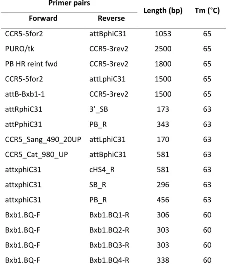
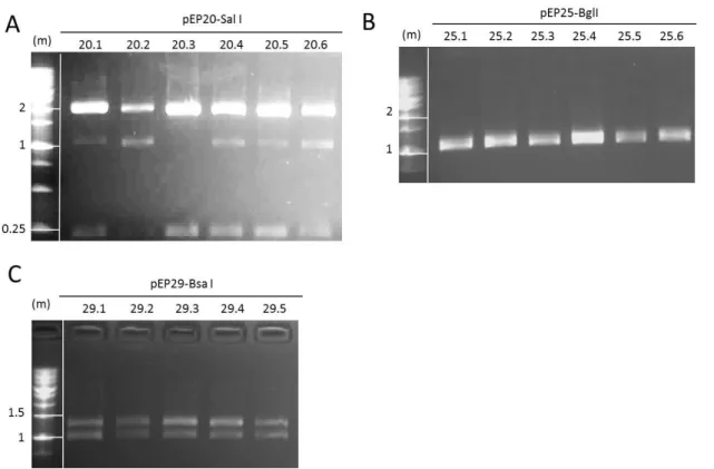
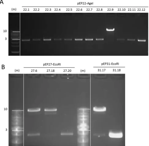
(14) LIST OF TABLES Table 1| Primer pairs used to verify each step of the synthesis of the CCR5 docking platform. ........................................................................................................... 51 Table 2| Transfection conditions in HeLa cells............................................................... 57 Table 3| Transfection conditions in hiPS cells. ............................................................... 59 Table 4| Recombination efficiencies at each of the three steps of the synthesis. ........ 90 Table 5| Summary of the number of loaded Bxb1 docking sites in HeLa 24.22 clones. 92 Table 6| Recombination efficiencies during hiPS loading. ............................................. 98 Table 7| Summary of the number of loaded Bxb1 docking sites in the hiPS 87.38 clone ........................................................................................................................ 101. LIST OF FIGURES Figure 1| Cell mechanism involved in double strand break repair. ................................. 4 Figure 2| Diagram of ZFNs. ............................................................................................... 6 Figure 3| Diagram of TALENs. .......................................................................................... 8 Figure 4| Diagram of CRISPR/Cas9. ................................................................................ 10 Figure 5| Site-specific recombination by serine integrases. .......................................... 14 Figure 6| Mechanism of serine integrase recombination.............................................. 15 Figure 7| piggyBac transposition.................................................................................... 20 Figure 8| Sleeping Beauty transposition. ....................................................................... 22 Figure 9| Differentiation and reprogramming processes. ............................................. 25 Figure 10| Diagram of the building strategy. ................................................................. 32 Figure 11| Diagram of the loading process. ................................................................... 33 Figure 12| Position of primers used to verify the integration of the different modules used during the process. ............................................................................. 49 Figure 13| Position of primers used to verify loading status in each one of the different docking units. ............................................................................................... 50 Figure 14|Diagram of the building strategy. Step 1. ...................................................... 61 Figure 15| Diagram of the building strategy. Step 2. ..................................................... 62 Figure 16| Diagram of the building strategy. Step 3. ..................................................... 63 Figure 17| Schematic representation of the recombination vectors. ........................... 64 Figure 18| First and second steps of phiC31 recombination vectors construction. ...... 65 Figure 19| Restriction fragment analysis of pEP20, pEP25 and pEP29. ......................... 66 Figure 20| Restriction fragment analysis of pEP21, pEP26 and pEP30. ......................... 68 Figure 21| Docking module assembly. ........................................................................... 70 Figure 22│ Restriction fragment analysis of pEP22, pEP27 and pEP31. ......................... 71 Figure 23│ Construction of vector pEP23. ...................................................................... 72 Figure 24│ Construction of vector pEP34. ...................................................................... 74.
(15) Figure 25│ Final step in the construction of the recombination vector pEP24. ............ 75 Figure 26| Final step in the construction of the recombination vector pEP28 ............. 76 Figure 27| Generation of the third docking vector pEP50 ............................................. 77 Figure 28| Generation of the testing vector pEP46. ...................................................... 78 Figure 29| Example of screening by PCR of the clones obtained by recombination at step 1. .......................................................................................................... 80 Figure 30| Excision of the selection cassette by PB transposase in HeLa Cl.24. ............ 81 Figure 31| HeLa Cl.24.22 sequence analysis. ................................................................. 82 Figure 32| Example of screening by PCR of the clones obtained by recombination at step 2. .......................................................................................................... 83 Figure 33| Excision of the selection cassette by SB transposase in HeLa Cl.24.22.21 and Cl.24.22.29 clones ........................................................................................ 84 Figure 34| Drug sensibility of HeLa Cl.24.22.21.3 and Cl.24.22.29.7 cells. .................... 85 Figure 35| HeLa Cl.24.22.21.3 sequence analysis. ......................................................... 86 Figure 36| PCR screening of the clones obtained by recombination at step 3. ............ 87 Figure 37| HeLa Cl.24.22.21.3.98 sequence analysis. .................................................... 88 Figure 38| Excision of the selection cassette by PB transposase in HeLa Cl. 24.22.21.3.98. .............................................................................................. 89 Figure 39| Determination of mCherry fluorescence by epifluorescence microscopy after pEP46 loading in HeLa Cl.24.22 cells. ................................................. 91 Figure 40| FACS analysis of mCherry expression after pEP46 loading in HeLa Cl.24.22 subclones. .................................................................................................... 92 Figure 41| Molecular characterization of docking sites loading in HeLa Cl.24.22 subclones. .................................................................................................... 93 Figure 42| hiPS cell karyotyping. .................................................................................... 94 Figure 43| Quantification of pluripotency markers by qRT-PCR.................................... 95 Figure 44| Characterization of the hiPS cell line IMEDEAi003-A by immunefluorescence studies. ................................................................................... 96 Figure 45| Identification of recombined clones by PCR screening in step 1 of platform construction in hiPS cells. ............................................................................ 97 Figure 46| Excision of the selection marker by PBc transposase in hiPS Cl.87 cells. ..... 98 Figure 47| hiPS Cl.87.38 cells sequence analysis. .......................................................... 99 Figure 48| Determination of mCherry fluorescence by epifluorescence microscopy after pEP46 loading in hiPS Cl.87.38 cells. ................................................. 100 Figure 49| FACS analysis of mCherry expression after pEP46 loading in hiPS Cl.87.38 subclones. .................................................................................................. 102.
(16) LIST OF ABBREVIATIONS AA: Amino Acids AAV: Adeno virus-associated AAVS1: the adeno-associated virus site 1 att: attachment bFGF: Basic Fibroblast Growth Factor bp: base pair CCR5: Chemokine (CC motif) receptor 5 CRISPR/Cas9: Clustered Regularly Interspaced Short Palindromic Repeats DAXX: Domain-associated ddH2O: Double Distilled H2O DICE: Dual Integrase Cassette Exchange DSBs: Double-Strand Breaks EEF1A1: eukaryotic translation elongation factor 1 alpha 1 ESC: Embryonic Stem Cell FACS: Fluorescence-activated cell sorting FBS: Fetal Bovine Serum GSHs: Genomic Safe Harbors HaHyPBase: Hyperactive PiggyBac transposase hiPS cells: Human Induced Pluripotent Stem cells HIV: Human Immunodeficiency Virus HR: homologous double repair Int: Integrase iPS: Induced Pluripotent Stem ITRs: Inverted Terminal Repeats Kb: kilo base KOSR: knockout Serum Replacement LB: Luria-Bertani LINEs: Long Interspersed Elements LTR: Long Terminal Repeat MLV: Murine Leukemia Virus.
(17) NEAA: Non-Essential Amino Acids NHEJ: Non-Homologous End Joined O.N: Overnight OPI: Overproduction Inhibition Phenomenon PAM: Protospacer Adjacent Motif PB: PiggyBac PBS: Phosphate-buffered Saline PSCs: Pluripotent Stem Cells RDF: Recombination Directionality Factor RT: Room Temperature RTs: Recombination Target sites RVDs: Repeat Variable Diresidues SAPTA: Scoring Algorithm for Predicting TALEN Activity SB: Sleeping Beauty SE: Selection Element SINEs: Short Interspersed Elements SM: Selection Marker SSRs: Site-Specific Recombinases TALE: Transcription Activator-like Effector TALEN: Transcription Activator-like Effector Nucleases Tm: melting Temperature ZD: Zin Ribbon Domain ZF: Zinc Finger ZFN: Zinc Finger Nucleases ZFP: Zinc Finger Protein.
(18) ABSTRACT. The goal of this project is to generate a safe harbor site in the genome of human cells, where information could be specifically inserted without the action of nucleases. The mechanism that we present here is based on two different site-specific recombinases phiC31 and Bxb1, as well as on the piggyBack and Sleeping Beauty transposons. Recombinase phiC31 is an integrase used by phages to establish the lysogenic life cycle. During integration, phiC31 drives recombination between the attP and the attB attachment sites on the phage and host genome, respectively. In naturally occurring phage infestations, the end result is an integrated phage genome flanked by new attL and attR sites, generated by recombination of the original, attP and attB sites. In our system, the phage genome is substituted by the acceptor sites that will constitute the core of the safe harbor locus. Under inducing conditions, the phage genome is excised via integrase-mediated recombination between attL and attR regenerating the attP and attB attachment sites. This action is directed by phiC31 in the presence of an accessory protein (the recombination directionality factor, RDF). The alternative use of phiC31, alone or together with RDF, allows for the indefinite repetition of the cycle and the subsequent incorporation into the targeted locus of as many attachment sites as needed. The whole mechanism is made possible by the coordinated and alternative use of piggyBac and Sleeping Beauty transposons that, at each step, remove residual DNA fragments (plasmid sequences, selection elements, etc.). Once the final configuration of the safe harbor locus is reached, the Bxb1 recombinase is used to upload the desired genetic information: markers, therapeutic genes, inducible system or even complete regulatory routes..
(19) RESUMEN. El objetivo principal de este proyecto es generar una plataforma de carga segura dentro del genoma de células humanas, donde la información pueda ser insertada de forma específica sin la necesidad de usar nucleasas. De este modo se reduciría la posibilidad de integraciones azarosas y se aumentaría la eficiencia de integración. Para este fin, el mecanismo propuesto en la presente Tesis Doctoral combina el uso de dos recombinasas diferentes, phiC31 y Bxb1, así como de los sistemas de transposición piggyBac y Sleeping Beauty. La recombinasa phiC31 es una integrasa encargada de la incorporación del ADN de fagos en el hospedador durante el ciclo lisogénico. La integración se realiza mediante recombinación entre dos sitios de unión específicos, attP presente en el fago y attB localizado en el genoma del hospedador. Durante el proceso de infección el genoma del fago queda integrado en el genoma del hospedador y flanqueado por dos nuevos sitios de reconocimiento específico, attL y attR, generados a partir de la recombinación de las secuencias originales, attP y attB. En nuestro sistema, el genoma del fago es sustituido por los sitios de carga que constituirán el núcleo de la plataforma de integración. Bajo condiciones de inducción el genoma del fago puede eliminarse del sitio de integración mediante recombinación entre los sitios attL y attR, regenerándose en el proceso los sitios attB y attP originales. Esta reacción inversa la cataliza phiC31 en presencia de una proteína accesoria denominada Factor de Direccionalidad de la Recombinación (RDF, por sus siglas en inglés). El uso alternativo de phiC31 sola o en combinación con RDF, hace posible la repetición indefinida del ciclo de carga y la consiguiente incorporación al locus genómico elegido de tantos sitios de recombinación como se consideren necesarios. El ensamblaje completo de la plataforma se consigue mediante el uso coordinado y alternativo de los transposones piggyBac y Sleeping Beauty que permite en cada paso la eliminación de los fragmentos de ADN no deseados (secuencias del plásmido, elementos de selección, etc.). Una vez constituida la plataforma de carga, la recombinasa Bxb1 es la responsable de mediar la carga de la información genética deseada: marcadores, genes terapéuticos, sistema inducible o incluso rutas regulatorias completas..
(20) RESUM. L’objectiu principal d’aquest projecte és generar una plataforma de càrrega segura dins el genoma, en cèl·lules humanes, on la informació pugui ser inserida de manera específica i sense necessitat d’usar nucleases. D’aquesta manera es reduiria la possibilitat d’integracions degudes a l’atzar i s’augmentaria l’eficiència d’integració. Amb aquest finalitat, el mecanisme proposat en la present Tesi doctoral combina l’ús de dues recombinases diferents, phiC31 i Bxb1, així com dels transposons piggyBac i Sleeping Beauty. La recombinasa phiC31 és una integrasa encarregada de la incorporació de l’ADN de fags a l’hoste durant el cicle lisogènic. Aquesta integració es realitza mitjançant la recombinació entre dos llocs d’unió específics, attP present en el fag i attB localitzat en el genoma de l’hoste. Durant el procés d’infecció el genoma del fag queda integrat en el genoma de l’hoste i flanquejat per dos llocs de reconeixement específic nous, attL i attR, generats a partir de la recombinació de les seqüències originals, attP i attB. En el nostre sistema, el genoma del fag és substituït pels llocs de càrrega que constituiran el nucli de la plataforma d'integració. Sota condicions d'inducció el genoma del fag pot eliminar del lloc d'integració mitjançant recombinació entre els llocs attL i attr, regenerant-se en el procés els llocs attB i attP originals. Aquesta reacció inversa la catalitza phiC31 en presència d'una proteïna accessòria anomenada Factor de Direccionalitat de la Recombinació (RDF, per les sigles en anglès). L'ús alternatiu de phiC31 sola o en combinació amb RDF, fa possible la repetició indefinida del cicle de càrrega i la consegüent incorporació al locus genòmic triat de tants llocs de recombinació com es considerin necessaris. L'acoblament complet de la plataforma s'aconsegueix mitjançant l'ús coordinat i alternatiu dels transposons piggyBac i Sleeping Beauty que permet a cada pas l'eliminació dels fragments d'ADN no desitjats (seqüències del plàsmid, elements de selecció, etc.). Un cop constituïda la plataforma de càrrega, la recombinasa Bxb1 és emprada per a la càrrega d'informació genètica desitjada: marcadors, gens terapèutics, sistemes induïble o inclús rutes reguladores completes..
(21)
(22) CONTENTS. 1. INTRODUCTION ............................................................................................................ 1 1.1. Genome editing methods ..................................................................................... 3 1.1.1. Double-strand DNA break repair ............................................................... 3 1.1.2. Site specific nucleases ................................................................................ 5 1.1.3. Site-specific recombinases ....................................................................... 12 1.1.3.1. Mechanism of serine site-specific recombinases ............................. 13 1.1.3.2. Serine integrase ................................................................................. 16 1.1.4. Transposon ................................................................................................ 18 1.2. Cellular systems................................................................................................... 23 1.2.1. Cell reprogramming ................................................................................. 23 1.3. Delivery vectors................................................................................................... 26 1.3.1. Viral vectors .............................................................................................. 26 1.3.2. Non-viral vectors....................................................................................... 27 1.4. Genomic Safe harbors ......................................................................................... 29 1.5. Aims of study ....................................................................................................... 31 2. MATERIALS AND METHODS....................................................................................... 34 2.1. Materials.............................................................................................................. 34 2.1.1. Laboratory equipment .............................................................................. 34 2.1.2. Plasmids .................................................................................................... 35 2.1.3. Primers ...................................................................................................... 38 2.1.4. Bacteria strains and mammalian cell lines .............................................. 40 2.1.4.1. Bacterial strains.................................................................................. 40 2.1.4.2. Mammalian cell lines ......................................................................... 40 2.1.5. Software .................................................................................................... 40 2.2. Methods .............................................................................................................. 42 2.2.1. Molecular biology methods ..................................................................... 42 2.2.1.1. RNA isolation and quantitative RT-PCR ............................................ 42 2.2.1.2. Cloning procedures ............................................................................ 44 2.2.1.2.1. Enzymatic digestion .................................................................. 44 2.2.1.2.2. DNA purification from agarose gels ......................................... 45 2.2.1.2.3. Ligation reactions ..................................................................... 45 2.2.1.2.4. Bacterial transformation .......................................................... 45 2.2.2 Karyotyping ................................................................................................ 46 2.2.3. Immunofluorescence and histochemistry ............................................... 46 2.2.4. Fluorescence activated cell analysis and sorting ..................................... 47 2.2.5. Genomic DNA extraction .......................................................................... 48.
(23) 2.2.6. PCR strategy for characterization of genomic loci following recombination .......................................................................................... 48 2.2.7. Sequencing ................................................................................................ 53 2.2.8. Cellular Biology methods.......................................................................... 53 2.2.8.1. Cell culture ......................................................................................... 53 2.2.8.2. hiPS cell derivation ............................................................................ 55 2.2.8.3. Transfection methods ........................................................................ 56 2.2.8.3.1. Lipofection HeLa cells ............................................................. 56 2.2.8.3.2. Nucleofection hiPS cells .......................................................... 58 3. RESULTS ...................................................................................................................... 60 3.1. Design of the building strategy ........................................................................... 60 3.2. Plasmids construction ......................................................................................... 64 3.2.1. Recombination plasmids .......................................................................... 64 3.2.2. Generation of Plasmids for phiC31 & Bxb1 recombinase assay ............. 78 3.3. Assembly of docking platform in the human cell line ....................................... 79 3.4. Proof of concept of the novel platform system via Bxb1 recombinase in the locus CCR5 in Hela cells .......................................................................................... 90 3.5. Generation of hiPS cell lines ............................................................................... 94 3.6. Assembly of the first docking module into the CCR5 locus of hiPS cells .......... 96 3.7. Loading test of the docking platform in hiPS cells ............................................. 99 4. DISCUSSION .............................................................................................................. 103 5. CONCLUSION ............................................................................................................ 115 6. REFERENCES ............................................................................................................. 116.
(24) 1. INTRODUCTION. Genome editing is a type of genetic engineering in which DNA is modified in the genome of single cells or organisms. Recent advances in genome editing technologies have improved the ability to make specific and easy changes in the genomes of cells and, therefore, to create new applications into all areas of biotechnology, including biopharmaceutical production (1), environment (2), studies of genome structure, regulation, and function (3; 4; 5), and clinical applications (6; 7). An ideal gene editing tool should present easy production, efficiency, targeting at multiple sites, high frequency of desired sequence changes in the target cell population and low toxicity. In addition, genome modifications should be heritable, and amenable to simple reading and testing the edition (8). In eukaryotic cells, the most frequently used genome editing technologies are based on programmable nucleases. These enzymes enable precise genome editing by introducing DNA Double-Strand Breaks (DSBs) at specific genomic locations. The most important programmable nucleases are Zinc Finger Nuclease (ZFN) (9; 10), Transcription Activator-Like Effector Nucleases (TALENs) (11; 12; 13) and Clustered Regularly Interspaced Short Palindromic Repeats (CRISPR/Cas9) (14; 15; 16; 17; 18; 19). Creating double-strand breaks may have undesired side effects, so an important limitation for the use of programmable nucleases is their potential off-target activity (20; 21; 22; 23), although in last few years the new generation of nucleases is showing low off-target cleavage frequency and more specificity (24; 25; 23; 26; 27; 28). Other molecular tools that are able to perform specific modifications in the genome are Site-Specific Recombinases (SSRs) and Transposons. SSRs have been used by the research community to induce reproducible site-specific genomic integration with high efficiency (29). Some site-specific recombination methods relay on recombination of two non-identical DNA sequences called, attachment sites (att), catalyzed by a phageencoded integrase protein (Int), that generates new, hybrid attachment sites as 1.
(25) products of the reaction (30; 31). In order to use this system in genome editing, an attachment site must already exist in the genome and a complementary attachment site in the DNA to be integrated into that locus (32; 33; 34). Transposable elements are DNA sequences that move from one location on the genome to another. Transposon DNA has Inverted Terminal Repeats (ITRs) that are recognized by specific transposases and moved by cut and paste mechanism in which the transposon is excised from one location and reintegrated elsewhere (35; 36; 37; 38). Neither SSRs or transposases require DNA synthesis or degradation, nor any cofactors such as ATP. Apart from the above-mentioned molecular tools, other critical parameters such as cell type, genome loci and delivery methods that profoundly affect the outcome of gene editing processes, have to be considered when dealing with genome editing. Among the different cell lines currently used in genome engineering, induced Pluripotent Stem (iPS) cells hold the biggest promise for their use in disease modelling (39; 40), therapeutic screens (41; 42) or clinical applications (43; 44). The iPS cells derived from somatic cells and transformed to an embryonic-like state by the addition of reprogramming factors (45) show similar molecular and functional features as Embryonic Stem Cells (ESCs) (46). Pluripotent stem cells (iPS cells and ESCs) are capable of unlimited self–renewal and reproduction of all adult cell types in the course of their differentiation (47; 48). ESCs research is ethically and politically controversial because it involves the destruction of human embryos (49), but iPS cells are produced from somatic cells which are abundant and are not associated to ethical restrictions (50). In addition, iPSCs permit autologous transplantation in regenerative therapies thus eliminating the need for immunosuppressive treatment (51). Stable and safe chromosomal integration of genetic payloads can help achieve longterm expression of transgenes (52). Genome Safe Harbors (GSHs) are secure locations in the genome where the newly integrated genetic material keeps their intended activity without adversely altering cellular functions (53; 54). Their location in selected 2.
(26) genes assumes that certain non-essential genes can be disrupted without pathological consequences (55). The most commonly GSHs used for targeted transgene addition in the human genome are the adeno-associated virus site 1 (AAVS1) (56), the chemokine (CC motif) receptor 5 gene (CCR5) (57), the human orthologue of the mouse ROSA26 locus (58) and the H11 locus (59; 60). Exogenous genetic material or genome editing tools can be delivered into cells as DNA (61; 62), RNA (63) or proteins (9; 11; 16). These biomolecules must first avoid extracellular barriers and once inside the cell, the DNA, RNA or protein must escape the endosomes and localize to the nucleus to edit the target gene (8). There are different vehicles used to ferry theses biomolecules in cells, tissues, and whole organs. These delivery methods include viral vectors, chemical, or physical procedure. (64).. 1.1.. Genome editing methods. As mentioned above, genome editing can be achieved using engineered nucleases such as ZFN, TALENs or CRISPR/Cas9 (26; 27; 28), site-specific recombinases (65; 66; 67) and transposons (68; 69). This section is devoted to the description of the process, as well as to a brief overview of the molecular and functional characteristics of the genome editing tools that make it possible.. 1.1.1.. Double-strand DNA break repair. In living cells and tissues, DNA can be damaged both by man-made mutagenic substances and natural agents and processes. Natural agents and processes include exogenous agents, such as sunlight and dietary mutagens, as well as endogenous agents, such as reactive oxygen species (70; 71). The potential damage includes: deletions, deamination, alkylation, dimerization, oxidation, single-strand breaks (72; 73) and DSB (73). Cells have to be able to fix their damaged DNA, otherwise,. 3.
(27) unrepaired alterations can result in the loss of genetic information, chromosomal aberrations, reduction in cell viability and, finally, in cell death. Cells have two primary DBS repair pathways: (i) Non-Homologous End Joining (NHEJ) and (ii) Homologous Recombination (HR) (74; 75; 76; 77) (Figure 1). (i). NHEJ modifies the broken DNA ends and ligates them together with no regard for generating deletions or insertions.. (ii). HR uses an undamaged DNA template to repair the break, leading to the reconstitution of the original sequence.. Figure 1| Cell mechanism involved in double strand break repair. (left) NHEJ generates gene disruption by deletion (red mark) or addition (yellow mark) of nucleotides. (right) HR generates gene correction (green mark) by the incorporation of a DNA fragment from a donor template.. Cellular repair mechanisms for DSBs are at the base of all techniques developed for the targeted introduction of genetic information into specific genomic locations (78; 79). In the early years, the principal limitations for the application of HR to the modification 4.
(28) of genomes in the laboratory were its very low efficiency (80; 81) and the high rate of random (non-targeted) integration of vector DNA (82; 80). The integration frequency of the targeting vector correlates with vector configuration, the location of the target in the genome, the length of the homology arms (83; 84), the amount of repetitive DNA sequences present in the arms (83; 85), and the method of transfection (81). Efficient recombination requires at least 500 bp of uninterrupted homology (86; 87; 83), with recombination frequency improving as the length of the homology arms increases (86; 87; 83; 88; 89; 84). But, in spite of substantial increases in length, the frequency of successful gene targeting was usually in the range of 105 to 106 prior to selection (84). Spontaneous recombination was inefficient, largely because it relayed on the spontaneous occurrence of DSBs in the region being targeted (82). Fortunately, the appearance of nucleases that perform double strand DNA breaks (90; 91; 92) at specific sites, like ZFNs, TALENs and CRISPR/Cas9, has significantly improved the frequency of homologous recombination, bringing it up to levels compatible even with clinical applications (93; 94; 95; 96; 97; 89).. 1.1.2.. Site specific nucleases. Nucleases are enzymes capable of recognizing and cleaving DNA sequences. Currently, three site-specific endonucleases are commonly used in the lab; ZFNs, TALENs and CRISPR/Cas9. These three types of nucleases share the same mechanism of action: they cleave chromosomal DNA in a site-specific manner, which triggers the endogenous DNA repair systems that results in targeted genome modification (98). ZFNs and TALENs are chimeric nucleases composed of programmable, sequencespecific DNA-binding modules linked to a nonspecific DNA cleavage domain, FokI. On the other hand, CRISPR/Cas9 utilizes a guide RNA to recognize the target (99).. 5.
(29) Zinc Finger Nucleases These programmable nucleases are derived from eukaryotic Cys2-His2 zinc finger proteins (ZFP) covalently linked to the nuclease domain derived from the type IIS restriction enzyme FokI (9). Two FokI molecules are involved in double-stranded DNA cleavage and dimerization occurs at the interface of two catalytic domains of FokI (100). Each zinc-finger (ZF) recognizes a 3 bp DNA sequence (101), and an array of 3-6 zinc-fingers are used to generate a single ZFN subunit that binds to DNA sequences of 4-30 bp in the genome, in plant and mammalian cells (102; 99). To cleave DNA sequences, two ZFN monomers must bind on opposite DNA strands in the appropriate orientation and at the correct distance, separated by spacers of 5-7 bp, from each other (103) (Figure 2).. Figure 2| Diagram of ZFNs. Each ZFN consists of two functional domains: (i) Zinc Finger Protein (ZFP: blue and red) comprising, in this example, a chain of 4 zinc finger modules. (ii) DNA-cleavage domain of the FokI nuclease.. The production of new ZFNs requires a complex and labour-intensive engineering of the zinc-finger domain (104). ZF design remains difficult due to position effects and a lack of straightforward ZFP design principles. A number of amino acid sequences in a given finger can specify a given triplet, but the activity of any given zinc finger is 6.
(30) strongly dependent on its position in the ZFP and the nature of the neighbouring zinc fingers (105; 106). In addition, the use of ZFNs can lead to undesired cleavage at different genome locations, so it is essential to test in advance their potential “offtarget” activity with bioinformatics tools (107). In general, off-target cleavage reduces efficiency of on-target modifications (108). Increasing the concentration of ZFNs and donor DNA could improve integration efficiency, but it increases the toxicity as well. ZFN toxicity can be lowered by selecting highly specific ZFPs, increasing the number of ZFPs within ZFNs (109) or by redesigning the FokI domain to avoid homodimerization (110; 111; 112).. Transcription Activator-Like Effector Nucleases Transcription Activator-Like Effector (TALEs) are derived from bacterial plant pathogens from the genus Xanthomas (11). TALEs contain N-terminal signals for bacterial type III secretion, variable numbers of tandem repeats that specify the target nucleotide sequence, nuclear localization signals, and a C-terminal region that is required for transcriptional activation (113). Within TALE structure, the central repeat DNA-binding domain consists of tandem arrays 33-35 amino acids (AAs) repeats. Each repeat is largely identical except for hypervariable AAs on positions 12 and 13, referred to as the Repeat Variable Diresidues (RVDs) (13; 114; 115) (Figure 3). TALE target specificity is determined by its sequence of RVDs, where each RVD binds to one nucleotide of the target sequence (11; 115). Among the most common RVDs in TALE are NN (Asn-Asn), HD (His-Asp), NG (Asn-Gly) and NI (Asn-Ile), which specify the nucleotides G, C, T and A, respectively (11; 13). The HD, NG and NI RVDs can also show a limited affinity for other bases (116). The most common RVD that specifies for G is NN, which also interacts with A. The NK RVD is rarely found in nature but has been show to interact with G (13). TALE containing NK are more specific for G-containing targets and less active than their NN-containing counterparts (117). In order to design TALENs with alternative specificity and less off-target, new combinations of the two key amino acids (at position 12 and 13) (non-RVD sites) have been studied (118). 7.
(31) TALENs are composed of a modified TALE comprising of four elements - a DNA-binding domain with a variable number of repeats, a N-terminal domain lacking the first 152 residues of the original TALE, but equipped with a new nuclear localization signal, a Cterminal domain retaining just 28 or 63 of the original residues, and the wild type nuclease domain of the FokI restriction endonuclease. (13). DNA cleavage by FokI proceeds via an enzyme dimerization step (100). Therefore, as in the case of ZFNs, TALENs can be further engineered to be active only as heterodimers using heterodimeric FokI variants (119). In this configuration, two distinct TALEN monomers are each designed to bind one target half-site resulting in cleavage within the DNA spacer sequence between the two half-sites (13; 120).. Figure 3| Diagram of TALENs. Each TALEN consists of two functional domains: (i) TALE consists of Nterminal domain (red), tandem arrays amino acids repeats (yellow, orange, brown and green), NLS (white) and C-terminal domain (dark blue). (ii) DNA-cleavage domain of the FokI nuclease.. Since their discovery, TALENs have been utilized to efficiently introduce targeted genetic modifications in a number of organisms such as Drosophila melanogaster (121; 122; 123), zebrafish (124; 125; 126), rats and mice (127; 128; 129; 130), non-human. 8.
(32) primates (131), as well as in different cell types, such as T cells (132; 133), K562 cells (134), ESCs, and iPS cells (135; 136; 137; 120). Although molecular cloning is required during their synthesis, TALENs can be rapidly designed and assembled with potentially high activity and specificity. TALENs are easier, cheaper and faster to generate than ZFNs (114; 138). Like with ZFNs there are bioinformatics programs that predict possible off-target localizations in the genome (104). E-TALEN (http://www.e-talen.org) was one of the first ones (139), but more recently, a new and improved web tool, SAPTA (Scoring Algorithm for Predicting TALEN Activity) (http://baolab.bme.gatech.edu/Research/BioinformaticTools/TAL_targeter.html) was published. SAPTA uses improved guidelines for TALEN design based on rules derived from experimentally testing 205 individual TALEN monomers, has been made available (140). Bioinformatics tools are also important to facilitate the screening of possible offtarget cleavage sites (104). In addition to these algorithms, other lines of research have focused on TALENs structural modifications to improve their efficiency. For example, the generation of mutant TALENs in the C-terminal domain (consisting of K788Q, R792Q, and R801Q) produced equal on-target cleavage but a 10-fold lower average off-target activity in human cells (141). The introduction of periodic mutations to residues at positions 4 and 32 (non-RVD sites) in each repeat (VT-TALEN) exhibited increased genome editing efficacy compared with the same TALEN without such mutations (CT-TALE) (142; 143).. Clustered Regularly Interspaced Short Palindromic Repeats-Cas9 RNA-guided nucleases are based on the naturally occurring Clustered Regularly Interspaced Short Palindromic Repeats (CRISPR)-Cas9 system, which provides, in bacteria and archaea, an adaptive immunity against viruses and plasmids by using CRISPR RNAs (crRNA) to guide the silencing of foreign nucleic acids (16; 15; 18; 14; 144; 145). The complex CRISPR/Cas9 is an endonuclease that uses a single guide RNA 9.
(33) (sgRNA) sequence within an RNA duplex, tracrRNA:crRNA, to pair with DNA target sequences, thus introducing a site-specific DSBs in it (19; 145; 146). The Cas9/sgRNA complex binds and cleaves double-stranded DNA containing a sequence match to the first 20 nucleotides of the sgRNA, if the target sequence is immediately adjacent to a protospacer adjacent motif (PAM: 5ʹ-NGG-3ʹ ) (147) (Figure 4).. Figure 4| Diagram of CRISPR/Cas9. Cas9 nuclease (light blue cloud) and sgRNA (guide RNA) consistent of tracrRNA (dark blue) and crRNA (green) complex. Red triangle: double-strand break region, 5’ to the protospacer-adjacent motif (PAM) (black).. The CRISPR/Cas system has been applied to modify single and/or multiple genes by either NHEJ or HR mediated repair in model organisms, like zebrafish (148; 149), Drosophila (150; 151), rats (152; 153), mice (152; 154; 155), non-human primates (156; 157), and is functional in human cells (18; 158; 137). CRISPR technology offers the possibility to generate large deletions in mammalian genomes (18; 137; 158) or achieve multiplex gene editing by simultaneous introduction of multiple sgRNAs (159). One of the disadvantages of CRISPR/Cas9 is that this system tolerates single and double mismatches on certain positions along the sgRNA-DNA complementary region, leading to undesired mutagenesis in human cells (HEK293, U2OS and K562 cells) (160). 10.
(34) There are ongoing efforts to increase the efficiency of CRISPR/Cas9 systems and to decrease their off-target effects (161; 162; 163; 164). The off-target frequency can be reduced by changing the method used to deliver the Cas9 and gRNA, through transient expression of Cas9 and gRNA by mRNA delivery (165), or using Cas9 protein instead of Cas9 expression from a plasmid (166). Another approach consist of truncations (167) and extensions (168) at the 5’ ends of gRNAs, generation of higher-fidelity version of Cas9 protein (25; 169) or incorporation of chemical modifications in gRNAs at specific sites in their DNA recognition sequence (162). Large genome size and the large number of potential nuclease cleavage sites have made it difficult to determine potential off-target sites, especially as genomic context can greatly influence the cleavage of identical sites at different loci (161). In order to identity potential off-target sites, there have been developments in scripts that systematically scan genomes and offer web-based bioinformatics tools (170; 171; 172; 104). Besides the problems associated with correct site recognition, observed in all types of nucleases, a very recent study identified a pre-existing adaptive immunity to Cas9 proteins in humans (173). This is critical point that must be taken into account before any clinical application of the CRISPR/Cas9 system is attempted in patients.. 11.
(35) 1.1.3.. Site-specific recombinases (SSRs). The superfamily of site-specific recombinases, also known as integrases, were originally found in phages, where they catalyze the site-specific recombination between phage attachment sites (attP) and bacterial attachment sites (attB). The attP and attB are relatively short, yet long enough to be specific on a genomic scale. SSRs can be grouped into two mechanistically distinct subfamilies: (31; 174): (i) tyrosine recombinases or λ integrase family, they use a conserved tyrosine catalytic residue to form a covalent bond between the recombinase and the DNA target (175), and (ii) serine recombinases, resolvase/invertase family. Serine recombinases use a conserved serine residue to establish the covalent link between the recombinase and its DNA target (176; 177). Tyrosine recombinases catalyze the recombination of two sequential single-strand cleavage-ligation steps at the recombination sites (178). Serine recombinases operate through a different mechanism than tyrosine recombinase. In a single step, they generate double-strand DNA cleavage, rotate the helices about a common axis and ligate at the recombination site (179; 180). SSRs recognize distinct sequence-specific motifs termed Recombination Target sites (RTs) (31; 181). Different kinds of tyrosine recombinases, such as Cre-loxP (33), FLP-FRT (182) and Drerox (183) have been shown to function in mammalian systems. Cre and FLP are sitespecific recombinases that specifically recognize the loxP and FRT DNA sequences, respectively. The reversible nature of Cre and FLP presents a limitation when their use in genome editing is considered. The target site for each of these recombinases is identical before and after the recombination reaction, which makes them potential targets for a reverse excision reaction when a second incision is attempted; furthermore, the excision is thermodynamically favoured. As a result, these enzymes are not efficient at inserting DNA, since the inserted DNA could be removed by the same enzyme (184). Contrary to that, serine integrase enzymes can be utilized to achieve site-specific insertion in which the same enzyme cannot reverse the insertion (179; 180).. 12.
(36) It is estimated that there are 30 members belonging to the large serine recombinase subfamily (176), including phiC31 (176), Bxb1 (185; 186), ϕRv1, A118, and TP901-1. There are fifteen phage-encoded serine recombinases for which the attachment sites are known. Nine of these fifteen recombinases have been shown to mediate accurate site-specific integration into genomic DNA in mammalian and prokaryotic cells (phiC31, Bxb1, φBT1, φC1, MR11, TP901-1, R4, A118, and φ RV) while six (TG1, φ 370.1, Wβ, BL3, SPBc and K38) have not yet been shown to be active outside of their native hosts (187). Comparing the fifteen serine recombinases for DNA manipulations in vertebrate cells, both Bxb1 and phiC31 demonstrated better recombinase activity than the others integrases. In addition, Bxb1 integrase presents higher recombination frequency and less damage than the next best recombinase, the phiC31 integrase (187).. 1.1.3.1 Mechanism of action of Serine Recombinases Serine site-specific recombinases mediate the integration and excision of the phage genome into and out of the host DNA using the same molecular mechanisms for both processes (188) (Figure 5 A). The att sites for serine integrases (Int) are small, 51 bp attB site and 50 bp attP attachment site are sufficient for recombination and to maintain site selectivity (34) (Figure 5 B). Although it has been reported that even smaller sites, 34 bp for attB and 39 bp for attP, are active in attP/attB recombination (189; 34). Serine integrase do not require accessory proteins to bind and rotate the attP and attB sites. After rotation, the result is an integrated phage genome flanked by the newly produced attL and attR sites (Figure 5 A), each containing half sites derived from the original attP and attB sites (Figure 5 B) (34; 190; 191).. 13.
(37) Figure 5| Site-specific recombination by serine integrases .. (A) Integration: A serine integrase. recombines an attP site (dark green) and an attB site (light green) to integrate a phage genome into a bacterial host genome. This reaction is directional and generates an attL (left) and an attR (right) which flank the integrated prophage. Excision: In the presence of a recombination directionality factor, the serine integrase can recombine attL and attR sites and restore the original attP and attB sites. (B) Attachment sites sequences of phiC31: An attB site (light green) consists of two half-sites, B and B’, on either side of a central dinucleotide core sequence (bold), and an attP site (dark green) consists of P and P’ half-sites, on either side of a central dinucleotide core sequence (bold). If the central dinucleotides in attP and attB sites match, they recombine to make attL and attR. Adapted from Merrick 2016.. The attP site contains P and P’ half sites and the attB site contains B and B’ half sites. Serine integrases bind their cognate attP and attB sites and bring them together to form a synapse containing an integrase tetramer (Figure 6). The Int/attP and Int/attB complexes are conformationally distinct due to the different positioning of a Zinc ribbon Domain (ZD) (191; 192). During recombination, a nucleophilic serine in each of the four subunits within a single synaptic complex attacks the DNA backbone at staggered positions on either side of the overlap sequence, forming a covalent 5’ phosphoserine bond and freeing a 3’ hydroxyl group (180; 179; 193). Afterwards, attP and attB sequences receive double stranded cuts on either side of the overlap sequence; half-sites B and P´ are joined to form attL sites, while P and B’ 0 are joined to form attR sites through 180° rotation in relation to the others (188; 191; 194). This reaction is highly directional in that attL and attR sites do not recombine in the presence of integrase alone (187; 190; 191; 195) (Figure 6).. 14.
(38) Figure 6| Mechanism of serine integrase recombination. Synapse formation: The serine integrase binds attB (light green) and attP (dark green) sites as a dimer, and brings the sites together to form a tetrameric protein-DNA synapse that is stabilized by interactions between coiled-coil motifs. Cleavage of att sites: nucleophilic serine residues in the integrase N-terminal domains (NTD) catalyze cleavage of all four DNA strands at the central dinucleotide, forming 5′-phosphoserine linkages between integrase subunits and DNA half-sites and generating 3′-dinucleotide overhangs. Rearrangement of half-sites: integrase subunits rotate 180° about a horizontal axis relative to the P and B-linked subunits. Religation of half-sites: If the 3’ overhangs of the rearranged half-sites match, they are religated to form attL and attR sites. Dissociation: the unique arrangement of Zinc ribbon domains (ZDs) (yellow arrow) in the attL and attR sites allows the CC motifs to form intra-molecular interactions that prevents the reaction from running efficiently in the reverse direction. NTD: N-terminal catalytic domain (dark grey); RD: recombinase domain (white); CC: coiled-coil motif (pale grey). Adapted from Merrick 2016.. When the phage again enters the lytic cycle, its genome is excised by integrasemediated recombination between the attL and attR sites, restoring in the process the attP and attB sites (Figure 5). Therefore, integration and excision of the phage genome need to be tightly regulated to prevent integration during the lytic cycle, to permit integration in a cell entering lysogeny and to enable excision during induction. Integrase is required for both integration and excision, but excision requires a phageencoded accessory protein, named recombination directionality factor (RDF), to control the activity of integrase. The RDF binds to the C-terminal domains of integrase dimers on attL and attR and positions them to make the tetrameric synapse (196; 197; 15.
(39) 198). In the absence of RDF, all phage integrases are unidirectional, mediating only integration. In the presence of the RDF the directionality of integrase is inverted, thus leading to activation of excision and blocking of integration (199; 200) The phiC31 recombinase is one of integrases used by Streptomyces phages (189), whereas Bxb1 recombinase integrates into Mycobacterium smegmatis groEL1 gene. Both are used to establish the lysogenic life cycle in bacteria and display the same recombination mechanism (185).. 1.1.3.2 Serine integrases PhiC31 PhiC31 is the most studied integrase. It is active in human cells, catalysing both intramolecular and intermolecular recombination (34; 201). In 2002, it was demonstrated that the phiC31 integrase system mediated genomic integration of coagulation factor (factor-IX) cDNA in mice at therapeutic levels (202). Subsequently, analogous experiments have been carried out with similar results in liver (203), hematopoietic stem cells (204), muscle (205), demonstrating the feasibility of phiC31 integrase system for gene therapy. Currently, phiC31 has been shown to catalyze the integration of up to 18 kb DNA fragments into mammalian cells (206). PhiC31 integrases consist of a conserved N-terminal domain essential for DNA cleavage, strand exchange, and protein-protein interactions (207) and a large Cterminal domain, which is required for DNA binding (196). Though the wild-type phiC31 integrase functions adequately in mammalian cells, it is not very efficient due its low level of activity (208). In an attempt to further improve phiC31 recombination efficiency, distinct mutations have been introduced in the N-terminal (209; 210) and the C-terminal domains (196; 197). In addition to the integrase itself, the attachment site is another element with an important effect on recombination efficiency. In the human genome there are native 16.
(40) sequences with partial identity to the wild attP site, named pseudo-attP sites, which could be used by phiC31 integrase to integrate exogenous DNA into the genome, although with low integration frequency (201; 211; 212). However, higher recombination efficiencies can be obtained by integrating canonical attP sites into the chromosomes of living mammalian cells, thus avoiding integration into the unfavoured pseudo attP sites (211; 213). To ensure a safe and efficient use of phiC31 in gene therapy, potential interactions between the integrase and endogenous elements in mammalian host cells have been studied. Several research groups have identified patterns of cell type dependent phiC31 activity. These differences have been associated to variances in chromatin structure affecting the accessibility of attP sites by the recombinase. They have also hypothesized the existence of other, yet undiscovered, cell-specific cofactors of phiC31 that could influence integrase activity (187; 214; 215; 216). It has been shown that the death domain-associated protein (DAXX protein) is an inhibitor of phiC31 (215; 217), and that different expression levels of DAXX (215) might regulate phiC31 activity. As mentioned above, RDFs are essential for the excision reaction of integrases. The phiC31 RDF is a 27.5 kDa protein encoded by the gp3 gene. Gp3 induces attL/attR recombination and inhibits attP/attB recombination in a stoichiometric rather than a catalytic manner. Kinetic analysis of the excision reaction suggests that gp3 modifies the interactions between Int, attL and attR to enable the formation of the excision synapse (195; 218). The attL/attR recombination by phiC31 plus gp3 had been proved in several mammalian cell systems with varying efficiencies (34; 219).. Bxb1 Like phiC31, Bxb1 recombinase has been used to insert a wide variety of genes, such as coagulation factor IX (203), vascular endothelial growth factor (220), ß-globin (204), αL-iduronidase (221) and full-length human dystrophin, (206; 222) into mammalian cells. It has been reported that Bxb1 integrase can insert sequences as long as 27 kb 17.
(41) (187; 206; 223). The integration is catalyzed by a 500 aa serine integrase that is composed of two domains, an N-terminal 150 aa catalytic domain, and a 350 aa Cterminal domain that confers DNA binding activity (224). Contrary to phiC31, neither modifications of Bxb1 structure nor evidence suggesting that Bxb1 could mediate integration at pseudo att sites in the human genome have been reported so far (225). The Bxb1 RDF, gp47, is a 30.6 kDa protein (255 aa) and contains a conserved domain of the protein phosphatase family. The biochemical mechanism of action of gp47 indicates that this protein does not directly bind to DNA, but instead controls integrase directionality through protein-protein interaction, similarly to gp3 function in the phiC31 system. Despite the weak interaction between gp47 and the attL/attR-bound integrase, gp47 can stimulate formation of the integrase:attL:attR synapse and the regeneration of attB and attP sites (218; 226). When using phiC31 and Bxb1 in the same system, it is important to ensure that both recombinases cannot interact with each other. There are several studies showing that phiC31 does not recognize the Bxb1 attachment sites; conversely, Bxb1 does not interact with phiC31 attachment sites, regardless of whether they are isolated or present in the same integration plasmid (59; 187; 227).. 1.1.4.. Transposons. Transposable elements are natural, non-viral gene-delivery vehicles that have the ability to move and replicate within genomes (228). Depending on whether they require reverse transcription for transposition or not, transposable elements can be classified into two major classes: Retrotransposons. Genetic information is transposed as RNA intermediates (35). Retroelements transpose through a replicative (copy-and-paste) process, in which the donor element itself does not move but, instead, a copy is produced and inserted elsewhere in the genome (229). This group includes the 18.
(42) Long Interspersed Elements (LINEs) (230), Short Interspersed Elements (SINEs) (231), and Long Terminal Repeat (LTR) retrotransposons (232). DNA transposons. Genetic information is transposed as DNA by a conservative (cut-and-paste) mechanism of transposition, in which the element gets excised from the donor locus and is subsequently reinserted in a different genomic location (35). This group includes elements such as piggyBac (PB) isolated from the cabbage looper moth Trichoplusia ni (36) and Sleeping Beauty (SB), reconstructed from inactive Tc1 transposons from fish (233). Transposons constitute a big proportion of the human genome. Almost half of it (45%) derives from retrotransposons, although there has been a marked decline in the overall activity of these elements in the hominid lineage. Around 2-3% of the genome comes from DNA transposons, which seems to be equally inactive, with no apparent activity in the past 50 million years (234; 235). The natural capacity of transposons to move genomic material in the genome has been adapted to be used as a genome editing tool to deliver genes like ß-globin (236; 237), coagulation factor IX (238; 239), μdistrophin (240; 241), to targeted cell or organisms, or as a method to produce insertional mutagenesis (242). Since the discovery of DNA transposition in maize (35) many different transposable elements have been described, but two of them, piggyBac and Sleeping Beauty, are the ones most commonly used for genome manipulation in vertebrate species (243). The mechanism of action is the same in both types of DNA transposons. In their natural form, they consist of a single gene encoding the transposase polypeptide flanked by inverted terminal repeats (ITRs) which contain binding sites for the transposase (36; 38). However, DNA sequences can also be mobilized if the transposase is supplied in trans. For example, by its expression from a separate plasmid vector, and the new DNA sequence to be transposed is flanked by the ITRs in their original orientation (233; 244). Transposases are able to perform excision and insertion process. Both transposition activities are carried out by the same catalytic 19.
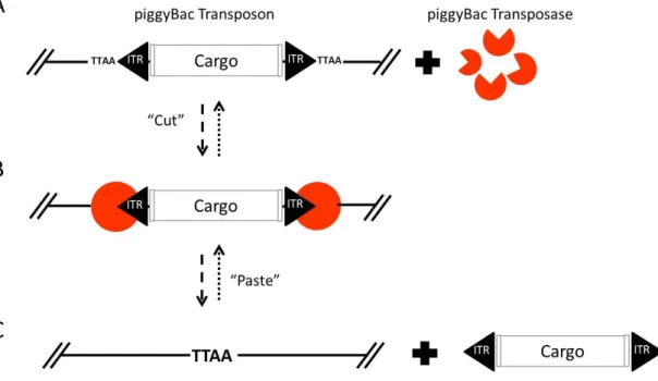
(43) domain of the transposase (244) (Figure 7-Figure 8). Beside all these similarities, there are differences between both transposases.. piggyBac PB operates using a precise cut-and-paste mechanism, targeting and duplicating TTAA tetranucleotide sequences upon insertion, and reforming a single target site upon excision (36). Excision by PB does not leave behind nucleotides at the donor site (Figure 7). The piggyBac system has been reported to be highly active when introduced into different mammalian genomes, including the human genome (245; 246).. Figure 7| piggyBac transposition. (A) piggyBac transposon elements (ITR) flanking DNA sequence (cargo) and PB transposase (orange). (B) PB transposase (orange) recognizes their ITR elements. (C) Excision and mobilization of cassette flanked by ITRs elements and TTAA PB recognition region in DNA receptor.. 20.
(44) In an attempt to increase its activity, a novel PB transposase has been generated. This new, hyperactive PB transposase (HAhyPBase) contains 7 aa substitutions with respect to the wild type version and shows more than a 10-fold higher rate of transposition than the mammalian codon-optimized piggyBac (247).. Sleeping Beauty SB transposons can integrate into TA-dinucleotide (TATA) pairs at any location in the genome. As a result of the staggered cut at the TATA target sites, the transposons are flanked by TA dinucleotides on both sides after integration (233; 248). Excision by SB from the donor site involves staggered, double stranded DNA breaks at each side of the transposon, which result in a small number of nucleotides at the termini of the transposon being left behind (249). Each ITR element of SB transposon consists of two Direct Repeats (DRs) (249). The outer DRs, termed Lo and Ro, are located at the left and right termini of the transposon, respectively, and the inner DRs, Li and Ri, are located nearby the cargo. All DRs contain binding sites for SB transposase. Both the outer and inner DRs are required for efficient transposition, but they are not interchangeable (233; 242; 250; 251) (Figure 8). Sleeping Beauty system shows efficient transposition in cells of a wide range of vertebrates, including humans (38; 252; 247; 253; 254).. 21.
(45) Figure 8| Sleeping Beauty transposition. (A) SB transposon elements (ITR) flanking DNA sequence (cargo) and SB transposase (orange). (B) SB transposase (orange) recognizes their ITR elements. (C) Excision and mobilization of cassette flanked by ITRs elements, leaving a TA motive at each end of the transposon’s original location.. In order to improve SB transposition efficiency, new hyperactive mutants of SB transposase were developed by incorporating phylogenetically conserved amino acids from related transposases belonging to the Tc1 transposon family, e.g. the amino-acid at position 243 in SB varies in the Tc1 family of transposases between M, Q or H, being M the one present in the original SB. The Q does not increase the activity of the enzyme, but a version of containing H (SB100X) confers a 100 fold increase in activity (255).. 22.
(46) 1.2.. Cellular systems. The era of efficient genome editing technologies has made possible, for the first time, the tailored modification of genetic information in living cells. The simplicity and adaptability of the new methods allows the creation of novel approaches in different research areas. Biopharmaceutical applications include the synthesis of stable cell lines with entire new genetic regulatory pathways, increased production of synthetic proteins, biosensors or even “intelligent networks” capable to detect endogenous stimuli and react to deficiencies in a host, upon transplantation (256). Another line of action is based on the ability of iPS cells to proliferate and give rise to practically any differentiated cell type in culture. Tissue specific cell derived from genetically modified iPS cells could be used to repopulate damaged organs or tissues in patients affected by hereditary diseases, cancer or age-related disorders. While stable cell lines, have been routinely used in laboratories for the last decades, iPS cells have been recently discovered and will be therefore describe in some detail in the following chapter.. 1.2.1.. Cell reprogramming. In 1958, Gurdon reported the concept and first example of rejuvenation and cellular reprogramming through somatic cell nuclear transfer (SCNT) in frogs (257). Cellular reprogramming refers to the process whereby cells travel back up their maturation path, process during which the gene expression profile of a cell type is converted into the gene expression profile of the other cell type. It took around 40 years before animal cloning could be attained in another vertebrate species, a sheep, showing that erasure of epigenetic memories that set somatic cell fate was possible even in mammals (258). During this time, embryonic stem cells (ES cells) were identified in mice (259). ES have the inherent capacity to generate all cell types in the organism, a property known as 23.
(47) pluripotency (259; 260) (Figure 9). In 1998, human embryonic stem cells were isolated and grown in the lab (49). The rejuvenation of a cell to the pluripotent state has also been shown by fusing somatic cells with embryonic stem cells (261; 262). These two approaches suggest that fertilized eggs and pluripotent stem cells contain hidden reprogramming factors that are able to erase the somatic memories. Other research demonstrated that transcription factor(s) are crucial for both the maintenance of cellular identity, and the determination of cell fate (263; 264; 265; 266) Based on those propositions, in 2006 Takahashi and Yamanaka announced the successful derivation of pluripotent stem cells from adult mouse fibroblast through the ectopic co-expression of only four genes (45). After screening a set of 24 gene candidates selected for being linked to ESC pluripotency, they proved that four factors were sufficient to reprogram adult fibroblasts into iPS cells. These reprogramming factors were octamer-binding transcription factor-3/4 (OCT3/4), SRY-related highmobility-group (HMG)-box protein-2 (SOX2), c-MYC and Kruppel-like factor-4 (KLF4) (Figure 9). The discovery showed the importance of transcription factor networks in cell fate determination, and helped to understand the nature of cellular reprogramming. iPS cells and ESCs show similar molecular and functional features (46), and they are cultured under the same conditions (46; 267; 268; 269; 270). Pluripotent stem cells (iPS cells and ESCs) are capable, when cultured, of unlimited self-renewal and differentiation into virtually all adult cell types (47; 48). These properties raised reasonable expectations that the cells could be useful tools for research on disease mechanisms, screenings for therapeutic drugs, and as therapeutic agents in regenerative medicine applications. The procedure of deriving human embryonic stem cells implies embryo disaggregation (49), which itself poses ethical controversies that hindered research progress. In addition, it is not possible to generate retroactively patient-specific therapeutic cells from embryos. Unlike ESCs, iPS cells are produced from somatic cells which are not associated with ethical controversies (50), and permit autologous transplantations avoiding the host immune response (51). 24.
(48) Figure 9| Differentiation and reprogramming processes. ESCs have the capacity to self-renew and to differentiate into cellular derivatives of the endodermal, ectodermal, and mesodermal lineages. iPS cells have equal capacities. Reprogramming allows us to turn any cell of the body into a pluripotent stem cell.. Since the seminal Yamanaka paper, a number of studies have demonstrated the feasibility of obtaining iPS cells from a wide variety of cell types, such as pancreatic β cells (271), neural stem cells (272), peripheral blood (273), fibroblast (274), etc. Recent years have also brought significant progress in increasing the efficiency and reducing the risks associated to this technique, making it more amenable to applications in the fields of regenerative medicine, disease modelling and drug discovery. Although the original gene set for reprogramming comprised four factors, follow-on studies have reduced, changed or increased the number of reprogramming factor required (275; 276; 277; 274). Additionally, modifications in the delivery of the reprogramming factor to the targeted cells have significantly improved the reliability of the process (278; 279; 280).. 25.
(49) 1.3.. Delivery vectors. Choosing the optimal vector/delivery system is a delicate process that must take into account many factors, like the targeted cells and their characteristics, the duration of expression required, and the size of the genetic material to be incorporated into the vector (281). The mechanisms to deliver the DNA, RNA or proteins into the cells can be generally categorized in viral or non-viral vectors. Viral delivery vectors substitute the non-desired parts of the viral genome by the genetic information to be delivered into the cells. Non-viral vectors typically make use of chemical or physical means to deliver biomolecules (DNA, RNA or protein) into living cells or organisms (98).. 1.3.1. Viral vectors Viral vectors take advantage of the natural ability of viruses to enter into and deliver genetic material to cells (282). The most commonly used vectors are retroviruses, lentivirus, adenoviruses, and adeno-associated viruses (AAV). Retroviral vectors. Replication-competent retrovirus vectors are based on murine leukaemia virus (MLV) (283). Retroviruses infect many types of cells in mammals. They need cells to divide in order to introduce their non-segmented RNA genome into the cell DNA and, therefore, will not infect many tissues where host cell growth and division have come to a standstill (284; 285). Retrovirus vectors have a relatively limited size capacity to carry genes, around 8 kb (286). Lentiviral vectors. Lentiviral vectors derived from the human immunodeficiency virus (HIV). Lentivirus are able to integrate their genomic RNA into dividing and non-dividing cells of human and other mammalian species (287). They may carry up to 9 kb of genetic information (288). Adenovirus vectors. Adenoviruses are non-enveloped (naked) viruses with an icosahedral nucleocapsid containing a double strand DNA genome. They have a broad range of vertebrate hosts; there are more than 100 serotypes characterized in 26.
(50) primates alone (289). Adenoviruses infect most cell types, so they are able to infect even non-dividing cells. This viral vector does not incorporate its genetic material into the host genome, so their DNA remains free in the cell nucleus. Their capacity to carry large DNA loads is not significantly better than other viral vectors, around 8 kb (64). Adeno-associated viral vectors (AAVs). AAVs are small, non-pathogenic and single stranded DNA viruses from the Parvovirus family (290). They infect a broad range of cells including both dividing and non-dividing cells (290). The virus requires additional genes to replicate, which are provided by adenovirus or herpes virus that carry cytostatic and cytotoxic genes to the target cells (291; 292; 290; 293). AAVs present a limited packaging capacity of around 5 kb (294). Although viral vectors are commonly used to transfer genes into cells and/or tissues (64), they have several limitations including immunogenicity, broad tropism, limited DNA packaging capacity, and difficulty of vector production and the unintended initiation of tumorigenic processes. Non-viral vector might resolve many of these limitations, particularly with respect to safety (98) and packaging capacity, a very important parameter when considering big payloads (68).. 1.3.2. Non-viral vectors Complex biomolecules like DNA, RNA and proteins must be able to reach specific cells (295; 296); and once on target, they have to cross several cellular barriers and avoid the cellular degradation machinery before reaching their destination inside the target (297; 298). All these requirements can be fulfilled by using non-viral vectors. Non-viral delivery tools are safe and specific, easy to produce in high quantities, and generally elicit low immunological responses (98). In addition, they are relatively inexpensive, can mobilize big molecules, and can be adapted to comply with specific needs to improve their efficiency (98).. 27.
(51) Non-viral vectors include physical and chemical methods. The simplest physical method is direct injection (299). Other, more complex ones include electroporation (300), sonoporation (301; 302) or magnetofection (303; 304). They are used to temporarily disrupt cell membranes by electrical field, ultrasonic frequency or magnetic field, respectively (300; 301; 304). These physical methods are efficient at enhancing membrane penetration in vitro, although their efficiency in vivo is significantly lower (98; 305; 64). Chemical methods are generally synthetic/natural biodegradable liposomes (Lipoplexes), polymers (Polyplexes) or lipopolymers (Lipopolyplex) (306). Briefly, the liposomes consist of cationic lipids that encapsulate the hydrophilic molecules to be transferred and subsequently fuse with the cell membrane and release the cargo into the cytoplasm of the cell (307; 308). The polymers use a net-positive charge to from electrostatic complexes with anionic molecules (309; 310), and the complex liposomes/polymers use a polymer to condense biomolecule and a lipid coat to aid entry into the cell (306; 311; 312; 313).. 28.
Figure

+7


Documento similar
In the “big picture” perspective of the recent years that we have described in Brazil, Spain, Portugal and Puerto Rico there are some similarities and important differences,
For the second case of this study, the features for training the SVM will be extracted from the brain sources estimated using the selected SLMs MLE, MNE and wMNE. The scenario is
No obstante, como esta enfermedad afecta a cada persona de manera diferente, no todas las opciones de cuidado y tratamiento pueden ser apropiadas para cada individuo.. La forma
The Dwellers in the Garden of Allah 109... The Dwellers in the Garden of Allah
The aim of this work is to search for immunological genetic markers that can explain the natural evolution of hepatitis C virus infection in the patient coinfected with the
Díaz Soto has raised the point about banning religious garb in the ―public space.‖ He states, ―for example, in most Spanish public Universities, there is a Catholic chapel
teriza por dos factores, que vienen a determinar la especial responsabilidad que incumbe al Tribunal de Justicia en esta materia: de un lado, la inexistencia, en el
AAV9-CnAβ1 decreases the expression of genes associated with heart failure and fibrosis in the remote myocardium post MI – AAV9-CnAβ1 administration at a dose of 3.5e10 VP,
