Volume 2012, Article ID 504292,7pages doi:10.1155/2012/504292
Review Article
Cytoplasmic Ribonucleoprotein Foci in Eukaryotes: Hotspots of
Bio(chemical)Diversity
Carla Layana,
1, 2Paola Ferrero,
1, 2and Rolando Rivera-Pomar
1, 21Centro Regional de Estudios Gen´omicos, Universidad Nacional de La Plata, CP 1888 Florencio Varela, Argentina 2Departamento de Ciencias B´asicas y Experimentales, Universidad Nacional del Noroeste de Buenos Aires,
Avenida Calchaqu´ı 5900, CP 1888 Buenos Aires, Pergamino, Argentina
Correspondence should be addressed to Rolando Rivera-Pomar,[email protected]
Received 1 February 2012; Accepted 22 March 2012 Academic Editor: Greco Hern´andez
Copyright © 2012 Carla Layana et al. This is an open access article distributed under the Creative Commons Attribution License, which permits unrestricted use, distribution, and reproduction in any medium, provided the original work is properly cited. The life of an mRNA from transcription to degradation offers multiple control check points that regulate gene expression. Transcription, splicing, and translation have been widely studied for many years; however, in recent years, new layers of posttranscriptional and posttranslational control have been uncovered. They involve the regulation of the metabolism of mRNA in cytoplasmic foci. They are collections of ribonucleoprotein complexes that, in most cases, remain still uncharacterized, except the processing bodies (PBs) and stress granules (SGs), which have been studied (and reviewed) in detail. A challenging prospective is to know how many different classes of foci exist, which functions they support, how are they formed, and how do they relate one to each other. Here, we present an update of the component of the different granules, a possible function, and hypothesis on their
in vivodynamics related to translational control.
1. Introduction
In recent years, several cytoplasmic foci/granules that contain proteins and RNA have been described. Two of them have been studied in more detail as they are related to mRNA silencing: stress granules (SG) and processing bodies (PB). SG are repressed mRNPs transiently induced in response
to cellular stress. They range from 0,5 to 5µm [1]. PB
are discrete RNP cytoplasmic foci of 0,1-2µm where the
machinery of RNA interference, degradation and storage locates. In PB the mRNAs are forming mRNP complexes either repressing translation, in degradation complexes or
stored for further use [2,3]. SG and PB have been shown to
share a growing number of proteins that are added in a day-to-day basis to the list of their components. SG, PB and other cytoplasmic foci are highly dynamic structures, although PB are quite stable over the time [4]; see also Supplementary Movie 1 available online at doi:10.1155/2012/504292. They are in a dynamic steady state with other mRNPs, such as polysomes in response to the translational state of the cell [5]. Although we do not intend to extensively review SG and PB, which have been matter of fine reviews in the last years
[6–10], we will overview their functions before we address neglected issues and hypothesis.
2. Stress Granules
Translation initiation is the key regulatory step of trans-lational control. Therefore, it is the most sensitive step to changes in the cellular environment, including stress. A key step in translation initiation inhibition is the
phosphoryla-tion of eIF2α, which results in an increase on the affinity of
eIF2-GDP for eIF2B, sequestering this factor to prevent new round of translational initiation [11]. During this process, translation is inhibited and polysomes become released from the mRNA leading to the accumulation of inactive mRNPs in SG. The SG are in equilibrium with active polysomes. Protein elongation inhibitors, such as cycloheximide, prevent the assembly of SG by blocking the polysomes in an inactive state, while protein initiation inhibitors promote the
formation of SG [8].Table 1 shows the components of SG
Table1: Components of stress granules.
Protein Function Interacting proteins Ago2 Cleaves interfered RNA RISC, FXR1
APOBEC3G Antiviral response ?
Ataxin-2 Translation PABP-1
Caprin-1 Cell growth G3BP
CPEB mRNA repression RCK, eIF4E, FXR1
DIS1 Unknown eIF3h
eIF3 Translation 40S, eIF4G
eIF4E Translation CPEB, Smaug, eIF4G, 4ET eIF4G Translation eIF4E, eIF3, PABP-1
FAST Translation TIA-1
FMRP, FXR1 Translation Ago2, RISC
FBP, KSRP mRNA degradation TIA-1
FUS/TLS Transcriptional control Transcriptional machinery
G3BP Ras signalling Caprin
HuR mRNA stabilization ?
IP5K Signalling ?
Lin28 Developmental control ?
LINE 1 ORF1p Transposon ?
MLN51 Splicing Exon junction
PABP-1 Translation eIF4G, eIF3, ataxina-2 RCK(p54) mRNA degradation GE-1, TTP
Plakophilin Adhesion G3BP, FXR1
PMR1 mRNA degradation TIA-1
Pumilio 2 mRNA silencing ?
Rap 55 mRNA silencing ?
Rpb4 Transcription ?
SRC3 Transcription TIA-1
Staufen mRNA silencing ?
SMN RNP assembly Complejo SMN
TDP-43 Transcription and splicing regulator eIF4G, eIF3, eIF2, ribosomal proteins, STAU-1, Xnr TIA-1(rox-8), TIAR mRNA silencing FAST, SRC3, PMR1, FBP
TRAF2 Signalling eIF4G
TTP, BRF-1 mRNA silencing RCK (p54)
YB-1 Cold shock ?
ZBP1 Localization ?
(1) Core components: stalled initiation complexes (pol-yadenylated mRNAs and translation factors eIF4E, eIF4A, eIF4G, eIF3, eIF2, PABP, and proteins of the small ribosome subunit).
(2) RNA-binding proteins associated to silencing and transcript stability: TIA-1, TIAR [12], FAST, Argonaute [13], CPEB, smaug, DExD/H-box RCK/p54 (o Dhh1), XRN1 [5].
(3) RNA-binding proteins associated to mRNA metabo-lism either translation of degradation such as G3BP [14] and Staufen [15].
The key concept regarding SG is that they are responsible of protecting the mRNA during cell stress, altering the com-position of the mRNPs in a reversible manner. As soon as the cell recovers, the mRNPs regain their translational capacity.
3. Processing Bodies
These structures have been described many times since 1997, when Bashkirov et al. observed that the exonuclease Xrn1 is located in small granular structures in the cytoplasm of mammalian cells and call them “Xrn1 foci” [17]. Later on, the decapping enzyme Dcp2 was also described to occur in cytoplasmic foci [18]. Contemporary, Eystathioy et al. have described that a protein associated to neuropathy named GW182 occurs in cytoplasmic speckles called GW bodies
[19,20]. Other RNA-related protein, the eIF4E-transporter,
was also localized in discrete cytoplasmic foci [16,21,22].
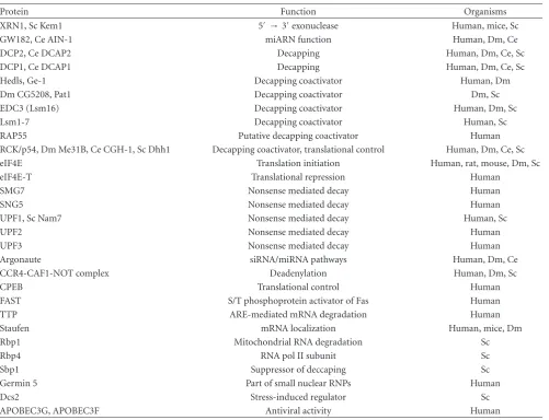
[image:2.600.54.552.88.555.2]Table2: Components of processing bodies.
Protein Function Organisms
XRN1, Sc Kem1 5′ →3′exonuclease Human, mice, Sc
GW182, Ce AIN-1 miARN function Human, Dm, Ce DCP2, Ce DCAP2 Decapping Human, Dm, Ce, Sc DCP1, Ce DCAP1 Decapping Human, Dm, Ce, Sc Hedls, Ge-1 Decapping coactivator Human, Dm Dm CG5208, Pat1 Decapping coactivator Dm, Sc EDC3 (Lsm16) Decapping coactivator Human, Dm, Sc Lsm1-7 Decapping coactivator Human, Sc RAP55 Putative decapping coactivator Human RCK/p54, Dm Me31B, Ce CGH-1, Sc Dhh1 Decapping coactivator, translational control Human, Dm, Ce, Sc eIF4E Translation initiation Human, rat, mouse, Dm, Sc eIF4E-T Translational repression Human
SMG7 Nonsense mediated decay Human
SNG5 Nonsense mediated decay Human
UPF1, Sc Nam7 Nonsense mediated decay Human, Sc
UPF2 Nonsense mediated decay Human
UPF3 Nonsense mediated decay Human
Argonaute siRNA/miRNA pathways Human, Dm, Ce CCR4-CAF1-NOT complex Deadenylation Human, Dm, Sc
CPEB Translational control Human
FAST S/T phosphoprotein activator of Fas Human TTP ARE-mediated mRNA degradation Human Staufen mRNA localization Human, mice, Dm Rbp1 Mitochondrial RNA degradation Sc
Rbp4 RNA pol II subunit Sc
Sbp1 Suppressor of deccaping Sc
Germin 5 Part of small nuclear RNPs Human
Dcs2 Stress-induced regulator Sc
APOBEC3G, APOBEC3F Antiviral activity Human
demonstrated that PB contains enzymes involved in the degradation of the mRNA. Later one further studies showed that they are also related to miRNA metabolism and can store mRNAs to bring them back to polysomes (reviewed in
[3,24]. They include, different than SG, neither ribosomal
proteins nor translation factors, except eIF4E. They do not present either the exosome components [25]. eIF4G and PABP were found in yeast PB, although at low level and in stress conditions resulting on glucose deprivation [26]. Proteins and mRNA can reversely go in and out of PB [25]. The relationship of PB and polysomes is demonstrated by the blocking of PB formation by cycloheximide. A summary of
the components in different organisms is shown inTable 2.
The occurrence of such large and diverse set of proteins (and the list continuously grows up) suggests that PBs are involved in a plethora of posttranslational processes regulating gene expression, such as mRNA degradation and silencing. mRNA degradation starts with the shortening of the poly-A tail— the deadenylation. In eukaryotes, there are several complexes involved in the process: PARN2-PARN3 initiates the pro-cess, which continues with the action of the CAF1-CCR4-NOT complex. Later on, mRNA degradation continues by
nucleolytic cleavage on both ends. 3′ → 5′ degradation is
catalyzed by the exosome and the SKI complex, while 5′ →
3′degradation requires previous decapping by DCP2 and the
coactivator DCP1 and the action of the exonuclease XRN1. All these enzyme localize in PB. There are several evidences indicating that mRNA degradation occurs in PB.
(i) The assembly of PB depends on mRNA, as RNase
treatment of the cells induces the disappearing of PB [27,28].
(ii) Inhibition or removal of the deadenylase Ccr4 reduces the number and size of PB, while the removal of the
downstream-acting enzymes Xrn1 and Dcp1 does not affect
the stability of PB [21].
(iii) mRNA degradation intermediates are present in PB [23].
Therefore, one can conclude that mRNA degradation occurs in PB and depends on the existence of degradation
enzymes and mRNA degradation intermediates [21,23,25,
29]. Many of the PB components are not restricted to the foci and also are present in the soluble cytoplasm and nuclei,
suggesting that the different processes might start before
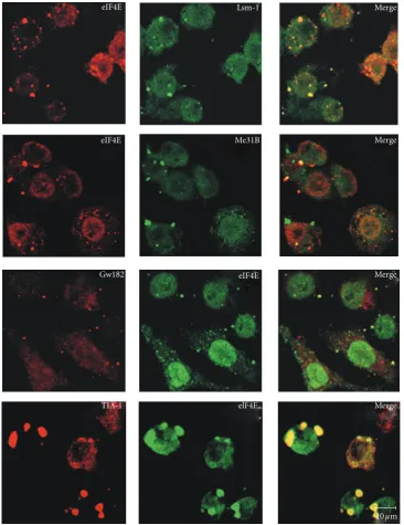
[image:3.600.53.555.86.470.2]Lsm-1 Merge
Me31B
Gw182
Merge
Merge
Merge elF4E
TIA-1
10µm eIF4E
eIF4E
eIF4E
Figure1: Colocalization of eIF4E with components of PBs and SGs shows a diversity of cytoplasmic foci quality. The experiment shows that,
in every case, the granules contain both components or either one or the other in different quantities. This would represent intermediates or different forms of eIF4E-containing foci.Drosophila melanogasterS2 cells were transfected with proteins fusion CFP-Lsm-1 or CFP-Me31B. GW182, eIF4E, and Rox8 (the TIA-1 ortholog inDrosophila) were revealed using antibodies against GW182, anti-V5, and anti-TIA-1, respectively. In the bottom panel (row 4), the cells were prestressed with arsenite for 30 minutes.
decay (NMD). The detection of premature termination in the cells by spotting an mRNA with an abnormal stop codon is mediated by a surveillance complex composed by UPF1, UPF2 y UPF3, additional proteins, namely, SMG1, and SMG5-7 [30–32]. As soon as the surveillance complex is assembled, the degradation enzymes (Dcp1, Xrn1) are recruited to the mRNA in PB. Although the degrading enzymes are located in PB, the mechanism of recruitment is
unknown. In silencing, there are two types of small mRNAs that regulate posttranscriptional gene expression: siRNAs
and miRNAs. Despite the different mechanism of silencing,
in both cases participate the protein Argonaute (Ago) and the RISC (RNA-induced silencing complex). In the case of siRNAs, Ago produces an endonucleolytic cleavage of the
mRNA to promote degradation by the 3′ → 5′ and 5′ →
[image:4.600.117.484.72.548.2]Polysomes
Translational machinery
Degradation or storage
Xm-1 Dcp Others GW182
rck/p54
Translational inactive mRNP Translational active mRNP
4G
4E 4E-T 4E-T
4G
4E
A
B
P
P
AAAAAAAA
A
B
P
P
Lsm1-7
Processing bodies
Figure2: Relationship among active polysomes and PBs. The recruitment of active polysomes to PB implies the removal from the mRNA
of the translation factors by translational repressors. Some of them have been demonstrated to interact with eIF4Ein vivo(rck/p54 and eIF4E-T, [16]). They further interact and/or recruit the enhancers of decapping Lsm1-7 or the miRNA-related protein GW182 to form the PB. Later on, they assemble the decapping and degradation enzymes and/or the proteins required for silencing and storage into PB. All the intermediate steps of this process can represent different populations of granules coexisting in the cell and visible with different morphology that might reflect a variety of components and/or diverse stoichiometry.
recruit Ago to direct the repressed mRNA to degradation mediated by the PB proteins GW182, CCR4-CAF1-NOT1, DCP2, DCP1, and XRN1.
4. The Cycle of an mRNA in the Cell: SG, PB,
Polysomes, and the Unknown Intermediates
From the previous analysis, one can establish many unsolved aspects on cytoplasmic foci function. One of them is the dynamic of the mRNP remodeling. The current model suggests an active movement of mRNPs from and to polysomes and from and to SG and PB [33]. However, how does it happen and the factors involved are not known. Translationally active mRNAs can interact, in response to errors in translational initiation or to specific recruitment of regulatory proteins, with translational repressors such as Dhh1, Pat 1, Lsm1-7, eIF4E-T. Those factors would promote the replacement of the translational machinery from the mRNA, promote the cap removal and determine degradation [33] or the accumulation of silenced mRNA in PB. Within PB, mRNPs could undergo further remodeling and define a path to follow, including their return to polysomes. In addition, PBs have been shown to interact and exchange
components or their own nature with SG (reviewed in [6,7])
in a process that may result in mRNPs intermediates of unknown nature. Evidence for the diversity of cytoplasmic foci and their components results from immunocytochem-istry and colocalization studies. A common factor present in most cytoplasmic mRNPs is the cap-binding protein eIF4E. eIF4E occurs in active polysomes as a translation initiation factor, in SG as part of the stalled initiation complex,
and in PB as the only translation factor present there in
multicellular eukaryotes. We observed inDrosophilaS2 cells
that eIF4E colocalizes with different pairs of markers, either
for PB (GW182, Lsm1, Me31B—an ortholog of the helicase rck/p54) or SG (TIA-1) and that the colocalization does not occur in all foci in the same way (PVF, CL, and RRP,
unpublished data and Figure 1). In some cases, the foci
contain one, the other, or both components. In the foci that show colocalization of both factors, the relative amount of each component may vary from foci to foci, as judged by confocal microscopy quantification of the colocalized factors (PVF, CL, RRP, unpublished observation). This implies that there are a diversity of granules. An appealing hypothesis
is that eIF4E is a common link among different mRNPs,
playing different roles depending on their interactors. One
plausible function could be that the accumulation of mRNPs in eIF4E-containing foci is a way to regulate the rate of
translation in different physiological states (cell cycle phases,
developmental stages, circadian rhythms). Moreover, it has been reported that, in mammalian cells, eIF4E interacts in PB with at least two factors, rck/p54 and eIF4E-T [21]. These are simultaneous interactions within the PB and imply
that both proteins could contact different domains of the
same eIF4E molecule or that they would represent different
populations of mRNPs or different functions within the same
PB. In either cases, the complexity of the interactions in
vivois more diverse than it has been expected. A model for
the remodeling of active mRNPs to silence and degradation
based on Andrei et al. [21] is depicted inFigure 2. This might
[image:5.600.148.451.71.289.2]would correlate with the large diversity of components and interactions within a cytoplasmic foci and the diversity of the foci within a cell. The understanding of the dynamics of mRNP is far from clear and unpredictable paths remain to be discovered. They will need further research and more
sophisticated methods forin vivostudies.
Acknowledgments
This work was supported by grants from CONICET (PIP 00318 to P. Ferrero) and ANPCyT (PICT-2008-1237 to RRP and P. Ferrero). P. Ferrero and R. Rivera-Pomar are investigators and CL doctoral fellow of the CONICET.
References
[1] M. G. Thomas, L. J. Martinez Tosar, M. A. Desbats, C. C. Leishman, and G. L. Boccaccio, “Mammalian staufen 1 is recruited to stress granules and impairs their assembly,”
Journal of Cell Science, vol. 122, no. 4, pp. 563–573, 2009. [2] S. P. Chan and F. J. Slack, “MicroRNA-mediated silencing
inside P-bodies,”RNA Biology, vol. 3, no. 3, pp. 97–100, 2006. [3] A. Eulalio, I. Behm-Ansmant, D. Schweizer, and E. Izaurralde,
“P-body formation is a consequence, not the cause, of RNA-mediated gene silencing,”Molecular and Cellular Biology, vol. 27, no. 11, pp. 3970–3981, 2007.
[4] A. Aizer and Y. Shav-Tal, “Intracellular trafficking and dynam-ics of P bodies,”Prion, vol. 2, no. 4, pp. 131–134, 2008. [5] N. Kedersha, G. Stoecklin, M. Ayodele et al., “Stress granules
and processing bodies are dynamically linked sites of mRNP remodeling,”Journal of Cell Biology, vol. 169, no. 6, pp. 871– 884, 2005.
[6] M. G. Thomas, M. Loschi, M. A. Desbats, and G. L. Boccaccio, “RNA granules: the good, the bad and the ugly,” Cellular Signalling, vol. 23, no. 2, pp. 324–334, 2011.
[7] J. R. Buchan and R. Parker, “Eukaryotic stress granules: the ins and outs of translation,”Molecular Cell, vol. 36, no. 6, pp. 932– 941, 2009.
[8] P. Anderson and N. Kedersha, “Stress granules: the Tao of RNA triage,”Trends in Biochemical Sciences, vol. 33, no. 3, pp. 141– 150, 2008.
[9] A. Eulalio, I. Behm-Ansmant, and E. Izaurralde, “P bodies: at the crossroads of post-transcriptional pathways,” Nature Reviews Molecular Cell Biology, vol. 8, no. 1, pp. 9–22, 2007. [10] R. Parker and U. Sheth, “P bodies and the control of mRNA
translation and degradation,”Molecular Cell, vol. 25, no. 5, pp. 635–646, 2007.
[11] S. Yamasaki and P. Anderson, “Reprogramming mRNA trans-lation during stress,”Current Opinion in Cell Biology, vol. 20, no. 2, pp. 222–226, 2008.
[12] N. L. Kedersha, M. Gupta, W. Li, I. Miller, and P. Anderson, “RNA-binding proteins TIA-1 and TIAR link the phospho-rylation of eIF-2α to the assembly of mammalian stress granules,”Journal of Cell Biology, vol. 147, no. 7, pp. 1431– 1442, 1999.
[13] A. K. Leung, J. M. Calabrese, and P. A. Sharp, “Quantitative analysis of Argonaute protein reveals microRNA-dependent localization to stress granules,” Proceedings of the National Academy of Sciences of the United States of America, vol. 103, no. 48, pp. 18125–18130, 2006.
[14] H. Tourri`ere, K. Chebli, L. Zekri et al., “The RasGAP-associated endoribonuclease G3BP assembles stress granules,”
Journal of Cell Biology, vol. 160, no. 6, pp. 823–831, 2003. [15] M. G. Thomas, L. J. Martinez Tosar, M. Loschi et al., “Staufen
recruitment into stress granules does not affect early mRNA transport in oligodendrocytes,”Molecular Biology of the Cell, vol. 16, no. 1, pp. 405–420, 2005.
[16] J. Dostie, F. Lejbkowicz, and N. Sonenberg, “Nuclear eukary-otic initiation factor 4E (eIF4E) colocalizes with splicing factors in speckles,”Journal of Cell Biology, vol. 148, no. 2, pp. 239–246, 2000.
[17] V. I. Bashkirov, H. Scherthan, J. A. Solinger, J. M. Buerstedde, and W. D. Heyer, “A mouse cytoplasmic exoribonuclease (mXRN1p) with preference for G4 tetraplex substrates,”
Journal of Cell Biology, vol. 136, no. 4, pp. 761–773, 1997. [18] E. van Dijk, N. Cougot, S. Meyer, S. Babajko, E. Wahle,
and B. S´eraphin, “Human Dcp2: a catalytically active mRNA decapping enzyme located in specific cytoplasmic structures,”
The European Molecular Biology Organization Journal, vol. 21, no. 24, pp. 6915–6924, 2002.
[19] T. Eystathioy, E. K. Chan, S. A. Tenenbaum, J. D. Keene, K. Griffith, and M. J. Fritzler, “A phosphorylated cytoplasmic autoantigen, GW182, associates with a unique population of human mRNAs within novel cytoplasmic speckles,”Molecular Biology of the Cell, vol. 13, no. 4, pp. 1338–1351, 2002. [20] T. Eystathioy, A. Jakymiw, E. K. Chan, B. S´eraphin, N. Cougot,
and M. J. Fritzler, “The GW182 protein colocalizes with mRNA degradation associated proteins hDcp1 and hLSm4 in cytoplasmic GW bodies,”RNA, vol. 9, no. 10, pp. 1171–1173, 2003.
[21] M. A. Andrei, D. Ingelfinger, R. Heintzmann, T. Achsel, R. Rivera-Pomar, and R. L¨uhrmann, “A role for eIF4E and eIF4E-transporter in targeting mRNPs to mammalian processing bodies,”RNA, vol. 11, no. 5, pp. 717–727, 2005.
[22] M. A. Ferraiuolo, S. Basak, J. Dostie, E. L. Murray, D. R. Schoenberg, and N. Sonenberg, “A role for the eIF4E-binding protein 4E-T in P-body formation and mRNA decay,”Journal of Cell Biology, vol. 170, no. 6, pp. 913–924, 2005.
[23] U. Sheth and R. Parker, “Decapping and decay of messenger RNA occur in cytoplasmic processing bodies,”Science, vol. 300, no. 5620, pp. 805–808, 2003.
[24] R. Parker and U. Sheth, “P bodies and the control of mRNA translation and degradation,”Molecular Cell, vol. 25, no. 5, pp. 635–646, 2007.
[25] M. Brengues, D. Teixeira, and R. Parker, “Movement of eukaryotic mRNAs between polysomes and cytoplasmic pro-cessing bodies,”Science, vol. 310, no. 5747, pp. 486–489, 2005. [26] M. Brengues and R. Parker, “Accumulation of polyadenylated mRNA, Pab1p, eIF4E, and eIF4G with P-bodies in Saccha-romyces cerevisiae,”Molecular Biology of the Cell, vol. 18, no. 7, pp. 2592–2602, 2007.
[27] V. I. Bashkirov, H. Scherthan, J. A. Solinger, J.-M. Buerstedde, and W.-D. Heyer, “A mouse cytoplasmic exoribonuclease (mXRN1p) with preference for G4 tetraplex substrates,”The Journal of Cell Biology, vol. 136, no. 4, pp. 761–773, 1997. [28] D. Ingelfinger, D. J. Arndt-Jovin, R. L¨uhrmann, and T. Achsel,
“The human LSm1-7 proteins colocalize with the mRNA-degrading enzymes Dcp1/2 and Xrn1 in distinct cytoplasmic foci,”RNA, vol. 8, no. 12, pp. 1489–1501, 2002.
[29] N. Cougot, S. Babajko, and B. S´eraphin, “Cytoplasmic foci are sites of mRNA decay in human cells,”Journal of Cell Biology, vol. 165, no. 1, pp. 31–40, 2004.
Current Opinion in Cell Biology, vol. 17, no. 3, pp. 316–325, 2005.
[31] N. Amrani, M. S. Sachs, and A. Jacobson, “Early nonsense: mRNA decay solves a translational problem,”Nature Reviews Molecular Cell Biology, vol. 7, no. 6, pp. 415–425, 2006. [32] F. Lejeune and L. E. Maquat, “Mechanistic links between
nonsense-mediated mRNA decay and pre-mRNA splicing in mammalian cells,”Current Opinion in Cell Biology, vol. 17, no. 3, pp. 309–315, 2005.
Submit your manuscripts at
http://www.hindawi.com
Hindawi Publishing Corporation
http://www.hindawi.com Volume 2014 Anatomy
Research International
Peptides
Hindawi Publishing Corporation
http://www.hindawi.com Volume 2014
Hindawi Publishing Corporation http://www.hindawi.com
International Journal of
Volume 2014
Zoology
Hindawi Publishing Corporation
http://www.hindawi.com Volume 2014 Molecular Biology International
Genomics
International Journal of
Hindawi Publishing Corporation
http://www.hindawi.com Volume 2014
The Scientiic
World Journal
Hindawi Publishing Corporationhttp://www.hindawi.com Volume 2014
Hindawi Publishing Corporation
http://www.hindawi.com Volume 2014
Bioinformatics
Advances inMarine Biology
Journal ofHindawi Publishing Corporation
http://www.hindawi.com Volume 2014
Hindawi Publishing Corporation
http://www.hindawi.com Volume 2014
Signal Transduction
Journal ofHindawi Publishing Corporation
http://www.hindawi.com Volume 2014
BioMed
Research International
Evolutionary Biology
International Journal of
Hindawi Publishing Corporation
http://www.hindawi.com Volume 2014
Hindawi Publishing Corporation
http://www.hindawi.com Volume 2014
Biochemistry Research International
Archaea
Hindawi Publishing Corporation
http://www.hindawi.com Volume 2014
Hindawi Publishing Corporation
http://www.hindawi.com Volume 2014
Genetics
Research International
Hindawi Publishing Corporation
http://www.hindawi.com Volume 2014
Advances in
Virology
Hindawi Publishing Corporation http://www.hindawi.com
Nucleic Acids
Journal ofVolume 2014
Stem Cells
International
Hindawi Publishing Corporation
http://www.hindawi.com Volume 2014
Hindawi Publishing Corporation
http://www.hindawi.com Volume 2014
Enzyme
Research
Hindawi Publishing Corporation
http://www.hindawi.com Volume 2014
International Journal of

![Figure 2: Relationship among active polysomes and PBs. The recruitment of active polysomes to PB implies the removal from the mRNAeIF4E-T, [16])](https://thumb-us.123doks.com/thumbv2/123dok_es/4760698.61210/5.600.148.451.71.289/figure-relationship-polysomes-recruitment-polysomes-implies-removal-mrnaeif.webp)
