RNPS 2235-145 © 2009-2013 Cardiocentro “Ernesto Che Guevara”, Villa Clara, Cuba. Todos los derechos reservados. 122
Sociedad Cubana de Cardiología
_______________
Caso Clínico
Rotura del septo interventricular después de infarto agudo de
miocardio con apertura y cierre intermitentes
MSc. Dr. Héctor Díaz Águila
, Dr. Yamir Santos Monzón, Dr. Alberto Fragoso Estévez,
Dr. Yonielis Rivero Nóbrega y Dr. Jorge L. Alonso Freire
Servicio de Medicina Intensiva. Hospital “Mártires del 9 de Abril”. Sagua la Grande, Villa Clara. Cuba.
Full English text of this article is also available
INFORMACIÓN DEL ARTÍCULO
Recibido: 12 de julio de 2012 Aceptado: 22 de agosto de 2012
Los autores declaran que no existen conflictos de intereses
Abreviaturas
IAM: Infarto agudo de miocardio
SIV: Septo interventricular
ECG: Electrocardiograma
Versiones On-Line: Español
-
Inglés H Díaz Águila
Hospital “Mártires del 9 de Abril” Carretera Circuito Norte Km. 2. Sagua la Grande. CP 52310. Villa Clara. Cuba
Correo electrónico:
RESUMEN
La rotura del septo interventricular es una grave complicación en pacientes que sufren infarto agudo de miocardio. Se presenta aproximadamente en el 1 % de los pacientes infartados, su mortalidad es elevada y el tratamiento de elección es la reparación quirúrgica. Se presenta un paciente anciano que ingresó en la Unidad de Cuidados Intensivos por infarto agudo de miocardio de cara anterior, que recibió tratamiento trombolítico con estreptokinasa recombinante cubana y 24 horas más tarde, presentó deterioro hemodinámico con cambios electrocardiográficos y apari-ción de soplo sistólico en la punta. Se realizó una ecocardiografía que mostró un de-fecto del septo interventricular con apertura y cierre intermitentes. Horas más tarde el paciente falleció por insuficiencia cardiocirculatoria, a pesar del tratamiento. Se presentan las imágenes ecocardiográficas y la pieza anatómica. Lo inusual del pre-sente caso fue la apertura y el cierre intermitentes del defecto interventricular. No se encontró ningún informe similar a en las bases de datos bibliográficas consultadas.
Palabras clave: Infarto de miocardio; Rotura Cardíaca Postinfarto; Choque Cardiogé-nico
Interventricular septal rupture after acute myocardial infarction with
intermittent opening and closing
ABSTRACT
ventricu-CorSalud 2013 Ene-Mar;5(1):122-126 123
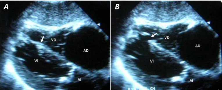
Figura 1. Rotura del septo interventricular. La flecha señala el defecto interventricular. A. Cerrado en sístole. B. Abierto en diástole. Leyenda: VD: ventrículo derecho; AD: aurícula derecha; VI: ventrículo izquierdo; AI: aurícula izquierda.
lar septal defect. No similar report was found in the bibliographic databases con-sulted.
Key words: Myocardial Infarction; Heart Rupture, Post-Infarction; Shock, Cardiogenic
INTRODUCCIÓN
El infarto agudo de miocardio (IAM) es un grave pro-blema de saluden los países desarrollados y en vías de desarrollo. Actualmente este accidente coronario afec-ta a más de 3.000.000 de personas cada año1. Los
avances en las medidas preventivas y en las técnicas diagnósticas y terapéuticas, han logrado disminuir la mortalidad hospitalaria del IAM de 25 a 30 % en los años 60, hasta un 6 % en la actualidad2.
Las principales causas de fallecimiento en pacientes con IAM son las arritmias cardíacas y las complica-ciones mecánicas que desarrollan shock cardiogénico. Estas últimas, en pacientes con IAM, provocan un grave deterioro hemodinámico con una elevada mor-talidad, aun cuando se realiza el tratamiento ade-cuado3.
Se presenta un paciente anciano con IAM que recibió tratamiento trombolítico con estreptokinasa recombinante cubana, y que desarrolló un shock
cardiogénico fatal después de la rotura del septo interventricular (SIV).
CASO CLÍNICO
Paciente masculino de 85 años de edad, con antece-dentes de aparente salud, que ingresa en la Unidad de
Cuidados Intensivos procedente de su área de salud por presentar un dolor retroesternal intenso, de carácter opresivo, con irradiación a la mandíbula, de comienzo súbito mientras se disponía a viajar a sus labores habituales, de aproximadamente hora y media de duración, y que se acompañó de sudoración pro-fusa y sensación de muerte; alivió con la adminis-tración de opiáceos.
Se realizó un electrocardiograma (ECG) de 12 de-rivaciones que mostró un ritmo sinusal, frecuencia cardíaca de 100 lpm; PR, 113 ms; QRS, 95 ms; QT, 320 ms; Eje QRS, -35°; presencia de onda Q en deriva-ciones I, II, III, aVF, y desde V2 a V6, con
despla-zamiento positivo del segmento ST de V2 a V6, y
trastorno difuso de la repolarización ventricular. La P en DII medía 0,035 seg y 0,01 mV. Se diagnosticó un
CorSalud 2013 Ene-Mar;5(1):122-126
124
Figura 2. Pieza anatómica del corazón. La flecha se encuen-tra en el sitio de la rotura del septo.
el infarto.
Después de 24 horas del ingreso, el paciente pre-sentó un cuadro clínico caracterizado por malestar general, aumento de las frecuencias cardíaca y respi-ratoria, y dolor retroesternal intenso. Se constataron hipotensión arterial grave (54/26 mmHg) y aumento de la frecuencia cardíaca (135 lpm); el examen físico reveló soplo holosistólico de gran intensidad en el borde esternal izquierdo sin signos de congestión venosa y ausencia de estertores pulmonares; llenado capilar retardado y gradiente térmico en tercio medio de las extremidades. Se diagnosticó shock cardiogé-nico y, desde el punto de vista clícardiogé-nico, se consideró que la causa podía ser una rotura de cuerda tendinosa de la válvula mitral o una rotura del SIV.
Se realizó ecocardiografía que reveló, en vista sub-xifoidea, dilatación de cavidades derechas y afina-miento del SIV hacia la región apical, con imagen cortada a ese nivel que recordaba el movimiento valvular con apertura y cierre del defecto (Figura 1). Los aparatos valvulares presentaban cambios degene-rativos (calcificación ligera) e impresionaban compe-tentes por el Doppler pulsado. Además, constatamos la ausencia de trombos o derrame pericárdico. El examen ecocardiográfico confirmó el diagnóstico clíni-co de rotura del SIV.
El paciente falleció al día siguiente. El estudio ne-crópsico demostró una intensa ateromatosis de la aorta y las arterias coronarias, signos de infarto agudo
de miocardio en la región anterior del ventrículo iz-quierdo y septo interventricular con rotura de 1,5 cm2
de superficie en su región apical (Figura 2).
COMENTARIO
La rotura del SIV ocurre en aproximadamente el 1 % de todos los pacientes con IAM y en el 0,2 % de los pacientes que reciben tratamiento trombolítico, en estos últimos se presenta en las primeras horas del accidente coronario, mientras que en los pacientes donde no se utiliza esta estrategia suele observarse más tardíamente (entre dos a cinco días)4. Su
frecuen-cia es mayor en mujeres, sobre todo en edades ma-yores y con topografía de cara anterior.
En pacientes con IAM de cara anterior, la rotura se observa en la región ántero-apical, mientras que aque-llos con IAM de cara inferior, el defecto interventri-cular se observa en la región basal del SIV5.
La mortalidad de esta complicación es muy elevada y el cierre quirúrgico es el tratamiento de elección. La mortalidad operatoria de estos pacientes es entre el 25 % en infartos agudos de miocardio de cara anterior y el 58 % de cara inferior6. La supervivencia sin cirugía
a corto plazo es excepcional; sin embargo, actualmen-te se discuactualmen-ten aspectos sobre el tratamiento de cierre del defecto interventricular, así como cuál es el mo-mento óptimo para la operación7. Algunos pacientes
con deterioro hemodinámico grave y gran cortocir-cuito de izquierda a derecha, se benefician con el cierre transitorio transcutáneo por catéter8 o
median-te dispositivos de asismedian-tencia ventricular izquierda, con el objetivo de mejorar las condiciones del paciente para su posterior reparación quirúrgica8. No obstante
a lo citado anteriormente, se han publicado cierres espontáneos de defectos del SIV como complicaciones del IAM9.
Trazos electrocardiográficos tomados en el mo-mento de la ruptura del SIV han mostrado aumo-mento de la frecuencia cardíaca, elevación del segmento ST, disminución de la profundidad de la onda Q y aumento del voltaje y de la duración de la onda P11.
El estudio ecocardiográfico es muy útil para el diag-nóstico de las complicaciones funcionales y mecánicas de pacientes con IAM, ya que puede realizarse de inmediato, junto a la cama del enfermo, y posee elevadas sensibilidad y especificidad12.
Otros estudios imagenológicos para la realización de pruebas funcionales, de perfusión y tomografía axial computarizada multicortes son de utilidad para el diagnóstico de la rotura del SIV en pacientes con IAM13. De igual forma, los estudios con resonancia
magnética nuclear han sido utilizados para el diagnós-tico y reparación quirúrgica de complicaciones mecá-nicas en pacientes con IAM14.
Niveles elevados de péptido natriurético cerebral (BNP, por sus siglas en inglés), han sido documentados en pacientes con IAM que han desarrollado ruptura del SIV; este marcador ha sido considerado un predictor de disfunción ventricular izquierda, compli-caciones mecánicas y muerte en pacientes con IAM15.
No obstante a todos los avances relacionados con el diagnóstico y el tratamiento de la rotura del SIV, su incidencia y mortalidad permanecen elevadas en pa-cientes que sufren de IAM.
REFERENCIAS BIBLIOGRÁFICAS
1. Lloyd-Jones D, Adams R, Carnethon M, De Simone G, Ferguson TB, Flegal K, et al. Heart Disease and Stroke Statistics 2009 Update: A report from the American Heart Association Statistics Committee and Stroke Statistics Subcommittee. Circulation 2009;119:e21-181.
2. Van de Werf F, Bax J, Betriu A, Blomstrom-Lundqvist C, Crea F, Falk V, et al. Management of acute myocardial infarction in patients presenting with persistent ST-segment elevation: the Task Force on the Management of ST-Segment Elevation Acute Myocardial Infarction of the European Society of Cardiology. Eur Heart J 2008;29(23): 2909-45.
3. Birnbaum Y, Fishbein MC, Blanche C, Siegel RJ. Ventricular septal rupture after acute myocardial infarction. N Engl J Med. 2002;347(18):1426-32. 4. Crenshaw B, Granger C, Birnbaum Y, Pieper K,
Morris DC, Kleiman NS, et al. Risk factors,
angio-graphic patterns, and outcomes in patients with ventricular septal defect complicating acute myo-cardial infarction. GUSTO-I (Global Utilization of Streptokinase and TPA for Occluded Coronary Arte-ries) trial investigators. Circulation. 2000:101(1):27-32.
5. Cummings RG, Reimer KA, Califf R, Hackel D, Boswick J, Lowe JE. Quantitative analysis of right and left ventricular infarction in the presence of postinfarction ventricular septal defect. Circulation. 1988;77(1):33-42.
6. Jones MT, Schofield PM, Dark JF, Moussalli H, Deiraniya AK, Lawson RA, et al. Surgical repair of acquired ventricular septal defect. Determinants of early and late outcome. J Thorac Cardiovasc Surg. 1987;93(5):680-6.
7. Curcio A, Martín J, Wilhelmi M, Soria JL. Doble com-plicación postinfarto agudo de miocardio: rotura del septo interventricular e insuficiencia mitral aguda. Rev Esp Cardiol. 1997;50(2):129-32.
8. Gregoric ID, Bieniarz MC, Arora H, Frazier OH, Kar B, Loyalka P. Percutaneous ventricular assist device support in a patient with a postinfarction ventri-cular septal defect. Tex Heart Inst J. 2008;35(1):46-9.
9. Williams RI, Ramsey MW. Spontaneous closure of an acquired ventricular septal defect. Postgrad Med J. 2002;78(921):425-6.
10.Coskun KO, Coskun ST, Popov AF, Hinz J, Schmitto JD, Bockhorst K, et al. Experiences with surgical treatment of ventricle septal defect as a post in-farction complication. J Cardiothorac Surg. 2009; 4:3. Disponible en:
http://www.cardiothoracicsurgery.org/content/4/1 /3
11.Kerr F, Haywood LJ. Electrocardiographic changes produced by interventricular septal rupture. Br Heart J. 1976;38(10):1098-100.
12.Soriano CJ, Pérez-Boscá JL, Canovas S, Ridocci F, Federico P, Echanove I, et al. Septal rupture with right ventricular wall dissection after myocardial infarction. Cardiovasc Ultrasound. 2005;3:33. Dis-ponible en:
http://www.cardiovascularultrasound.com/content /3/1/33
CorSalud 2013 Ene-Mar;5(1):122-126
126
perfusion and function by multidetector computed tomography. Can J Cardiol. 2008;24(3):e21-2. 14.Vogel-Claussen J, Skrok J, Fishman EK, Lima JA,
Shah AS, Bluemke DA. Cardiac CT and MRI guide surgery in impending left ventricular rupture after acute myocardial infarction. J Cardiothorac Surg. 2009;4:42. Disponible en:
http://www.biomedcentral.com/content/pdf/1749
-8090-4-42.pdf
________________
Case Report
Interventricular septal rupture after acute myocardial infarction
with intermittent opening and closing
Héctor Díaz Águila
, MD, MSc; Yamir Santos Monzón, MD; Alberto Fragoso Estévez, MD;
Yonielis Rivero Nóbrega, MD and Jorge L. Alonso Freire, MD
Intensive Care Unit. Mártires del 9 de Abril Hospital. Sagua la Grande, Villa Clara, Cuba.
Este artículo también está disponible en español
ARTICLE INFORMATION
Received: july 12, 2012 Accepted: August 22, 2012
Authors have no competing interests
Acronyms
IAM: Infarto agudo de miocardio
SIV: Septo interventricular
ECG: Electrocardiograma
On-Line versions: Spanish
-
EnglishH Díaz Águila
Hospital “Mártires del 9 de Abril” Carretera Circuito Norte Km. 2. Sagua la Grande. CP 52310. Villa Clara. Cuba
E-mail address:
ABSTRACT
Ventricular septum rupture is a serious complication in patients with acute myocar-dial infarction. It occurs in approximately 1% of heart attack patients; its mortality rate is high and surgical repair is the treatment of choice. The case of an elderly male patient who was admitted to the Intensive Care Unit for acute anterior myocardial infarction is reported. This patient received thrombolytic therapy with Cuban recom-binant streptokinase and 24 hours later presented hemodynamic deterioration with electrocardiographic changes and appearance of systolic murmur at the apex. Echo-cardiography was performed which showed a ventricular septal defect with inter-mittent opening and closing. Despite treatment, the patient died of circulatory failure hours later. Echocardiographic images and the anatomical specimen are shown. What wasunusualin thiscase wasthe intermittentopeningand closingof the ventricu- lar septal defect. No similar report was found in the bibliographic databases con-sulted.
Key words: Myocardial Infarction; Heart Rupture, Post-Infarction; Shock, Cardiogenic
Rotura del septo interventricular después de infarto agudo de
miocar-dio con apertura y cierre intermitentes
RESUMEN
CorSalud 2013 Jan-Mar;5(1):122-126 123
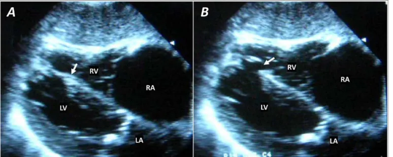
Figure 1. Interventricular septum rupture. The arrow indicates the ventricular septum defect. A. Closed in systole. B. Open in diastole. Caption: RV: right ventricle; RA: right atrium, LV: left ventricle; LA: left atrium.
encontró ningún informe similar a en las bases de datos bibliográficas consultadas.
Palabras clave: Infarto de miocardio; Rotura Cardíaca Postinfarto; Choque Cardiogé-nico
INTRODUCTION
Acute myocardial infarction (AMI) is a major health problem both in developed and developing countries. Currently, it affects more than 3,000,000 people each year1. Advances in preventive measures and diagnostic
and therapeutic techniques have reduced hospital mortality of AMI from 25 and 30% in the 1960s to 6% nowadays2.
The main causes of death in patients with AMI are cardiac arrhythmias and mechanical complications that lead to cardiogenic shock. The latter, in patients with AMI, cause a severe hemodynamic compromise with high mortality, even when the correct treatment is performed3.
The case of an elderly patient with AMI who received thrombolytic treatment with Cuban recom-binant streptokinase, and who developed a fatal car-diogenic shock after a rupture of the interventricular septum (IVS), is reported.
CASE REPORT
A male patient, 85 years old, with a history of appa-rent health, was admitted to the Intensive Care Unit
referred from the health area by presenting a severe oppressive retrosternal pain, radiating to the jaw, with a sudden onset while he was going to perform his daily activities. It lasted about an hour and a half, and was accompanied by profuse sweating and feeling of death; relieved with opioid administration.
A 12-lead electrocardiogram (ECG) was performed. It showed sinus rhythm, heart rate of 100 bpm; PR, 113 ms; QRS, 95 ms; QT 320 ms; QRS axis, -35°; pre-sence of Q waves in leads I, II, III, aVF, and from V2 to V6, with ST positive displacement from V2 to V6, and diffuse disorder of ventricular repolarization. The P wave in DII was 0.035 sec and 0.01 mV. An acute coronary syndrome with ST elevation was diagnosed, and thrombolysis with Cuban recombinant strepto-kinase was performed, according to the protocol of the service. There were no incidents during drug administration, except isolated premature ventricular beats. The ECG performed after thrombolysis showed no changes compared to the initial one, therefore, it was assumed that the treatment was not effective for the recanalization of the infarct-related artery.
Figure 2. Anatomical specimen of the heart. The arrow is at the site of the rupture of the septum.
symptoms characterized by malaise, increased heart and respiratory rates, and intense retrosternal pain. Severe hypotension (54/26 mmHg) and increased heart rate (135 bpm) were found. Physical examina-tion revealed intense holosystolic murmur at the left sternal border with no signs of venous congestion and absence of pulmonary rales; delayed capillary refill and temperature gradient in the mid-third of the extremities. Cardiogenic shock was diagnosed and, from a clinical standpoint, it was considered that the cause could be a chordal rupture of the mitral valve or a rupture of the IVS.
An echocardiography was performed which re-vealed, in subxiphoid view, right chambers dilatation and thinning of the IVS in the apical region, with an image cut at that level that was reminiscent of valve movement with opening and closing of the defect (Figure 1). The valvular apparatus showed degenera-tive changes (slight calcification) and seemed to be competent according to the pulsed Doppler. More-over, the absence of thrombus or pericardial effusion was established. Echocardiographic examination con-firmed the clinical diagnosis of interventricular septum rupture.
The patient died the following day. The autopsy showed a severe atherosis of the aorta and the coro-nary arteries, signs of acute myocardial infarction in the anterior region of the left ventricle, and a rupture of 1.5 cm2 in the apical region of the interventricular
septum (Figure 2).
COMMENT
IVS rupture occurs approximately in 1% of all patients with AMI and in 0.2% of patients receiving thrombo-lytic treatment; in the latter, it occurs in the early hours of the coronary accident, however, it is often seen later (between two to five days)4 in patients
where this strategy is not used. Its frequency is higher in women, especially at older ages and with anterior wall location.
In patients with anterior wall AMI, the rupture is observed in the anteroapical region, while in those with inferior wall AMI, the ventricular septum defect is observed in the basal region of IVS5.
The mortality of this complication is very high, and the surgical closure is the treatment of choice. The operative mortality of these patients is between 25%, in anterior wall acute myocardial infarction, and 58%, in inferior wall infarctions6. Survival without short
term surgery is exceptional; however, aspects of the treatment of ventricular septum defect closure are currently discussed, as well as the optimal time for surgery7. Some patients with severe hemodynamic
compromise and a large left to right shunt, benefit from the temporary transcutaneous closure by cathe-ter8 or by left ventricular assist devices, with the aim
of improving the patient's conditions for subsequent surgical repair8. Notwithstanding the above
men-tioned, there have been reports of spontaneous closures of ventricular septum defects that presented as complications of AMI 9.
Clinical diagnosis is made by detecting a murmur of recent onset that is intense, pansystolic, more audible in the lower left sternal area, with presence of thrill, and accompanied by a significant clinical deterioration, often with biventricular dysfunction. Hemodynamic manifestations in these patients are due to the acute overload of pressure in the heart’s right chambers, caused by the defect of the IVS. The sudden onset or worsening of cardiogenic shock, increasing dyspnea and signs of pulmonary and splanchnic engorgement have also been described10.
ECG traces taken at the time of the rupture of the IVS have shown increased heart rate, ST segment elevation, decrease in the depth of the Q wave and increase of the voltage and duration of the P wave11.
pa-CorSalud 2013 Jan-Mar;5(1):122-126 125 tients, as it can be performed immediately at the
bed-side, and has high sensitivity and specificity12.
Other imaging tests for functional and perfusion testing and multislice computed tomography are use-ful for diagnosing IVS rupture in patients with AMI13.
Similarly, the nuclear magnetic resonance studies have been used for the diagnosis and surgical repair of mechanical complications in patients with AMI 14.
Elevated levels of brain natriuretic peptide (BNP) have been documented in patients with AMI who have shown IVS rupture. This marker has been considered a predictor of left ventricular dysfunction, mechanical complications and death in patients with AMI15.
In spite of all the developments related to the diag-nosis and treatment of IVS rupture, its incidence and mortality remain high in patients suffering from AMI.
REFERENCES
1. Lloyd-Jones D, Adams R, Carnethon M, De Simone G, Ferguson TB, Flegal K, et al. Heart Disease and Stroke Statistics 2009 Update: A report from the American Heart Association Statistics Committee and Stroke Statistics Subcommittee. Circulation 2009;119:e21-181.
2. Van de Werf F, Bax J, Betriu A, Blomstrom-Lundqvist C, Crea F, Falk V, et al. Management of acute myocardial infarction in patients presenting with persistent ST-segment elevation: the Task Force on the Management of ST-Segment Elevation Acute Myocardial Infarction of the European Society of Cardiology. Eur Heart J 2008;29(23): 2909-45.
3. Birnbaum Y, Fishbein MC, Blanche C, Siegel RJ. Ventricular septal rupture after acute myocardial infarction. N Engl J Med. 2002;347(18):1426-32. 4. Crenshaw B, Granger C, Birnbaum Y, Pieper K,
Morris DC, Kleiman NS, et al. Risk factors, angio-graphic patterns, and outcomes in patients with ventricular septal defect complicating acute myo-cardial infarction. GUSTO-I (Global Utilization of Streptokinase and TPA for Occluded Coronary Arte-ries) trial investigators. Circulation. 2000:101(1):27-32.
5. Cummings RG, Reimer KA, Califf R, Hackel D, Boswick J, Lowe JE. Quantitative analysis of right and left ventricular infarction in the presence of postinfarction ventricular septal defect. Circulation. 1988;77(1):33-42.
6. Jones MT, Schofield PM, Dark JF, Moussalli H,
Deiraniya AK, Lawson RA, et al. Surgical repair of acquired ventricular septal defect. Determinants of early and late outcome. J Thorac Cardiovasc Surg. 1987;93(5):680-6.
7. Curcio A, Martín J, Wilhelmi M, Soria JL. Doble com-plicación postinfarto agudo de miocardio: rotura del septo interventricular e insuficiencia mitral aguda. Rev Esp Cardiol. 1997;50(2):129-32.
8. Gregoric ID, Bieniarz MC, Arora H, Frazier OH, Kar B, Loyalka P. Percutaneous ventricular assist device support in a patient with a postinfarction ventri-cular septal defect. Tex Heart Inst J. 2008;35(1):46-9.
9. Williams RI, Ramsey MW. Spontaneous closure of an acquired ventricular septal defect. Postgrad Med J. 2002;78(921):425-6.
10.Coskun KO, Coskun ST, Popov AF, Hinz J, Schmitto JD, Bockhorst K, et al. Experiences with surgical treatment of ventricle septal defect as a post in-farction complication. J Cardiothorac Surg. 2009; 4:3. Available at:
http://www.cardiothoracicsurgery.org/content/4/1 /3
11.Kerr F, Haywood LJ. Electrocardiographic changes produced by interventricular septal rupture. Br Heart J. 1976;38(10):1098-100.
12.Soriano CJ, Pérez-Boscá JL, Canovas S, Ridocci F, Federico P, Echanove I, et al. Septal rupture with right ventricular wall dissection after myocardial infarction. Cardiovasc Ultrasound. 2005;3:33. Avai-lable at:
http://www.cardiovascularultrasound.com/content /3/1/33
13.Ghersin E, Lessick J, Abadi S, Agmon Y, Adler Z, Engel A, et al. Ventricular septal rupture com-plicating myocardial infarction: comprehensive assessment of cardiac coronary arteries, anatomy, perfusion and function by multidetector computed tomography. Can J Cardiol. 2008;24(3):e21-2. 14.Vogel-Claussen J, Skrok J, Fishman EK, Lima JA,
Shah AS, Bluemke DA. Cardiac CT and MRI guide surgery in impending left ventricular rupture after acute myocardial infarction. J Cardiothorac Surg. 2009;4:42. Available at:
http://www.biomedcentral.com/content/pdf/1749 -8090-4-42.pdf
Ventricular Function, Early Outcome and