Short communication
Kinetics-like equation for cerebrospinal fluid-blood diffusion models
José Alejandro Rodríguez-Pérez1*
Alejandro Mirabal-Viel1
Vanessa Pérez-del-Vallín1
Christian Meijides-Mejías1
William Castillo-González1
Jairo Lumpuy-Castillo1
Alberto Juan Dorta-Contreras1
1Laboratorio Central de Líquido Cefalorraquídeo (LABCEL) Universidad de Ciencias
Médicas de La Habana, Cuba.
*Autor para la correspondencia: [email protected]
ABSTRACT
Introduction: The diffusion of proteins from the blood to the cerebrospinal fluid is
influenced by its molecular weight and by the intrinsic properties and biological
properties of the protein.
Methods: Paired samples of serum and cerebrospinal fluid were taken from normal
subjects to quantify albumin and proteins of the lectin pathway of the complement
system. The distribution of these with regard to the value of QAlbúmin = (Albumin in
serum / albumin in cerebrospinal fluid) was evaluated because this protein is used as a
marker of the passage of the barrier.
Results: It was observed that some of these describe a saturation pattern which resembles
the consideration of two constants that will help to characterize the behavior of these
proteins by spreading to the cerebrospinal fluid: the maximum Q of the protein, which is
the maximum proportion found empirically between the concentrations in blood and
cerebrospinal fluid and the value Kcdw which is the value of the average diffusion speed
of Q albumin when the semi-maximal value of the Q of the protein under study is
obtained.
Conclusions: Empirically obtained constants will help the characterization and
differentiation of the diffusion of these new proteins as they pass from the blood to the
cerebrospinal fluid.
Keywords: cerebrospinal fluid-blood; Q albumin; proteins.
Recibido: 30/12/2018 Aprobado: 04/02/2019
INTRODUCTION
Complement system includes three pathways: classical, alternative and lectin pathway.
The last one is present since from the invertebrates, but it is not yet well defined. It is still
discovering new structures and new evidences of its component’s functions.
So far there are five different forms of pattern recognition (PRM): mannose binding lectin
(MBL); H-, L- and M-ficolin (also known as Ficolin-3, -2 and -1, respectively); and
collectin K (CL-K1): an MBL (MASP) -1 and -2 are associated with the serine protease
to activate the complement. In addition, CL-K1 and collectin L 1 (CL-L1) are related to
heteromers that are also associated with MASPs and activate complement. (1)
These initiators are not capable of initiating the enzymatic cascade by themselves; it
Although MASP-2 is capable of self-activation, recent studies have shown that MASP-1
is the exclusive activator of MASP-2 under physiological conditions. (1,2) MASP3 and the
MAp44 are components with regulatory functions in this pathway.
These proteins are found in the highest percentage in the blood and they diffuse through
the blood-brain barrier, but they can also be synthesized in the central nervous system.
These proteins participate in enzymatic reactions in the lectin pathway, where a balance
is established between the activated and non-activated forms and between the soluble
forms and those associated with other surfaces.
When the variation of Q albumin as a measure of protein diffusion between blood and
CSF is plotted against the quotient diffusion of some of the lectin pathway, a typical
curved curve is observed that resembles the enzymatic reactions shape, such as
Michaelis-Menten.reaction.
The objective of this work is to evaluate the behavior of the lectin pathway component
MASP2, MASP3 and MAp44, based on their observed dynamics mimicking enzymatic
activity.
METHODS
MASP2 was quantified in 56 serum and CSF samples by the enzyme-linked
immunosorbent assay (ELISA) kits (Hycult Biotech, Uden, the Netherlands). (3)
Plasma levels of MASP3 and Map44 were measured using time-resolved
immunofluorometric in duplicates. All assays and specific antibodies for the assays were
produced in-house. Detailed description for each assay can be found in the reference. (4)
Albumin in serum and in CSF was quantified by nephelometric assay using Behring
Statistical package MedCalc version 13.3.3.0 and GraphPad Prism version 5.01 was used
to perform statistics and graphs.
RESULTS
The passage of proteins from the blood to the CSF is characterized from the values of Q
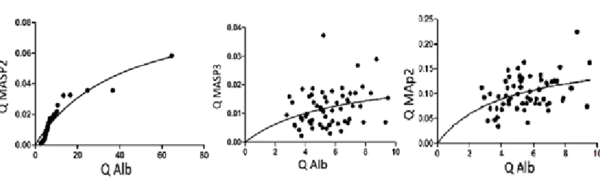
Albumin. The distribution of the QMASP2, QMASP3 and QMap44 are shown in Fig. 1.
Fig. 1- Variation of QMASP2, MASP3 and QMAp44 with Q albumin.
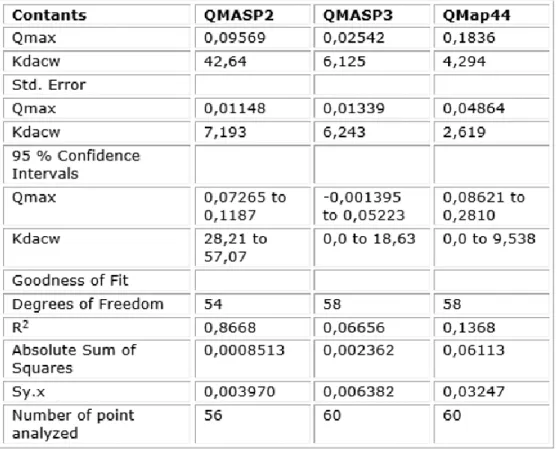
In the following table 1 you can see the new constants Qmax and Kdacw calculated from
Table 1- Kdacw and Q max of MSAP2. MASP3 and Map44
DISCUSSION
The passage of these components of lectin pathway from blood to CSF has allowed
characterize these proteins as blood derived with the possibility of being synthesized in
the CNS (1) because they follow the established statements for these types of proteins.
MASP2 has a behavior when it is distributed based on the speed of diffusion between the
blood and the CSF that resembles a reaction of the Michaelis-menten type. This favors
that this curve can be used to characterize this protein whose tracing resembles a reaction
of enzymatic activity. The same thing happens with the regulation complement molecules
MASP3 and MAp44.(5,6)
By this way it was possible to characterize the new constants Kdacw and Qmax for each
The Qmax is the maximum value experimentally obtained from the diffusion of the
protein when it passes from blood to CSF. It is the maximum relationship between these
proteins when they pass from blood to CSF as a function of its serum concentration. It is
the maximum diffusion speed between blood and CSF
The Kdacw is the Q value of the evaluated protein from the passage through the
blood-LCR barrier that corresponds to the half-maximal speed-It can be experimentally
obtained from Qmax / 2 and is a constant value depends on the molecule which is being
evaluated.
With these new constants, the passage of these proteins from the serum to the CSF can be
better defined and allows improving theoretical and practical analysis of this dynamics in
blood-CSF barrier.
REFERENCES
1. Pihl R, Jensenius JC, Thiel S. MASP-2. The Complement Facts Book. Academic Press
2018: p. 79-87.
2. Yaseen S, Demopulos G, Dudler T, Yabuki M, Wood C L, Cummings W J, Tjoelker L,
et al. Lectin pathway effector enzyme mannan-binding lectin-associated serine protease-2
can activate native complement C3 in absence of C4 and/or C2 . The FASEB Journal
2017 31:5,2210-2219
3. Dorta-Contreras A J, Padilla-Docal B, Iglesias González I M, Martínez-Larrarte J P,
Castillo-González W, González-Losada C, González-Argote J, et al. MAp44: diffusion
from blood to cerebrospinal fluid and intrathecal synthesis . The FASEB Journal 2016
30:1_supplement, 970.1-970.1
4. Degn SE, Jensen L, Gál P, Dobó J, Holmvad SH, Jensenius JC, et al. Biological
5. Dorta-Contreras A J, Padrón-González A A, González-Losada C, Lumpuy-Castillo J,
Rodriguez-Pérez J A, Ramos-Robledo A, Martínez-Reyes J, et al. MASP-3: a new
leptomeningeal protein in the lectin pathway. The FASEB Journal 2018
32:1_supplement, 741.5-741.5
6. Padrón-González AA, González-Losada C, Lumpuy-Castillo J, Rodriguez-Pérez J A,
Ramos-Robledo A, Castillo-González W, Dorta-Contreras A J. MASP-3 aggregation and
its blood to cerebrospinal fluid diffusion. The FASEB Journal 2018 32:1_supplement,