Pulmonary vascular clearance of harmful endogenous
macromolecules in a porcine model of acute liver failure
Geir I. Nedredal,* Kjetil Elvevold,† Marcio F. Chedid,‡ Lars M. Ytrebø,§
Christopher F. Rose,|| Sambit Sen,¶ Bård Smedsrød,† Rajiv Jalan,** Arthur Revhaug*
* Department of Digestive Surgery, University Hospital Northern Norway. † Vascular Biology Research Group, Department of Medical Biology, University of Tromsø. ‡ Liver and Pancreas Transplant and Hepatobiliary Surgery Unit, Hospital de Clínicas de Porto Alegre, Federal University of Rio Grande do Sul (UFRGS), Porto Alegre, Brazil. § Department of Anesthesia and Intensive Care, University Hospital Northern Norway. || Hepato-Neuro Laboratory, CRCHUM, Universite de Montreal, Canada. ¶ Department of Gastroenterology, Luton & Dunstable University Hospital, Luton, UK. ** Institute of Liver and Digestive Health, Royal Free Hospital, London, UK.
A B S T R A C T A B S T R A C T A B S T R A C T A B S T R A C T A B S T R A C T
Background. Background.Background. Background.
Background. Pulmonary complications are common in acute liver failure (ALF). The role of the lungs in the uptake of harmful solu-ble endogenous macromolecules was evaluated in a porcine model of ALF induced by hepatic devascularization (n = 8) vs. controls (n = 8). In additional experiments, pulmonary uptake was investigated in healthy pigs. Fluorochrome-labeled modified albumin (MA) was applied to investigate the cellular uptake. Results.Results.Results.Results. As compared to controls, the ALF group displayed a 4-fold net increasedResults. lung uptake of hyaluronan, and 5-fold net increased uptake of both tissue plasminogen activator and lysosomal enzymes. Anatomical distribution experiments in healthy animals revealed that radiolabeled MA uptake (taken up by the same receptor as hyaluronan) was 53% by the liver, and 24% by the lungs. The lung uptake of LPS was 14% whereas 60% remained in the blood. Both fluorescence and electron microscopy revealed initial uptake of MA by pulmonary endothelial cells (PECs) with later translocation to pulmonary in-travascular macrophages (PIMs). Moreover, the presence of PIMs was evident 10 min after injection. Systemic inflammatory mark-ers such as leukopenia and increased serum TNF-α levels were evident after 20 min in the MA and LPS groups. Conclusion.Conclusion.Conclusion.Conclusion.Conclusion. Significant lung uptake of harmful soluble macromolecules compensated for the defect liver scavenger function in the ALF-group. In-fusion of MA induced increased TNF-α serum levels and leukopenia, similar to the effect of the known inflammatory mediator LPS. These observations suggest a potential mechanism that may contribute to lung damage secondary to liver disease.
Key words. Key words.Key words. Key words.
Key words. Systemic inflammatory response syndrome. Hyaluronic acid. Liver sinusoidal endothelial cells. Pulmonary intravascu-lar macrophages.
May-June, Vol. 15 No. 3, 2016: 427-435
INTRODUCTION
Pulmonary complications in patients with liver related diseases are common.1 In acute liver failure (ALF), lung
complications may comprise acute lung injury and adult respiratory distress syndrome. Clinical and radiological evidence of pulmonary edema may occur in patients with liver failure.2 The pathophysiological mechanisms of lung
injury in ALF are not completely elucidated.3 Systemic
in-flammatory response syndrome (SIRS) and sepsis are asso-ciated with ALF4 with many factors contributing to the
risk of developing sepsis in ALF, such as impaired func-tion of circulating leukocytes and acute lung injury.1
Elevated blood levels of hyaluronic acid (HA) are re-ported in ALF.5 In ALF induced by paracetamol, one study
demonstrated elevated circulatory levels of HA in both survivors and non-survivors, and significantly greater lev-els among the non-survivors.6 Under physiologic
condi-tions, serum HA exists as a high-molecular weight substance but changes to a low molecular weight sub-stance in disease states.7 Low molecular weight HA is
known to elicit acute lung injury via Toll-like receptors-2
The Official Journal of the Mexican Association of Hepatology, the Latin-American Association for Study of the Liver and
the Canadian Association for the Study of the Liver
Manuscript received: Manuscript received:Manuscript received:
and 4 with subsequent MyD88 mediated inflammatory re-sponse.8
The highly endocytic liver sinusoidal endothelial cells (LSECs) which make up the liver sinusoidal walls take up colloids and soluble macromolecules from the blood, whereas Kupffer cells clear larger particles (> 200 nm) by phagocytosis.9 Chronic liver diseases induce
vas-cular clearance of particles by the lungs,10 which has been
attributed to pulmonary intravascular macrophages (PIMs).11 The existence of PIMs in healthy humans is
controversial and believed to be inducible in certain pathological conditions such as cirrhosis.12 During ALF
and SIRS a variety of inflammatory substances, including HA and lipopolysaccharide (LPS), are released.5 These
substances may play a role in the lung damage induced by liver failure.
The aim of this study was to assess the lung uptake of harmful soluble macromolecules in a porcine model of ALF. We also aimed to investigate the kinetics of cellular absorption and clearance of modified albumin (MA), a sol-uble probe that is taken up by the same receptor as HA,13
and LPS by the lungs.
MATERIAL AND METHODS
Materials
All animals received human care in accordance with the institution’s guidelines. Chemicals were obtained from Sigma-Aldrich (St. Louis, MO) unless otherwise stated. Modification of serum albumin was done with formalde-hyde as described.14 Modified Albumin (MA) (0.8 mg/mL)
was incubated with the fluorochrome FITC in a sodium carbonate buffer (0.5 mol/L, pH 9.5) in a protein/dye weight ratio of 5:1 at 4 °C overnight. Unbound fluoro-chrome was removed by dialysis against PBS without Ca2+
or Mg2+. E. coli lipopolysaccharide (serotype 0111; B4).
Monoclonal goat anti-mouse IgG, TRITC-conjugate, was obtained from Zymed, San Francisco, CA. Monoclonal rabbit anti-FITC was from DAKO A/S, Denmark. Mono-clonal rabbit anti-TRITC was from Molecular Probes Inc., OR. Anti-angiotensin antibody (RP183). Monodis-perse polymer particles (MDPPs) with a diameter of 2.0
μm were obtained from SINTEF, University of Trondhe-im, Norway.
Animal preparations
All animals (Sus scrofa domesticus) received care accord-ing to the criteria outlined in the Guide for the Care and Use of LaboratoryAnimals prepared by the National Academy of Sciences and published by the NIH (publication 86-23, revised 1985).
All animals were fasted overnight with free access to wa-ter. Anesthesia was induced by giving 10 mg/kg ketamine and 0.5 mg/kg atropine intramuscularly. Further anesthesia induction was performed via an ear vein intravenous bolus of 10 mg/kg pentobarbital sodium and 10 microg/kg fenta-nyl. All animals had a tracheotomy performed with subse-quent intubation and connection to a volume-controlled ventilator (Servo 900, Elema-Schönander, Stockholm, Swe-den). FiO2 was kept at 0.5. Tidal volumes were adjusted by means of repeated arterial blood gas analyses to maintain PaCO2 between 4.5 to 5.0 kPa during surgery. Ventilation was not altered after zero hours. A 16-G central venous catheter (Secalon T, Ohmeda, Swindon, UK) was intro-duced into the left external jugular vein. An arterial line was placed in the left carotid artery. A Swan-Ganz catheter (Bax-ter Healthcare, Irvine, CA) was inserted into the pulmonary artery via the right external jugular vein. Continuous total intravenous anesthesia was maintained with 4 mg/kg/h pentobarbital sodium, 0.02 mg/kg/h fentanyl, and 0.3 mg/kg/ h midazolam. The depth of the anesthesia was assessed by corneal reflex and heart rate. The thermo dilution method was applied for measuring cardiac output, where 5 mL of cold (< 5 °C) normal saline was injected (Com-1, Ameri-can Edwards Laboratories, Santa Ana, CA). Cardiac output was measured in triplicate and the results expressed as the mean value. A single investigator made all measurements over a period of < 4 s at end-expiration. Heating pads and blankets were used to keep normal body temperature (38.5 ± 1 °C). All animals were euthanized with an overdose of intravenous pentobarbital sodium and potassium chloride.
Study outline 1 – Lung mediated clearance of blood borne macromolecules
Sixteen pigs weighing between 23 to 30 kg were ran-domly allocated into an ALF group (n = 8) or a sham-op-erated control group (n = 8) (Figure 1). ALF was induced by ligation of the hepatic arteries and the proximal portal vein. Following transection of the distal portal vein, the distal end of the portal vein was anastomosed end-to-side to the vena cava as described earlier.15 The controls
under-went sham surgery: laparotomy and mobilization of the portal vein. Blood samples were drawn from the tip of the Swan-Ganz catheter within the pulmonary artery, and the arterial line placed in the left carotid artery. The samples were analyzed for soluble endogenous macromolecules such as HA, tissue plasminogen activator (t-PA) and lyso-somal enzymes (acid phosphatase, hexosaminidase, acid li-pase and aryl sulfatase). Net lung uptake was calculated by subtracting the venous concentration from the arterial concentration and multiplying with the cardiac output:
Study outline 2 – Lung mediated clearance of intravenously administered macromolecules
and particles in healthy subjects
Thirteen pigs were randomly allocated into 3 groups that were injected with either MA (n = 5) or LPS (n = 4), or a control group that did not receive any macromolecule (n = 4) (Figure 1).
• Distribution of macromolecules in healthy ani-mals. Organ distribution of intravenously adminis-tered radiolabeled trace amounts of MA and LPS were determined as follows. Approximately 100 x 106 cpm 125I-MA, or 125I-LPS were injected into the left jugular
vein. Blood samples were collected in 2 mL aliquots from an intravenous catheter positioned in the right jugular vein with its tip in the right atrium. The organs were dissected out and weighed after one hour. Radio-activity was measured in a γ-counter (Cobra II, Pack-ard Instrument Co., CT).
• Investigation of lung uptake. Based on dose-re-sponse curves and matching mean pulmonary artery pressure profiles, the concentrations of 3 mg/kg MA and 5 microg/kg LPS were chosen. An MA dose > 3 mg/kg triggered a lethal cor pulmonale. Both MA and LPS were diluted in normal saline, and given as an infusion over 30 sec via the left external jugular vein. A median sternotomy was performed prior to infusion to gain ac-cess to the lung parenchyma. Thirty mg (approximately 10 billion) of an intravenous bolus of TRITC-labeled monodisperse polymer particles (MDPPs) were ad-ministered to saturate the PIM uptake.
Immunocytochemistry
To examine the cellular localization of intravenously infused FITC-labeled MA, multiple small lung biopsies
were taken prior to, and after the infusion for fluores-cence microscopy and immuno-transmission electron microscopy. Fluorescence microscopy biopsies were fixed in 4% paraformaldehyde and embedded in paraffin-blocks. Transmission electron microscopy biopsies were prepared as described.16 Briefly, small cubes were cut out
of the lung tissue, fixed in McDowell’s fixative and incu-bated in 2.3 mol/l sucrose overnight before immersion in liquid nitrogen on aluminum pins. Prior to immunolabe-ling, sections were retrieved in sucrose and mounted on carbon-coated grids. Sections were blocked for 15 min in 1% cold water fish skin gelatin, followed by incubation with anti-TRITC antibodies (diluted in cold water fish skin gelatin), washing in PBS and incubation with protein A-gold (University of Utrecht, The Netherlands)17
dilut-ed in cold water fish skin gelatin for 15 min. The sections were examined in a JEOL JEM 1010 transmission electron microscope (Tokyo, Japan) at 80 kV.
Assays
Plasma HA concentrations were measured with a radio-immunometric assay (Pharmacia Diagnostics AB, Swe-den). Lysosomal enzyme assays (acid phosphatase, hexosaminidase, acid lipase and aryl sulfatase) were per-formed as described.18 Enzyme activity, expressed in U,
was expressed as the number of substrate molecules trans-formed (1U = microM product transtrans-formed per microl serum/h).
Statistics
Two-way (time and group) analysis of variance for re-peated measurements was applied to test whether any sta-tistical significance existed between the groups followed by the LSD post hoc comparison test. An overall signifi-cance in analyses of variance for repeated measurements
Figure 1. Figure 1.Figure 1.
Figure 1.Figure 1. Outline 1 shows a large ani-mal model of ALF (n = 8) and controls (n = 8). ALF was induced by hepatic arterial devascularization and creation of end-to-side portacaval anastomosis. Controls underwent only laparotomy and mobilization of the portal vein. Outline 2 shows intravenous injections of the sol-uble macromolecules MA (n = 5), the partially insoluble LPS (n = 4), and controls (n = 4). ALF: acute liver failure. MA: modified albumin. LPS: lipopolysac-charide.
Outline 1 Outline 1 Outline 1 Outline 1 Outline 1
Laparotomy
0 h 6 h
ALF
Control
Outline 2 Outline 2 Outline 2 Outline 2 Outline 2
Clearance kinetics & cellular uptake
0 h 1 h
MA
LPS
Figure 2. Figure 2. Figure 2. Figure 2.
Figure 2. Lung clearance of soluble endogenous macromolecule in ALF. Lung uptake was given by the aortic concentrations subtracted by the pulmonary ar-tery concentrations, and multiplied with the cardiac output. A positive value denoted uptake of circulating soluble macromolecule by the lungs, while a negative value denoted secretion. In ALF, note the initial large clearance by the lungs (2 to 4 h), and thereafter reduction of uptake indicating a saturation of the lung uptake. A.A.A.A.A. Hyaluronan. B.B.B.B.B. t-PA. C.C.C. Hexosaminidase. D.C.C. D.D.D.D. Aryl sulfatase. E.E.E.E.E. Acid phosphatase, and F.F.F.F.F. Acid lipase. ALF: acute liver failure. HA: hyaluronan. t-PA: tissue plasminogen activator.
0 h 2 h 4 h 6h 0 h 2 h 4 h 6h
A. A. A. A.
A. Hyaluronan B. B. B. B. B. t-PA
ALF Control
g/min
4
3
2
1
0
-1
C. C. C. C.
C. Hexosaminidase D.D.D.D.D. Aryl sulfatase
8
6
4
2
0
-2
0 h 2 h 4 h 6h
E. E. E. E.
E. Acid phosphatase F.F.F.F.F. Acid lipase
ng/min
U/min
3 2 1 0 -1
0 h 2 h 4 h 6h
U/min
4 3 2 1 0 -1
U/min (x10
–2)
0 h 2 h 4 h 6h
0 h 2 h 4 h 6h
U/min (x10
–4)
ALF Control
ALF Control
ALF Control
ALF Control
ALF Control 8
6 4 2 0 -2 -4 -6 4
3
2
1
0
(F-test P < 0.05) may be attributable to either the effect of time (PT) or the interaction of group and time (PGT). PGT < 0.05 denoted a significant difference between the groups dependent of time, while PG < 0.05 denoted a sig-nificant difference between the groups independent of time. If both PGT and PG were significant, then only PGT was denoted since PGT is more robust than PG. PT<0.05 denoted a significant change in time for the groups. We used the SPSS statistical package (Chicago, Il).
RESULTS
Study outline 1
Uptake of endogenous HA across the lungs in the ALF group was significantly greater than the control group (Figure 2A). The HA lung uptake rate 2 h after ALF induc-tion was over 30-fold greater compared with the control group (PGT < 0.05) with a subsequent 10-fold decrease 6 h after ALF onset compared with the control group (PGT < 0.05). No significant increase in rate of uptake after the different time intervals was observed in the control group. This suggests an increase and a subsequent saturation of lung uptake in ALF. This notion is strengthened by the over 10-fold increase in HA concentrations in the pulmo-nary artery in the ALF group, an increase from 0.53 ± 0.43 mg/L at 0 h to 5.60 ± 0.42 mg/L at 6 h (PGT < 0.05). No significant increase was observed in the control group; 0.20 ± 0.57 mg/L at 0 h and 0.83 ± 0.32 mg/L at 6 h.
As with HA, t-PA uptake across the lung also increased 30-fold after 2 h (PGT < 0.05) (Figure 2B) that subsequently decreased throughout the experimental period. After ALF induction, there was a 5-fold increase in t-PA concentra-tions in the pulmonary artery at 6 h (PGT < 0.05). No sig-nificant increase was observed in the control group at 6 h.
There was also a 30-fold increase in t-PA lung flux after 2 h (PGT < 0.05) (Figure 2B). This uptake decreased throughout the experimental period. After ALF induction, there was a 5-fold increase in t-PA concentrations in the pulmonary artery, from 1.52 ± 0.20 ng/mL at 0 h to 7.93 ± 0.59 ng/mL at 6 h whereas no significant increase was ob-served in the control group; from 0.53 ± 0.12 ng/mL at 0 h to 0.72 ± 0.06 ng/mL at 6 h (PGT < 0.05).
Lysosomal enzymes were also taken up by the lungs in the ALF group. The greatest rate of uptake was observed for hexosaminidase, 7-fold more than the control group (PGT < 0.05) (Figure 2C). The uptake of aryl sulfatase in-creased gradually (PGT < 0.05) (Figure 2D), while acid phosphatase had a 3-fold greater uptake than the control group (PGT < 0.05) (Figure 2E). Acid lipase showed a continuous lung uptake with no indication of saturation in the ALF group compared with the control group (PGT < 0.05) (Figure 2F).
Study outline 2
• Clearance and distribution of macromolecules in healthy animals. The anatomical distributions of trace amounts of radiolabeled MA (a soluble probe that is taken up by the same receptor as HA13), and LPS
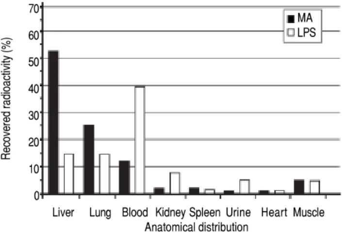
were investigated in 13 healthy animals: MA group (n = 5), LPS group (n = 4), and control group (n = 4) (Figure 1). Sixty minutes after injection, the organs were analyzed for anatomical distribution of radioactivity (Figure 3). The liver was the main site of uptake for MA (52%), while LPS uptake was similar in both the lungs and the liver (15%). Moreover, more than 90% of the in-jected doses were recovered in the organs listed. • Investigation of cellular uptake in the lungs.
PECs endocytosed detectable amounts of MA already 10 min after intravenous injection (Figure 4). After 10 min, note the presence of PIMs, with subsequent translocation of the green fluorescence from PECs to PIMs. Particulate matter (MDPP, 2 microns in diame-ter) was phagocytosed by PIMs (Figure 4). FITC-MA and TRITC-MDPP colocalized in PIMs after 60 min. The same pattern of translocation from PECs to PIMs was confirmed with electron microscopy (Figure 5), which also showed that PECs containing MA was co-labeled with the endothelial marker angiotensin con-verting enzyme (Figure 5A). Moreover, after 10 min MA translocated and aggregated in phagolysosomes of the PIMs (Figures 5B and 5C).
Figure 3. Figure 3.Figure 3.
Figure 3.Figure 3. Clearance and anatomical distribution of intravenously adminis-tered macromolecules in healthy animals. Trace amounts of radiolabeled MA, and partially insoluble LPS were injected into the left jugular vein. Or-gans were analyzed for anatomical distribution 60 min after injection. Note that almost 40% of LPS remained in the blood after 60 min. More than 90% of the injected doses were recovered in the organs listed. Results are ex-pressed as percent total radioactivity recovered. Organs that were analyzed with no uptake and therefore not included in the figure, were aorta, thyroi-dea, pancreas, thymus, stomach, duodenum, lymph nodes, pancreas, and colon. MA: modified albumin. LPS: lipopolysaccharide.
70 60 50 40 30 20 10 0
Recovered radioactivity (%)
Liver Lung Blood Kidney Spleen Urine Heart Muscle Anatomical distribution
Figure 4. Figure 4. Figure 4. Figure 4.
• Systemic inflammatory markers. Serum TNF-α was detectable (4.0 ± 2.7 pg/mL) 40 min after MA injection, i.e. 20 min after the translocation of MA from PECs to PIMs. After 60 min, TNF-α levels in-creased to 32.0 ± 7.3 pg/mL (PGT < 0.05). Even greater TNF-α levels were measured after injection of LPS; baseline TNF-α secretion was 0.0 ± 0.0 pg/ mL and after 60 min 411.8 ± 70.1 pg/mL (PGT < 0.05). In the control group, the TNF-α was undetec-table after 60 min.
After 60 min, the white blood cell count (WBC x 1,000/ mm3) decreased from 18.6 to 8.3 in the LPS group,
consistent with a leukopenia (PGT < 0.05). The same pattern was observed in the MA group, where WBC decreased from 19.1 to 14.1 (PGT < 0.05). In the con-trol group, the WBC remained relatively unchanged from 18.1 to 19.7 after 60 min (PGT > 0.05).
Potential interfering factors associated with Study outline 2
No serum TNF-α secretion was observed after a 3 mg/kg bolus of bovine serum albumin (i.e. no modification of albumin) administration, suggesting no cross-reaction be-tween species. The blood drawn in the MA-group showed no pyrogenes as demonstrated with a limulus amebocyte lysate assay. Blood cultures after 60 min from Study outline 2 showed no bacterial growth.
DISCUSSION
Significance of
lung uptake of soluble macromolecule in ALF
In healthy subjects, the soluble macromolecules inves-tigated in this study are known to be endocytosed by the LSECs.9 It has been shown that serum levels of HA are
significantly greater in paracetamol induced ALF among non-survivors vs. survivors.6 The soluble macromolecule
t-PA is a major determinant of fibrinolytic activity in blood, cleaving plasminogen into plasmin and causing degradation of fibrin in blood clots, and potentially con-tributing to the bleeding diathesis observed in ALF. In-creased serum levels of lysosomal enzymes in ALF, due to either decreased LSEC scavenger capacity, increased re-lease, or a combination may add to the complications of ALF such as acute lung injury, renal failure and myocardial dysfunction.19 Moreover, a study revealed that ALF caused
by paracetamol overdose, non-survivors had 10-fold great-er levels of lysosomal enzymes in the blood compared with survivors.20
Chronic liver disease, on the other hand, is known to induce vascular clearance of particles by the lungs.10
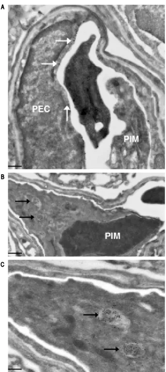
Figure 5. Figure 5.Figure 5.
Figure 5.Figure 5. Ultrastructural studies in lung cells following intravenous admin-istration of macromolecule. Immuno-gold staining visualized both large gold particles (MA, horizontal arrows) and small gold particles (anti-angiotensin converting enzyme, vertical arrow) labeling the surface of PEC (< 10 min) (AAAAA). Of note, there was no accumulation of MA in the phagolysosomes of the PIM, and the PIM was not labeled with anti-angiotensin converting en-zyme. There is a blood clot within the capillary. Beyond 10 min, MA howev-er accumulated in the phagolysosomes of PIMs indicated by arrows (BBBBB). Greater magnification of a segment of B showed accumulation of MA within phagolysosomes more clearly (CCCCC). Scale bars. A.A.A.A.A. 2 microm. B.B.B.B.B. 1 microm. C.
C.C.
C.C. 500 nm. MA: modified albumin. PECs: pulmonary capillary endothelial cells. PIM: pulmonary intravascular macrophage.
A A A A A
B B B B B
However, there is a paucity of data on lung clearance in ALF. One study showed that the lungs took up radi-olabeled E. coli permanently in a rodent ALF model with 90% hepatic resection.21 Chronic liver failure
studies in rodents have shown increased lung uptake of lysosomal enzymes,22 radiolabeled E. coli.,23 and
LPS.24 This is in line with our results where the lungs
compensate for the loss of liver scavenger function by LSECs.
Proinflammatory effects of soluble macromolecules
Our study revealed that MA, which is a soluble probe that is taken up by the same receptor as HA,13 can
elicit a similar inflammatory response as to the one ob-served with LPS. PIMs are known to secrete TNF-α upon stimulation, while depletion of PIMs reduce TNF-α secretion.25 Minute amounts of LPS are potent
triggers of an inflammatory response, hence the 12-fold greater TNF-α release with LPS than with MA after 60 min (infused LPS amount was 600-fold less than the MA amount). This reflects normal homeostasis, where minute amounts of pro-inflammatory endogenous mol-ecules in most mammals would be lethal. The concen-trations of low molecular weight HA required to induce Toll-like receptor-4 mediated signaling in den-dritic cells are several orders of magnitude greater than for LPS.26 A study performed showed a 1/3 increase of
TNF-α secretion in the systemic circulation with pro-inflammatory heparan sulfate stimulation as compared with LPS stimulation in mice.27
Our findings suggest a role for PECs in vascular clear-ance of soluble macromolecules. Furthermore, our re-sults showed that LPS is cleared both by the liver and lungs with the greatest fraction remaining in the blood most likely bound to LPS-binding proteins.28
CONCLUSION
In conclusion, this study shows for the first time that lung uptake of harmful soluble macromolecules occurs in ALF. The significant lung uptake of harmful soluble macromolecules compensated for the defect liver scav-enger function in the ALF-group. Our findings suggest a role for PECs in vascular clearance of soluble macro-molecules. Moreover, PECs may induce an inflammato-ry response, similar to the effect of the known inflammatory mediator LPS. These observations suggest a potential mechanism that may contribute to lung dam-age secondary to liver disease.
ABBREVIATIONS
• ALF: acute liver failure. • HA: hyaluronic acid. • LPS: lipopolysaccharide.
• LSECs: liver sinusoidal endothelial cells. • MA: modified albumin.
• MDPP: monodisperse polymer particles. • PECs: pulmonary endothelial cells.
• PIMs: pulmonary intravascular macrophages. • SIRS: systemic inflammatory response syndrome.
CONFLICTS OF INTEREST
None.
AWARD
This manuscript reports on the work that received a Travel Award for the Oral Presentation “Uptake of Solu-ble Macromolecules by Pulmonary Capillary Endothelial Cells - Implications for Lung Complications in Liver Disease” performed at the 18th International Liver Trans-plantation Society (2012 Travel Award - ILTS - San Fran-cisco CA).
FINANCIAL SUPPORT
Supported by the Norwegian Research Council.
AUTHORSHIP
GIN: participated in study conception and design, ex-periment performances, acquisition of the research data, analysis and interpretation of data and writing of the man-uscript. KE and MFC participated in study conception and design, analysis and interpretation of data and writing of the manuscript; LMY, CFR, SS, BS, RJ, and AR par-ticipated in study conception and design, analysis and in-terpretation of data, and critical revision of the manuscript.
ACKNOWLEDGEMENT
We express our gratitude to Ellinor Hareide and Victo-ria Nilsen for excellent analytical service, Hege Hagerup for technical assistance, and Dr. Tom Wilsgaard for statis-tical advice.
REFERENCES
1. Bernal W, Wendon J. Acute liver failure. N Engl J Med
2. Trewby P, Warren NR, Contini S, Crosbie WA, Wilkinson SP, Laws JW, Williams R. (1978). Incidence and pathophysiology of pulmonary edema in fulminant hepatic failure. Gastroen-terology 1978: 74: 859-65.
3. Audimoolam VK, McPhail MJ, Wendon JA, Willars C, Bernal
W, Desai SR, Auzinger G. Lung injury and its prognostic sig-nificance in acute liver failure. Crit Care Med 2014: 42: 592-600. doi: 10.1097/01.ccm.0000435666.15070.d5.
4. Rolando N, Wade J, Davalos M, Wendon J, Philpott-Howard
J, Williams R. The systemic inflammatory response syn-drome in acute liver failure. Hepatology 2000; 32: 734-9. doi: 10.1053/jhep.2000.17687.
5. Bramley PN, Rathbone BJ, Forbes MA, Cooper EH,
Los-owsky MS. Serum hyaluronate as a marker of hepatic derangement in acute liver damage. J Hepatol 1991; 13: 8-13.
6. Williams AM, Langley PG, Osei-Hwediah J, Wendon JA,
Hughes RD. Hyaluronic acid and endothelial damage due to paracetamol-induced hepatotoxicity. Liver Int 2003; 23: 110-5.
7. McKee CM, Penno MB, Cowman M, Burdick MD, Strieter RM,
Bao C, Noble PW. Hyaluronan (HA) fragments induce chem-okine gene expression in alveolar macrophages. The role of HA size and CD44. J Clin Invest 1996; 98: 2403-13. doi: 10.1172/JCI119054.
8. Jiang D, Liang J, Fan J, Yu S, Chen S, Luo Y, Prestwich GD,
et al. Regulation of lung injury and repair by Toll-like receptors and hyaluronan. Nat Med 2005; 11: 1173-9.
9. Sorensen KK, McCourt P, Berg T, Crossley C, Le Couteur D,
Wake K, Smedsrod B. The scavenger endothelial cell: a new player in homeostasis and immunity. Am J Physiol Regul Integr Comp Physiol 2012; 303: R1217-R1230.
10. Keyes JW, Jr., Wilson GA, Quinonest JD. An evaluation of lung uptake of colloid during liver imaging. J Nucl Med 1973; 14: 687-91.
11. Brain JD, Molina RM, DeCamp MM, Warner AE. Pulmonary in-travascular macrophages: their contribution to the mononu-clear phagocyte system in 13 species. Am J Physiol 1999; 276: L146-L154.
12. Schneberger D, Aharonson-Raz K, Singh B. Pulmonary intra-vascular macrophages and lung health: what are we
miss-ing? Am J Physiol Lung Cell Mol Physiol 2012; 302:
L498-L503. doi: 10.1152/ajplung.00322.2011.
13. McCourt PA, Smedsrod BH, Melkko J, Johansson S. Charac-terization of a hyaluronan receptor on rat sinusoidal liver en-dothelial cells and its functional relationship to scavenger
receptors. Hepatology 1999: 30: 1276-86. doi: 10.1002/
hep.510300521.
14. Mego JL, McQueen JD. The Uptake and Degradation of In-jected Labeled Proteins by Mouse-Liver Particles. Biochim Biophys Acta 1965; 100: 136-43.
15. Ytrebo LM, Nedredal GI, Langbakk B, Revhaug A. An experi-mental large animal model for the assessment of bioartificial
liver support systems in fulminant hepatic failure. Scand J Gastroenterol 2002; 37: 1077-88.
16. Tokuyasu KT. Application of cryoultramicrotomy to immuno-cytochemistry. J Microsc 1986; 143: 139-49.
17. Slot JW, Geuze HJ. Sizing of protein A-colloidal gold probes for immunoelectron microscopy. J Cell Biol 1981: 90: 533-6. 18. Elvevold KH, Nedredal GI, Revhaug A, Smedsrod B. Scaven-ger properties of cultivated pig liver endothelial cells. Comp Hepatol 2004; 3: 4. doi: 10.1186/1476-5926-3-4.
19. Lefer AM. The role of lysosomes in circulatory shock. Life Sci 1976: 19: 1803-9.
20. Gove CD, Wardle EN, Williams R. Circulating lysosomal en-zymes and acute hepatic necrosis. J Clin Pathol 1981; 34: 13-6.
21. Wang X, Andersson R, Ding J, Norgren L, Bengmark S. Re-ticuloendothelial system function following acute liver failure induced by 90% hepatectomy in the rat. HPB Surg 1993; 6: 151-62.
22. Holmberg JT, Bergqvist L, Hultberg B, Hagerstrand I, Ihse I, Ryden S. Radiolabelled colloid uptake distribution and pulmo-nary contents and localization of lysosomal enzymes in cholestatic rats. Scand J Gastroenterol 1986; 21: 291-9. 23. Katz S, Grosfeld JL, Gross K, Plager DA, Ross D, Rosenthal
RS, Hull M, et al. Impaired bacterial clearance and trapping in obstructive jaundice. Ann Surg 1984; 199: 14-20.
24. Chang SW, Ohara N. Chronic biliary obstruction induces pul-monary intravascular phagocytosis and endotoxin sensitivity in rats. J Clin Invest 1994; 94: 2009-19.
25. Singh B, Pearce JW, Gamage LN, Janardhan K, Caldwell S. Depletion of pulmonary intravascular macrophages inhibits acute lung inflammation. Am J Physiol Lung Cell Mol Physiol 2004; 286: L363-L372.
26. Termeer C, Benedix F, Sleeman J, Fieber C, Voith U, Ahrens T, Miyake K, et al. Oligosaccharides of Hyaluronan activate dendritic cells via toll-like receptor 4. J Exp Med 2002; 195: 99-111.
27. Johnson GB, Brunn GJ, Platt JL. Cutting edge: an endog-enous pathway to systemic inflammatory response syn-drome (SIRS)-like reactions through Toll-like receptor 4. J Immunol 2004; 172: 20-4.
28. Schumann RR, Leong SR, Flaggs GW, Gray PW, Wright SD, Mathison JC, Tobias PS, et al. Structure and function of lipopol-ysaccharide binding protein. Science 1990; 249: 1429-31.
Correspondence and reprint request: Geir I. Nedredal, M.D., PhD.
Division of Transplantation, Department of Surgery, University of California, San Francisco Medical Center.
505 Parnassus Ave, San Francisco, CA 94143. USA. Tel.: +1 415 734-6355