Role of skeletal muscle proteoglycans during myogenesis
9
0
0
Texto completo
(2) 290. E. Brandan, J. Gutierrez / Matrix Biology 32 (2013) 289–297. HSPGs (Anastasi et al., 1997; Deakin and Lyon, 1999). Studies carried out during skeletal muscle differentiation indicate that the four syndecans are downregulated (Larrain et al., 1997a,b; Fuentealba et al., 1999; Gutierrez et al., 2006). Since FGF-2 and HGF are stimulators of skeletal muscle cell proliferation and strong inhibitors of muscle differentiation, membrane bound syndecans and glypican-1 potentially play unique, but pivotal, roles in muscle cell physiology. HSPGs also play a critical role during skeletal muscle regeneration, a highly complex and regulated process that involves muscle precursor proliferation, migration and differentiation. Furthermore, the different localisations of the HSPGs, give them another level of complexity, besides their localisation in the ECM and plasma membrane, in this later compartment they also present a special distribution which determine some of their functions.. activation and differentiation (Cornelison et al., 2001; Casar et al., 2004a,b; Cornelison et al., 2004). Myoblasts with inhibited syndecan-3 expression grafted into regenerating muscle had a normal proliferation rate but an impaired capacity to fuse and form skeletal muscle fibres (Casar et al., 2004a,b). It has been demonstrated that in the absence of syndecan-3, there is increased cell death, delayed onset of differentiation, and markedly reduced numbers of Pax7(+) satellite cells. Interestingly, syndecan-3-null satellite cell is rescued by ectopic expression of the constitutively active Notch intracellular domain and is required for Notch processing by ADAM17/tumour necrosis factor-alphaconverting enzyme (TACE) and signal transduction, supporting the idea that syndecan-3 and Notch cooperate in regulating the homeostasis of the satellite cell population and myofibre size (Pisconti et al., 2010).. 2.1.1. Syndecan-1 In human skeletal formation RNA transcript for syndecan-1 peaked at week 13 of gestation, after which a significant decrease was observed. However, perlecan expression levels were undetectable (Sogos et al., 1998). Similar results were found during the formation of mouse limb muscles. Syndecan-1 is expressed at day 10.5 and decreases after day 14.5, whereas perlecan increases their expression at day 12.5 of development (Olguin and Brandan, 2001). In the turkey pectoralis major muscle the peak of syndecan-1 expression is early and reached between embryonic days 18 and 20, previous than glypican (Liu et al., 2004). Sindecan-1 and -3 are critical modulators of FGF-2 biological activity (Larrain et al., 1998; Fuentealba et al., 1999). Since FGF-2 is a potent inhibitor of skeletal muscle differentiation (Brunetti and Goldfine, 1990), the downregulation of both syndecans (Fig. 1) seems to be a key step required to silence the muscle inhibitory signal mediated by FGF-2, allowing by this way the skeletal muscle differentiation (Larrain et al., 1997a,b; Larrain et al., 1998; Fuentealba et al., 1999). Turkey derived myogenic satellite cells overexpressing syndecan-1, maintain a rounded morphology and are unable to fuse to form multinucleated myotubes after differentiation is triggered (Velleman et al., 2004).. 2.1.4. Syndecan-4 During the formation of mouse limb muscles syndecan-4 increase their expression at day 12.5, peaked at day 13.5 of development and down regulated thereafter (Olguin and Brandan, 2001). Syndecan-4 presence in adult skeletal muscle fibres is also restricted to satellite cells, as mentioned for syndecan-3, and in vitro studies show that its levels are downregulated during the muscle differentiation (Gutierrez et al., 2006). Interestingly, syndecan-4 expression is regulated by electrical activity, influencing the adhesion properties of skeletal myotubes during differentiation (Ugarte et al., 2010). Syndecan-4 associates with focal adhesions in adherent cells, and it has been described as a marker of satellite cells (Cornelison et al., 2001; Ugarte et al., 2010). Syndecan-4-null mice have no apparent defects, however fail to regenerate suggesting a unique requirement for syndecan-4 in satellite cell function during muscle regeneration (Cornelison et al., 2001). Recently it has been described that in satellite cells, syndecan-4 forms a complex with Frizzled-7 (Fzd7), a non-canonical Wnt receptor, and the ECM protein fibronectin facilitating Wnt-7a signalling which is required for the symmetric expansion of the satellite cell pool required for regenerative myogenesis (Bentzinger et al., 2013).. 2.1.2. Syndecan-2 Much less attention has been given to evaluate the expression of syndecan-2 in skeletal muscle. One study performed to evaluate the expression of syndecan-2 during the mouse embryo development, indicates that this occurred almost exclusively on mesenchymal cells, which would derivate mainly in connective tissue cells. No syndecan-2 was detected in epithelia, neural or muscle cells (David et al., 1993). More recent in vitro studies of skeletal muscle differentiation, indicate that the expression of syndecan-2 is downregulated, similar to syndecan-1 and syndecan-3, suggesting common but not necessarily overlapping functions during this process (Fig. 1) (Larrain et al., 1997a,b; Fuentealba et al., 1999; Gutierrez et al., 2006). Studies focused to evaluate the role of syndecan-2 in skeletal muscle are required. 2.1.3. Syndecan-3 During the development of mouse limb muscles, syndecan-3 increases their expression at day 12.5 of development (Olguin and Brandan, 2001). Its expression is temporally and spatially coincident with the expression of myogenin, an essential muscle regulatory factor (Rudnicki and Jaenisch, 1995). In the adult skeletal muscle fibres, the expression of syndecan-3 is restricted to the satellite cells (Cornelison et al., 2001) and as mentioned in myoblast its expression after inducing skeletal muscle differentiation decrease quite rapidly (Fig. 1) (Fuentealba et al., 1999). In vitro evidences indicate that in the absence of syndecan-3 the muscle differentiation process is enhanced due to a diminished sensibility to FGF-2 (Fuentealba et al., 1999). In vivo its absence is associated with a dystrophic–fibrotic muscle phenotype characterised by impaired locomotion, fibrosis, and hyperplasia of myonuclei and satellite cells, indicating a key role for this HSPG in satellite cell maintenance,. 2.1.5. Glypican-1 Glypican-1 has been observed in skeletal muscle during late embryonic and early postnatal stages, but absent in developing heart, lung liver, dermis or vascular endothelium at the stages studied (Litwack et al., 1998). During the mouse limb skeletal muscle development the expression of glypican is detected in day 10.5 then increases its levels at day 12.5 keeping unchanged throughout the evaluation period, in contrast to the mentioned changes in syndecan-1 and -3 expression (Olguin and Brandan, 2001). Myoblast induced to differentiate in vitro, recapitulates this in vivo observations, the expression of the syndecans is down regulated meanwhile the levels of glypican remain unchanged (Fig. 1) (Campos et al., 1993; Brandan et al., 1996; Gutierrez et al., 2006; Gutierrez and Brandan, 2010). The glypican-1null mice do not present an obvious muscle phenotype (Jen et al., 2009) and deeper analysis in these mice is required. As mentioned in the syndecan-4-null mice the affected muscle phenotype become evident during the muscle regeneration after an acute injury (Cornelison et al., 2001). Besides the different expression patterns of glypican and syndecans, they also show another important difference, glypican-1 is the only HSPG localised in lipid raft domains in myoblasts, whereas the syndecans are located in non-raft domains (Brandan et al., 1996; Gutierrez and Brandan, 2010). Glypican-1 in the lipid raft domains sequesters FGF-2 away from its receptors (FGFR-I and FGFR-IV) which are located in non-raft domains (Gutierrez and Brandan, 2010) acting like a FGF-2 signalling inhibitor. Therefore, the receptors localise and interact with the syndecans in the same membrane domain where the FGF-2 signalling ternary complex occurs (Fig. 2). Myoblasts that do not express glypican-1 exhibit defective differentiation, with an increased.
(3) E. Brandan, J. Gutierrez / Matrix Biology 32 (2013) 289–297. 291. Table 1 Expression and function of proteoglycans during skeletal muscle development, differentiation and regeneration. Proteoglycan. Development. Muscle differentiation. Muscle regeneration and disease. Neuromuscular junction. Syndecan-1. ↑ In human embryonic at week 13 (Sogos et al., 1998). ↑ In mouse limb bud at day 10.5 (Olguin and Brandan, 2001). ↑ In turkey pectorals between days 18 and 20 (Liu et al., 2004). ○ Undetected in embryonic mouse (David et al., 1993). ↑ In mouse limb bud at day 12.5 (Olguin and Brandan, 2001).. ↓ During myogenesis (Larrain et al., 1997a,b, 1998). ○ Overexpression abolishes muscle differentiation (Larrain et al., 1998; Velleman et al., 2004).. N.D.. N.D.. ↓ During myogenesis (Gutierrez et al., 2006). ↓ During myogenesis (Fuentealba et al., 1999). ○ When silenced in myoblasts increases myogenesis (Fuentealba et al., 1999). ○ In adult muscles is a marker of satellite cells (Cornelison et al., 2001).. N.D.. N.D.. ↑ During regeneration (Casar et al., 2004a,b). ○ Fibrotic–dystrophic phenotype in syndecan-3-null mice (Cornelison et al., 2004) and increase cell death and reduced number of Pax7(+) satellite cells (Pisconti et al., 2010). ↓ Syndecan-3 negative myoblasts less fusion with regenerating myofibres (Casar et al., 2004a,b). ↓ Regeneration in syndecan-4-null mice (Cornelison et al., 2004).. N.D.. ↑ After damage (Casar et al., 2004a,b). ○ No evident muscle phenotype in glypican-1-null mice (Yi-Huei et al., 2009).. N.D.. ↑ After damage (Casar et al., 2004a,b). ○ Fast fibres enriched muscles affected in the perlecan-null mice (Xu et al., 2010). ○ Binds histone H1 in the ECM (Henriquez et al., 2002).. ↓ AChE in perlecan-null mice (Arikawa-Hirasawa et al., 2002). ○ Binds AChE in the basal lamina (Brandan et al., 1985). ○ Binds to the dystroglycan complex (Peng et al., 1998). ○ Regulates Wnt signalling activities (Kamimura et al., 2013). N.D.. Syndecan-2 Syndecan-3. Syndecan-4. ↑ In mouse limb bud between days 12.5 and 13.5 (Olguin and Brandan, 2001).. Glypican-1. ↑ In mouse limb bud since day 10.5 (Olguin and Brandan, 2001). ↑ In turkey pectoral muscle at day 20 (Liu et al., 2004).. Perlecan. ○ Undetectable at week 13 in human embryos (Sogos et al., 1998). ↑ In mouse limb bud since day 12.5 (Olguin and Brandan, 2001).. Decorin. ↑ In mouse limb bud since day 12.5 (Olguin and Brandan, 2001). ↑ In foetal skeletal muscles compared to neonates and adults (Nishimura et al., 2007). ○ Involved in the establishment of critical myoblast density (Olguin et al., in press).. Biglycan. ↑ In human foetal muscle at week 21 (Lechner et al., 2006). ↑ In mouse embryonic muscle at day 16 and between postnatal day P1 and P7 (Lechner et al., 2006).. Agrin. –. N.D.; not determined.. ↓ During myogenesis (Gutierrez et al., 2006). ○ In adult muscles is a marker of satellite cells (Cornelison et al., 2001). ↓ In myotubes by electrical activity (Ugarte and Brandan, 2010). ○ Complex with Frizzled-7 and fibronectin facilitating Wnt-7a signalling (Bentzinger et al., 2013). ○ Constant expression during myogenesis (Brandan et al., 1996; Gutierrez et al., 2006; Gutierrez and Brandan, 2010). ○ When silenced in myoblast increase myogenesis (Gutierrez and Brandan, 2010). ○ Only HSPG in lipid raft domain (Gutierrez and Brandan, 2010). ↓ During myogenesis (Larrain et al., 1997a,b). ○ Binds histone H1 in myoblast surface (Henriquez et al., 2002).. ↑ During myogenesis (Brandan et al., 1991). ○ When silenced in myoblast increase myogenesis (Cabello-Verrugio, 2007). ↑ Myoblast proliferation and myogenesis (Kishioka et al., 2008). ○ Expression in satellite cells required to maintain quiescence (Nishimura et al., 2008). ↓ During myogenesis (Casar et al., 2004a,b).. –. ↑ During regeneration in biglycan-null mice (Casar et al., 2004a,b). ↓ In mdx skeletal muscle (Bowe et al., 2000; Caceres et al., 2000; Fadic et al., 2006).. ↓ Transiently induced during muscle regeneration associated with newly formed myofibres (Casar et al., 2004a,b). ↑ In mdx skeletal muscle (Bowe et al., 2000; Caceres et al., 2000; Fadic et al., 2006). ○ Constituent of the dystrophin glycoprotein complex (Rafii et al., 2006). ↑ Utruphin in mdx (Amenta et al., 2011). ○ Agrin-null mice die at birth (Gautam et al., 1996).. N.D.. ↓ Musk levels in the biglycan-null mice (Amenta et al., 2012). ○ Binds MusK participating in the clustering of AChR (Amenta et al., 2012).. ○ Required to maintain adult NMJ (Samuel et al., 2012). ○ Binds to Lrp4 promoting association with MusK and stimulating MusK activity (Zhang et al., 2011). ↑ HGF agrin-dependent AChR clustering (Madhavan and Peng, 2006). ○ Agrin-null mice show immature NMJ (Yang et al., 2001)..
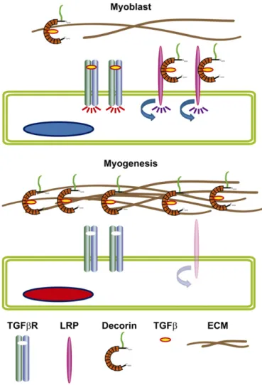
(4) 292. E. Brandan, J. Gutierrez / Matrix Biology 32 (2013) 289–297. adhesion to collagen type IV is inhibited by blocking this substrate with exogenous perlecan core protein or by pre-incubation of the cells with antibodies against murine perlecan (Villar et al., 1999). It has been described that histone H1 localises in the plasma membrane or is present in the ECM of myoblasts or regenerating skeletal muscles and binds specifically to perlecan. This histone H1 incorporated into the ECM strongly stimulated myoblast proliferation via a HS-dependent mechanism (Henriquez et al., 2002). Studies of the skeletal muscle of perlecan-null mice indicate that this HSPG is critical to regulate the muscle mass of fast fibre-enriched muscles, probably regulating the inhibitory signalling of myostatin on fast muscle hypertrophy (Xu et al., 2010). The above evidence strongly indicates pivotal roles for HSPGs during muscle formation, differentiation and regeneration, interacting with growth factors and receptors through HS chains. HS-6-O-endosulfatases (Sulfs) during muscle regeneration has been described. These Sulfs are critical to promote muscle regeneration, as Sulf double mutant mice exhibit delayed myogenic differentiation and prolonged Pax7 expression after skeletal muscle injury (Langsdorf et al., 2007). Sulfs repress FGF-2 mediated signalling in activated satellite cells, likely controlling proliferation and differentiation transition during regeneration (Langsdorf et al., 2007). 2.2. Chondroitin/dermatan sulphate proteoglycans Fig. 1. The expression of syndecans but not glypican-1 is down-regulated during the skeletal muscle differentiation. Proliferating myoblasts (cells with blue nuclei) express all four syndecans and glypican-1. After triggering myogenesis, they express myogenin (red nuclei). Then, myogenin expressing cells elongate and align to fuse forming multinucleated differentiated myotubes that express muscle specific genes (cells with green nuclei). During myogenesis the expression of syndecans is progressively lost whereas the expression of glypican-1 remains unchanged.. binding of FGF-2 to its receptors and augmented signalling. These myoblasts show decreased expression of myogenin, myosin and myoblast fusion index. All of these effects can be reverted by re-expressing glypican-1(Brandan et al., 1996; Gutierrez and Brandan, 2010). In myoblasts, glypican-1 is required for HGF mediated signalling, recruiting or stabilising the MET receptor in lipid raft domains where it is activated with the consequent triggering of downstream targets, such as the extracellular signal-regulated kinase 1 and 2 (ERK1/2) and the phosphoinositide 3-kinase–Akt (Akt) (Gutierrez and Brandan, manuscript under preparation). This evidence suggests that membranebound HSPGs might exert different functions, even opposite, depending on the ligands and their localisation in the myoblast plasma membrane. Thus, syndecan-1 and -3 act as co-receptors of FGF-2 in non-raft domains, while glypican-1 in lipid raft domains sequesters FGF-2 away from the FGFRs. Interestingly, the same glypican-1 in the lipid-enriched domain could regulate HGF-dependent signalling (Fig. 2). 2.1.6. Perlecan During development perlecan increases their expression at day 12.5 of development, later than syndecan-1and glypican-1 (Olguin and Brandan, 2001). This HSPG is expressed in myoblasts but is downregulated during terminal muscle differentiation (Larrain et al., 1997a,b). The expression of HSPGs during skeletal muscle regeneration indicates that four major species; perlecan, glypican-1, syndecan-3 and -4 are transiently upregulated. The first three were detected at the surface or basal lamina of newly formed myotubes (Casar et al., 2004a,b). Perlecan is a component of the basal lamina that surrounds skeletal muscle fibres and at the cell surface of myoblasts, interacting with collagen type IV through the core protein, from where it can be solubilised by detergents; whereas, heparin which specifically competes with HS GAGs for ligands, high salt or RGD peptides are unable to do it. Myoblast. Embryonic chick skeletal muscle has been shown to synthesize a distinct PG of large size with relatively high 6-O-sulfated chondroitin sulphate (CS) GAGs. Further analysis of this PG indicates that tryptic digestion gives rise to fragments with an average of two CS chains per peptide (Carrino and Caplan, 1982, 1989). The large PG synthesized earlier in myogenesis has been identified as the CS-PG, versican (Carrino et al., 1999). Among the small PGs synthesized at later embryonic stages correspond to the small CS/DS-PG, decorin (Carrino et al., 1999). These data are consistent with the notion that skeletal muscle regeneration involves a recapitulation of embryonic events and give further support to the hypothesis that large skeletal muscle CS-PG followed by small CS/DS-PG play a role in some early aspect of myogenesis. Among the CS/DS-PGs, the small leucine-rich PGs (SLRPs), decorin and biglycan, are associated with important processes connected with myogenesis. Decorin is the most abundant PG in skeletal muscle, mainly associated with the ECM that surrounds bundles of myofibres at the perymisium (Andrade and Brandan, 1991; Brandan et al., 1992; Melo and Brandan, 1993; Velleman et al., 1997) and with the plasma membrane (Hausser et al., 1998; Brandan et al., 2006a,b; Cabello-Verrugio and Brandan, 2007; Cabello-Verrugio et al., 2012). Biglycan directly associates with individual myofibres and is concentrated in certain NMJ bound to α-dystroglycan as part of a myofibre–ECM anchoring complex (Bowe et al., 2000). 2.2.1. Decorin As indicated, during limb skeletal muscle formation decorin expression was temporally and spatially coincident with differentiating myogenin-positive cells (Olguin and Brandan, 2001). In vitro studies show that during skeletal muscle differentiation decorin expression increases (Brandan et al., 1991) suggesting that this PG may play a role during myogenesis. Among the different functions described for decorin (Brandan et al., 2008; Schaefer and Iozzo, 2008; Iozzo and Schaefer, 2010), this PG in skeletal muscle seems to be involved in the establishment and/or coordination of a critical myoblast density, through inhibition of migration that permits normal muscle differentiation during embryonic myogenesis (Olguin et al., in press). During prenatal skeletal muscle formation, the mRNA expression pattern of decorin and myostatin, a ligand for decorin associated with myogenesis inhibition, was similar. Myostatin was located in the muscle fibres and decorin in the periphery of the fibres. Decorin sequesters myostatin.
(5) E. Brandan, J. Gutierrez / Matrix Biology 32 (2013) 289–297. 293. 2008; Cabello-Verrugio et al., 2012). This decorin requirement seems to be necessary for myogenesis since silence decorin expression accelerates skeletal muscle differentiation by decreasing sensitivity to TGFβ signalling (Riquelme et al., 2001; Cabello-Verrugio and Brandan, 2007) (Fig. 3).. Fig. 2. The plasma membrane HSPGs differentially regulate the cell response to FGF-2 and HGF during the muscle differentiation process. In proliferating myoblasts (upper, cell with blue nucleus) syndecans act as FGF-2 co-receptors, forming a ternary complex with the FGFRs (syndecan-FGF2-FGFR) required for FGF-2 signalling. In the plasma membrane lipid raft domains, glypican-1 the only HSPG associated with these microdomains, binds and sequesters FGF-2 away from FGFRs acting as a negative regulator of FGF-2 signalling. Glypican also is required to stabilize the HGF transducing receptor, MET, in the lipid raft domains where it is activated. At the beginning of the myogenic process the expression levels of syndecans, FGFRs and MET are down regulated and glypican remains unchanged. This changes the balance from a proliferative anti-myogenic FGF-2 and HGF dependent state to a pro-myogenic where both muscle inhibitory signals diminish allowing differentiation.. in the ECM during the development of skeletal muscle, silencing the myogenic inhibitory signal of myostatin (Miura et al., 2006; Nishimura et al., 2007). Also, myoblasts overexpressing decorin show an enhanced differentiation, explained by the suppression of myostatin activity (Kishioka et al., 2008). mRNA levels of decorin are higher in reserve cell than in differentiated myotubes and growing myoblasts, suggesting a potential role of decorin to maintain the quiescence of myogenic cells (Nishimura et al., 2008). Decorin binds several ligands and receptors such as transforming growth factor type β (TGFβ), the mentioned myostatin, insulin growth factor (IGF), connective tissue growth factor (CTGF), collagens, fibronectin, thrombospondin-1, epidermal growth factor receptor (EGF-R), MET, low density lipoprotein-related-receptor (LRP-1) among others (Iozzo, 1997, 1999; Brandan et al., 2008; Iozzo and Schaefer, 2010; Vial et al., 2011). Along them highlight TGFβ (Hildebrand et al., 1994; Schonherr et al., 1998; Cabello-Verrugio et al., 2012). The upregulation of decorin during skeletal muscle differentiation (Brandan et al., 1991) likely sequester TGFβ from its transducing receptors (Droguett et al., 2006). Like myostatin, TGFβ also is a potent inhibitor of myogenesis (Florini et al., 1991) and the removal by decorin during muscle differentiation seems to be necessary. However, TGFβ regulation by decorin is more complex. Under proliferative conditions, the myoblast require decorin for a full TGFβ cell response in a mechanism dependent on LRP-1 receptor (Riquelme et al., 2001; Brandan et al., 2006a,b; Cabello-Verrugio and Brandan, 2007; Brandan et al.,. 2.2.2. Biglycan Developmental studies indicates that the expression of biglycan reaches a peak at week 21 in human foetal muscle and in developing mouse muscle biglycan can be detected since day 16 (Lechner et al., 2006). The onset and progression of skeletal muscle regeneration are controlled by a complex set of interactions between muscle precursor cells and their environment. In contrast to the high levels of decorin present in the ECM of adult skeletal muscle, biglycan expression is lower (Brandan et al., 1992). After skeletal muscle damage, a transient and dramatic upregulation of biglycan is observed and associated with newly formed myotubes (Goetsch et al., 2003; Casar et al., 2004a,b). The role of biglycan during the skeletal muscle regenerative process has been tested in biglycan-null mice indicating that skeletal muscle maintains its regenerative capacity but shows a delay in fibre growth and a diminished expression of embryonic myosin, a marker of myofibre formation during regeneration, despite the normal expression of MyoD and myogenin. These observations suggest that one of the roles of biglycan could be associated with myogenic events of myofibre maturation that occurred after myogenin expression and cell fusion (Casar et al., 2004a,b). Since biglycan also binds TGFβ, it is possible that biglycan upregulation during skeletal muscle regeneration could be to immobilised TGFβ active forms, allowing muscle regeneration (Droguett et al., 2006). 3. Participation of proteoglycans at the neuromuscular junction PGs have essential and fundamental roles in the structure and physiology of the NMJ. Different functions for HSPGs at the NMJ have been demonstrated (Yamaguchi, 2002). Thus, aggregates of acetylcholine receptors (AChRs) associate with plaques of basal lamina HSPGs on the surface of skeletal muscle fibres. This correlation indicates that the spatial organisation of AChR and HSPGs is coordinately regulated and suggests that interactions between these two species may contribute to the localisation of AChR at the NMJ (Anderson and Fambrough, 1983). The essential role of HSPGs in the anchorage of the collagenic-tailed form of acetylcholinesterase (AChE) to the NMJ junction has been demonstrated (Brandan and Inestrosa, 1984; Brandan et al., 1985; Brandan and Inestrosa, 1986). AChE is solubilised from highly enriched NMJ-derived ECM by heparin and treatments with heparitinase but not chondroitinase ABC, which specifically break down HS and CS/DS GAG chains, respectively (Brandan et al., 1985). Agrin, another HSPG (Winzen et al., 2003) and synaptic laminin is required to maintain adult NMJ (Samuel et al., 2012). Agrin is a large HSPG first isolated from the Torpedo electric organ (McMahan, 1990) synthesized in the motoneurons, transported along axons, and released in the synaptic basal lamina, which surrounds the muscle fibre. In the mouse, the muscle fibres are formed around embryonic days 12–14 (Cossu and Biressi, 2005), at this moment AChRs take place at sites that will form the NMJ that initially is independent of innervation (Mishina et al., 1986; Lin et al., 2001; Lin et al., 2005). Formation of a complete and stable NMJ occurs between embryonic days 16–17 (Ontell and Kozeka, 1984), and this process depends on the presence of the neurally produced agrin which is 1000-fold more active than the muscle agrin isoform to induce AChR clusters (McMahan, 1990; Gesemann et al., 1995). Agrin-null mice die at birth, showing an immature NMJ (Gautam et al., 1996), with branched motor neurons unable to reach or interact with pre-patterned AChRs to establish the stable NMJ (Yang et al., 2001). Agrin exerts this effects through the binding to the.
(6) 294. E. Brandan, J. Gutierrez / Matrix Biology 32 (2013) 289–297. Thus, biglycan is important for the maintenance of muscle cell integrity and plays a direct role in regulating the expression and sarcolemmal localisation of the intracellular signalling proteins and NMJ stability (Fig. 4). 4. Proteoglycans in skeletal muscular dystrophic diseases. Fig. 3. The TGFβ dependent signalling is regulated by decorin and LRP during the skeletal muscle differentiation. In myoblasts, TGFβ full inhibitory response depends on the binding of two types of cell membrane receptor, the canonical transducing receptors (TGFβR), activating the Smad dependent pathway (red signal) and the decorin/LRP-1 receptor complex which upon endocitosis activates PI3K dependent pathway (purple signal). Decorin sequesters TGFβ in the ECM acting as a reservoir of the ligand. During the differentiation process, the expression of decorin increases whereas LRP-1 decrease, augmenting the amount of decorin concentration in the ECM. Thus, TGFβ is sequestered away from the TGFβR decreasing the TGFβ dependent signalling and allowing the myogenic process. This proposed model is also applied to other anti myogenic cytokine such as myostatin.. N-terminal region of Lrp4 and MusK, a muscle-specific receptor tyrosine kinase present on the muscle cell surface and critical for synapse stability, and stimulates MusK kinase activity (Zhang et al., 2011). HGF a HS-binding growth factor surrounds muscle fibres and also localises at NMJ and promotes AChR clustering and synaptogenic signalling in muscle. Addition of HGF potentiates agrin-dependent AChR clustering in the muscle (Madhavan and Peng, 2006). In perlecan-null mice, AChE is absent from the NMJ (Arikawa-Hirasawa et al., 2002). AChE binds to HSPGs (Brandan and Inestrosa, 1984; Brandan et al., 1985; Brandan and Inestrosa, 1986) specifically to perlecan, which in turn binds to the dystroglycan complex through α-dystroglycan (Peng et al., 1998). It has been shown that perlecan participates regulating pre- and postsynaptic Wnt signalling activities at the NMJ (Kamimura et al., 2013). Biglycan is one of the members of the dystrophin glycoprotein complex required for myofibre anchoring to the ECM (Rafii et al., 2006). Using biglycan-null mice, the expression and sarcolemmal localisation of the intracellular signalling proteins dystrobrevin-1 and -2, alpha- and β1-syntrophin and nNOS are reduced (Mercado et al., 2006). Biglycan also binds Musk and its levels are selectively reduced at biglycan-null skeletal muscle synapses (Amenta et al., 2012).. One of the features of several skeletal muscle dystrophies is the accumulation of ECM around skeletal muscle fibres and interstitial space. This corresponds to fibrosis, which is regularly associated with an inflammatory process characterised by the continuous replacement of functional cells by an excess of ECM proteins, persistent and severely affecting tissues or organs (Bowe et al., 2000; Caceres et al., 2000; Schiaffino and Partridge, 2008; Wynn, 2008; Serrano and Munoz-Canoves, 2010). Among the PGs that are increased in skeletal muscular dystrophies or cells isolated from DMD skeletal muscle biopsies, the CS/DS-PGs are the most abundant (Gosselin et al., 2004; Zanotti et al., 2005; Fadic et al., 2006). Decorin and biglycan are augmented in skeletal muscle samples from the animal model of DMD, the mdx mouse (Bowe et al., 2000; Caceres et al., 2000; Fadic et al., 2006). Only one report described an increase in HSPGs in mdx in whole skeletal muscles (Alvarez et al., 2002). It has been indicated that HSPGs are elevated in the mdx satellite cells compared with cells from normal animals. These satellite cells synthesize ten times more HSPGs than normal satellite cells with no significant differences in muscle fibroblasts (Litwack et al., 1998). Remarkably biglycan regulates utrophin expression, the synaptic form of dystrophin, in immature muscle and that recombinant human biglycan (rhBGN) increases utrophin expression in cultured myotubes (Amenta et al., 2011). Quite notable systemically delivered rhBGN up-regulates utrophin at the sarcolemma and reduces muscle pathology in the mdx mouse model of DMD, suggesting that rhBGN could be a therapy for DMD (Amenta et al., 2011). The relevance of the augmentation in PGs in the dystrophic disease can be hypothesised in at least two ways; as part of an exacerbated accumulation of ECM constituents characteristic of the fibrotic process or as an increase of these SLRPs decreasing the bio-availability of profibrotic growth factors such as TGFβ (Droguett et al., 2006) and CTGF as part of a possible protective mechanism (Gao and Brigstock, 2003; Vial et al., 2008; Chen and Lau, 2009; Huang and Brigstock, 2011; Vial et al., 2011). The exact physiological/pathological relevance of this PG accumulation requires further investigation. 5. Future perspectives Several roles are played by PGs during skeletal muscle formation, adult muscle regeneration and they are also associated with acute and chronic muscle damage. Their role in skeletal muscle repair associated with skeletal muscular dystrophies is just emerging, offering the ground for future studies designed to provide insight into the molecular interactions among these PGs with versatile growth factors such as FGF-2, HGF, TGFβ and CTGF and their astounding common receptors. Very provocative and exciting are the results indicating some new roles for PGs as inducers of critical molecules associated with macromolecular complex severely affected in skeletal muscular dystrophies. Future studies focused in understanding the interactions among these molecules and the mechanisms involved in these novel and unexpected functions will permit the discovery of new targets for a deep understanding of skeletal muscle PG biology and potential treatment of skeletal muscular diseases. Acknowledgments This study was supported by research grants from CARE PFB12/ 2007, FONDECYT 1110426, CONICYT 79090027, FONDECYT 11110010, and Fundación Chilena para Biología Celular Proyecto MF-100..
(7) E. Brandan, J. Gutierrez / Matrix Biology 32 (2013) 289–297. 295. Fig. 4. Participation of proteoglycans at the neuromuscular junction. Several PGs are critical at the neuromuscular junction (see text for functional details): Biglycan interacts with α-dystroglycan and α- and γ-sarcoglycans. Furthermore, biglycan interacts with Musk and is important for maintaining synapse stability. Perlecan at the basal lamina anchors asymmetric form of AChE and bind dystroglycan complex through α-dystroglycan. Agrin interacts with Musk, through LRP-4 (not shown in the figure) stimulating MusK kinase activity.. References Alvarez, K., Fadic, R., Brandan, E., 2002. Augmented synthesis and differential localization of heparan sulfate proteoglycans in Duchenne muscular dystrophy. J. Cell. Biochem. 85, 703–713. Amenta, A.R., Yilmaz, A., Bogdanovich, S., McKechnie, B.A., Abedi, M., Khurana, T.S., Fallon, J.R., 2011. Biglycan recruits utrophin to the sarcolemma and counters dystrophic pathology in mdx mice. Proc. Natl. Acad. Sci. U. S. A. 108, 762–767. Amenta, A.R., Creely, H.E., Mercado, M.L., Hagiwara, H., McKechnie, B.A., Lechner, B.E., Rossi, S.G., Wang, Q., Owens, R.T., Marrero, E., Mei, L., Hoch, W., Young, M.F., McQuillan, D.J., Rotundo, R.L., Fallon, J.R., 2012. Biglycan is an extracellular MuSK binding protein important for synapse stability. J. Neurosci. 32, 2324–2334. Anastasi, S., Giordano, S., Sthandier, O., Gambarotta, G., Maione, R., Comoglio, P., Amati, P., 1997. A natural hepatocyte growth factor/scatter factor autocrine loop in myoblast cells and the effect of the constitutive Met kinase activation on myogenic differentiation. J. Cell Biol. 137, 1057–1068. Anderson, M.J., Fambrough, D.M., 1983. Aggregates of acetylcholine receptors are associated with plaques of a basal lamina heparan sulfate proteoglycan on the surface of skeletal muscle fibers. J. Cell Biol. 97, 1396–1411. Andrade, W., Brandan, E., 1991. Isolation and characterization of rat skeletal muscle proteoglycan decorin and comparison with the human fibroblast decorin. Comp. Biochem. Physiol. B 100, 565–570. Arikawa-Hirasawa, E., Rossi, S.G., Rotundo, R.L., Yamada, Y., 2002. Absence of acetylcholinesterase at the neuromuscular junctions of perlecan-null mice. Nat. Neurosci. 5, 119–123. Bentzinger, C.F., Wang, Y.X., von Maltzahn, J., Soleimani, V.D., Yin, H., Rudnicki, M.A., 2013. Fibronectin regulates wnt7a signaling and satellite cell expansion. Cell Stem Cell 12, 75–87. Bernfield, M., Gotte, M., Park, P.W., Reizes, O., Fitzgerald, M.L., Lincecum, J., Zako, M., 1999. Functions of cell surface heparan sulfate proteoglycans. Annu. Rev. Biochem. 68, 729–777. Bottaro, D.P., Rubin, J.S., Faletto, D.L., Chan, A.M., Kmiecik, T.E., Vande Woude, G.F., Aaronson, S.A., 1991. Identification of the hepatocyte growth factor receptor as the c-met proto-oncogene product. Science 251, 802–804. Bowe, M.A., Mendis, D.B., Fallon, J.R., 2000. The small leucine-rich repeat proteoglycan biglycan binds to alpha-dystroglycan and is upregulated in dystrophic muscle. J. Cell Biol. 148, 801–810.. Brandan, E., Inestrosa, N.C., 1984. Binding of the asymmetric forms of acetylcholinesterase to heparin. Biochem. J. 221, 415–422. Brandan, E., Inestrosa, N.C., 1986. The synaptic form of acetylcholinesterase binds to cell-surface heparan sulfate proteoglycans. J. Neurosci. Res. 15, 185–196. Brandan, E., Maldonado, M., Garrido, J., Inestrosa, N.C., 1985. Anchorage of collagentailed acetylcholinesterase to the extracellular matrix is mediated by heparan sulfate proteoglycans. J. Cell Biol. 101, 985–992. Brandan, E., Fuentes, M.E., Andrade, W., 1991. The proteoglycan decorin is synthesized and secreted by differentiated myotubes. Eur. J. Cell Biol. 55, 209–216. Brandan, E., Fuentes, M.E., Andrade, W., 1992. Decorin, a chondroitin/dermatan sulfate proteoglycan is under neural control in rat skeletal muscle. J. Neurosci. Res. 32, 51–59. Brandan, E., Carey, D.J., Larrain, J., Melo, F., Campos, A., 1996. Synthesis and processing of glypican during differentiation of skeletal muscle cells. Eur. J. Cell Biol. 71, 170–176. Brandan, E., Cabello-Verrugio, C., Retamal, C., Marzolo, M.P., 2006. The low density lipoprotein receptor-related protein/2 macroglobulin receptor, LRP, functions as an endocytic receptor for decorin. J. Biol. Chem. (Revised form). Brandan, E., Retamal, C., Cabello-Verrugio, C., Marzolo, M.P., 2006. The low density lipoprotein receptor-related protein functions as an endocytic receptor for decorin. J. Biol. Chem. 281, 31562–31571. Brandan, E., Cabello-Verrugio, C., Vial, C., 2008. Novel regulatory mechanisms for the proteoglycans decorin and biglycan during muscle formation and muscular dystrophy. Matrix Biol. 27, 700–708. Brunetti, A., Goldfine, I.D., 1990. Role of myogenin in myoblast differentiation and its regulation by fibroblast growth factor. J. Biol. Chem. 265, 5960–5963. Cabello-Verrugio, C., Brandan, E., 2007. A novel modulatory mechanism of transforming growth factor-beta signaling through decorin and LRP-1. J. Biol. Chem. 282, 18842–18850. Cabello-Verrugio, C., Santander, C., Cofre, C., Acuna, M.J., Melo, F., Brandan, E., 2012. The internal region leucine-rich repeat 6 of decorin interacts with low density lipoprotein receptor-related protein-1, modulates transforming growth factor (TGF)-betadependent signaling, and inhibits TGF-beta-dependent fibrotic response in skeletal muscles. J. Biol. Chem. 287, 6773–6787. Caceres, S., Cuellar, C., Casar, J.C., Garrido, J., Schaefer, L., Kresse, H., Brandan, E., 2000. Synthesis of proteoglycans is augmented in dystrophic mdx mouse skeletal muscle. Eur. J. Cell Biol. 79, 173–181..
(8) 296. E. Brandan, J. Gutierrez / Matrix Biology 32 (2013) 289–297. Campos, A., Nunez, R., Koenig, C.S., Carey, D.J., Brandan, E., 1993. A lipid-anchored heparan sulfate proteoglycan is present in the surface of differentiated skeletal muscle cells. Isolation and biochemical characterization. Eur. J. Biochem. 216, 587–595. Carey, D.J., 1997. Syndecans: multifunctional cell-surface co-receptors. Biochem. J. 327, 1–16. Carrino, D.A., Caplan, A.I., 1982. Isolation and preliminary characterization of proteoglycans synthesized by skeletal muscle. J. Biol. Chem. 257, 14145–14154. Carrino, D.A., Caplan, A.I., 1989. Structural characterization of chick embryonic skeletal muscle chondroitin sulfate proteoglycan. Connect Tissue Res. 19, 35–50. Carrino, D.A., Sorrell, J.M., Caplan, A.I., 1999. Dynamic expression of proteoglycans during chicken skeletal muscle development and maturation. Poult. Sci. 78, 769–777. Casar, J.C., Cabello-Verrugio, C., Olguin, H., Aldunate, R., Inestrosa, N.C., Brandan, E., 2004. Heparan sulfate proteoglycans are increased during skeletal muscle regeneration: requirement of syndecan-3 for successful fiber formation. J. Cell Sci. 117, 73–84. Casar, J.C., McKechnie, B.A., Fallon, J.R., Young, M.F., Brandan, E., 2004. Transient upregulation of biglycan during skeletal muscle regeneration: delayed fiber growth along with decorin increase in biglycan-deficient mice. Dev. Biol. 268, 358–371. Chen, C.C., Lau, L.F., 2009. Functions and mechanisms of action of CCN matricellular proteins. Int. J. Biochem. Cell Biol. 41, 771–783. Cornelison, D., Filla, M., Stanley, H., Rapraeger, A., Olwin, B., 2001. Syndecan-3 and syndecan-4 specifically mark skeletal muscle satellite cells and are implicated in satellite cell maintenance and muscle regeneration. Dev. Biol. 239, 79–94. Cornelison, D.D., Wilcox-Adelman, S.A., Goetinck, P.F., Rauvala, H., Rapraeger, A.C., Olwin, B.B., 2004. Essential and separable roles for Syndecan-3 and Syndecan-4 in skeletal muscle development and regeneration. Genes Dev. 18, 2231–2236. Cossu, G., Biressi, S., 2005. Satellite cells, myoblasts and other occasional myogenic progenitors: possible origin, phenotypic features and role in muscle regeneration. Semin. Cell Dev. Biol. 16, 623–631. Couchman, J.R., 2003. Syndecans: proteoglycan regulators of cell-surface microdomains? Nat. Rev. Mol. Cell Biol. 4, 926–937. David, G., Bai, X.M., Van der Schueren, B., Marynen, P., Cassiman, J.J., Van den Berghe, H., 1993. Spatial and temporal changes in the expression of fibroglycan (syndecan-2) during mouse embryonic development. Development 119, 841–854. Deakin, J.A., Lyon, M., 1999. Differential regulation of hepatocyte growth factor/scatter factor by cell surface proteoglycans and free glycosaminoglycan chains. J. Cell Sci. 112, 1999–2009. Droguett, R., Cabello-Verrugio, C., Riquelme, C., Brandan, E., 2006. Extracellular proteoglycans modifies TGF-beta bio-availability attenuating its signaling during skeletal muscle differentiation. Matrix Biol. 25, 332–341. Fadic, R., Mezzano, V., Alvarez, K., Cabrera, D., Holmgren, J., Brandan, E., 2006. Increase in decorin and biglycan in Duchenne muscular dystrophy: role of fibroblasts as cell source of these proteoglycans in the disease. J. Cell. Mol. Med. 10, 758–769. Filmus, J., Selleck, S.B., 2001. Glypicans: proteoglycans with a surprise. J. Clin. Invest. 108, 497–501. Filmus, J., Capurro, M., Rast, J., 2008. Glypicans. Genome Biol. 9, 224. Florini, J.R., Ewton, D.Z., Magri, K.A., 1991. Hormones, growth factors, and myogenic differentiation. Annu. Rev. Physiol. 53, 201–216. Fuentealba, L., Carey, D.J., Brandan, E., 1999. Antisense inhibition of syndecan-3 expression during skeletal muscle differentiation accelerates myogenesis through a basic fibroblast growth factor-dependent mechanism. J. Biol. Chem. 274, 37876–37884. Gao, R., Brigstock, D.R., 2003. Low density lipoprotein receptor-related protein (LRP) is a heparin-dependent adhesion receptor for connective tissue growth factor (CTGF) in rat activated hepatic stellate cells. Hepatol. Res. 27, 214–220. Gautam, M., Noakes, P.G., Moscoso, L., Rupp, F., Scheller, R.H., Merlie, J.P., Sanes, J.R., 1996. Defective neuromuscular synaptogenesis in agrin-deficient mutant mice. Cell 85, 525–535. Gesemann, M., Denzer, A.J., Ruegg, M.A., 1995. Acetylcholine receptor-aggregating activity of agrin isoforms and mapping of the active site. J. Cell Biol. 128, 625–636. Gillies, A.R., Lieber, R.L., 2011. Structure and function of the skeletal muscle extracellular matrix. Muscle Nerve 44, 318–331. Goetsch, S.C., Hawke, T.J., Gallardo, T.D., Richardson, J.A., Garry, D.J., 2003. Transcriptional profiling and regulation of the extracellular matrix during muscle regeneration. Physiol. Genomics 14, 261–271. Gosselin, L.E., Williams, J.E., Deering, M., Brazeau, D., Koury, S., Martinez, D.A., 2004. Localization and early time course of TGF-beta 1 mRNA expression in dystrophic muscle. Muscle Nerve 30, 645–653. Gutierrez, J., Brandan, E., 2010. A novel mechanism of sequestering fibroblast growth factor 2 by glypican in lipid rafts, allowing skeletal muscle differentiation. Mol. Cell. Biol. 30, 1634–1649. Gutierrez, J., Osses, N., Brandan, E., 2006. Changes in secreted and cell associated proteoglycan synthesis during conversion of myoblasts to osteoblasts in response to bone morphogenetic protein-2: role of decorin in cell response to BMP-2. J. Cell. Physiol. 206, 58–67. Hausser, H., Schonherr, E., Muller, M., Liszio, C., Bin, Z., Fisher, L.W., Kresse, H., 1998. Receptor-mediated endocytosis of decorin: involvement of leucine-rich repeat structures. Arch. Biochem. Biophys. 349, 363–370. Henriquez, J.P., Casar, J.C., Fuentealba, L., Carey, D.J., Brandan, E., 2002. Extracellular matrix histone H1 binds to perlecan, is present in regenerating skeletal muscle and stimulates myoblast proliferation. J. Cell Sci. 115, 2041–2051. Hildebrand, A., Romaris, M., Rasmussen, L.M., Heinegard, D., Twardzik, D.R., Border, W.A., Ruoslahti, E., 1994. Interaction of the small interstitial proteoglycans biglycan, decorin and fibromodulin with transforming growth factor beta. Biochem. J. 302, 527–534. Huang, G., Brigstock, D.R., 2011. Integrin expression and function in the response of primary culture hepatic stellate cells to connective tissue growth factor (CCN2). J. Cell. Mol. Med. 15, 1087–1095. Iozzo, R.V., 1997. The family of the small leucine-rich proteoglycans: key regulators of matrix assembly and cellular growth. Crit. Rev. Biochem. Mol. Biol. 32, 141–174.. Iozzo, R.V., 1999. The biology of the small leucine-rich proteoglycans. Functional network of interactive proteins. J. Biol. Chem. 274, 18843–18846. Iozzo, R.V., Schaefer, L., 2010. Proteoglycans in health and disease: novel regulatory signaling mechanisms evoked by the small leucine-rich proteoglycans. FEBS J. 277, 3864–3875. Jen, Y.H., Musacchio, M., Lander, A.D., 2009. Glypican-1 controls brain size through regulation of fibroblast growth factor signaling in early neurogenesis. Neural Dev. 4, 33. Jenniskens, G.J., Hafmans, T., Veerkamp, J.H., van Kuppevelt, T.H., 2002. Spatiotemporal distribution of heparan sulfate epitopes during myogenesis and synaptogenesis: a study in developing mouse intercostal muscle. Dev. Dyn. 225, 70–79. Jenniskens, G.J., Veerkamp, J.H., van Kuppevelt, T.H., 2006. Heparan sulfates in skeletal muscle development and physiology. J. Cell. Physiol. 206, 283–294. Kamimura, K., Ueno, K., Nakagawa, J., Hamada, R., Saitoe, M., Maeda, N., 2013. Perlecan regulates bidirectional Wnt signaling at the Drosophila neuromuscular junction. J. Cell Biol. 200, 219–233. Kishioka, Y., Thomas, M., Wakamatsu, J., Hattori, A., Sharma, M., Kambadur, R., Nishimura, T., 2008. Decorin enhances the proliferation and differentiation of myogenic cells through suppressing myostatin activity. J. Cell. Physiol. 215, 856–867. Kresse, H., Hausser, H., Schonherr, E., 1994. Small proteoglycans. EXS 70, 73–100. Langsdorf, A., Do, A.T., Kusche-Gullberg, M., Emerson Jr., C.P., Ai, X., 2007. Sulfs are regulators of growth factor signaling for satellite cell differentiation and muscle regeneration. Dev. Biol. 311, 464–477. Larrain, J., Alvarez, J., Hassell, J.R., Brandan, E., 1997. Expression of perlecan, a proteoglycan that binds myogenic inhibitory basic fibroblast growth factor, is down regulated during skeletal muscle differentiation. Exp. Cell Res. 234, 405–412. Larrain, J., Cizmeci-Smith, G., Troncoso, V., Stahl, R.C., Carey, D.J., Brandan, E., 1997. Syndecan-1 expression is down-regulated during myoblast terminal differentiation. Modulation By growth factors and retinoic acid. J. Biol. Chem. 272, 18418–18424. Larrain, J., Carey, D.J., Brandan, E., 1998. Syndecan-1 expression inhibits myoblast differentiation through a basic fibroblast growth factor-dependent mechanism. J. Biol. Chem. 273, 32288–32296. Lechner, B.E., Lim, J.H., Mercado, M.L., Fallon, J.R., 2006. Developmental regulation of biglycan expression in muscle and tendon. Muscle Nerve 34, 347–355. Lin, W., Burgess, R.W., Dominguez, B., Pfaff, S.L., Sanes, J.R., Lee, K.F., 2001. Distinct roles of nerve and muscle in postsynaptic differentiation of the neuromuscular synapse. Nature 410, 1057–1064. Lin, W., Dominguez, B., Yang, J., Aryal, P., Brandon, E.P., Gage, F.H., Lee, K.F., 2005. Neurotransmitter acetylcholine negatively regulates neuromuscular synapse formation by a Cdk5-dependent mechanism. Neuron 46, 569–579. Litwack, E.D., Ivins, J.K., Kumbasar, A., Paine-Saunders, S., Stipp, C.S., Lander, A.D., 1998. Expression of the heparan sulfate proteoglycan glypican-1 in the developing rodent. Dev. Dyn. 211, 72–87. Liu, X., Nestor, K.E., McFarland, D.C., Velleman, S.G., 2002. Developmental expression of skeletal muscle heparan sulfate proteoglycans in turkeys with different growth rates. Poult. Sci. 81, 1621–1628. Liu, X., McFarland, D.C., Nestor, K.E., Velleman, S.G., 2004. Developmental regulated expression of syndecan-1 and glypican in pectoralis major muscle in turkeys with different growth rates. Dev. Growth Differ. 46, 37–51. Madhavan, R., Peng, H.B., 2006. HGF induction of postsynaptic specializations at the neuromuscular junction. J. Neurobiol. 66, 134–147. McMahan, U., 1990. The agrin hypothesis. Cold Spring Harb. Symp. Quant. Biol. 55, 407–418. Melo, F., Brandan, E., 1993. Decorin is specifically solubilized by heparin from the extracellular matrix of rat skeletal muscles. FEBS Lett. 319, 249–252. Mercado, M.L., Amenta, A.R., Hagiwara, H., Rafii, M.S., Lechner, B.E., Owens, R.T., McQuillan, D.J., Froehner, S.C., Fallon, J.R., 2006. Biglycan regulates the expression and sarcolemmal localization of dystrobrevin, syntrophin, and nNOS. FASEB J. 20, 1724–1726. Mishina, M., Takai, T., Imoto, K., Noda, M., Takahashi, T., Numa, S., Methfessel, C., Sakmann, B., 1986. Molecular distinction between fetal and adult forms of muscle acetylcholine receptor. Nature 321, 406–411. Miura, T., Kishioka, Y., Wakamatsu, J., Hattori, A., Hennebry, A., Berry, C.J., Sharma, M., Kambadur, R., Nishimura, T., 2006. Decorin binds myostatin and modulates its activity to muscle cells. Biochem. Biophys. Res. Commun. 340, 675–680. Nishimura, T., Oyama, K., Kishioka, Y., Wakamatsu, J., Hattori, A., 2007. Spatiotemporal expression of decorin and myostatin during rat skeletal muscle development. Biochem. Biophys. Res. Commun. 361, 896–902. Nishimura, T., Nozu, K., Kishioka, Y., Wakamatsu, J., Hattori, A., 2008. Decorin expression in quiescent myogenic cells. Biochem. Biophys. Res. Commun. 370, 383–387. Olguin, H., Brandan, E., 2001. Expression and localization of proteoglycans during limb myogenic activation. Dev. Dyn. 221, 106–115. Olguin, H., Santander, C., Brandan, E., 2013. Inhibition of myoblast migration, via decorin expression is critical for normal skeletal muscle differentiation. Dev. Biol. (in press). Olwin, B.B., Rapraeger, A., 1992. Repression of myogenic differentiation by aFGF, bFGF, and K-FGF is dependent on cellular heparan sulfate. J. Cell Biol. 118, 631–639. Ontell, M., Kozeka, K., 1984. Organogenesis of the mouse extensor digitorum logus muscle: a quantitative study. Am. J. Anat. 171, 149–161. Peng, H., Ali, A., Daggett, D., Rauvala, H., Hassell, J., Smalheiser, N., 1998. The relationship between perlecan and dystroglycan and its implication in the formation of the neuromuscular junction. Cell Adhes. Commun. 5, 475–489. Pisconti, A., Cornelison, D.D., Olguin, H.C., Antwine, T.L., Olwin, B.B., 2010. Syndecan-3 and Notch cooperate in regulating adult myogenesis. J. Cell Biol. 190, 427–441. Rafii, M.S., Hagiwara, H., Mercado, M.L., Seo, N.S., Xu, T., Dugan, T., Owens, R.T., Hook, M., McQuillan, D.J., Young, M.F., Fallon, J.R., 2006. Biglycan binds to alpha- and gamma-sarcoglycan and regulates their expression during development. J. Cell. Physiol. 209, 439–447..
(9) E. Brandan, J. Gutierrez / Matrix Biology 32 (2013) 289–297 Rapraeger, A.C., Krufka, A., Olwin, B.B., 1991. Requirement of heparan sulfate for bFGFmediated fibroblast growth and myoblast differentiation. Science 252, 1705–1708. Riquelme, C., Larrain, J., Schonherr, E., Henriquez, J.P., Kresse, H., Brandan, E., 2001. Antisense inhibition of decorin expression in myoblasts decreases cell responsiveness to transforming growth factor beta and accelerates skeletal muscle differentiation. J. Biol. Chem. 276, 3589–3596. Rudnicki, M.A., Jaenisch, R., 1995. The MyoD family of transcription factors and skeletal myogenesis. Bioessays 17, 203–209. Samuel, M.A., Valdez, G., Tapia, J.C., Lichtman, J.W., Sanes, J.R., 2012. Agrin and synaptic laminin are required to maintain adult neuromuscular junctions. PLoS One 7, e46663. Sarrazin, S., Lamanna, W.C., Esko, J.D., 2011. Heparan sulfate proteoglycans. Cold Spring Harb. Perspect. Biol. 3. Schaefer, L., Iozzo, R.V., 2008. Biological functions of the small leucine-rich proteoglycans: from genetics to signal transduction. J. Biol. Chem. 283, 21305–21309. Schiaffino, S., Partridge, T., 2008. Skeletal muscle repair and regeneration. Advances in Muscle Research, 3. Springer, Netherlands (pp. XIV-380). Schonherr, E., Broszat, M., Brandan, E., Bruckner, P., Kresse, H., 1998. Decorin core protein fragment Leu155-Val260 interacts with TGF-beta but does not compete for decorin binding to type I collagen. Arch. Biochem. Biophys. 355, 241–248. Serrano, A.L., Munoz-Canoves, P., 2010. Regulation and dysregulation of fibrosis in skeletal muscle. Exp. Cell Res. 316, 3050–3058. Sogos, V., Balaci, L., Ennas, M.G., Dell'era, P., Presta, M., Gremo, F., 1998. Developmentally regulated expression and localization of fibroblast growth factor receptors in the human muscle. Dev. Dyn. 211, 362–373. Ugarte, G., Santander, C., Brandan, E., 2010. Syndecan-4 and beta1 integrin are regulated by electrical activity in skeletal muscle: implications for cell adhesion. Matrix Biol. 29, 383–392. Velleman, S.G., Patterson, R.A., Nestor, K.E., 1997. Identification of decorin and chondroitin sulfate proteoglycans in turkey skeletal muscle. Poult. Sci. 76, 506–510. Velleman, S.G., Liu, X., Coy, C.S., McFarland, D.C., 2004. Effects of syndecan-1 and glypican on muscle cell proliferation and differentiation: implications for possible functions during myogenesis. Poult. Sci. 83, 1020–1027. Vial, C., Zuniga, L.M., Cabello-Verrugio, C., Canon, P., Fadic, R., Brandan, E., 2008. Skeletal muscle cells express the profibrotic cytokine connective tissue growth. 297. factor (CTGF/CCN2), which induces their dedifferentiation. J. Cell. Physiol. 215, 410–421. Vial, C., Gutiérrez, J., Santander, C., Cabrera, D., Brandan, E., 2011. Decorin interacts with CTGF/CCN2 through LRR12 inhibiting its biological activity. J. Biol. Chem. 286, 24242–24252. Villar, M.J., Hassell, J.R., Brandan, E., 1999. Interaction of skeletal muscle cells with collagen type IV is mediated by perlecan associated with the cell surface. J. Cell. Biochem. 75, 665–674. Winzen, U., Cole, G.J., Halfter, W., 2003. Agrin is a chimeric proteoglycan with the attachment sites for heparan sulfate/chondroitin sulfate located in two multiple serine–glycine clusters. J. Biol. Chem. 278, 30106–30114. Wynn, T.A., 2008. Cellular and molecular mechanisms of fibrosis. J. Pathol. 214, 199–210. Xu, Z., Ichikawa, N., Kosaki, K., Yamada, Y., Sasaki, T., Sakai, L.Y., Kurosawa, H., Hattori, N., Arikawa-Hirasawa, E., 2010. Perlecan deficiency causes muscle hypertrophy, a decrease in myostatin expression, and changes in muscle fiber composition. Matrix Biol. 29, 461–470. Yamaguchi, Y., 2002. Glycobiology of the synapse: the role of glycans in the formation, maturation, and modulation of synapses. Biochim. Biophys. Acta 1573, 369–376. Yang, X., Arber, S., William, C., Li, L., Tanabe, Y., Jessell, T.M., Birchmeier, C., Burden, S.J., 2001. Patterning of muscle acetylcholine receptor gene expression in the absence of motor innervation. Neuron 30, 399–410. Yayon, A., Klagsbrun, M., Esko, J.D., Leder, P., Ornitz, D.M., 1991. Cell surface, heparin-like molecules are required for binding of basic fibroblast growth factor to its high affinity receptor. Cell 64, 841–848. Zanotti, S., Negri, T., Cappelletti, C., Bernasconi, P., Canioni, E., Di Blasi, C., Pegoraro, E., Angelini, C., Ciscato, P., Prelle, A., Mantegazza, R., Morandi, L., Mora, M., 2005. Decorin and biglycan expression is differentially altered in several muscular dystrophies. Brain 128, 2546–2555. Zhang, W., Coldefy, A.S., Hubbard, S.R., Burden, S.J., 2011. Agrin binds to the N-terminal region of Lrp4 protein and stimulates association between Lrp4 and the first immunoglobulin-like domain in muscle-specific kinase (MuSK). J. Biol. Chem. 286, 40624–40630..
(10)
Figure

Documento similar
Develop and implement a computational framework designed for the real-time physical simu- lation of biomechanical tissue, specifically, skeletal muscle belly, that must consider
Gibala, “A practical model of low-volume high- intensity interval training induces mitochondrial biogenesis in human skeletal muscle: potential mechanisms, ” The Journal of
In this regard, since skeletal muscle is the only tissue that is clinically affected in all patients with GSDV, the control tissue was skeletal muscle biopsies from aged and
Thus, the aim of the present study was to evaluate the role of various determinants on SMM, sarcopenia, and sarcopenic obesity in nine countries (China, Finland, Ghana, India, Mex-
(1998) Arterial heparan sulfate proteoglycans inhibit vascular smooth muscle cell proliferation and phenotype change in vitro and neointimal formation in vivo.. (2001) Relationship
At the same time, ZEB1 can switch from a transcriptional repressor to an activator by binding to p300/CBP suggesting another potential mechanism by which ZEB1 regulates
This article has been accepted for publication and undergone full peer review but has not been through the copyediting, typesetting, pagination and proofreading process which may
Also, a very recent report demonstrates that the loss in self-renewal detected in satellite cells from aged mice can be overcome by p38α/β inhibition (Bernet and Rudnicki,
