Edited by:
Linda Popplewell, Royal Holloway, University of London, United Kingdom
Reviewed by:
Muhammad Jawad Hassan, National University of Sciences and Technology, Pakistan Virginia Arechavala-Gomeza, BioCruces Health research Institute, Spain
*Correspondence:
Capucine Trollet [email protected]
Specialty section:
This article was submitted to Genetic Disorders, a section of the journal Frontiers in Genetics
Received:31 October 2017
Accepted:22 March 2018
Published:10 April 2018
Citation:
Cordova G, Negroni E, Cabello-Verrugio C, Mouly V and Trollet C (2018) Combined Therapies for Duchenne Muscular Dystrophy to Optimize Treatment Efficacy. Front. Genet. 9:114. doi: 10.3389/fgene.2018.00114
Combined Therapies for Duchenne
Muscular Dystrophy to Optimize
Treatment Efficacy
Gonzalo Cordova
1, Elisa Negroni
1, Claudio Cabello-Verrugio
2,3, Vincent Mouly
1and
Capucine Trollet
1*
1Sorbonne Université, Institut National de la Santé et de la Recherche Médicale, Association Institut de Myologie, Centre de
Recherche en Myologie, UMRS974, Paris, France,2Laboratorio de Patologías Musculares, Fragilidad y Envejecimiento,
Departamento de Ciencias Biológicas, Facultad de Ciencias Biológicas, Universidad Andres Bello, Santiago, Chile,
3Millennium Institute on Immunology and Immunotherapy, Santiago, Chile
Duchene Muscular Dystrophy (DMD) is the most frequent muscular dystrophy and one
of the most severe due to the absence of the dystrophin protein. Typical pathological
features include muscle weakness, muscle wasting, degeneration, and inflammation.
At advanced stages DMD muscles present exacerbated extracellular matrix and fat
accumulation. Recent progress in therapeutic approaches has allowed new strategies to
be investigated, including pharmacological, gene-based and cell-based therapies. Gene
and cell-based therapies are still limited by poor targeting and low efficiency in fibrotic
dystrophic muscle, therefore it is increasingly evident that future treatments will have to
include “combined therapies” to reach maximal efficiency. The scope of this mini-review is
to provide an overview of the current literature on such combined therapies for DMD. By
“combined therapies” we mean those that include both a therapy to correct the genetic
defect and an additional one to address one of the secondary pathological features of
the disease. In this mini-review, we will not provide a comprehensive view of the literature
on therapies for DMD, since many such reviews already exist, but we will focus on the
characteristics, efficiency, and potential of such combined therapeutic strategies that
have been described so far for DMD.
Keywords: gene therapy, cell therapy, muscle, Duchenne muscular dystrophy, dystrophin, fibrosis, inflammation, atrophy
INTRODUCTION
between the extracellular matrix and the cytoskeletal actin
in muscle fibers through the dystrophin-associated protein
complex (DAPC) (Ervasti and Sonnemann, 2008). Dystrophin
deficiency leads to the rupture of the muscle fiber membrane
during contraction (Allen and Whitehead, 2011) and causes
impaired intracellular signaling (Constantin, 2014). At the
cellular level, the muscles of DMD patients show evidence of
necrosis, degeneration and regeneration, myofiber atrophy, fatty
accumulation, fibrosis, and inflammation (Spencer and Tidball,
2001; Alvarez et al., 2002; Desguerre et al., 2009a,b; Serrano and
Muñoz-Cá-noves, 2010; Zhou and Lu, 2010; Villalta et al., 2011).
Different approaches (gene-based, cell-based, nano-particles, and
pharmacological) have been developed to restore a functional
dystrophin to DMD muscles (Negroni et al., 2016; Chamberlain
and Chamberlain, 2017; Nance et al., 2017). These strategies are
promising and several clinical trials are on-going or have been
conducted on DMD patients: between 1995 and 2018, 127 clinical
trials are found on clinicaltrials.gov, with 57% pharmacological
approaches, 28% gene-based (22% antisense oligonucleotide
based exon skipping, 6% AAV gene addition), and 3%
based approaches. To maximize the efficiency of gene- and
cell-based approaches, future therapies will have to take into account
the state of the muscle tissue and the secondary modifications
associated to the genetic defect. For example, the integrity of the
sarcolemma of muscle fibers, essential for efficient and long term
gene therapy, is severely compromised in DMD (McElhanon
and Bhattacharya, 2018), which leads to the concomitant loss
of the therapeutic agent (Le Hir et al., 2013). In addition, in
dystrophic muscle the continuous breakdown of muscle fibers
causes inflammation and fibrosis (Serrano and Muñoz-Cánoves,
2017). Such a hostile environment will be detrimental for the
efficacy of cell-based therapies and exacerbated extracellular
matrix will affect the accessibility of all therapeutic agents to the
muscle fibers (gene, cell, or pharmacological).
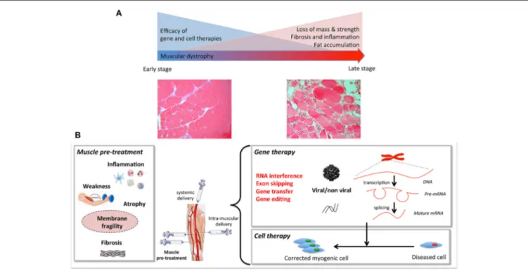
The aim of this review is to highlight pre-clinical studies in
DMD that have tested two therapeutic strategies in combination
(
Figure 1
). We will only focus on studies that have used
“combined therapies”: one to correct the genetic defect, and a
second to improve the status of the recipient muscle. To improve
the dystrophic environment of the recipient tissue, strategies are
being developed to eliminate the barriers that limit the access of
the therapeutic vector to the fibers and to limit degeneration—
even temporarily—to allow dystrophin to reach therapeutic
expression levels. “Muscle conditioning” treatments ameliorating
the status of the targeted muscle are being developed. Such
improvements
per se
are beneficial to the muscle and will also
improve the efficacy of gene or cell-based therapy. The final goal
of any combined therapy should be to improve the efficacy of
the single target therapies. Since an extensive literature on the
different single target strategies developed for DMD already exist,
we will not describe these here, nor shall we discuss animal
models used for the same reason.
Improving Dystrophin Expression Using
Combined Therapy
Exon skipping approaches have already shown promising results
in animal models. This therapy is based on the use of
antisense oligonucleotides (AON), that will interfere with the
normal splicing process removing the mutation-carrying exons,
allowing the production of a truncated but still functional
dystrophin (Nakamura, 2017). Indeed the dystrophin structure
with its central rod-domain made of 24 spectrin-like repeats,
can tolerate large internal deletions while maintaining most
of its function. In an elegant study, Peccate et al. recently
demonstrated that a pre-treatment of the skeletal muscle of
mdx
mice (the most common mouse model for DMD;
Bulfield et al.,
1984) with peptide-phosphorodiamidate morpholino (PPMO)
antisense oligonucleotides targeting dystrophin was beneficial
for a subsequent AAV-based exon-skipping therapy (Peccate
et al., 2016). This pre-treatment allowed temporary restoration
of dystrophin at the sarcolemma, improving membrane integrity
to reduce the loss of vector genome after AAV injection and
improve the efficiency of gene therapy. This study emphasizes
the strong potential of combined approaches to improve the
benefit of AAV-based therapies since without pre-treatment the
viral vector would be lost when the muscle fibers degenerate. For
DMD, such pre-treatment would allow the use of lower and thus
safer vector doses for a higher level of dystrophin expression in
the long term. Such pre-treatment aiming at improving muscle
fiber integrity could benefit also to other muscular dystrophies
with degenerative features. The efficiency of exon-skipping can
also be targeted. Using high-throughput screening, Kendall
et al. identified Dantrolene—currently used to treat malignant
hyperthermia—as a “skipping enhancer” (Kendall et al., 2012).
This drug delivered to
mdx
mice by intraperitoneal injections
enhanced antisense oligonucleotide (AON)-mediated DMD exon
skipping. The use of such an enhancer will improve AON
treatment by increasing the therapeutic value of AON, reducing
the dose needed, and thus lowering the costs and potential
toxicity. Finally, nanotechnologies have also been used to deliver
therapeutic agents, such as antisense nucleotides (for a review see
Falzarano et al., 2014). Such tools might in the future be used in
combined therapeutic strategies.
Stimulation of Muscle Growth and
Regeneration
If muscle wasting has already progressed, dystrophin expression
in the surviving fibers will not be sufficient to restore function.
Maintaining and stimulating higher levels of muscle regeneration
could potentially have a beneficial effect in dystrophic muscles.
The first attempt of a combined therapy stimulating muscle
growth came from
Abmayr et al. (2005), who used the
co-expression of Insulin-like Growth factor-1 (IGF-1)—a known
inducer of muscle hypertrophy, strength and regeneration
(Philippou and Barton, 2014)—together with the expression of
a functional microdystrophin (
µ
Dys) in
mdx
mice. Muscles
treated with this combined therapy, showed increased muscle
mass and specific force compared to untreated or to muscles
treated with
µ
Dys alone. A similar approach was used by
Rodino-Klapac et al. (2013)
by combining follistatin—an inhibitor of
myostatin (Sharma et al., 2015)—to increase muscle mass and
strength, and
µ
Dys. They showed a potent synergistic effect
FIGURE 1 | (A)Muscular dystrophies fibers, including loss of mass, weakness, fat, and extracellular matrix accumulation. Gene and cell based therapies will have to overcome the progressive degeneration of muscle fibers. When these histological changes become prominent, combined strategies are needed.(B)Muscle pre- or co-treatment may target inflammation, atrophy, membrane fragility, muscle weakness, and/or atrophy to pre-condition the tissue to increase efficiency of gene and cell therapy.
to improve muscle weakness (Kemaladewi et al., 2011;
Lu-Nguyen et al., 2017) with promising results. Similar approaches
to interfere with the myostatin pathways have also been used in
combined therapies: RNA interference for the Activin Receptor
type IIb (AcvRIIB) (Dumonceaux et al., 2010)—the receptor for
Myostatin—or a soluble version of AcvRIIB (Hoogaars et al.,
2012) have been used in combination with AAV-U7 based exon
skipping resulting in a beneficial effect increasing both muscle
mass and strength.
Controlling Fibrosis, Inflammation, and
Atrophy
Fibrosis, inflammation and muscle atrophy are among the most
important complications associated with muscular dystrophy,
and they can severely compromise the efficiency of gene or cell
therapy by limiting access to the dystrophic muscle. Fibrosis
can be defined as the increased expression and accumulation
of Extracellular Matrix (ECM) proteins, such as fibronectin and
collagen, which contributes to muscle dysfunction (Serrano and
Muñoz-Cánoves, 2017). Transforming Growth Factor type
β
(TGF-
β
) is a potent pro-fibrotic cytokine that contributes to the
pathogenesis of several fibrotic disorders, including muscular
dystrophies (Bernasconi et al., 1999). Interestingly, it has been
found that TGF-
β
induces the expression of Connective Tissue
Growth Factor (CTGF/CCN2) in fibroblasts (Igarashi et al., 1993)
and the pro-fibrotic effects of TGF-
β
may be CTGF-dependent
(Grotendorst, 1997; Leask and Abraham, 2004; Leask et al., 2004).
The expression of microRNA-29—a family of microRNAs whose
downregulation is associated with fibrosis—not only decreased
TGF-
β
1 and ECM proteins expression but also completely
restored muscle strength in dystrophic muscle when combined
with
µ
Dys treatment (Heller et al., 2017). Similarly, reducing
CTGF expression genetically or blocking CTGF with neutralizing
antibodies, decreased fibrosis, and increased muscle strength and
the efficiency of cell therapy (Morales et al., 2013b). Combining
different cell types in cell therapy has also been shown to
improve the fibrotic environment in dystrophic mice. Gargioli
et al. showed that a pre-treatment using modified tendon
fibroblasts, expressing angiogenic factors such as placenta growth
factor and an antifibrotic treatment using MMP-9, improved
microcirculation, reduced collagen and fat tissue deposition,
decreased leukocyte infiltration, increased fiber numbers and
improved cell-therapy in aged
α
-Sarcoglycan null mice, a model
Several different combined approaches have been used to
decrease inflammation and improve therapeutic outcome:
anti-inflammatory prednisolone combined with AON exon
skipping treatment has been shown to increase dystrophin
expression (Verhaart et al., 2012). A study by
Cabrera et al.
(2014)
showed that andrographolide—an inhibitor of NF-
κ
B
(pro-inflammatory pathway implicated in atrophy and fibrosis;
Li et al., 2008)—reduces the expression of fibrotic factors and
ECM proteins, while increasing muscle strength and cell therapy
efficacy. Another study showed that treatment with HCT 1026—
a non-steroidal anti-inflammatory drug capable of releasing
nitric oxide (NO)—increased the efficiency of cell therapy
in
mdx
mice and in a mouse model of limb girdle muscular
dystrophy (Brunelli et al., 2007). NO deficiency in DMD, due to
the disappearance of nNOS linked to the dystrophin complex, is
also an important issue since it is a potent regulator of skeletal
muscle physiology and regeneration, and could also be targeted
in combined therapies (Timpani et al., 2017).
Muscular atrophy is a common feature of DMD and many
pathological processes discussed in this mini-review contribute
to muscle wasting (Shin et al., 2013). One of the pathways
involved in the regulation of muscle mass is the
Renin-Angiotensin System (RAS) (Cabello-Verrugio et al., 2012a,
2015). Several of its components are upregulated in dystrophic
muscles (Sun et al., 2009) where they can also trigger a fibrotic
response. Pharmacological modulation of RAS can be used to
decrease atrophy (Burks et al., 2011), decrease fibrosis
(Cabello-Verrugio et al., 2012b; Morales et al., 2013a; Acuna et al.,
2014) and ameliorate cardiac complications related to MD (Allen
et al., 2013; Sabharwal et al., 2014). Several studies have used
combined therapies using Losartan, an inhibitor or the AT-1
receptor. Losartan treatment has been shown to increase the
efficiency of myoblast cell therapy (Fakhfakh et al., 2012) and
Adipose-Derived Stem Cell therapy (Lee et al., 2015). However,
a study by
Lee et al. (2014)
showed that although combined
therapy with Losartan and exon skipping was beneficial in
terms of muscle regeneration, the efficiency of exon skipping
was lower in Losartan treated mice due to decreased
in vivo
morpholino penetration. In this study, Losartan was added
prior to morpholino treatment, it would be interesting to see
what happens when losartan treatment is started after the exon
skipping treatment. Moreover, as Losartan seems to increase
sarcolemma stability, it could be interesting to see the effect of
viral gene therapy after Losartan treatment.
In Vitro
Modification of Myogenic Cells
Among “combined therapies” those combining gene- and
cell-therapies should be mentioned, even though none of the two
strategies improve the status of the recipient muscle. Most
combined gene- and cell-based studies developed so far consist in
genetic modification of adult stem cells harvested from patients
or dystrophic models to produce a functional dystrophin protein.
In order to accommodate DNA packaging limitations in a range
of viral vectors, synthetic mini- and micro-dystrophin versions
have been engineered (Athanasopoulos et al., 2004) and tested
in cell transplantation studies using lentiviral vectors. Several
transduced types of myogenic cells, e.g., mouse, canine, primate,
and human muscle precursors (Ikemoto et al., 2007; Quenneville
et al., 2007b; Pichavant et al., 2010), murine side population (SP)
cells (Bachrach et al., 2004), canine (Sampaolesi et al., 2006),
and human (Dellavalle et al., 2007) mesoangioblasts/pericytes,
have been tested in dystrophic models. The group of J. Tremblay
demonstrated that the use of electroporation combined with the
introduction of a phiC31 integrase led to the stable expression
of full-length dystrophin in murine and human MPCs, even
if this technique is less efficient than viral vector transduction
(Quenneville et al., 2007a). Kazuki et al. have also validated
the use of a human artificial chromosome (HAC) to restore
full-length dystrophin in mouse and human iPS cells (Kazuki
et al., 2010), while genomic integration of the full-length human
dystrophin has been achieved in iPS cells (Farruggio et al., 2017)
and mesangioblasts (Loperfido et al., 2016). Also, a full-length
dystrophin was efficiently expressed in dog mesoangioblasts
using piggyBac transposons (Loperfido et al., 2016). The same
technique was used to modify mouse mesoangioblasts prior to
transplantation in
mdx
mice, showing a good level of dystrophin
expression, increased number of satellite cells, reduction in
fibrosis, and increased muscle function (Iyer et al., 2018). Exon
skipping has also been tested in combination with cell therapy
approaches using targeted antisense sequences vectorised in U7
snRNA constructs in skin fibroblasts (Chaouch et al., 2009) or
CD133
+
cells of DMD patients (Benchaouir et al., 2007).
More recently, direct targeting of a morbid allele has been
challenged using nucleases
in vitro
and
in vivo:
meganucleases,
Zinc-finger nucleases, TALENs, and CRISPR have all been used
for genome editing to correct DMD cells carrying deletions and
out-of-frame mutations in dystrophin gene (Ousterout et al.,
2013, 2015b; Popplewell et al., 2013; Young et al., 2016; Gee et al.,
2017; Pini et al., 2017; Reinig et al., 2017; Wang et al., 2017; Zhu
et al., 2017). A recent study also described a multiplexed strategy
using a lentiviral vector capable of editing multiple sequences at
a time, allowing the correction of up to 62% of mutations causing
DMD (Ousterout et al., 2015a). While none of these approaches
have yet been used to our knowledge in combined therapies, they
could also profit from such strategies.
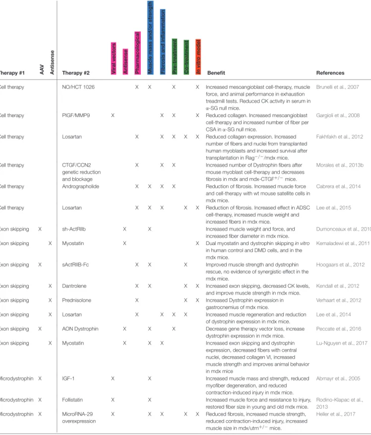
CONCLUDING REMARKS
Combined Therapies for DMD
There is now an increased interest in developing combined
therapies for DMD (
Table 1
). A combined therapy is most often
designed to treat the secondary consequences of the muscular
dystrophy that decrease the efficiency of single therapies, e.g.,
inflammation, fibrosis, or degeneration. If efficient, this therapy
should have two effects: (1) improve the muscle phenotype
TABLE 1 |Overview of synergistic therapies tested in muscular dystrophies.
Cell therapy NO/HCT 1026 X X X X Increased mesoangioblast cell-therapy, muscle
force, and animal performance in exhaustion treadmill tests. Reduced CK activity in serum in α-SG null mice.
Brunelli et al., 2007
Cell therapy PIGF/MMP9 X X X X Reduced collagen. Increased mesoangioblast
cell-therapy and increased number of fiber per CSA inα-SG null mice.
Gargioli et al., 2008
Cell therapy Losartan X X X X X Reduced collagen expression. Increased
number of fibers and nuclei from transplanted human myoblasts and increased survival after transplantation in Rag−/−/mdx mice.
Fakhfakh et al., 2012
Cell therapy CTGF/CCN2
genetic reduction and blockage
X X X Increased number of Dystrophin fibers after mouse myoblast cell-therapy and decreases fibrosis in mdx and mdx-CTGF+/−mice.
Morales et al., 2013b
Cell therapy Andrographolide X X X X Reduction of fibrosis. Increased muscle force and cell-therapy with wt mouse satellite cells in mdx mice.
Cabrera et al., 2014
Cell therapy Losartan X X X X X Reduction of fibrosis. Increased effect in ADSC
cell-therapy, increased muscle weight and increased fibers in mdx mice.
Lee et al., 2015
Exon skipping X sh-ActRIIb X X Increased muscle weight and force, and
increased fiber diameter in mdx mice.
Dumonceaux et al., 2010
Exon skipping X Myostatin X X Dual myostatin and dystrophin skippingin vitro
in human control and DMD cells, and in the mdx mice.
Kemaladewi et al., 2011
Exon skipping X sActRIIB-Fc X X X Improved muscle strength and dystrophin
rescue, no evidence of synergistic effect in the mdx mice.
Hoogaars et al., 2012
Exon skipping X Dantrolene X X X X Increased exon skipping, decreased CK levels,
and improve muscle strength in mdx mice.
Kendall et al., 2012
Exon skipping X Prednisolone X X X Increased Dystrophin expression in
gastrocnemius of mdx mice.
Verhaart et al., 2012
Exon skipping X Losartan X X X X Increased muscle regeneration and reduction
of dystrophin expression in mdx mice.
Lee et al., 2014
Exon skipping X AON Dystrophin X X X Decrease gene therapy vector loss, increase
dystrophin expression in mdx mice.
Peccate et al., 2016
Exon skipping X Myostatin X X X Increased exon skipping and dystrophin
expression, decreased fibers with central nuclei, decreased collagen VI, increased muscle strength and improves animal behavior in mdx mice
Lu-Nguyen et al., 2017
Microdystrophin X IGF-1 X X Increased muscle mass and strength, reduced
myofiber degeneration, and reduced contraction-induced injury in mdx mice.
Abmayr et al., 2005
Microdystrophin X Follistatin X X Increased muscle force and resistance to injury,
restored fiber size in young and old mdx mice.
Rodino-Klapac et al., 2013
Microdystrophin X MicroRNA-29 overexpression
X X X X X Reduced fibrosis, increased muscle strength, reduced contraction-induced injury, increased muscle size in mdx/utrn+/−mice.
Heller et al., 2017
a-SG, alpha-sarcoglycan; DMD, Duchenne Muscular Dystrophy; MDC1A, congenital muscular dystrophy type 1A; AAV, adeno-associated virus; AON, antisense oligonucleotide; NO, Nitric oxide.
Pink, type of treatment (viral vectors, antsiense, or pharmacological).
Blue, status of the muscle (muscle mass and/or strength, fibrosis, and inflammation). Green, whether this is a pre or a co treatment.
no side-effects (Ricotti et al., 2016)—shows a synergistic effect
when combined with other therapies, like cell therapy.
Of course, the advances in combined therapies should not
stop the efforts that have been conducted to ameliorate single
target therapies for DMD since they will eventually also benefit to
these combined therapies. Future technical advances in distinct
approaches will help to improve combined therapeutic assays
that will eventually lead to the effective treatment or even a cure
for DMD.
Early Detection and Treatment vs.
Symptomatic Patients
It is important to make this distinction. The early detection of
dystrophin deficiency and a precise genetic diagnosis
(Aartsma-Rus et al., 2016) will allow the treatment to be started before
the onset of fibrosis, chronic damage, and inflammation in
the muscle. A genetic correction might then be enough to
avoid the progression of the disease. With this in mind, a
screening test for the presence of a fully functional dystrophin
for all male newborns could potentially result in an invaluable
social and monetary benefit for the families and the health
care system (Landfeldt et al., 2016; Ryder et al., 2017).
However, the diagnosis for sporadic mutations is usually done
when the patients start to show their first symptoms in
early childhood and, at this moment, muscles already show
extensive damage, inflammation and fibrosis. In this case,
secondary effects of the gene deficiency should be addressed in
combined therapies to enhance the correction of the genetic
defect.
AUTHOR CONTRIBUTIONS
All authors listed have made a substantial, direct, and intellectual
contribution to the work, and approved it for publication.
ACKNOWLEDGMENTS
This work was supported by the Centre National pour la
Recherche Scientifique, the Association Française contre les
Myopathies [Research Program 17110], the University Paris
VI Pierre et Marie Curie, the Institut National de la Santé
et de la Recherche Médicale, the ECOS-CONICYT program
(action #C16S02), Fondecyt 1161646, Millennium Institute on
Immunology and Immunotherapy [P09-016- F].
REFERENCES
Aartsma-Rus, A., Ginjaar, I. B., and Bushby, K. (2016). The importance of genetic
diagnosis for Duchenne muscular dystrophy. J. Med. Genet.53, 145–151.
doi: 10.1136/jmedgenet-2015-103387
Abmayr, S., Gregorevic, P., Allen, J. M., and Chamberlain, J. S. (2005). Phenotypic improvement of dystrophic muscles by rAAV/microdystrophin
vectors is augmented by Igf1 codelivery. Mol. Ther. 12, 441–450.
doi: 10.1016/j.ymthe.2005.04.001
Acuña, M. J., Pessina, P., Olguin, H., Cabrera, D., Vio, C. P., Bader, M., et al. (2014). Restoration of muscle strength in dystrophic muscle by
angiotensin-1-7 through inhibition of TGF-beta signalling.Hum. Mol. Genet.23, 1237–1249.
doi: 10.1093/hmg/ddt514
Allen, D. G., and Whitehead, N. P. (2011). Duchenne muscular dystrophy–what causes the increased membrane permeability in skeletal muscle?Int. J. Biochem. Cell Biol.43, 290–294. doi: 10.1016/j.biocel.2010.11.005
Allen, H. D., Flanigan, K. M., Thrush, P. T., Dvorchik, I., Yin, H., Canter, C., et al. (2013). A randomized, double-blind trial of lisinopril and losartan for the
treatment of cardiomyopathy in Duchenne muscular dystrophy.PLoS Curr.5.
doi: 10.1371/currents.md.2cc69a1dae4be7dfe2bcb420024ea865
Alvarez, K., Fadic, R., and Brandan, E. (2002). Augmented synthesis and differential localization of heparan sulfate proteoglycans in Duchenne muscular dystrophy.J. Cell. Biochem.85, 703–713. doi: 10.1002/jcb.10184
Athanasopoulos, T., Graham, I. R., Foster, H., and Dickson, G. (2004). Recombinant adeno-associated viral (rAAV) vectors as therapeutic tools for
Duchenne muscular dystrophy (DMD).Gene Ther.11 (Suppl. 1), S109–S121.
doi: 10.1038/sj.gt.3302379
Bachrach, E., Li, S., Perez, A. L., Schienda, J., Liadaki, K., Volinski, J., et al. (2004). Systemic delivery of human microdystrophin to regenerating mouse dystrophic muscle by muscle progenitor cells.Proc. Natl. Acad. Sci. U.S.A.101, 3581–3586. doi: 10.1073/pnas.0400373101
Benchaouir, R., Meregalli, M., Farini, A., D’Antona, G., Belicchi, M., Goyenvalle, A., et al. (2007). Restoration of human dystrophin following transplantation of
exon-skipping-engineered DMD patient stem cells into dystrophic mice.Cell
Stem Cell1, 646–657. doi: 10.1016/j.stem.2007.09.016
Bernasconi, P., Di Blasi, C., Mora, M., Morandi, L., Galbiati, S., Confalonieri, P., et al. (1999). Transforming growth factor-beta1 and fibrosis in
congenital muscular dystrophies. Neuromuscul. Disord. 9, 28–33.
doi: 10.1016/S0960-8966(98)00093-5
Brunelli, S., Sciorati, C., D’Antona, G., Innocenzi, A., Covarello, D., Galvez, B. G., et al. (2007). Nitric oxide release combined with nonsteroidal antiinflammatory
activity prevents muscular dystrophy pathology and enhances stem cell therapy.
Proc. Natl. Acad. Sci. U.S.A.104, 264–269. doi: 10.1073/pnas.0608277104 Bulfield, G., Siller, W. G., Wight, P. A., and Moore, K. J. (1984). X
chromosome-linked muscular dystrophy (mdx) in the mouse.Proc. Natl. Acad. Sci. U.S.A.81, 1189–1192. doi: 10.1073/pnas.81.4.1189
Burks, T. N., Andres-Mateos, E., Marx, R., Mejias, R., Van Erp, C., Simmers, J. L., et al. (2011). Losartan restores skeletal muscle remodeling and
protects against disuse atrophy in sarcopenia.Sci. Transl. Med. 3:82ra37.
doi: 10.1126/scitranslmed.3002227
Bushby, K., Finkel, R., Birnkrant, D. J., Case, L. E., Clemens, P. R., Cripe, L., et al. (2010). Diagnosis and management of Duchenne muscular dystrophy, part 1:
diagnosis, and pharmacological and psychosocial management.Lancet Neurol.
9, 77–93. doi: 10.1016/S1474-4422(09)70271-6
Cabello-Verrugio, C., Córdova, G., and Salas, J. D. (2012a). Angiotensin II:
role in skeletal muscle atrophy. Curr. Protein Pept. Sci. 13, 560–569.
doi: 10.2174/138920312803582933
Cabello-Verrugio, C., Morales, M. G., Cabrera, D., Vio, C. P., and Brandan, E. (2012b). Angiotensin II receptor type 1 blockade decreases CTGF/CCN2-mediated damage and fibrosis in normal and dystrophic skeletal muscles.J. Cell. Mol. Med.16, 752–764. doi: 10.1111/j.1582-4934.2011.01354.x
Cabello-Verrugio, C., Morales, M. G., Rivera, J. C., Cabrera, D., and Simon, F. (2015). Renin-angiotensin system: an old player with novel functions in skeletal
muscle.Med. Res. Rev.35, 437–463. doi: 10.1002/med.21343
Cabrera, D., Gutiérrez, J., Cabello-Verrugio, C., Morales, M. G., Mezzano, S., Fadic, R., et al. (2014). Andrographolide attenuates skeletal muscle dystrophy inmdx
mice and increases efficiency of cell therapy by reducing fibrosis.Skelet. Muscle
4:6. doi: 10.1186/2044-5040-4-6
Chamberlain, J. R., and Chamberlain, J. S. (2017). Progress toward Gene
Therapy for Duchenne Muscular Dystrophy. Mol. Ther. 25, 1125–1131.
doi: 10.1016/j.ymthe.2017.02.019
Chaouch, S., Mouly, V., Goyenvalle, A., Vulin, A., Mamchaoui, K., Negroni, E., et al. (2009). Immortalized skin fibroblasts expressing conditional MyoD as a renewable and reliable source of converted human muscle cells to assess therapeutic strategies for muscular dystrophies: validation of an exon-skipping
approach to restore dystrophin in Duchenne muscular dystrophy cells.Hum.
Gene Ther.20, 784–790. doi: 10.1089/hum.2008.163
Constantin, B. (2014). Dystrophin complex functions as a scaffold
for signalling proteins. Biochim. Biophys. Acta 1838, 635–642.
doi: 10.1016/j.bbamem.2013.08.023
myogenic precursors distinct from satellite cells.Nat. Cell Biol.9, 255–267. doi: 10.1038/ncb1542
Desguerre, I., Christov, C., Mayer, M., Zeller, R., Becane, H. M., Bastuji-Garin, S., et al. (2009a). Clinical heterogeneity of Duchenne muscular dystrophy (DMD): definition of sub-phenotypes and predictive criteria by long-term follow-up.
PLoS ONE4:e4347. doi: 10.1371/journal.pone.0004347
Desguerre, I., Mayer, M., Leturcq, F., Barbet, J. P., Gherardi, R. K., and Christov, C. (2009b). Endomysial fibrosis in Duchenne muscular dystrophy: a
marker of poor outcome associated with macrophage alternative activation.J.
Neuropathol. Exp. Neurol.68, 762–773. doi: 10.1097/NEN.0b013e3181aa31c2 Dumonceaux, J., Marie, S., Beley, C., Trollet, C., Vignaud, A., Ferry, A., et al.
(2010). Combination of myostatin pathway interference and dystrophin rescue
enhances tetanic and specific force in dystrophicmdxmice.Mol. Ther.18,
881–887. doi: 10.1038/mt.2009.322
Ervasti, J. M., and Sonnemann, K. J. (2008). Biology of the striated
muscle dystrophin-glycoprotein complex. Int. Rev. Cytol. 265, 191–225.
doi: 10.1016/S0074-7696(07)65005-0
Fakhfakh, R., Lamarre, Y., Skuk, D., and Tremblay, J. P. (2012). Losartan enhances the success of myoblast transplantation.Cell Transplant.21, 139–152. doi: 10.3727/096368911X576045
Falzarano, M. S., Passarelli, C., and Ferlini, A. (2014). Nanoparticle delivery of antisense oligonucleotides and their application in the exon skipping
strategy for Duchenne muscular dystrophy.Nucleic. Acid Ther.24, 87–100.
doi: 10.1089/nat.2013.0450
Farruggio, A. P., Bhakta, M. S., du Bois, H., Ma, J., and P Calos, M. (2017). Genomic integration of the full-length dystrophin coding sequence in Duchenne muscular dystrophy induced pluripotent stem cells.Biotechnol. J.12. doi: 10.1002/biot.201600477
Gargioli, C., Coletta, M., De Grandis, F., Cannata, S. M., and Cossu, G. (2008). PlGF-MMP-9-expressing cells restore microcirculation and efficacy
of cell therapy in aged dystrophic muscle. Nat. Med. 14, 973–978.
doi: 10.1038/nm.1852
Gee, P., Xu, H., and Hotta, A. (2017). Cellular reprogramming, genome editing, and alternative CRISPR Cas9 technologies for precise gene
therapy of Duchenne muscular dystrophy. Stem Cells Int. 2017:8765154.
doi: 10.1155/2017/8765154
Grotendorst, G. R. (1997). Connective tissue growth factor: a mediator of
TGF-beta action on fibroblasts. Cytokine Growth Factor Rev. 8, 171–179.
doi: 10.1016/S1359-6101(97)00010-5
Guiraud, S., Aartsma-Rus, A., Vieira, N. M., Davies, K. E., van Ommen, G. J., and Kunkel, L. M. (2015). The pathogenesis and therapy of
muscular dystrophies. Annu. Rev. Genomics Hum. Genet. 16, 281–308.
doi: 10.1146/annurev-genom-090314-025003
Heller, K. N., Mendell, J. T., Mendell, J. R., and Rodino-Klapac, L. R. (2017). MicroRNA-29 overexpression by adeno-associated virus suppresses fibrosis
and restores muscle function in combination with micro-dystrophin. JCI
Insight2:93309. doi: 10.1172/jci.insight.93309
Hoogaars, W. M., Mouisel, E., Pasternack, A., Hulmi, J. J., Relizani, K., Schuelke, M., et al. (2012). Combined effect of AAV-U7-induced dystrophin exon
skipping and soluble activin Type IIB receptor inmdxmice.Hum. Gene Ther.
23, 1269–1279. doi: 10.1089/hum.2012.056
Igarashi, A., Okochi, H., Bradham, D. M., and Grotendorst, G. R. (1993). Regulation of connective tissue growth factor gene expression in human
skin fibroblasts and during wound repair. Mol. Biol. Cell 4, 637–645.
doi: 10.1091/mbc.4.6.637
Ikemoto, M., Fukada, S., Uezumi, A., Masuda, S., Miyoshi, H., Yamamoto, H., et al. (2007). Autologous transplantation of SM/C-2.6(+) satellite cells transduced
with micro-dystrophin CS1 cDNA by lentiviral vector intomdxmice.Mol.
Ther.15, 2178–2185. doi: 10.1038/sj.mt.6300295
Iyer, P. S., Mavoungou, L. O., Ronzoni, F., Zemla, J., Schmid-Siegert, E., Antonini, S., et al. (2018). Autologous cell therapy approach for Duchenne muscular
dystrophy using piggybac transposons and mesoangioblasts.Mol. Ther.26,
1093–1108. doi: 10.1016/j.ymthe.2018.01.021
Juban, G., and Chazaud, B. (2017). Metabolic regulation of macrophages during
tissue repair: insights from skeletal muscle regeneration. FEBS Lett. 591,
3007–3021. doi: 10.1002/1873-3468.12703
Kazuki, Y., Hiratsuka, M., Takiguchi, M., Osaki, M., Kajitani, N., Hoshiya, H., et al. (2010). Complete genetic correction of ips cells from Duchenne muscular
dystrophy.Mol. Ther.18, 386–393. doi: 10.1038/mt.2009.274
Kemaladewi, D. U., Hoogaars, W. M., van Heiningen, S. H., Terlouw, S., de Gorter, D. J., den Dunnen, J. T., et al. (2011). Dual exon skipping in myostatin
and dystrophin for Duchenne muscular dystrophy.BMC Med. Genomics4:36.
doi: 10.1186/1755-8794-4-36
Kendall, G. C., Mokhonova, E. I., Moran, M., Sejbuk, N. E., Wang, D. W., Silva, O., et al. (2012). Dantrolene enhances antisense-mediated exon skipping in
human and mouse models of Duchenne muscular dystrophy.Sci. Transl. Med.
4:164ra160. doi: 10.1126/scitranslmed.3005054
Koenig, M., Hoffman, E. P., Bertelson, C. J., Monaco, A. P., Feener, C., and Kunkel, L. M. (1987). Complete cloning of the Duchenne muscular dystrophy (DMD) cDNA and preliminary genomic organization of the DMD gene in normal and affected individuals.Cell50, 509–517. doi: 10.1016/0092-8674(87)90504-6 Kunkel, L. M., Monaco, A. P., Hoffman, E., Koenig, M., Feener, C., and Bertelson,
C. (1987). Molecular studies of progressive muscular dystrophy (Duchenne).
Enzyme38, 72–75. doi: 10.1159/000469192
Landfeldt, E., Lindgren, P., Bell, C. F., Guglieri, M., Straub, V., Lochmüller, H., et al. (2016). Quantifying the burden of caregiving in Duchenne muscular dystrophy.
J. Neurol.263, 906–915. doi: 10.1007/s00415-016-8080-9
Leask, A., and Abraham, D. J. (2004). TGF-beta signaling and the fibrotic response.
FASEB J.18, 816–827. doi: 10.1096/fj.03-1273rev
Leask, A., Denton, C. P., and Abraham, D. J. (2004). Insights into the
molecular mechanism of chronic fibrosis: the role of connective
tissue growth factor in scleroderma. J. Invest. Dermatol. 122, 1–6.
doi: 10.1046/j.0022-202X.2003.22133.x
Lee, E. J., Kim, A. Y., Lee, E. M., Lee, M. M., Min, C. W., Kang, K. K., et al. (2014).
Therapeutic effects of exon skipping and losartan on skeletal muscle ofmdx
mice.Pathol. Int.64, 388–396. doi: 10.1111/pin.12190
Lee, E. M., Kim, A. Y., Lee, E. J., Park, J. K., Lee, M. M., Hwang, M., et al. (2015). Therapeutic effects of mouse adipose-derived stem cells and losartan
in the skeletal muscle of injuredmdxmice. Cell Transplant.24, 939–953.
doi: 10.3727/096368914X678599
Le Hir, M., Goyenvalle, A., Peccate, C., Précigout, G., Davies, K. E., Voit, T., et al. (2013). AAV genome loss from dystrophic mouse muscles during
AAV-U7 snRNA-mediated exon-skipping therapy.Mol. Ther.21, 1551–1558.
doi: 10.1038/mt.2013.121
Li, H., Malhotra, S., and Kumar, A. (2008). Nuclear factor-kappa B
signaling in skeletal muscle atrophy. J. Mol. Med. 86, 1113–1126.
doi: 10.1007/s00109-008-0373-8
Loperfido, M., Jarmin, S., Dastidar, S., Di Matteo, M., Perini, I., Moore, M., et al. (2016). piggyBac transposons expressing full-length human dystrophin enable genetic correction of dystrophic mesoangioblasts.Nucleic Acids Res.44, 744–760. doi: 10.1093/nar/gkv1464
Lu-Nguyen, N., Malerba, A., Popplewell, L., Schnell, F., Hanson, G., and Dickson, G. (2017). Systemic antisense therapeutics for dystrophin and myostatin exon
splice modulation improve muscle pathology of adultmdxmice.Mol. Ther.
Nucleic Acids6, 15–28. doi: 10.1016/j.omtn.2016.11.009
McElhanon, K. E., and Bhattacharya, S. (2018). Altered membrane
integrity in the progression of muscle diseases. Life Sci. 192, 166–172.
doi: 10.1016/j.lfs.2017.11.035
McNally, E. M. (2007). New approaches in the therapy of
cardiomyopathy in muscular dystrophy. Annu. Rev. Med. 58, 75–88.
doi: 10.1146/annurev.med.58.011706.144703
Miyatake, S., Shimizu-Motohashi, Y., Takeda, S., and Aoki, Y. (2016). Anti-inflammatory drugs for Duchenne muscular dystrophy: focus on
skeletal muscle-releasing factors. Drug Des. Dev. Ther. 10, 2745–2758.
doi: 10.2147/DDDT.S110163
Monaco, A. P., Bertelson, C. J., Liechti-Gallati, S., Moser, H., and Kunkel, L. M. (1988). An explanation for the phenotypic differences between
patients bearing partial deletions of the DMD locus.Genomics 2, 90–95.
doi: 10.1016/0888-7543(88)90113-9
Morales, M. G., Cabrera, D., Céspedes, C., Vio, C. P., Vazquez, Y., Brandan, E., et al. (2013a). Inhibition of the angiotensin-converting enzyme decreases skeletal muscle fibrosis in dystrophic mice by a diminution in the expression and activity of connective tissue growth factor (CTGF/CCN-2).Cell Tissue Res.
353, 173–187. doi: 10.1007/s00441-013-1642-6
Morales, M. G., Gutierrez, J., Cabello-Verrugio, C., Cabrera, D., Lipson, K. E.,
Goldschmeding, R., et al. (2013b). Reducing CTGF/CCN2 slows downmdx
muscle dystrophy and improves cell therapy.Hum. Mol. Genet.22, 4938–4951.
Nakamura, A. (2017). Moving towards successful exon-skipping therapy
for Duchenne muscular dystrophy. J. Hum. Genet. 62, 871–876.
doi: 10.1038/jhg.2017.57
Nance, M. E., Hakim, C. H., Yang, N. N., and Duan, D. (2017). Nanotherapy
for Duchenne muscular dystrophy. Wiley Interdiscip. Rev. Nanomed.
Nanobiotechnol.10:e1472. doi: 10.1002/wnan.1472
Negroni, E., Bigot, A., Butler-Browne, G. S., Trollet, C., and Mouly, V. (2016). Cellular Therapies for Muscular Dystrophies: frustrations and clinical
successes.Hum. Gene Ther.27, 117–126. doi: 10.1089/hum.2015.139
Ousterout, D. G., Kabadi, A. M., Thakore, P. I., Majoros, W. H., Reddy, T. E., and Gersbach, C. A. (2015a). Multiplex CRISPR/Cas9-based genome editing for correction of dystrophin mutations that cause Duchenne muscular dystrophy.
Nat. Commun.6:6244. doi: 10.1038/ncomms7244
Ousterout, D. G., Kabadi, A. M., Thakore, P. I., Perez-Pinera, P., Brown, M. T., Majoros, W. H., et al. (2015b). Correction of dystrophin expression in cells from Duchenne muscular dystrophy patients through genomic excision of exon 51 by zinc finger nucleases.Mol. Ther.23, 523–532. doi: 10.1038/mt.2014.234 Ousterout, D. G., Perez-Pinera, P., Thakore, P. I., Kabadi, A. M., Brown, M. T., Qin,
X., et al. (2013). Reading frame correction by targeted genome editing restores dystrophin expression in cells from Duchenne muscular dystrophy patients.
Mol. Ther.21, 1718–1726. doi: 10.1038/mt.2013.111
Peccate, C., Mollard, A., Le Hir, M., Julien, L., McClorey, G., Jarmin, S., et al. (2016). Antisense pre-treatment increases gene therapy efficacy in dystrophic muscles.
Hum. Mol. Genet.25, 3555–3563. doi: 10.1093/hmg/ddw201
Philippou, A., and Barton, E. R. (2014). Optimizing IGF-I for
skeletal muscle therapeutics. Growth Horm. IGF Res. 24, 157–163.
doi: 10.1016/j.ghir.2014.06.003
Pichavant, C., Chapdelaine, P., Cerri, D. G., Dominique, J. C., Quenneville, S. P., Skuk, D., et al. (2010). Expression of dog microdystrophin in mouse and dog
muscles by gene therapy.Mol. Ther.18, 1002–1009. doi: 10.1038/mt.2010.23
Pini, V., Morgan, J. E., Muntoni, F., and O’Neill, H. C. (2017). Genome editing and muscle stem cells as a therapeutic tool for muscular dystrophies.Curr Stem Cell
Rep.3, 137–148. doi: 10.1007/s40778-017-0076-6
Popplewell, L., Koo, T., Leclerc, X., Duclert, A., Mamchaoui, K., Gouble, A., et al. (2013). Gene correction of a Duchenne muscular dystrophy mutation
by meganuclease-enhanced exon knock-in. Hum. Gene Ther.24, 692–701.
doi: 10.1089/hum.2013.081
Quenneville, S. P., Chapdelaine, P., Rousseau, J., and Tremblay, J. P. (2007a). Dystrophin expression in host muscle following transplantation of muscle
precursor cells modified with the phiC31 integrase.Gene Ther.14, 514–522.
doi: 10.1038/sj.gt.3302887
Quenneville, S. P., Chapdelaine, P., Skuk, D., Paradis, M., Goulet, M., Rousseau, J., et al. (2007b). Autologous transplantation of muscle precursor cells modified with a lentivirus for muscular dystrophy: human cells and primate models.Mol. Ther.15, 431–438. doi: 10.1038/sj.mt.6300047
Reinig, A. M., Mirzaei, S., and Berlau, D. J. (2017). Advances in the treatment of Duchenne muscular dystrophy: new and emerging Pharmacotherapies.
Pharmacotherapy37, 492–499. doi: 10.1002/phar.1909
Ricotti, V., Spinty, S., Roper, H., Hughes, I., Tejura, B., Robinson, N., et al. (2016). Safety, tolerability, and pharmacokinetics of SMT C1100, a 2-Arylbenzoxazole utrophin modulator, following single- and multiple-dose administration to
pediatric patients with Duchenne muscular dystrophy.PLoS ONE11:e0152840.
doi: 10.1371/journal.pone.0152840
Roberts, R. G., Gardner, R. J., and Bobrow, M. (1994). Searching for the 1 in
2,400,000: a review of dystrophin gene point mutations.Hum. Mutat.4, 1–11.
doi: 10.1002/humu.1380040102
Rodino-Klapac, L. R., Janssen, P. M., Shontz, K. M., Canan, B., Montgomery, C. L., Griffin, D., et al. (2013). Micro-dystrophin and follistatin co-delivery restores
muscle function in aged DMD model. Hum. Mol. Genet. 22, 4929–4937.
doi: 10.1093/hmg/ddt342
Ryder, S., Leadley, R. M., Armstrong, N., Westwood, M., de Kock, S., Butt, T., et al. (2017). The burden, epidemiology, costs and treatment for Duchenne
muscular dystrophy: an evidence review. Orphanet J. Rare Dis. 12:79.
doi: 10.1186/s13023-017-0631-3
Sabharwal, R., Cicha, M. Z., Sinisterra, R. D., De Sousa, F. B., Santos, R. A., and Chapleau, M. W. (2014). Chronic oral administration of Ang-(1-7) improves skeletal muscle, autonomic and locomotor phenotypes in muscular dystrophy.
Clin. Sci.127, 101–109. doi: 10.1042/CS20130602
Sampaolesi, M., Blot, S., D’Antona, G., Granger, N., Tonlorenzi, R., Innocenzi, A., et al. (2006). Mesoangioblast stem cells ameliorate muscle function in
dystrophic dogs.Nature444, 574–579. doi: 10.1038/nature05282
Serrano, A. L., and Muñoz-Cá-noves, P. (2010). Regulation and
dysregulation of fibrosis in skeletal muscle.Exp. Cell Res.316, 3050–3058.
doi: 10.1016/j.yexcr.2010.05.035
Serrano, A. L., and Muñoz-Cánoves, P. (2017). Fibrosis development in early-onset
muscular dystrophies: mechanisms and translational implications.Semin. Cell
Dev. Biol.64, 181–190. doi: 10.1016/j.semcdb.2016.09.013
Sharma, M., McFarlane, C., Kambadur, R., Kukreti, H., Bonala, S., and Srinivasan, S. (2015). Myostatin: expanding horizons.IUBMB Life67, 589–600. doi: 10.1002/iub.1392
Shin, J., Tajrishi, M. M., Ogura, Y., and Kumar, A. (2013). Wasting
mechanisms in muscular dystrophy.Int. J. Biochem. Cell Biol.45, 2266–2279.
doi: 10.1016/j.biocel.2013.05.001
Spencer, M. J., and Tidball, J. G. (2001). Do immune cells promote the
pathology of dystrophin-deficient myopathies? Neuromuscul. Disord. 11,
556–564. doi: 10.1016/S0960-8966(01)00198-5
Sun, G., Haginoya, K., Dai, H., Chiba, Y., Uematsu, M., Hino-Fukuyo, N., et al. (2009). Intramuscular renin-angiotensin system is activated in human muscular dystrophy.J. Neurol. Sci.280, 40–48. doi: 10.1016/j.jns.2009.01.020 Tidball, J. G. (2005). Inflammatory processes in muscle injury and
repair. Am. J. Physiol. Regul. Integr. Comp. Physiol. 288, R345–353.
doi: 10.1152/ajpregu.00454.2004
Timpani, C. A., Hayes, A., and Rybalka, E. (2017). Therapeutic strategies to address neuronal nitric oxide synthase deficiency and the loss of nitric oxide bioavailability in Duchenne muscular dystrophy.Orphanet J. Rare Dis.12:100. doi: 10.1186/s13023-017-0652-y
Tinsley, J. M., Fairclough, R. J., Storer, R., Wilkes, F. J., Potter, A. C., Squire, S. E., et al. (2011). Daily treatment with SMTC1100, a novel small molecule utrophin
upregulator, dramatically reduces the dystrophic symptoms in themdxmouse.
PLoS ONE6:e19189. doi: 10.1371/journal.pone.0019189
Verhaart, I. E., Heemskerk, H., Karnaoukh, T. G., Kolfschoten, I. G., Vroon, A., van Ommen, G. J., et al. (2012). Prednisolone treatment does not interfere with 2’-O-methyl phosphorothioate antisense-mediated exon skipping in Duchenne
muscular dystrophy.Hum. Gene Ther.23, 262–273. doi: 10.1089/hum.2011.127
Villalta, S. A., Rinaldi, C., Deng, B., Liu, G., Fedor, B., and Tidball, J.
G. (2011). Interleukin-10 reduces the pathology of mdx muscular
dystrophy by deactivating M1 macrophages and modulating macrophage
phenotype. Hum. Mol. Genet. 20, 790–805. doi: 10.1093/hmg/
ddq523
Wang, J. Z., Wu, P., Shi, Z. M., Xu, Y. L., and Liu, Z. J. (2017). The AAV-mediated and RNA-guided CRISPR/Cas9 system for gene therapy of DMD and BMD.
Brain Dev.39, 547–556. doi: 10.1016/j.braindev.2017.03.024
Young, C. S., Hicks, M. R., Ermolova, N. V., Nakano, H., Jan, M., Younesi, S., et al. (2016). A Single CRISPR-Cas9 deletion strategy that targets the majority of DMD patients restores dystrophin function in
hiPSC-derived muscle cells.Cell Stem Cell18, 533–540. doi: 10.1016/j.stem.2016.
01.021
Zhou, L., and Lu, H. (2010). Targeting fibrosis in Duchenne muscular dystrophy.J. Neuropathol. Exp. Neurol.69, 771–776. doi: 10.1097/NEN.0b013e3181e9a34b Zhu, P., Wu, F., Mosenson, J., Zhang, H., He, T. C., and Wu, W.
S. (2017). CRISPR/Cas9-mediated genome editing corrects dystrophin mutation in skeletal muscle stem cells in a mouse model of muscle
dystrophy. Mol. Ther. Nucleic Acids 7, 31–41. doi: 10.1016/j.omtn.2017.
02.007
Conflict of Interest Statement: The authors declare that the research was conducted in the absence of any commercial or financial relationships that could be construed as a potential conflict of interest.