Rev Electron Biomed / Electron J Biomed 2007;3:1.
ISSN: 1697-090X
Inicio Home
Comité Editorial Editorial Board
Comité Científico Scientific Committee
Normas para los autores
Instruction to Authors
Derechos de autor / Copyright
Contacto/Contact:
Rev Electron Biomed / Electron J Biomed 2007;3:1-70 Septiembre - Diciembre de 2007 / September - December 2007
EDITORIALS / EDITORIALES
3-7.- FISIOLOGÍA DEL RIÑÓN ARTIFICIAL
8-12.- RENAL PHISIOLOGY
Carlos G. Musso, Gerardo Torres Torres
Servicio de Nefrología. Hospital Italiano de Buenos Aires. Argentina. Servicio de Nefrología. Hospital General Yagüe.
Complejo Asistencial de Burgos. España
ORIGINALS / ORIGINALES
13-17.- HISTOLOGICAL STUDIES OF THE EFFECTS OF RED PEPPER ON THE STOMACH OF ADULT WISTAR RATS.
K. O. Kendabie, Josiah O. Adjene.
Department of Anatomy, School of Basic Medical Sciences, University of Benin, Edo State, Nigeria.
18-28.- DAÑO OXIDATIVO A LÍPIDOS Y PROTEÍNAS EN LA INSUFICIENCIA RENAL CRÓNICA EXPERIMENTAL.
Miriela Betancourt Valladares, Ygber Luis González de la Cruz, Elizabeth Vidor Guerra, Madelín Miranda Naranjo, María Josefina Méndez.
Instituto Superior de Ciencias Médicas Carlos J. Finlay. Facultad de Estomatología. Departamento de Ciencias Básicas.
Camaguey. Cuba.
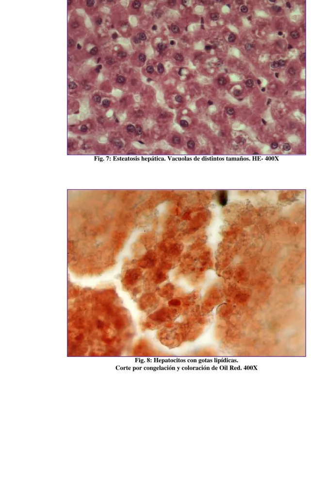
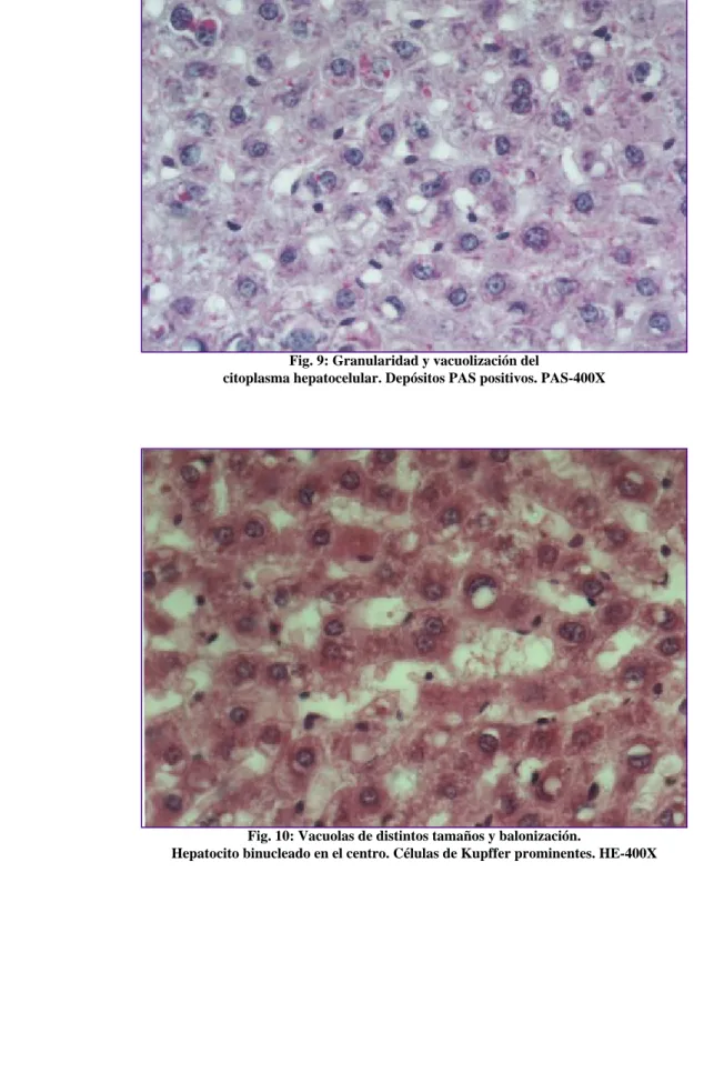
29-39.- HÍGADO GRASO NO ALCOHÓLICO EN RATAS MACHOS DE UNA LÍNEA CON DIABETES GENÉTICA Stella Maris Daniele, Juan Carlos Picena, Silvana Marisa Montenegro, María Cristina Tarrés, Stella Maris Martínez.
Facultad de Ciencias Bioquímicas, Facultad de Ciencias Médicas, Consejo de Investigaciones. Universidad Nacional de Rosario. Rosario, Argentina
40-45.- EFFECTS OF ZINGIBER OFFICINALE ON LIVER FUNCTION OF MERCURIC CHLORIDE-INDUCED HEPATOTOXICITY IN ADULT WISTAR RATS.
Ezeuko VC, Nwokocha CR, Mounmbegna PE, Nriagu CC
Department of Anatomy and Biochemistry. Madonna University, Elele Campus. Rivers State. Departmant of Physiology, Delta State University, Abraka, Delta State. Nigeria
CASE REPORTS / CASOS CLÍNICOS
46-49.- GAUCHER´S DISEASE: A RARE CAUSE OF FANCONI SYNDROME?
Musso CG, Reynaldi J, Navarro M, Vilas M, Jáuregui R, Imperiali N, Algranati L
Servicio de Nefrología. Centro Médico Agustín Rocca Hospital Italiano de Buenos Aires. Argentina 50-54.- ENFERMEDAD DE GAUCHER: UNA CAUSA INFRECUENTE DE SÍNDROME DE FANCONI ? Musso CG, Reynaldi J, Navarro M, Vilas M, Jáuregui R, Imperiali N, Algranati L
Servicio de Nefrología. Centro Médico Agustín Rocca Hospital Italiano de Buenos Aires. Argentina
55-58.- EMBOLISMO DE LÍQUIDO AMNIÓTICO. PRESENTACIÓN DE UN CASO Y REVISIÓN DE LA LITERATURA.
Silvia Maria Ferro Montes, Roberto Guzmán Parrado
Anestesiología y Reanimación y Ginecostetricia. Hospital Docente Ginecostétrico Ramón González Coro. Ciudad de la Habana, Cuba
http://biomed.uninet.edu/2007/n3/index.html
INTERNET REVIEWS / REVISIONES EN INTERNET
59-65.- INMUNOTOLERANCIA EN TUMORES GASTROINTESTINALES: HLA-G E INDOLAMINA 2,3- DIOXIGENASA
Ana Sofía López González, Carlos García-Girón, Mónica Cavia, Ana López-Muñoz, María García-González, María Jesús Coma, Pilar Muñiz
Unidad de Investigación. Unidad de Oncología Médica. Hospital General Yagüe. Complejo Asistencial de Burgos.
Área de Bioquímica y Biología Molecular. Dpto. de Biotecnología y Ciencia de los Alimentos. Universidad de Burgos.
España
LETTERS TO THE EDITOR / CARTAS AL EDITOR
66-70.- CAQUEXIA DE RUSSELL ASOCIADA A ASTROCITOMA PILOCITICO: PRESENTACIÓN DE UN CASO Leonardo Domínguez De la Ossa, Luís Rafael Moscote Salazar.
Sección de Neurocirugía. Facultad de Medicina. Universidad de Cartagena. Cartagena de Indias, Colombia
Rev Electron Biomed / Electron J Biomed 2007;3:3. Editorial: FISIOLOGÍA DEL RIÑÓN ARTIFICIAL
ISSN: 1697-090X
Inicio Home
Indice del volumen Volume index
Comité Editorial Editorial Board
Comité Científico Scientific
Committee
Normas para los autores
Instruction to Authors
Derechos de autor Copyright
Contacto/Contact:
Rev Electron Biomed / Electron J Biomed 2007;3:3-7
Editorial:
FISIOLOGÍA DEL RIÑÓN ARTIFICIAL
Carlos G. Musso 1 y Gerardo Torres Torres 2
1 Servicio de Nefrología. Hospital Italiano de Buenos Aires.
Argentina
2 Servicio de Nefrología. Hospital General Yagüe. Complejo Asistencial de Burgos. España
carlos.musso @ hospitalitaliano.org.ar
English version
Introducción
Las técnicas depurativas extrarrenales se basan en el encuentro indirecto de la sangre del paciente con un baño dialítico. Dicho encuentro, realizado a través de una membrana semipermeable (membrana dialítica), permite el intercambio de
sustancias entre ambos compartimientos: sanguíneo y dializador. Es entonces durante dicho proceso que sustancias tóxicas como la urea son removidas del
organismo y sustancias necesarias para el mismo, como el bicarbonato ingresan a él.
Si dicho encuentro tiene lugar fuera del cuerpo del paciente (en el filtro de
hemodiálisis) requiriéndose para lograrlo un circuito de circulación extracorpórea de la sangre y un circuito de agua para diálisis: se trata de un procedimiento de
hemodiálisis, mientras que si el mismo se realiza dentro del organismo (cavidad peritoneal), oficiando el peritoneo de filtro, y realizándose recambios periódicos de baño dialítico peritoneal: se trata de un procedimiento de diálisis peritoneal
1.
Fisiología del riñón artificial: métodos
El riñón artificial ofrece una serie de métodos de depuración / infusión cada uno de los cuales permite extracción / incorporación corporal de un tipo particular de sustancia. Los métodos antes mencionados son: Difusión, ultrafiltración, convección, adsorción, bufferación y calefacción. Desarrollamos a continuación cada uno de ellos:
Difusión: consiste en el pasaje de solutos a través de una membrana semipermeable del compartimiento en el que se encuentra en mayor concentración hacia aquel en
http://biomed.uninet.edu/2007/n3/editorial.html
donde se encuentra en menor concentración. En general se basa en el uso de membranas de baja permeabilidad al agua y, que permiten sólo el pasaje de
pequeñas moléculas (< 500 daltons: ejemplo la remoción dialítica de la urea, potasio, etc (1).
Ultrafiltración: consiste en el pasaje de agua de un compartimiento a otro motivado por una diferencia osmótica entre los mismos a favor del
Convección: se basa en el transporte de solutos de un compartimiento a otro como consecuencia del pasaje de fluido a través de una membrana de alta permeabilidad hidráulica. Este proceso permite la remoción de moléculas de tamaño mediano (hasta 30.000 dalton) entre las cuales se encuentran muchos de los mediadores de la
inflamación
1. Para que esta remoción sea significativa la cantidad de fluido
desplazado debe ser elevado (35 ml/Kg / h) Un ejemplo típico es la hemofiltración
2. Teóricamente un método convectivo puede eliminar solutos (moléculas pequeñas) al ser arrastradas por el pasaje del solvente, pero para que ella sea significativa debería ser la remoción del fluido tan grande que en la práctica se debe recurrir al uso de métodos difusivos
3. Los métodos convectivos remueven mediadores que participan en el síndrome de respuesta de la inflamatoria sistémica: TNF, IL 6, etc. Dicha depuración sería lograda gracias a la remoción de un alto volumen de líquido (para algunos autores 50 litros/día), así como por la adsorción de los mismos a la
membrana del hemofiltro (requerimiento de un frecuente recambio del mismo). Sin embargo estudios han demostrado que pese a esta efectiva depuración, no se logran modificaciones significativas en los niveles séricos de estos mediadores, ni en la mortalidad de los pacientes tratados. Por otra parte durante estos procedimientos son también removidos mediadores de acción protectora (anti-inflamatoria) tales como la IL 10.
3-5Adsorción: Este proceso consiste en la remoción de solutos por medio de su adhesión a la membrana del filtro. Dicha propiedad es aprovechada para depurar la sustancia en cuestión del compartimento sanguíneo, como sucede con algunos tóxicos que pueden ser removidos mediante el uso de filtros con carbón activado. Un ejemplo es la hemoperfusión
1.
Bufferación: La solución buffer a utilizar puede estar hecha a base de bicarbonato o lactato (el cual se convierte en bicarbonato, en proporción equimolar, a nivel
hepático). No obstante se debe usar bicarbonato y no lactato en algunas situaciones tales como acidosis láctica, insuficiencia hepática y post - operatorio inmediato de transplante hepático
6-7.
Calefación: En las técnicas continuas debe controlarse la temperatura corporal y evaluar si se calentará el líquido de reposición ya que existe pérdida de calor y riesgo de hipotermia
8.
Esquemas de prescripción
Los métodos ofrecidos por el riñón artificial pueden ser aplicados en forma simple o combinada siguiendo esquemas de distinta duración, frecuencia y velocidad de bomba dependiendo del objetivo que se desee alcanzar. A continuación
desarrollamos cada uno de estos esquemas alternativos:
Rev Electron Biomed / Electron J Biomed 2007;3:5. Editorial: FISIOLOGÍA DEL RIÑÓN ARTIFICIAL
❍
Velocidad de las bombas extracorpóreas:
Por lo general, en el paciente crítico se utilizan procedimientos lentos (con velocidad de bombas de sangre y dializado lentas) a fin de no propiciar con el tratamiento la inestabilización hemodinámica del paciente.
Se prefiere entonces en estos casos el uso de procedimientos que tienen mayor probabilidad de ser mejor tolerados hemodinámicamente: tales como los semicontinuos y continuos. Entre las razones por las cuales los métodos convectivos implican un menor riesgo de hipotensión arterial se encuentran:
la lenta remoción de fluido que permite el rellenado (refilling) del
compartimiento intravascular, la lenta remoción de soluto del intravascular que evita el masivo pasaje osmótico de líquido del intravascular hacia el compatimento intracelular (como sucede con la hemodiálisis), la reducción de la temperatura corporal de 2-3 grados (vasoconstricción), la remoción de mediadores con propiedades cardio-depresoras (en recambios de alto volúmen).
Estos métodos permiten lograr mayores volúmenes de ultrafiltración con mejor tolerancia hemodinámica, situación que conduce a un mejor manejo del volumen incorporado por el paciente a través de la alimentación
parentenal y del suministro de drogas intravenosas
3, 8.
❍
Tiempo y Frecuencia de las sesiones:
Desde esta perspectiva los métodos sustitutivos de la función renal pueden ser intermitentes como la hemodiálisis trisemanal, semicontinuos: diarios y prolongados en el tiempo entre 8-18 horas, como por ejemplo la diálisis prolongada de baja eficiencia (SLED) o la diálisis diaria prolongada.
Finalmente, están los procedimientos continuos que se caracterizan por ser diarios y de 24-48 horas de duración., con requerimiento adicional de líquido de reposición de modo que permita un alto volumen de desplazamiento de fluido. Ejemplo: hemofiltración veno venosa, hemodiafiltración, etc.
Con respecto a la velocidad de las bombas de sangre y baño dialítico, 300 ml/min y 500 ml/min respectivamente suelen ser las velocidades para los procedimientos intermitentes, con velocidades iguales o menores a las anteriores para los procedimientos semicontinuos, y finalmente de
velocidades más lentos (100 ml/min y 300 ml/min respectivamente) para los procedimientos continuos.
Cabe destacar que en todo procedimiento de depuración extrarrenal los solutos extraídos son removidos del compartimiento sanguíneo al baño de diálisis, proceso que genera a su vez un gradiente favorable para el pasaje de dichos solutos del compartimiento intracelular (que es en definitiva aquel que se desea depurar) al compartimiento intra-vascular. Este fenómeno aumenta el gradiente de solutos entre el compartimiento intra-vascular y el dialítico, aumentando en consecuencia la eficiencia del proceso depurativo. Sin embargo, en los procedimientos intermitentes (hemodiálisis) el pasaje de solutos entre el compartimiento sanguíneo y el dializador resulta más rápido que el suscitado entre el compartimiento intracelular y el
sanguíneo. Este fenómeno explica por qué en procedimientos intermitentes la mayor remoción de solutos se logra en las primeras 2 horas de la sesión, que es el momento de mayor gradiente sanguíneo - dializador; así como por qué luego de interrumpir una sesión dialítica sigue habiendo pasaje de solutos desde el compartimiento intracelular al intra-vascular, por el cual se produce aproximadamente 30 minutos post-desconexión un ascenso de 10-20% de los niveles séricos del soluto en remoción (fenómeno de rebote)
1.
http://biomed.uninet.edu/2007/n3/editorial.html
Por el contrario en los procedimientos lentos la velocidad de pasaje de solutos entre los distintos compartimientos se realiza aproximadamente a la misma velocidad, de modo que la depuración de soluto es prácticamente constante a lo largo de todo el procedimiento y en consecuencia los métodos lentos carecen prácticamente de fenómeno de rebote post - desconexión.
Es muy importante evitar la inestabilidad hemodinámica durante los tratamientos de depuración extrarrenal: pues al dializar un paciente hipotenso se corre el riesgo de dializar sólo su compartimiento intra-vascular y no su intracelular, que es en definitiva el objetivo del tratamiento sustitutivo. Esto se debe a que en un paciente hipotenso, sus tejidos al estar mal prefundidos, acumulan los solutos, pues su pasaje al compartimiento vascular es escaso, en consecuencia no son adecuadamente dializados del organismo. Al finalizar la sesión dialítica y mejorar la situación hemodinámica del paciente, los tejidos ahora mejor perfundidos comienzan a pasar los solutos al compartimento intravascular lo cual genera un marcado ascenso de los niveles séricos del soluto (urea), reflejando la baja eficiencia alcanzada durante la sesión dialítica
3.
Tipos de circuitos
En los métodos de depuración extrarrenal, basados en circuitos de circulación extra- corpórea, estos sistemas requieren de un circuito de salida de la sangre del cuerpo y otro para su reingreso una vez ya depurada. Mientras que los circuitos de reingreso son siempre venosos, los de salida pueden ser arteriales o venosos.
En el primer caso es la propia circulación del paciente el motor del sistema mientras que en el segundo caso se requiere de una bomba para movilizar extracorporeamente la sangre. Se trata de los circuitos arterio-venoso y veno-venoso respectivamente. El primero de estos circuitos presenta un especial riesgo de sangrado, isquemia del miembro involucrado, así como de eventos trombóticos y/o troboembólicos. Los circuitos veno-venosos presentan muchas menos complicaciones, requieren una sola canalización (venosa) al poderse utilizar catéteres doble lumen, pueden mantener un flujo sanguíneo contínuo a pesar de presentarse hipotensión arterial dado que
emplean una bomba de sangre extracorpórea. Por las razones antes mencionadas los accesos preferidos son los veno-venosos (3,9)
Conclusión:
El conocimiento de la fisiología del riñón artificial permite comprender cuales son tanto sus potenciales beneficios terapéuticos como sus limitaciones al momento de tener que sustituir la función renal nativa.
REFERENCIAS
1.- Van Stone J, Daugirdas J. Physiologic principles. In Daugirdas J, Ing T
(Eds).Handbook of dialysis. Boston. Little Brown & Company. 1994: 13-29
2.- Ronco C, Bellomo R, Hotel P, Brendolan A, Dan M, Piccini P, La Greca
G. Effects of different doses in continuous veno-venous hemofiltration on
Rev Electron Biomed / Electron J Biomed 2007;3:7. Editorial: FISIOLOGÍA DEL RIÑÓN ARTIFICIAL
outcomes of acute renal failure: a prospective randomised trial. Lancet 2000;
355: 26-30
3.- Junco E, Verde E. Depuración extrarrenal en el paciente agudo:fracaso renal agudo, alteraciones hidroelectroliticas, ácido-base e intoxicaciones. En Valderrábano F. (Ed). Tratado de hemodiálisis. Barcelona. Editorial Médica JIIMS.1999: 421-438
4.- Augustine J, Sandy D, Seifert T, Paganini E. A randomized controlled trial comparing intermittent with continuos diálisis in patients with ARF.
2004; 44 (6): 1000-1007
5.- Bellomo R, Ronco C. Continuous renal replacement therapy in the intensive care unit. Int Care Med 1999; 25: 781-789
6.- Barenbrock M, Hausberg M, Matzkies F, de la Motte S, Schaeffer M.
Effects of bicarbonate and lactate -buffered replacement fluid on
cardiovascular outcome in CVVH patients. Kidney Int 2000; 58: 1751-1757.
7.- Mas A, Salmerón J. Sistemas de sustitución hepática bioartificial. En Net A, Reglan A (Eds). Depuración extrarrenal en el paciente grave. Barcelona.
Masson. 2004: 39-50.
8.- Sánchez Izquierdo JA, Toral D. Utilidad de las técnicas continuas de reemplazo renal en la enfermedad traumática. En Net A, Reglan A (Eds).
Depuración extrarrenal en el paciente grave. Barcelona. Masson. 2004: 265- 279
9.- Bellomo R, Ronco C. Continuous versus intermittent renal replacement therapy in the intensive care unit. Kidney Int 1998; 53: S 182-185
http://biomed.uninet.edu/2007/n3/editorial.html
ISSN: 1697-090X
Inicio Home
Indice del volumen Volume index
Comité Editorial Editorial Board
Comité Científico Scientific
Committee
Normas para los autores
Instruction to Authors
Derechos de autor Copyright
Contacto/Contact:
Rev Electron Biomed / Electron J Biomed 2007;3:8-12
Editorial:
ARTIFICIAL KIDNEY PHYSIOLOGY
Carlos G. Musso 1 and Gerardo Torres Torres 2
1 Nephrology Department. Hospital Italiano de Buenos Aires.
Argentina
2 Nephrology Department. Hospital General Yagüe.
Complejo Asistencial de Burgos. España carlos.musso @ hospitalitaliano.org.ar
Versión en español
Introduction
The extrarrenal depurative techniques are based on the indirect contact of the patient´s blood with the dialysate. Such contact, performed via a semipermeable membrane (dialytic membrane), allows for the exchange of substances in both: the blood and dialytic compartment. It is during this process that toxic substances such as urea are removed from the body and substances that are necessary, such as
bicarbonate enter it. If such contact happens outside the patient´s body, in fact in the hemodialysis filter (for which an external blood and dialysate circulation circuit are necessary): this is a hemodialytic procedure, while if this procedure happens inside the body (peritoneal cavity), with the peritoneum acting as a filter: this procedure corresponds to peritoneal dialysis
1.
Physiology of the artificial kidney: methods
The artificial kidney offers a series of depurative/infusion methods, each of which allows for the extraction/incorporation of particular substances in the body. These methods are: difusion, ultrafiltration, convection, adsorption, buffer system, heating.
Difusion: it is the passage of solutes through a semi permeable membrane of the
compartment where it is most concentrated to where it is less concentrated. In
general it is based on the use of low permeability membranes, which only allows the
passage of small molecules (< 500 daltons: for example: urea, potassium, etc)
1Ultrafiltration: it is the passage of water from one compartment to the other
Rev Electron Biomed / Electron J Biomed 2007;3:9. Editorial: ARTIFICIAL KIDNEY PHYSIOLOGY
motivated by an osmotic gradient between them.
Convection: it is based on the transportation of solutes from one compartment to the other as a consequence of the passage of fluid through a high permeability
membrane. This process allows for the removal of medium sized molecules (up to 30.000 daltons) among which are most of the inflammation mediator
1. In order for this removal to be significant the amount of fluid displaced has to be high (35 ml/Kg / h). A typical example is hemofiltration
2. Theoretically, a convective method can also eliminate solutes (small molecules) when they are dragged through the solvent passage, but for this to be significant, the removal of fluid should be so much that in practise it makes it necessary to use diffusive methods for achieving this objective
3. The convective methods remove mediators that participate in the systemic
inflammatory response syndrome: TNF, IL 6, etc. Such depuration would be achieved through the removal of a high volume of liquid (for some authors 50 litres/day), and through the adsorption of these molecules in the hemofilter
membrane (it is necessary to change the filters often in order to make this effect to be significant). However, research has shown that despite this effective depuration, no significant modifications are achieved in the serum levels of the mediators, not even in the mortality of the treated patients. On the other hand, these procedures also remove protecting mediators (anti-inflammatory ones) such as the IL 10.
3-5Adsorption: This process consists of the removal of solutes through its adhesion to the filter membrane. Such property is used to depurate the substance in question from the blood compartment, as it happens with some toxic substances which can be removed by using filters with charcoal
1.
Buffer System: The buffer solution to be used can be based on bicarbonate or lactate base (which becomes bicarbonate, in equimolar proportion, at hepatic level).
Nevertheless, bicarbonate must be used instead of lactate in some situations such as lactic acidosis, hepatic insufficiency and post - surgical treatment immediately after hepatic transplant
6-7.
Heating: in continuous techniques body temperature must be controlled and it should be evaluated whether to heat the replacing solution since there is loss of heat and subsequent risk of hypothermia
8.
Artificial kidney physiology: prescription schemes
The methods offered by the artificial kidney can be applied in a simple or combined way following schemes of different duration, frequency and pump speed depending on the objective you want to achieve.
❍
Speed of external pumps:
In general, in critical patients it is common to use slow procedures (with blood and dialysate slow speed pumps) so as not to induce hypotension.
Thus, in this situation is better to use procedures which have more chances of being tolerated: such as semi continuous and continuous therapies. Among the reasons why convective methods involve a lower risk of arterial
hypotension are; the slow removal of fluids which allow the refilling of the intravascular compartment, the slow removal of the intravascular solutes which prevents the massive osmotic passage of intravascular liquid to the
http://biomed.uninet.edu/2007/n3/editorial-en.html
intracellular compartment (as it happens in hemodialysis), decrease of body temperature in 2-3 degrees (vasoconstriction), the removal of mediators with heart-depressor properties (only in high volume exchanges).
These methods allow to use bigger volume of ultrafiltration with better hemodynamic tolerance, which leads to a better handling of the volume absorbed by the patient through parentenal feeding and from intravenous drugs
3, 8.
❍
Time and frequency of the sessions:
From this perspective, the substitutive methods of the renal function can be intermittent such as hemodialysis three times a week, semicontinuos: daily and prolonged in time between 8-18 hours, such as low efficiency prolonged dialysis (SLED) or daily prolonged dialysis. Finally, there are the continuous procedures which are daily and last 24-48 hours, with an additional
requirement of fluid restitution so it allows a high volume of fluid
displacement. Example: veno venous hemofiltration, hemodyafiltration, etc.
Regarding the speed of the blood pumps and the dialysate, 300 ml/min and 500 ml/min respectively usually are the speeds of the intermittent procedures, with speeds similar or lower to the previous ones for semicontinuous
procedures, and finally slower speeds (100 ml/min y 300 ml/min respectively) for continuous treatments.
It is worth noticing that in every extra renal depuration procedure the solutes extracted are removed from the blood compartment to the dialyzed, which in turn creates a favourable gradient for the passage of such solutes from the intracellular compartment (which is the one we wish to depurate) to the intra-vascular one. This phenomenon increases the gradient of solutes between the intravascular and dialytic compartments, thus increasing the efficiency of the depurative process. However, in intermittent procedures (hemodyalisis) the passage of solutes between the blood and the dialytic compartments is faster than the one between the intracellular and the blood compartment. This phenomenon explains why in intermittent procedures the most significant removal of solutes is achieved in the first 2 hours of the session, which is the moment of the bigger blood-dialysate gradient; it also explains why after interrupting the dialytic session there are still solutes passing from the intracellular to the intravascular compartment, in which approximately 30 minutes after
disconnection the serum levels of solutes being removed increased in a 10-20%
(rebound effect)
1.
On the contrary, in slow procedures the speed of the passage of solutes between the different compartments happens approximately at the same speed, so the depuration of the solutes is virtually constant throughout the procedure and consequently the slow methods do not cause rebound effect after disconnection.
It is very important to keep hemodynamic stability during extrarenal depuration
treatments: since when a hypotense patient is dialyzed it is common to dialyse only
the intravascular and not the intracellular compartment, which is the objective of the
substitutive treatment. This happens because in a hypotense patient, as their tissues
are badly perfused they accumulate solutes, since there is little passage of them to the
vascular compartment, thus they are not dialyzed properly out of the body. When
the dialytic session is finished and the hemodynamic condition of the patient
improves, the tissues that are now better perfused begin to pass solutes to the
Rev Electron Biomed / Electron J Biomed 2007;3:11. Editorial: ARTIFICIAL KIDNEY PHYSIOLOGY
intravascular compartment which generates a significant increase in serum levels of the solutes (urea), reflecting the low efficiency reached during the dialytic session
3.
Artificial kidney physiology: types of circuits
In the extra-renal depurative methods, based on extra body circulation circuits, these systems require a circuit for blood goes of the body and another for its reentry once it has been depurated.
While the reentry circuits are always venous systems, the exit circuit can be an arterial or venous one. In the first case the motor of the system is the circulation of the patient itself while in the second case it is necessary to use a pump to mobilize the blood. These are the arterial-venous and venous-venous circuits respectively. The first of these circuits presents a special risk of bleeding, ischemia of the part of the body involved, as well as thrombotic events. Veno-venous circuits present a lot less complications, they require only one access (a venous one) since it is possible to use a double lumen catheter, they can hold a continuous blood flow despite presenting the patient arterial hypotension since it uses an extra-body blood pump. Because of all the above exposed reasons the preferred access is the veno-venous one. (3,9)
Conclusion:
The knowledge of the physiology of the artificial kidney enables us to understand which are its potential therapeutic benefits as well as its limitations when substituting the native renal function.
REFERENCIAS
1.- Van Stone J, Daugirdas J. Physiologic principles. In Daugirdas J, Ing T (Eds).Handbook of dialysis. Boston. Little Brown & Company. 1994: 13-29 2.- Ronco C, Bellomo R, Hotel P, Brendolan A, Dan M, Piccini P, La Greca G. Effects of different doses in continuous veno-venous hemofiltration on outcomes of acute renal failure: a prospective randomised trial. Lancet 2000;
355: 26-30
3.- Junco E, Verde E. Depuración extrarrenal en el paciente agudo:fracaso renal agudo, alteraciones hidroelectroliticas, ácido-base e intoxicaciones. En Valderrábano F. (Ed). Tratado de hemodiálisis. Barcelona. Editorial Médica JIIMS.1999: 421-438
4.- Augustine J, Sandy D, Seifert T, Paganini E. A randomized controlled trial comparing intermittent with continuos diálisis in patients with ARF.
2004; 44 (6): 1000-1007
5.- Bellomo R, Ronco C. Continuous renal replacement therapy in the intensive care unit. Int Care Med 1999; 25: 781-789
6.- Barenbrock M, Hausberg M, Matzkies F, de la Motte S, Schaeffer M.
Effects of bicarbonate and lactate -buffered replacement fluid on
http://biomed.uninet.edu/2007/n3/editorial-en.html
cardiovascular outcome in CVVH patients. Kidney Int 2000; 58: 1751-1757.
7.- Mas A, Salmerón J. Sistemas de sustitución hepática bioartificial. En Net A, Reglan A (Eds). Depuración extrarrenal en el paciente grave. Barcelona.
Masson. 2004: 39-50.
8.- Sánchez Izquierdo JA, Toral D. Utilidad de las técnicas continuas de reemplazo renal en la enfermedad traumática. En Net A, Reglan A (Eds).
Depuración extrarrenal en el paciente grave. Barcelona. Masson. 2004: 265- 279
9.- Bellomo R, Ronco C. Continuous versus intermittent renal replacement
therapy in the intensive care unit. Kidney Int 1998; 53: S 182-185
Electron J Biomed 2007;3:13. Kendabie and Adjene. EFFECTS OF RED PEPPER ON THE STOMACH OF ADULT WISTAR RATS
ISSN: 1697-090X Inicio Home
Indice del volumen Volume index
Comité Editorial Editorial Board
Comité Científico Scientific Committee
Normas para los autores
Instruction to Authors
Derechos de autor Copyright Contacto/Contact:
HISTOLOGICAL STUDIES OF THE EFFECTS OF RED PEPPER ON THE STOMACH
OF ADULT WISTAR RATS.
K. O. Kendabie, Josiah O. Adjene.
Department of Anatomy, School of Basic Medical Sciences, University of Benin, Edo State, Nigeria.
Rev Electron Biomed / Electron J Biomed 2007;3:13-17
Comment of the reviewer Prof. Pilar Muñiz Rodríguez PhD. Área de Bioquímica y Biología Molecular. Dpto. de Biotecnología y Ciencia de los Alimentos. Universidad de Burgos. España
Comment of the reviewer Erhan Suleymanoglu PhD. G.U.E.F., Department of Pharmaceutical Chemistry, Gazi University. Gazi Mahallesi, Ankara. Turkey.
ABSTRACT:
Histological effects of red pepper commonly used as spice in food on the stomach of adult wistar rats were carefully investigated.
The rats of both sexes (n=24), average weight of 200g were randomly assigned into two treatments (n=16) and control (n=6) groups. The rats in the treatments groups received 1g and 2g of red pepper thoroughly mixed with 20g of their feeds for 7 and 14 days, while the control rats received equal amounts of feeds without the red pepper added. The rats were fed with grower's mash purchased from Edo feeds and flour mill Ltd, Ewu, Edo State and were given water liberally. The rats were sacrificed on day eight and fifteen of the experiment respectively.
The stomach was carefully dissected out and quickly fixed in 10% formol saline for routine histological procedure after H & E method.
The histological findings after H&E methods indicated that the treated sections of the stomach showed some level of cellular hypertrophy, congestion of blood vessels degenerative changes disruption and distortion of the cytoarchitecture of the stomach.
These findings indicate that red pepper may have some deleterious effects on the microanatomy of the stomach of adult wistar rat at higher doses. It is recommended that further studies aimed at corroborating these findings be carried out.
Key words: Red pepper. Histological effects. Stomach and wistar rats.
RESUMEN:
Han sido investigados los efectos histológicos de la pimienta roja, especie comúnmente utilizada como condimento de alimentos, en el estómago de ratas wistar adultas. 24 ratas de ambos sexos de un peso promedio de 200 gramos fueron asignados
aleatoriamente a dos grupos: experimental (n = 16) y control (n = 6). Las ratas del grupo experimental recibieron 1g y 2g de la pimienta roja, mezclada con 20 gramos de la dosis de pienso para 7 y 14 días, mientras que el grupo de ratas control recibió iguales cantidades de alimentos, pero sin pimienta roja añadida. El pienso de rata procedía de Edo feeds and flour mill Ltd., Ewu, Edo State, y se les proporcionó a los animales agua sin restricciones. Los animales fueron sacrificados en el día ocho y quince de la prueba, respectivamente.
El estómago fue cuidadosamente disecado y fijado rápidamente en una solución salina de formol al 10% para estudio histológico
http://biomed.uninet.edu/2007/n3/kendabie.html
y siendo procesado rutinariamente y teñido con Hematoxilina y Eosina.
Los hallazgos histológicos en los cortes de estómago mostraron cierto grado de hipertrofia celular, congestión de los vasos sanguíneos y cambios degenerativos, con alteraciones y distorsión de la citoarquitecture del estómago.
Estos resultados indican que la pimienta roja puede tener algunos efectos perjudiciales sobre la anatomía microscópica del estómago de las ratas Wistar adultas a dosis más altas. Serían recomendables nuevos estudios encaminados a corroborar estos resultados.
Palabras clave: Pimienta roja. Efectos histológicos. Estómago y ratas wistar.
INTRODUCTION
Pathological processes frequently involve the body's normal responses to abnormal environmental influences. Such noxious external influences as pathogenic microorganisms, trauma, dietary deficiencies and hereditary factors acting alone or in a complex interaction with environmental factors, case diseases1. Various environmental chemicals, industrial pollutants and food additives have been implicated as causing harmful effects2. Most food additives act either as preservatives, or enhancer of palatability. Spices, in particular black pepper, red pepper, and chili powder, may produce indigestion, but they do not seem to seriously injure the stomach3. Some investigators have reported that there is no difference in rates of inflammation of the stomach in heavy consumers of spice and no difference in the rate of ulcer heating in those patients consuming large amount of red pepper daily4,5. In experiments on rats, the active ingredient in pepper, capsaicin, was found to protect the stomach mucosa from damage caused by alcohol or aspirin6,7.
Capsaicin is contained in a different spicy vegetable among which is cayenne pepper, which was used in this experiment.
Cayenne pepper is a member of the family solanaceae genus: capsicum of vegetables and with botanical name - capsicum frutescence. The common name, cayenne, was actually given to this pepper because of its cultivation in a town that bears the same name in French Quiana on the North-East Coast of South America. The Scoville heat unit of cayenne pepper ranges 30k to 50k8. Red pepper is the mostly used spicy for food throughout the world, especially in Central America, Latin America, Africa and Asia9. In moderately large quantities, it is one of the most ingredients used in meals in Nigeria, especially the Southern part.
Chemically, the red pepper contains approximately 0.14% capsaicin10-11, a crystalline colourless compound which is an active principle that accounts for its pungency.
It has been reported that the gastro protection afforded by prostaglandin E2, cholecystokinin gastrin, the proton pump inhibits lansoprazole, and the antacid, hydrotalcit is reduced or abolished in capsaicin pretreated rats12. In 1999, Holzer reported that capsaicin lessen ablation effect produced by aspirin, and that neurotoxin dosage of capsaicin defunctionalizes nociceptive afferents, including those innervating the stomach, this producing a chemical knockout of neurons that participate in the maintenance of the gastric mucosal integrity13. In the stomach of the rats capsaicin has been reported to cause no change on the basal mucosal blood flow14. While it was also stated by another investigator that in the stomach of the rat capsaicin do not cause any change in the basal acid and pepsin15.
The stomach is the most dilated portion of the alimentary canal, located in the epigastriun and the left hypochondruim regions of the abdomen. It is lined by simple columnar epithelial cell and functions in the degradation and digestion of food materials in the body. The stomach also functions in the preventions of gastric ulceration due to the presence of the numerous mucous secreting glands. Since red pepper contains capsaicin which was found to protects the stomach mucosa from damage caused by alcohol or aspirin, it is therefore relevant to investigate some of its histological effects on the stomach. It is probable that the adverse effects of red pepper may affect the normal histological structure of the stomach, and hence this investigation.
MATERIALS AND METHODS
Animals: Twenty four adult wistar rats of both sexes with average weight of 200g were randomly assigned into three groups, (two treatments n=16 and control n=6). The rats were obtained and maintained in the animal holdings of the department of anatomy, school of basic medical sciences, University of Benin, Edo State, Nigeria. They were fed with grower's marsh obtained from Edo feed and flour mill limited, Ewu, Edo State and given water liberally. The control group consisted of six rats while the two treatments groups consisted of six rats each red pepper was obtained from the Edaiken market, Uselu, Benin City.
Red pepper administration: Red pepper was obtained from the Edaiken market, Uselu, Benin City, dried and grinded in the department of anatomy, school of basic medical sciences, University of Benin Edo State. The rats in the treatments groups were divided into two main groups (A, B), with each group receiving 1.0g and 2.0g of red Pepper mixed with 20g of grower's march respectively. The red pepper was thoroughly mixed with the feeds in a container fixed to the floor of the experimental cage to avoid spillage of the feeds during their feeding. The quantity of feeds consumed was known by weighing before and after each day of the experiment. The control rats received equal amount of feeds without red pepper added for the same period of the experiment: the rats were sacrificed by cervical dislocation method on the eight and fifteenth days of the experiments respectively. The stomach was quickly dissected out and fixed in 10% formal saline for routine histological techniques.
Histological study: The tissue were dehydrated in an ascending grade of alcohol (ethanol), cleared in xylene and embedded in paraffin wax, serial sections of 7 microns thick were obtained using a rotator microtome. The deparaffused sections were stained routinely with haematoxylin and eosin. Photomicrographs of the desired section were obtained using digital research
photographic microscope in the department of anatomy, school, of basic medical sciences, university of Benin for further
Electron J Biomed 2007;3:15. Kendabie and Adjene. EFFECTS OF RED PEPPER ON THE STOMACH OF ADULT WISTAR RATS observations.
RESULTS
The stomach of the control section showed normal histological features with the mucosa lined with simple columnar epithelial cells, lamina propria showing some highly packed glandular secretory cells, and some blood vessels in the section of the control stomach.
The stomach of the treated sections showed some level of cellular hypertrophy associated with congestion of some blood vessels.
There is also some degree of degenerative changes distortion and necrotic debris associate with the treated sections which is more marked in the treatment sections receiving 2g of red pepper
DISCUSSION
The results of the haematoxylin and eosin staining (H&E) reactions showed increased cellular hypertrophy and degenerative changes in the stomach of the treatment groups. The increased in cellular hypertrophy of the treatments group as reported in this study may have been as a result of cellular proliferation caused by the intake of food mixed with the red pepper. It may then be inferred from the present results that higher dose and prolonged intake of red pepper resulted in degenerative changes observed in the glandular epithelium of the stomach. The actual mechanism by which red pepper induced cellular degeneration observed in this experiment needs further investigation.
Degenerative changes have been reported to result in cell death, which is of two types, namely apoptotic and necrotic cell death.
These two types differ morphologically and biochemically16. Pathological or accidental cell death is regarded as necrotic and could result from extrinsic insults to the cell such as osmotic, thermal toxic and traumatic effects17. In this experiment, red pepper could have acted as toxins to the cell of the stomach. The process of cellular necrosis involves disruption and distortion of membrane's structural and functional integrity which was also a landmark of this experiment.
In cellular necrosis, the rate of progression depends on the severity of the environmental insults. The greater the severity of insults, the more rapid the progression of neuronal injury18. The principle holds true for toxicological insults to the brain and other organs19. It may be inferred from the present results that prolonged intake of red pepper resulted in increase toxic effects on the stomach with that of higher dose more marked. This work is in consonance with the research work carried out by Eweka et al, (2007) that monosodium glutamate used as food additive causes cellular hypertrophy, vacuotations and distortions in the epithelia of the stomach20.
The observed congestion of blood vessel in the treated groups suggested the occurrence of inflammatory, reaction which was as a result of tissue damage. This could be explained by the fact that red pepper inflicted chemical injury on the gastric mucosa which was followed by arterial relaxation around the stomach. The capillary network around the stomach then becomes engorged with rapidly flowing blood accompanied by lymphocytic infiltration. This work is in line with some studies carried out on the effect of red pepper on rat gastric mucosa resulting to some abnormality ranging from oedema submucosal
haemorrhages, congestion of blood vessels and exfoliation of gastric surface epithelial cells21-22.
CONCLUSION AND RECOMMENDATION
In conclusion, our study revealed that chronic administration of red pepper to adult wistar rats caused cellular hypertrophy, congestion of blood vessels, degenerative changes, disruption and distortion of the cytoarchitecture of the stomach. This resulted in some necrotic debris associated with the treated sections.
With these results, it is probable that the functions of the stomach may be adversely affected. It is recommended that further studies aimed at corroborating these findings be carried out.
REFERENCES
1. Allen, G.H: The genetic basis of diseases in general pathology, Churchill Iivingstone Medical Division Longman Co.
Ltd, New York 1989 pp. 35056.
2. Moore K.L: Congenital Malformations due to environmental, Developing Humans W.B. Saunders Co. Ltd Philadelphia 2003 2nd Ed. Chap. 8. pp. 173-183.
3. Graham D: Spicy food and the stomach, evaluation by video endoscopy. JAMA 1988. 260:3473-75
4. Tyagi K: Gastric mucosal morphology in tropics and influence of spices, tea and smoking Nutr metab 1974 17: 129- 135.
5. Kumar, N: Do Chilies influence healing of duodenal ulcer? BMJ 1984 288: 1803-4
6. Holzer, P: Intragastric capsaicin protects against aspirin - induced lesion formation and bleeding in rat gastric mucosa. Gastroenterology 1989 Jun: 96(6): 1425-33.
7. Holzer, P: Stimulation of afferent nerve endings by intragastric capsaicin protects against ethanol - induced damage of http://biomed.uninet.edu/2007/n3/kendabie.html
gastric mucosa. Neurosience 1988 27:981-7.
8. Berkley R; Peppers A cook book, New York, Simon and Schuster 1992
9. Nopaintaya, W and Nye S.W: Duodenal mucosal response to the pungent principle of Hot pepper (capsaicin) in rat:
light and Electron microscopic study. Toxicology and applied pharmacology 1974. 30 (1): 149-161
10. Sir sat S.M. and Khanolkar, V.R. Sub mucous fibrosis of the palate in diet-preconditoned wistar rats (induction by local painting by capsaicin. An optional and electron microscope study J. Arch pathol 1960 70:171-179.
11. Watt, J.M and Breyer - Bandwijk M.G: The medicinal and poisonous plants of southern and Eastern Africa 2nd Edition Living Stone 1962 942-943
12. Holzer et al: Neural emergency system in the stomach Gastroenterology 1998: 114: 823-839.
13. Holzer P: Implications of tachykinins and calcitomn gene-related peptides in inflammatory bowel disease digestion 1999 59: 269 - 283.
14. Raybould, H.E; Steminic Eysselein, V.E; Yoneda, M; Holzer, P: Selective ablation of spinal afferent neurons containing CGRP attenuates gastric hyperemic response to acid. Peptides 1992 13:249-254
15. Lippe, I.T; Pabst, MA; Holzer, P: Intragastric capsaicin enhances rat gastric acid elimination and mucosal blood flow by afferent neurons stimulation Br. J. Pharmaco 1989 96:91-100.
16. Wyllie, A.H: Glucocorticoid-induced thymocyte apoptosis is associated and endogenous endonuclease activation nature, London 1980; 284: 555-556
17. Farber, J. L; Chein, K.R; Mittnacht, S: The pathogenesis of irreversible cell injury in ischemia; American Journal of pathology 1981; 102:271-281
18. Ito, U; Sparts, M; Walker, J.T; and Warzo, I: Experimental cerebral ischemia in Magolian Gerbils (1) Light microscope observations. Acta neurophatology USA. 1975; 209 - 223.
19. Martins, L.J; Deobler, J.A; Shih, T; Anthony, A: Cytophotometric analysis of thalamic neuronal RNA 1984; 35: 1593- 1600
20. Eweka, A.O; Om'Iniabohs, F.A.E; Adjene, J.O: Histological studies of the effects of monosodium Glutamate on the stomach of adult Wistar Rats Ann Biomed Sci 2007, 6:1: 45-52
21. Viramivatti Vikit el al: Effect of capsicum solution on human gastric mucosa as observed gastroscopically. Am. J.
Gastroenterology 1972; 58:225-232.
22. Desai, H.G; Venugo Polan, K; Antia, F.P: Effect of Chili powder on DNA content of gastric aspirates 1973; 14:974- 976.
Comment of the reviewer Prof. Pilar Muñiz Rodríguez PhD. Biochemical and Molecular Biology Section. Biotechnology and Food Science Department. University of Burgos. España
In the present study a stomach histological investigation was carried out on the effects of feeding rats with a diet containing red pepper.
Red pepper is one of the spices that has been used all over the world from old times. It varies physiological activities, especially the suppression of blood lipid levels and the antibacterial activity, and it has an inhibitory effect on cecal bifidobacteria, etc.
Furthermore, its active material, capsaicin, is irritating and inflammatory and may cause gastric ulcers and mucosal lesions. The present study reporting histological data on the effect of red pepper on the stomach can be regarded as an interesting
contribution to previous studies
Comment of the reviewer Erhan Suleymanoglu PhD. G.U.E.F., Department of Pharmaceutical Chemistry, Gazi University. Gazi Mahallesi, Ankara. Turkey.
Having considered the current concerns regarding the effects of diet on developing stomach pathology, the evaluation of the dietary risk factors for stomach disorders becomes essential. The mechanism by which dietary pepper affects various gastric features is poorly understood, as is the ability of certain compounds to relieve possible symptoms. Therefore, it is worth
Electron J Biomed 2007;3:17. Kendabie and Adjene. EFFECTS OF RED PEPPER ON THE STOMACH OF ADULT WISTAR RATS
investigating the ability of black pepper, red pepper and other relevant chilli species to determine gastric surface hydrophobicity and to induce or relieve visceral pain in rat model systems.
The study presented herein examines the effects on stomach of adult Wistar rats of red pepper consumption, one of the most popular ingredients in authors' native Nigeria, as well as elsewhere. The experimental design and results are clearly presented.
The histological findings show certain level of cellular hypertrophy, congestion of blood vessels, degenerative changes and distortion of the cytoarchitecture of the stomach, following the administration of red pepper. The presented work is of interest also for comparative purposes with the results of other research groups and thus deserves to be published.
Corresponding author: Josiah O. Adjene
Department of Anatomy, School of Basic Medical Sciences, University of Benin, Edo State, Nigeria.
Received August 31, 2007 Published October 28, 2007
http://biomed.uninet.edu/2007/n3/kendabie.html
ISSN: 1697-090X Inicio Home
Indice del volumen Volume index
Comité Editorial Editorial Board
Comité Científico Scientific Committee
Normas para los autores
Instruction to Authors
Derechos de autor Copyright Contacto/Contact:
DAÑO OXIDATIVO A LÍPIDOS Y PROTEÍNAS EN LA INSUFICIENCIA RENAL CRÓNICA EXPERIMENTAL.
Miriela Betancourt Valladares, Ygber Luis González de la Cruz, Elizabeth Vidor Guerra, Madelín Miranda Naranjo, María Josefina Méndez
Instituto Superior de Ciencias Médicas Carlos J. Finlay.
Facultad de Estomatología. Departamento de Ciencias Básicas.
Camaguey. Cuba.
Rev Electron Biomed / Electron J Biomed 2007;3:18-28
Comentario del revisor Prof. Pilar Muñiz Rodríguez PhD. Área de Bioquímica y Biología Molecular. Dpto. de Biotecnología y Ciencia de los Alimentos. Universidad de Burgos. España
Comentario del revisor Dr. Abdías Hurtado Aréstegui. Servicio de Nefrologia. Hospital Arzobispo Loayza. Universidad Peruana Cayetano Heredia. Lima. Peru
ABSTRACT:
An experimental trial in 40 Wistar rats was done. The renal failure was induced by surgical ablation of 5/6 of the renal mass to 30 rats; 3 groups were formed and followed over a period of 2, 4, and 6 weeks. The remaining group of rats was used as control.
Functional remainder state was evaluated by measurement of the Glomerular Filtration Rate (GFR) and effective Renal Plasmatic Flow (RPF), Filtration Fraction (FF) was also calculated. Lipid and protein oxidative damage were evaluated on the renal tissue. As markers of oxidative stress the levels of Malonildialdehyde (MDA) and Advanced oxidation protein products (AOPP) were determined. Progressive decreasing of GFR, RPF and FF were noted. MDA levels rose through the time, AOPP concentrations was also higher over the nephrectomized rats. The relationship between progression of experimental chronic renal failure and oxidative stress was showed.
Key words: Chronic Renal Failure, Glomerular filtration, Renal Plasmatic Flow, Filtration Fraction, oxidative stress
RESUMEN:
Se realizó un estudio experimental en 40 ratas Wistar provocando insuficiencia renal crónica (IRC) por ablación de 5/6 de la masa renal a 30 de ellas; las que fueron divididas en 3 grupos y evolucionadas durante 2, 4 y 6 semanas comparadas con las 10 ratas restantes sanas.
Para evaluar el estado funcional remanente se midió la intensidad de filtración glomerular (IFG), el flujo plasmático renal efectivo (FPR), y se calculó la fracción de filtración (FF). Se evaluó el daño oxidativo a lípidos y proteínas en homogenado de tejido renal. Para este fin se determinaron los niveles de malonildialdehído (MDA) y de productos avanzados de oxidación de proteínas (PAOP) como indicadores de daño oxidativo.
Se constató la disminución progresiva de la IFG y el FPR y disminución de la FF. Los niveles de MDA se elevaron en función del tiempo y los de PAOP fueron mayores en las ratas nefrectomizadas. Se concluyó la implicación del estrés oxidativo en la progresión de la IRC experimental.
Palabras clave: Insuficiencia renal crónica, filtrado glomerular, flujo plasmático renal efectivo, fracción de filtración, estrés oxidativo.
Electron J Biomed 2007;3:19. Betancourt y col. DAÑO OXIDATIVO A LÍPIDOS Y PROTEÍNAS EN LA IRC EXPERIMENTAL.
INTRODUCCIÓN
Dentro de las enfermedades crónicas no transmisibles la insuficiencia renal se destaca como un prioritario problema de salud por su repercusión importante sobre la calidad de vida de los que la padecen. Se entiende como insuficiencia renal crónica (IRC) la reducción lenta y progresiva de la filtración glomerular, que se asocia a medida que avanza, a la pérdida de las restantes funciones ejercidas por el riñón. Una de las observaciones clínicas más relevantes en los últimos años, corroborada en modelos experimentales, ha sido que independientemente de la causa que determina el daño renal inicial y a partir de cierto grado de destrucción tisular, la función renal puede continuar deteriorándose, sin que medie en ello ningún estímulo nocivo adicional1. Las causas fundamentales de IRC, según la Asociación Europea de Diálisis y Trasplante (EDTA) son las glomerulonefritis; las nefropatías tubulointersticiales crónicas; la diabetes; la nefroangiosclerosis y las nefropatías autoinmunes2.
El daño ocasionado por estas enfermedades provoca cambios adaptativos en las nefronas remanentes, que inicialmente
compensan la pérdida de las funciones que se ven comprometidas por la afectación de la masa renal; pero a la vez constituyen un factor de progresión de la enfermedad hacia su etapa terminal3-4.
Brenner y colaboradores formularon hace un par de décadas, una hipótesis unificadora que sostiene que cuando hay pérdida de nefronas, las remanentes se adaptan hipertrofiándose e hiperfiltrando, para compensar la pérdida inicial de la función y más tarde, van a la glomeruloesclerosis, que destruye nuevas unidades y perpetua el ciclo hasta la destrucción final del parénquima renal3. El estudio secuencial de la morfología y función renal tras la remoción de la masa renal en ratas ha mostrado que efectivamente, hay una hiperfiltración e hipertrofia renal inicial, seguida de una caída de la función y tamaño renal en el tiempo5.
La teoría de la hiperfiltración, original y muy bien avalada experimentalmente, aún tiene puntos oscuros por lo que se han propuesto múltiples mecanismos para explicar la vasodilatación de la arteriola aferente y parece tratarse de un fenómeno multifactorial con implicación de diversos mediadores. Teniendo en cuenta que dicha teoría no explica en su totalidad la progresión de la IRC es necesario admitir la implicación de otros mecanismos2.
Hoy sabemos que la progresión hacia la insuficiencia renal terminal de la mayoría de las enfermedades renales, es producto de los efectos de la angiotensina II. La hormona se acopla a dos receptores de alta afinidad: el tipo 1 (AT1) y el tipo2 (AT2). La señalización a través del receptor AT1 resulta en vasoconstricción, estimulación del crecimiento, activación de los fibroblastos y miocitos y aumento de la expresión de varios factores proliferativos que promueven el crecimiento celular y la fibrosis. La señalización a través del AT2 resulta en vasodilatación, respuesta antiproliferativa y aumento de la apoptosis6.
La angiotensina II también estimula el estrés oxidativo. Tal estrés potencia el rol vasoconstrictor del péptido debido al aumento del catabolismo del NO6. Es esta una de las razones por las se ha empezando a considerar que el estrés oxidativo es otro elemento a tener en cuenta en la patogénesis de la IRC progresiva. Estudios realizados en pacientes con distintos grados de IRC, sugieren que los enfermos renales están en una situación de estrés oxidativo, si los comparamos con individuos sanos y que el grado de estrés oxidativo está correlacionado con el grado de IRC7.
El estrés oxidativo aparece en células y tejidos cuando se rompe el equilibrio que existe entre sustancias prooxidantes y antioxidantes a favor de las primeras. Las principales sustancias oxidantes en los sistemas biológicos son los radicales libres derivados del oxígeno (EROs)8.
Que las EROs están implicadas en el daño renal progresivo se basa en varias líneas de evidencias:
● En la IRC está aumentada la producción de EROs.
● Distintas estrategias antioxidantes ejercen efectos beneficiosos en modelos de injuria renal crónica.
● El estrés oxidativo puede inducir en el riñón sano cambios similares a los vistos en la IRC.
La mayor parte de los estudios realizados se han basado en la medición directa de productos oxidantes e indirectamente midiendo productos de peroxidación lipídica en los tejidos renales7, 9-10.
Los estudios que relacionan el estrés oxidativo con la IRC motivaron la realización de la presente investigación, en la que se provocó IRC experimental en ratas utilizando el modelo de nefrectomía subtotal por ablación de 5/6 de la masa renal y se evaluó el comportamiento secuencial de 2 marcadores de estrés oxidativo en el homogenado de riñón a medida que declinaba la función del órgano. El estudio se realizó a las 2, 4 y 6 semanas post ablación para determinar los cambios paulatinos en dichos
marcadores, comparados con un grupo control sin intervenir relacionando los mismos con indicadores del estado funcional remanente.
MATERIAL Y MÉTODOS
Se trabajó con un grupo de 40 ratas Wistar, con un peso aproximado de 180 gramos. La muestra se dividió en 4 grupos de 10 animales cada uno, un grupo CONTROL (C) constituido por ratas que no fueron sometidas a proceder quirúrgico y tres grupos EXPERIMENTALES (E) que fueron sometidos a nefrectomía subtotal por ablación de 5/6 de la masa renal, eliminando el riñón derecho y ligando 2 ramas de la arteria renal izquierda, sin aplicar ningún tratamiento. El grupo experimental E- I se evolucionó durante 2 semanas después de la nefrectomía sub-total, el grupo experimental E- II se evolucionó durante 4 semanas y el grupo experimental E- III durante 6 semanas.
http://biomed.uninet.edu/2007/n3/betancourt.html
El último día de evolución de cada animal se realizó el aclaramiento plasmático de ácido paramino-hipúrico (PAH) e inulina (I) para medir el flujo plasmático renal efectivo (FPR) y la intensidad de filtración glomerular (IFG) respectivamente, por el método multicompartimental. Con los resultados de IFG y FPR se calculó la fracción de filtración (FF). El comportamiento de la IFG, el FPR y la FF permitieron evaluar las modificaciones funcionales a medida que progresaba la insuficiencia renal. La masa renal fue extraída y congelada; posteriormente se hizo el homogenado del tejido y se utilizó el sobrenadante para las
determinaciones relacionadas con el estrés oxidativo, que incluyeron la determinación del daño oxidativo a lípidos y proteínas [niveles de malonildialdehído (MDA) y concentración de productos avanzados de oxidación de proteínas (PAOP)
respectivamente].
Proceder para la medición de los aclaramientos plasmáticos
Se obtuvieron 12 muestras de sangre donde el PAH y la I fueron determinados en filtrados liberados de proteínas por sulfato de cadmio, utilizando para el PAH la técnica fotocolorimétrica de Bratton y Marshall, modificada por Homer W. Smith y para la inulina el método directo del resorcinol sin tratamiento alcalino. Con las concentraciones plasmáticas de PAH e Inulina se calcularon los aclaramientos mediante el análisis de descomposición multicompartimental de las curvas de desaparición respectivas de dichas sustancias en el plasma11.
Los resultados fueron expresados en mililitros por minuto por 100 gramos de peso rata (mL/min/100g)
Técnica para la determinación de los niveles de malonildialdehído (MDA).
La determinación de malonildialdehído se realizó a partir de una técnica espectrofotométrica basada en la reacción de dos moléculas del reactivo cromogénico N - methyl - 2 - phenyindole con una molécula de MDA (producto final de la peroxidación lipídica) a 45ºc que conduce a un cromóforo estable con un máximo de absorbancia a 486 nm12. Los resultados se expresaron en micromoles por litro (µmol/L)
Técnica para la determinación de productos avanzados de oxidación de proteínas (PAOP).
La concentración de PAOP es expresada como equivalentes de cloramina T (patrón) en condiciones acídicas a 340 nm en presencia de ioduro de potasio, siguiendo la transformación de los iones yodo a yodo biatómico que provocan estos PAOP13. Los resultados se expresaron en micromoles por litro (µmol/L)
Análisis estadístico.
Para resumir la información de las variables se utilizaron medidas de resumen y dispersión como la media y la desviación estándar. Se realizó ANOVA de una vía de efecto fijo para estudiar el cambio de las variables en el tiempo y la trayectoria entre los grupos. Se realizó el análisis de regresión lineal para establecer relación entre la función renal y los marcadores de estrés oxidativo. Se utilizó para el análisis el paquete estadístico SPSS para Windows.
RESULTADOS
Los resultados obtenidos en los animales del grupo control (C) manifestaron en las variables flujo plasmático renal efectivo (FPR), intensidad de filtración glomerular (IFG) y fracción de filtración (FF) valores que corresponden a cifras normales, de manera que contra los mismos pueden ser comparados los resultados de los animales de los grupos experimentales (E) sometidos a la nefrectomía subtotal de 5/6 de la masa renal.
Las medias obtenidas para las variables FPR, IFG y FF se muestran en los gráficos del 1 al 3.
En el Gráfico 1 se muestran los resultados obtenidos para el FPR, cuyo valor va declinando, aunque se hace significativo el cambio a las 4 semanas post nefrectomía respecto a los animales sanos.
Electron J Biomed 2007;3:21. Betancourt y col. DAÑO OXIDATIVO A LÍPIDOS Y PROTEÍNAS EN LA IRC EXPERIMENTAL.
En el gráfico 2 se pueden observar los cambios en la IFG, cuyo valor disminuyó incluso de manera significativa a las dos semanas post nefrectomía (grupo E- II) respecto a los animales sin nefrectomizar y continuó disminuyendo hasta las 6 semanas.
http://biomed.uninet.edu/2007/n3/betancourt.html
Fueron pequeños los cambios en la fracción de filtración, variable que solo mostró disminución significativa a las 6 semanas post nefrectomía (grupo E- III) respecto al grupo control, lo que se observa en el Gráfico 3.
Electron J Biomed 2007;3:23. Betancourt y col. DAÑO OXIDATIVO A LÍPIDOS Y PROTEÍNAS EN LA IRC EXPERIMENTAL.
Los gráficos 4 y 5 muestran las medias de las variables relacionadas con el daño oxidativo.
Las concentraciones de malonildialdehído (MDA) aumentaron en los grupos experimentales, siendo significativo dicho aumento desde las 4 semanas post nefrectomía respecto a los controles. También fue significativo el cambio entre las 4 y 6 semanas (Gráfico 4).
http://biomed.uninet.edu/2007/n3/betancourt.html
La determinación de los productos avanzados de oxidación de proteínas (PAOP) mostró un aumento significativo al comparar los grupos E con el grupo control. (Gráfico 5)