Otras secciones de este sitio:
☞ ☞ ☞ ☞
☞ Índice de este número ☞
☞ ☞ ☞
☞ Más revistas ☞
☞ ☞ ☞
☞ Búsqueda
Others sections in this web site:
☞ ☞ ☞ ☞
☞ Contents of this number
☞ ☞ ☞ ☞
☞ More journals
☞ ☞ ☞ ☞ ☞ Search
Artículo:
Exocrinopatía mostrada en el ratón hipotímico CD1 et/et que semeja al síndrome de Sjögren
Derechos reservados, Copyright © 2003: Facultad de Medicina Veterinaria y Zootecnia, UNAM
Veterinaria México
Número
Number2
Abril-Junio
April-June2003
Volumen
Volume 34
edigraphic.com
Recibido el 8 de marzo de 2002 y aceptado 3 de septiembre de 2002.El presente trabajo es parte del proyecto caracterización de la mutante et/et derivada del ratón CD1, que ha recibido apoyo del programa DGAPA, UNAM, PAPIIT IN224201.
* Laboratorio de Inmunología L 313, Facultad de Estudios Superiores Zaragoza, Universidad Nacional Autónoma de México, Campus II, Batalla 5 de mayo s/n, Col. Ejército de Oriente, 09230, México, D. F.
** Laboratorio de Farmacología y Toxicología, Departamento de Farmacia, Escuela Nacional de Ciencias Biológicas, Instituto Politécnico Nacional, Prolongación de Carpio y Plan de Ayala, Col. Plutarco Elías Calles,11340, México, D. F.
*** Laboratorio de Inmunología Celular, Departamento Michael Heidelberger, Escuela Nacional de Ciencias Biológicas, Instituto Politécnico Nacional, Prolongación de Carpio y Plan de Ayala, Col. Plutarco Elías Calles,11340, México, D. F.
Correspondencia: Rubén Marroquín Segura, Laboratorio de Inmunología L 313, Facultad de Estudios Superiores Zaragoza, Universidad Nacional Autónoma de México, Campus II, Batalla 5 de mayo s/n, Col. Ejército de Oriente, 09230, México, D. F., Tel. 0155 5623 0758, fax 0155 5773 6335. E-mail: [email protected]
Exocrinopatía mostrada en el ratón hipotímico CD1 et/et
que semeja al síndrome de Sjögren
Exocrinopathy in the CD1 et/et hypothymic mouse
resembling Sjögren syndrome
Rubén Marroquín-Segura* María de los Ángeles Lara Hernández* Ricardo Calvillo Esparza* Martha Mercedes García Burciaga** María Eugenia Castro Mussot*** José Luis Alfredo Mora Guevara* Maurilio Flores Pimentel*
Abstract
In this paper, the lacrimal and submaxilar glands of the new naked mutant mice known as et/et were compared to +/+ and heterozygous et/+ mice. Results showed that et/et mice present lower glandular lacrimal indexes (relative lacrimal gland weights) than +/+ mice (P = 0.0061). It was found that et/et and et/ + showed lower indexes than +/+ mice (P = 0.0002) when comparing glandular submaxilar indexes. When analyzing the influence of age on the glandular indexes in the et/et mice, it was found that the maximum index
of the lacrimal glands occurred at an age of around 25 weeks, while the smallest occurred at around the 68th
week. In as regards age and glandular submaxilar indexes, in et/et mice the maximum index was found in the
19th week, with the lowest in the 68th week of life. The males of the et/et group presented higher mean
glandular lacrimal indexes than the females (10.093 vs 4.558), P = 0.0001, whereas no sex differences were
found in the glandular submaxilar indexes, P = 0.132. When comparing absolute gland weights among different animal groups, the results were similar to those found when using relative gland weights (indexes). In the histopathologic study of the et/et, et/+ and +/+ glands, the et/et mice presented different degrees of leucocyte infiltration, which were moderate (comprised mainly of monocytes and maintaining architectural detail) after the 20th week of age, and very severe (abundant cell infiltrate and loss of parenchyma) by the 68th
week; whereas et/+ and +/+ animals did not show infiltrated glands at any age. The et/et mice developed inflammatory exocrinopathy resembling that of patients with Sjögren’s syndrome.
Key words: ET/ET MICE, LACRIMAL GLANDS, SALIVARY GLANDS, EXOCRINOPATHY, Key words: ET/ET MICE, LACRIMAL GLANDS, SALIVARY GLANDS, EXOCRINOPATHY,Key words: ET/ET MICE, LACRIMAL GLANDS, SALIVARY GLANDS, EXOCRINOPATHY, Key words: ET/ET MICE, LACRIMAL GLANDS, SALIVARY GLANDS, EXOCRINOPATHY,Key words: ET/ET MICE, LACRIMAL GLANDS, SALIVARY GLANDS, EXOCRINOPATHY, XEROPHTHALMIA, XEROSTOMIA, SJÖGREN’S SYNDROME, AUTOIMMUNE DISEASE.
130 130 130 130 130
edigraphic.com
Introducción
os antecedentes del síndrome de Sjögren (SS) se remontan hasta 1892 cuando Johann von Mikulicz Radecki describió a un paciente con crecimiento bilateral de las glándulas salivales y lagrimales.1 El examen microscópico mostró atrofia del parénquima acinar e infil-trado difuso por linfocitos. Posteriormente se identificaron nuevos casos con crecimiento de las glándulas salivales debido a infiltrado por células linfoides. En 1933 Herick Sjögren describió la asociación de queratoconjuntivitis sicca, sequedad de boca (xerostomía) y artritis reumatoide (AR); esta asociación de síntomas (síntomas sicca) se conoce como SS.2 Bloch et al. propusieron subdividir al SS en primario y secundario;3 el SS primario (SSp) no muestra asociación con ninguna otra enfermedad, su sustrato fisio-patológico implica la infiltración linfocitaria crónica de las glándulas salivales y lagrimales mediada por mecanismos relacionados con la autoinmunidad; con frecuencia el SSp puede tener manifestaciones extraglandulares no específi-cas como neumonitis, nefritis intersticial y tiroiditis. El SS secundario incluye a los pacientes con síntomas sicca aso-ciados con una enfermedad en particular AR, lupus erite-matoso sistémico (LES), esclerodermia o dermatomiositis.4 La etiología del SS es desconocida, pero todo proceso autoinmune requiere la coincidencia de al menos cuatro
Introduction
he history of Sjögren’s Syndrome (SS) dates back to 1892 when Johann von Mikulicz Radecki described a patient with bilateral salivary and lacrimal gland growth.1 Histopatholo-gy showed atrophy of the acinar parenchyma and diffuse lymphocyte infiltrate. Later nine new cases were identified, each with salivary gland growth which could be attributed to lymphoid cell infiltrate. In 1933, Herick Sjögren described a concurrent kera-toconjunctivitis sicca, dry mouth (xerostomia) and rheumatoid arthritis (RA). This group of symptoms (sicca symptoms) has since been known as SS.2 Bloch et al. proposed a subdivision of SS into primary and secondary types.3 Primary SS (pSS) is not associated with any other illness, its physiopathology compris-es chronic lymphocyte infiltration of the salivary and lacrimal glands mediated by mechanisms of autoimmunity, and frequently includes non-specif-ic extraglandular problems, such as pneumonia, terstitial nephritis and thyroiditis. Secondary SS in-cludes those patients with sicca symptoms associat-ed to another illness, in particular RA, systemic lu-pus erythematosus (SLE), scleroderma and dermat-omyositis.4
Resumen
En este trabajo se estudiaron comparativamente las glándulas lagrimales y submaxilares de los ratones mutantes desnudos de la cepa et/et, de los et/+ (heterocigóticos) y +/+. Los resultados mostraron que los índices glandulares lagrimales (pesos relativos de las glándulas lagrimales) son menores en los ratones et/et comparados con los índices de los ratones +/+ (P = 0.0061). Al comparar los índices glandulares de las submaxilares se encontró que los et/et y los et/+ mostraron menores índices que los ratones +/+ (P = 0.0002). Al analizar la influencia de la edad en los ratones et/et sobre los índices glandulares, se encontró que el índice máximo de las lagrimales está alrededor de la semana 25 y el menor índice se mostró a las 68 semanas de edad; en relación con la edad y los índices glandulares de las submaxilares, en los et/et se encontró el índice máximo a las 19 semanas de edad y el menor índice a las 68 semanas de vida. Los machos del grupo et/et presentaron una media de índices lagrimales mayor a la de las hembras (10.093 vs 4.558), P = 0.0001, mientras que en los índices de las glándulas submaxilares no se observaron diferencias entre ellos P = 0.132. Cuando se compararon los pesos absolutos de las glándulas, entre los diferentes grupos de animales, los resultados fueron similares a los encontrados usando los pesos relativos de las glándulas. En el estudio histopatológico de las glándulas de los ratones et/et, et/+ y +/+, se encontró que los et/et presentaron glándulas con diferentes grados de infiltración leucocitaria moderada (se mostró un infiltrado principalmente de mononucleares, observándose parte de la arquitectura de la glándula) después de la semana 20 de vida y muy severa (infiltrado celular muy abundante, con pérdida total del parénquima glandular) en la semana 68 de vida; mientras los animales et/+ y +/+ no mostraron infiltrados en glándulas a ninguna edad. Los ratones et/et desarrollaron una exocrinopatía inflamatoria que semeja a la de los pacientes con síndrome de Sjögren.
Palabras clave: RATONES ET/ET, GLÁNDULAS LAGRIMALES, GLÁNDULAS SALIVALES, Palabras clave: RATONES ET/ET, GLÁNDULAS LAGRIMALES, GLÁNDULAS SALIVALES, Palabras clave: RATONES ET/ET, GLÁNDULAS LAGRIMALES, GLÁNDULAS SALIVALES, Palabras clave: RATONES ET/ET, GLÁNDULAS LAGRIMALES, GLÁNDULAS SALIVALES, Palabras clave: RATONES ET/ET, GLÁNDULAS LAGRIMALES, GLÁNDULAS SALIVALES, EXOCRINOPATÍA, XEROFTALMIA, XEROSTOMÍA, SÍNDROME DE SJÖGREN, ENFERMEDAD EXOCRINOPATÍA, XEROFTALMIA, XEROSTOMÍA, SÍNDROME DE SJÖGREN, ENFERMEDAD EXOCRINOPATÍA, XEROFTALMIA, XEROSTOMÍA, SÍNDROME DE SJÖGREN, ENFERMEDAD EXOCRINOPATÍA, XEROFTALMIA, XEROSTOMÍA, SÍNDROME DE SJÖGREN, ENFERMEDAD EXOCRINOPATÍA, XEROFTALMIA, XEROSTOMÍA, SÍNDROME DE SJÖGREN, ENFERMEDAD AUTOINMUNE.
V V V V
Vet. M M M M Méx., 34 (2) 2003 131131131131131
edigraphic.com
The etiology of SS is unknown but all autoimmunediseases require the concurrence of at least four well-documented factors, namely: a) genetic predisposition, b) an induction factor, which can include anything from infection with certain microorganisms, drugs or even radiation, c) hormonal factors (which may account for the greater incidence of SS seen in women than in men), and d) psychoneuroimmunologic factors, such as stress.5 There are three main theories that have been proposed to explain the mechanisms implicated in the altered immune response that occurs in SS, these are: anomalies in peripheral lymphocyte regulation, anomalies in T and B cell selection and, an immune response against self-antigens.6,7 The diagnosis of SS is carried out via a biopsy of the minor salivary gland in which a determi-nation of the number of foci of lymphocyte infiltration must be made in a 4 mm2 area. A focus is considered to be made up of 50 or more lymphocytes, according to Fox.8 Two or more foci are considered to be diagnostic for SS, while less severe infiltration are suggestive of disease. Lip biopsy has an 82% sensibility and 86% specificity.9 The et/et nude mouse was observed in 1985 in a non-consanguine closed colony of albino mice of the CD1 strain at the Zaragoza College of Superior Studies (Fa-cultad de Estudios Superiores – FES) mouse colony of the Universidad Nacional Autonoma de Mexico. Alope-cia was the result of a simple autosomic recessive gene, and the “et” symbol was adopted to refer to the mutant strain. The et/et mice were considered to be hypothymic due to the fact that the male of this strain presented a rudimentary thymus presenting a weight approximate-ly half of that of euthymic +/+ animals; while females presented a structure which was similar to a lymph node in place of the thymus, and whose weight was lower than the thymus weight in euthymic female +/+ mice.10 The mutant mouse has the following character-istics: it is nude, has low fertility, high mortality, a short lifespan, is hypothymic and commonly develops a spon-taneous uveoretinitis which is clinically apparent.11 The et/et mice have greater numbers of CD3+ and CD8+ cells, when compared to et/+ and +/+ mice.12 However, hypothymia in et/et mice does not affect the cellular immune response against Mycobacterium lepraemurium,
when compared with +/+ and et/+ mice.13
The objective of the present study was to compare gland size and histological appearance in et/et and +/+ mice, as well as the heterozygous et/+ carrier, to determine whether gland alterations were associ-ated with the et/et strain, including hypothymia and ocular lesions, among others.
Material and methods
One hundred and thirty-four male and female, 10 to 50-week-old mice from the +/+, et/+ and et/et strains,
factores bien documentados: a) la predisposición genéti-ca, b) factores inductores que pueden ser desde una infección por microorganismos, fármacos hasta radiacio-nes, c) factores hormonales que posiblemente sean los responsables de que el SS sea más frecuente en mujeres que en hombres, y d) los factores psiconeuroinmunológi-cos como el estrés.5 Entre los mecanismos implicados en la respuesta inmune alterada del SS se han propuesto tres líneas principales: anomalías en la regulación linfocitaria periférica, en la selección del repertorio de células T y B y en la generación de una respuesta inmune contra antíge-nos propios.6,7 Para establecer el diagnóstico del SS se realiza la biopsia de la glándula salival menor en la que se debe hacer una graduación en términos de número de focos de infiltración linfocitaria en una área de 4 mm2; un foco linfocitario está constituido por 50 linfocitos o más según Fox.8 En términos anatomopatológicos la presencia de dos o más focos por área se considera diagnóstico de SS, grados menores de infiltración son sugestivos de esta enfermedad. La biopsia del labio tiene una sensibilidad de 82% y una especificidad de 86%.9
El ratón desnudo et/et fue observado en 1985 en una colonia cerrada no consanguínea de ratones albinos de la cepa CD1 en el Bioterio de la Facultad de Estudios Superio-res (FES) Zaragoza de la Universidad Nacional Autónoma de México. La alopecia es el resultado de un gen recesivo autosómico simple, el símbolo “et” se ha adoptado para referir a la forma mutante. Los ratones et/et son considera-dos hipotímicos debido a que se encontró que los machos de esta cepa presentan un timo rudimentario que tiene aproximadamente la mitad del peso que presentan los animales eutímicos (+/+); mientras que las hembras et/et presentan una estructura parecida a un ganglio linfático en lugar del timo, y el peso de esta estructura es más bajo que el peso del timo de las hembras eutímicas +/+.10 Este ratón mutante tiene las siguientes características: desnudo, ferti-lidad baja, mortaferti-lidad alta, vida corta, hipotímico y es común que desarrollen espontáneamente todo el cuadro clínico de una uveorretinitis espontánea.11 Los ratones et/et muestran mayor cantidad de células CD3+ y CD8+, com-parados con los ratones et/+ y +/+.12 Sin embargo, la hipotimia en los ratones et/et no afecta sensiblemente la competencia inmune celular a Mycobacterium lepraemu-rium, que es similar a la de los ratones +/+ y et/+.13
El propósito de esta investigación fue comparar el tama-ño de las glándulas, así como su estado histopatológico en los ratones et/et con las de los ratones +/+ y las de los portadores heterocigóticos et/+, con la finalidad de esta-blecer si las alteraciones en las glándulas se asocian a la cepa et/et, como son: hipoiímia, lesiones en ojos, etcétera.
Material y métodos
manteni-132 132 132 132 132
edigraphic.com
were kept in standard fashion, including a controlledlight-dark cycle, woodchip bedding changes four times a week and ad libitum feed and water. The et/et mice were obtained by mating et/et males with et/+ fe-males, separating the et/et progeny from their furred siblings by using their alopecia as the distinguishing feature.
Animals were identified according to their geno-type and breeding background into: normal CD1 mice, nude hypothymic et/et mice, and heterozygous, or carrier, et/+ mice. They were distributed in the follow-ing manner: 20 +/+ mice, 16 et/+ mice and 98 et/et mice for the final assay. Strain, gender, age, body weight, gland weight and visible ocular lesions includ-ing inflammation, cataracts and number of eyes lost (ptisis bulbi), were noted for all mice. Ocular lesions were assigned a severity score in a 0 to 3 range in the following manner: 0, healthy eyes; 1, inflamed eyes; 2, cataracts; and, 3, lost eyes. The highest rank of severity was always assigned, for example if a mouse had one inflamed eye while the other eye had a cataract, the assigned value was 2. Mice were weighed and anesthe-tized in ether chambers, then terminally bled via an axillar incision. Submaxilar and lacrimal glands were extracted using microsurgery equipment. Glands were immediately weighed on an analytical balance. Due to the varying ages of the animals it was convenient to calculate the total glandular index (TGI), that is, the relative gland weight. TGI was calculated in the follow-ing manner: TGI = (sum of gland weight / animal’s weight) × 100.14 Absolute gland weight was also calcu-lated. All glands were placed in a 10% formaldehyde-PBS solution, paraffin embedded, cut into 5 µm-thick sections using a rotating microtome and stained using hematoxylin-eosin (HE). Histopathology for salivary and lacrimal glands was evaluated using a light micro-scope and Fox’s8 classification criteria, which establish that a positive diagnosis for Sjörgen’s syndrome can be made when there are two or more lymphocyte infil-trate foci in any glandular lobule in a 4 mm2 area, a focus being considered as a cluster of at least 50 lym-phocytes.
Statistical parametric tests were selected based upon normal data distribution and variance homogeneity (P > 0.05, Barlett’s test). Non-parametric tests were ap-plied when variances were not homogenous (P ≤ 0.05, Bartlett’s test) and not normally distributed.15
The analysis of the results of the gross ocular lesions in mice from each strain were evaluated using Kruskal-Wallis analysis of variance since the numbering as-signed to the different levels of lesions does not comply with normality precepts.
Glandular index (relative value) results and abso-lute values were evaluated using an analysis of vari-ance for one factor (mouse genotype) followed by a
dos bajo las condiciones convencionales de bioterio, con control de ciclo de luz-oscuridad, cambio de cama de viruta de madera cuatro veces por semana y con libre acceso al agua y alimento. Los ratones et/et se obtuvieron cruzando machos et/et con hembras et/+; los críos et/et se separan de sus hermanos con pelo, de los cuales se diferencian al nacer por la ausencia de vibrisas (vibrissae). Los animales fueron identificados según su genotipo y antecedentes de crianza en: animales normales CD1, rato-nes desnudos hipotímicos et/et y los heterocigóticos o portadores et/+. Se distribuyeron de la siguiente manera de acuerdo con el parámetro por analizar en: 20 ratones +/ +, 16 et/+ y 98 et/et para el ensayo total. Para cada ratón se registró la cepa, el sexo, la edad, peso corporal, peso de las glándulas y lesiones visibles en ojos como: inflamación, cataratas y ojos perdidos (ptisis bulbi). Se asignó a las lesiones en ojos, un rango de 0 a 3 de acuerdo al grado de daño, de la siguiente manera: 0, ojos sanos; 1, ojos inflama-dos; 2, ojos con cataratas; y 3, ojos perdidos, asignando siempre el valor máximo. Por ejemplo, si un ratón mostra-ba un ojo inflamado y otro con catarata se damostra-ba el número 2. Los ratones se pesaron y se anestesiaron en cámara de éter, se sangraron en blanco por incisión axilar. Se extraje-ron las glándulas submaxilares y lagrimales con material de microcirugía. Las glándulas se pesaron inmediatamen-te en una balanza analítica. Debido a que se manejaron diferentes edades en los animales fue conveniente calcular el índice glandular total (IGT), que es el peso relativo de las glándulas que se calculó de la siguiente manera: IGT = (suma del peso de las glándulas / peso del animal) × 100,14 y además se calculó el peso absoluto de las glándulas. Todas las glándulas se colocaron en una solución de formalina al 10% en PBS, se empotraron en parafina, se cortaron seccio-nes de 5 µm de grosor usando un microtomo rotatorio y se tiñeron con hematoxilina y eosina (HE). La histopatología de las glándulas salivales y lagrimales fue evaluada con ayuda de un microscopio de luz, y de acuerdo con el criterio de clasificación de Fox,8 que establece que la presencia de dos o más focos de infiltrado linfocitario, por área de 4 mm2, en cualquier lóbulo glandular, se considera diagnóstico del síndrome de Sjögren. Se define un foco linfocitario como un racimo de por lo menos 50 linfocitos.
Las pruebas paramétricas estadísticas usadas se se-leccionaron de acuerdo con la distribución de datos y homogeneidad de varianzas (P > 0.05, por la prueba de Bartlett) y que los datos mostraran una distribución normal; mientras que las pruebas no paramétricas se aplicaron cuando las varianzas no mostraron homoge-neidad (P ≤ 0.05, por la prueba de Bartlett) y que los datos no siguieron una distribución normal.15
V V V V
Vet. M M M M Méx., 34 (2) 2003 133133133133133
edigraphic.com
Tukey test. Student’s t test was used for genderdifferences within the et/et group. Results for age analysis and glandular indexes was carried out us-ing an analysis of variance for one factor (mouse genotype) followed by a Tukey test.
The SPSS statistical package for MS Windows was used for the Kruskal-Wallis, analysis of variance for one factor and Student’s t tests.16
Results
Kruskal-Wallis analysis of variance results for gross ocular lesions observed in the three groups of mice
Los resultados de los valores de los índices glandu-lares (valores relativos) y los valores absolutos se eva-luaron mediante el análisis de varianza de un factor (genotipo de ratones) seguido de la prueba de Tukey, mientras que para la diferencia por sexos dentro del grupo et/et se aplicó la prueba t de Student. Para el análisis de resultados de la edad y los índices glandu-lares se realizó un análisis de varianza de un factor, seguido de la prueba de Tukey.
Los análisis estadísticos de Kruskal Wallis, análisis de varianza de un factor y la t de Student, se realizó usando un paquete estadístico SPSS ver. 10 para MS
Windows.16
Cuadro 1 Cuadro 1 Cuadro 1 Cuadro 1 Cuadro 1
FRECUENCIA DE LAS LESIONES MACROSCÓPICAS EN LOS OJOS DE LOS RATONES GROSS OCULAR LESIONS FREQUENCY IN MICE
Strain No lesion Inflamed Cataracts Lost
CD1 20 0 0 0
et/+ 16 0 0 0
et/et 93 24 20 25
n=198
+/+ = Albino mice CD1 et/+ = Heterozygous carrier mice et/et = Nude hypothymic mice
Kruskal-Wallis analysis of variance between groups showed the et/et group to be different to the CD1 and et/+ groups, P = 0.0001.
Cuadro 2 Cuadro 2 Cuadro 2 Cuadro 2 Cuadro 2
MEDIAS DE LOS ÍNDICES GLANDULARES TOTALES EN TODAS LAS CEPAS MEAN OF THE TOTAL GLANDULAR INDEXES FOR ALL STRAINS
Strain +/+ et/+ et/et
IGTlag 15.185 ± 0.781a 14.443 ± 1.487 11.654 ± 0.525
IGTsub 11.270 ± 1,127b 6.093 ± 0.763 8.150 ± 0.353
+/+ = Albino mice CD1 et/+ = Heterozygous carrier mice et/et = Nude hypothymic mice
IGTlag = total glandular index for lacrimal glands IGTsub = total glandular index for submaxilar glands Means ± standard deviation.
a P = 0.0061 +/+ vs. et/et
b P=0.0002 +/+ vs. et/+ and et/et
134 134 134 134 134
edigraphic.com
demonstrated that the most lesions were seen in theet/et strain (P = 0.0002) (Table 1). The analysis of variance for one factor applied to the TGIlac data between groups showed a significant (P = 0.0061) difference, which, when analyzed with a Tukey test, showed the greatest indexes in the +/+ group, when compared to the et/et group (Table 2). As regards TGIsub, the analysis of variance for one factor showed a significant (P = 0.0002) difference between groups, which, when analyzed with a Tukey test also found a greater glandular index in the +/+ group which dif-fered from that of the et/+ and et/et groups (Table 2).
Resultados
Al realizar el análisis de varianza de Kruskal Wallis, a los resultados obtenidos en las lesiones macroscópicas obser-vadas en los ojos de los ratones de los tres grupos, las lesiones se asociaron con la cepa et/et (P = 0.0002) (Cuadro 1). El análisis de varianza de un factor aplicado a los datos de los IGTlag entre los grupos mostró una diferencia significativa (P = 0.0061) y cuando se aplicó la prueba de Tukey, el grupo +/+ presentó los mayores índices compa-rados con los et/et (Cuadro 2). Con respecto a los IGTsub, el análisis de varianza de un factor mostró una diferencia Cuadro 3
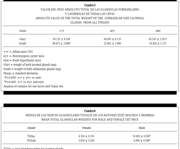
Cuadro 3Cuadro 3 Cuadro 3 Cuadro 3
VALOR DEL PESO ABSOLUTO TOTAL DE LAS GLÁNDULAS SUBMAXILARES Y LAGRIMALES DE TODAS LAS CEPAS
ABSOLUTE VALUE OF THE TOTAL WEIGHT OF THE SUBMAXILAR AND LACRIMAL GLANDS FROM ALL STRAINS
Strain +/+ et/+ et/et
Glact 65.125 ± 4.509 64.093 ± 8.133 43.165 ± 2.161a
Gsubt 46.615 ± 3.846b 25.862 ± 3.089 29.424 ± 1.275
+/+ = Albino mice CD1 et/+ = Heterozygous carrier mice et/et = Nude hypothymic mice
Glact = weight of both lacrimal glands (mg) Gsubt = weight of both submaxilar glands (mg) Means ± standard deviation.
aP=0.0001 +/+ y et/+ vs. et/et bP=0.0001 +/+ vs. et/+ and et/et
Analysis of variance for one factor and Tukey test.
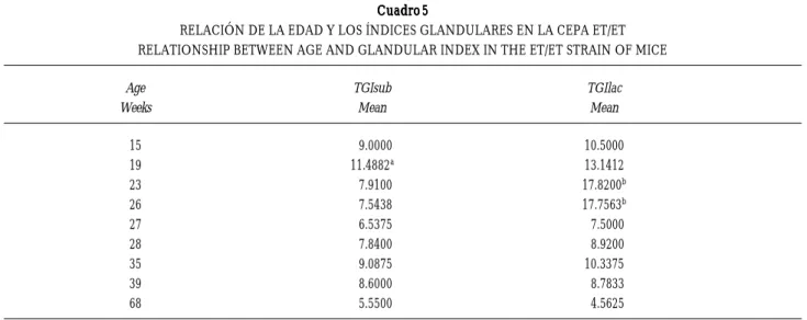
Cuadro 4 Cuadro 4Cuadro 4 Cuadro 4 Cuadro 4
MEDIAS DE LOS ÍNDICES GLANDULARES TOTALES EN LOS RATONES ET/ET MACHOS Y HEMBRAS MEAN TOTAL GLANDULAR WEIGHTS FOR MALE AND FEMALE T/ET MICE
Gender Females Males
TGIlac 4.556 ± 0.353 10.093 ± 0.590a
TGIsub 5.816 ± 0.526 6.900 ± 0.508b
TGIlac = total glandular index for lacrimal glands TGIsub = total glandular index for submaxilar glands Means ± standard deviation.
V V V V
Vet. M M M M Méx., 34 (2) 2003 135135135135135
edigraphic.com
An analysis of variance for one factor was also donefor absolute gland weights, showing et/et mice to have lighter lacrimal glands when compared to the et/+ and +/+ mice (P = 0.0001). For absolute weight of the submaxilar glands, those from +/+ mice were heavi-er that those of the et/+ and et/et mice (P = 0.0001) (Table 3).
Student’s t test between the males and females of the et/et group found that the values for TGIlac were lower in females than males (P = 0.0001) (Table 4). The same test for the TGIsub of the et/et mice did not show a significant (P = 0.132) differ-ence between males and females (Table 4). Analysis of variance contrasting age with the glandular in-dexes for the submaxilar glands showed greater indexes at week 19, with the lowest values occur-ring at week 68 of age. An analysis of variance for one factor on the et/et group comparing age and lacrimal glandular indexes showed a general de-crease with age, the greatest indexes being ob-served at weeks 23 and 26, with the lowest indexes at week 68 (Table 5).
There is less pathological histology for gland size in et/et mice than in the glands from +/+ mice (as detailed by glandular indexes). Histological analy-sis of lacrimal and submaxilar glands from et/+ and +/+ animals at 40 weeks of age was normal. The lacrimal gland from one female et/+ mice at 40 weeks of age showed well-defined stroma and
aci-significativa entre los grupos (P = 0.0002), y cuando se aplicó la prueba de Tukey también se encontró que el grupo +/+ presentó los mayores índices de estas glándu-las y fue diferente a los grupos et/+ y et/et (Cuadro 2). Se realizó un análisis de varianza de un factor tomando los valores de los pesos absolutos de las glándulas y se encon-tró que los ratones et/et mostraron menor peso de las glándulas lagrimales comparados con los pesos de las glándulas de los ratones et/+ y +/+ (P = 0.0001), y respecto al peso absoluto de las glándulas submaxilares, los ratones +/+ mostraron también un mayor peso que las glándulas de los ratones et/+ y et/et (P = 0.0001) (Cuadro 3).
En el grupo de ratones et/et se realizó una prueba t de Student entre machos y hembras y se encontró que los IGTlag son menores en las hembras que en los machos (P = 0.0001) (Cuadro 4). Mientras que para los IGTsub de los ratones et/et, la prueba t de Student no mostró diferencia significativa entre machos y hembras P = 0.132 (Cuadro 4). Se realizó un análisis de varianza contrastando la edad con los índices de las glándulas submaxilares y se observó que los índices mayores se registraron en la semana 19 mientras los valores meno-res se ubicaron en la semana 68 de vida. También se realizó un análisis de varianza de un factor en el grupo et/et relacionando la edad y los índices glandulares lagrimales, el análisis mostró una tendencia general a disminuir con la edad, los índices mayores se observa-ron en las semanas 23 y 26 mientras que a la semana 68 se mostraron los índices menores (Cuadro 5).
Cuadro 5 Cuadro 5 Cuadro 5 Cuadro 5 Cuadro 5
RELACIÓN DE LA EDAD Y LOS ÍNDICES GLANDULARES EN LA CEPA ET/ET RELATIONSHIP BETWEEN AGE AND GLANDULAR INDEX IN THE ET/ET STRAIN OF MICE
Age TGIsub TGIlac
Weeks Mean Mean
15 9.0000 10.5000
19 11.4882a 13.1412
23 7.9100 17.8200b
26 7.5438 17.7563b
27 6.5375 7.5000
28 7.8400 8.9200
35 9.0875 10.3375
39 8.6000 8.7833
68 5.5500 4.5625
TGIsub = total glandular index in submaxilar glands TGIlac = total glandular index in lacrimal glands
136 136 136 136 136
edigraphic.com
Figura 1A. Figura 1A.Figura 1A. Figura 1A.
Figura 1A. Glándula lagrimal nor-mal de ratón macho et/+ de 40 semanas de edad. Estroma y pa-rénquima acinar bien definidos. 1000X tinción HE.
Normal lacrimal gland from a 40-week-old male et/+ mouse. 400X HE stain. Stroma and aci-nar parenchyma are well de-fined.
En relación con los estudios histopatológicos el ta-maño de las glándulas de los ratones et/et es menor que el de las glándulas de los animales +/+ (como lo refieren los índices glandulares), y en el examen histo-patológico realizado a los ratones et/+ y +/+ aun a las 40 semanas de edad, las glándulas lagrimales y sub-maxilares se observan normales. La glándula lagrimal de una hembra et/+ de 40 semanas de vida, mostró nar parenchyma (Figure 1A). In Figure 1B one can
see interlobular ducts and well-defined acinar pa-renchyma in tissue from the submaxilar gland tak-en from the same female mouse. Figure 2A shows histological tissue from the lacrimal gland of a fe-male et/et mouse at 27 weeks of age, where lobular architectural alterations, areas of fibrosis and mono-nuclear cells can be observed. Figure 2B is of tissue
Figura 1B. Figura 1B. Figura 1B. Figura 1B.
V V V V
Vet. M M M M Méx., 34 (2) 2003 137137137137137
edigraphic.com
Figura 2A. Figura 2A. Figura 2A. Figura 2A.
Figura 2A. Glándula lagrimal de hembra et/et de 27 semanas de edad. Se observan alteraciones en la arquitectura lobular con zonas de fibrosis e infiltrado de mononucleares. 400X tinción HE. Lacrimal gland from a 27-week-old female et/et mouse. Lobular architectural alterations can be seen, as web as fibrosis and mono-nuclear infiltrate. 400X HE stain.
Figura 2B. Figura 2B. Figura 2B. Figura 2B.
Figura 2B. Glándula lagrimal de hembra et/et de 68 semanas de edad. Infiltrado importante de mononucleares y células plas-máticas. 1000X tinción HE. Lacrimal gland from a 68-week-old female et/et mouse. Consid-erable mononuclear and plasma cell infiltrate. 400X HE stain. estroma y parénquima acinar bien definido (Figura 1A). En la Figura 1B en la glándula submaxilar de la misma hembra se observan los conductos interlobula-res y el parénquima acinar bien definido. La Figura 2A muestra un corte histológico de la glándula lagrimal de una hembra et/et de 27 semanas de edad, donde se observan alteraciones de la arquitectura lobular, zonas de fibrosis e infiltrado de células mononucleares. La Figura 2B es una glándula lagrimal de hembra et/et de from a lacrimal gland taken from a female et/et
138 138 138 138 138
edigraphic.com
Figura 3A. Figura 3A. Figura 3A. Figura 3A.
Figura 3A. Glándula submaxi-lar de hembra et/et de 27 sema-nas de edad. Alteraciones en el parénquima acinar con fibrosis e infiltrado de mononucleares. 400X tinción HE.
Submaxilar gland from a 27-week-old female et/et mouse. Acinar parenchymal alterations with fibrosis and mononuclear infiltrate. 1000X HE stain.
Figura 3B. Figura 3B. Figura 3B. Figura 3B.
Figura 3B. Glándula submaxilar de hembra et/et de 27 semanas de edad. Infiltrado de células plasmáticas y mononucleares. 1000X tinción HE.
Submaxilar gland from a 27-week-old female et/et mouse. Plasma cell and mononuclear infiltrate. 400X HE stain.
Discussion
Gross ocular lesions are greater in the et/et group, which is the same group that had the lowest glandu-lar indexes and leucocyte infiltrates in lacrimal glands. These lesions could be the result of lacrimal gland destruction following inflammation, an in-flammatory process or reduced secretion from the gland with subsequent poor lubrication and corneal
V V V V
Vet. M M M M Méx., 34 (2) 2003 139139139139139
edigraphic.com
opacity. Some eyes showed corneal ulceration andcataracts leading to a degenerative process with even-tual eye loss (ptisis bulbi),17 such that older et/et mice had a greater number of eyes lost.11 Within the et/+ group only one animal with lesions was found, while no lesions were seen in the +/+ group. Glandular indexes for the et/et group were the lowest (Table 2). The lower number of lacrimal glands in the et/et mice, as compared to the et/+ and the +/+ mice, coupled with the histological findings of lympho-cyte infiltration and glandular destruction, explain the xerophthalmia seen in et/et mice. Submaxilar glands showed that et/+ mice, the gene carriers, showed similar glandular indexes to those seen for et/et mice, both being lower than those seen for +/+ mice (Table 2). This indicates that the presence of the gene has an effect on gland size, making them small-er. However, glandular alterations in et/+ mice did not include lymphocyte infiltration (Figure 1A and 1B), as was seen for the et/et mice (Figures 2A, 2B, 3A and 3B). This suggests that for the infiltrative pro-cess and damage to occur, the homozygous condi-tion must be present, along with other factors, given that some female et/et mice never develop lesions even at an advanced age.11 Female et/+ mice had to be used, instead of et/et mice, to obtain et/et progeny given the low fertility achieved with et/et mice and the inability of the et/et females to suckle their young.10 This suckling inability suggests deficient milk production due to the destruction of mammary glands. The lacrimal and submaxilar gland involu-tion seen in et/et mice probably reflects the thymus involution that also occurs in these animals, since, though born with a normal thymus, both males and females develop hypothymia by week 20.10 This could also be responsible for the low fertility of et/et female mice since there are reports that a thymectomy at
birth can induce autoimmune oophoritis.18 Some
histological changes were found in the submaxilar and lacrimal glands of the hypothymic et/et nude mice, consisting of periductal and perivascular mono-nuclear and plasma cell infiltrates (Figures 2A, 2B, 3A and 3B). Fox’s8 criteria for establishing a histolog-ical diagnosis of Sjörgen’s Syndrome requires the presence of at least two lymphocyte clusters in a 4 mm2 area of glandular tissue; however, we consider that these mice presented these infiltrates in an even smaller area, as can be seen in Figures 2 and 3, such
that in a 4 mm2 area there would be more than two
clusters.
The glandular histology for the et/et mice ranges from animals without any infiltrates, through moder-ate infiltration (where despite considerable lympho-cyte infiltration, the architectural structure of the gland is maintained), to severe cellular infiltrates,
Discusión
140 140 140 140 140
edigraphic.com
comprised mainly by mononuclear cells, withaddi-tional acinar disarray, loss of interlobular ducts, and in some cases, complete destruction of glandular tis-sue. Since these acini produce serous secretions, the absence of ducts would explain the xerostomia and keratoconjunctivitis seen in humans with SS.19 The TGIsub values for the et/et strain are highest at week 19, and then gradually decrease with increasing age, while TGIlac values are highest at weeks 23 and 26. More serious lesions can be appreciated in the et/et strain from week 22, with a concurrent increase in severity as age increases. The +/+ and et/+ mice had normal glandular histology and their acini were well defined even in animals at 40 weeks of age (Figures 1A and 1B).
When studying the gender influence of the TGIlag values of the et/et group of mice, we found that females have lower glandular indexes than do males (Table 4). The decreased size of the gland in females must be a consequence of the destruction that takes place following the immune response. In human SS something similar occurs, since there are reports of a nine times greater incidence in women than in men.20 Hormones play a very important role in SS, since a series of experimental models obtained following the administration of drug hormones suggest that test-osterone, progesterone and corticosterone have an immunosuppressive effect, while estrogen activity can boost the immune system.21 It is thought that the exocrinopathy demonstrated in et/et mice is the di-rect result of the “et” gene, which is recessive and thus requires a homozygous condition, as well as some already mentioned risk factors, for the glandular dam-age to become apparent. The et/et mice showed a similar pattern to that seen in primary human SS and, as such, could be used as an animal model for study-ing this disease.
el diagnóstico del síndrome de Sjögren, por histopatología, deben presentarse en cualquier lóbulo de la glándula examinada la presencia de cuando menos dos focos linfo-citarios en una área de 4 mm2, se piensa que los animales mostraron esos infiltrados en áreas mucho menores, pues en un campo de seco fuerte se observan infiltrados severos, como lo muestran las Figuras 2 y 3, por lo que en un área de 4 mm2 la cantidad de focos sería mayor de dos.
La histopatología de las glándulas en los ratones et/et van desde animales sin infiltraciones linfocitarias, pasando por infiltraciones moderadas (donde se mostró una infil-tración celular importante, sin modificar la arquitectura de la glándula), hasta infiltrados severos de células principal-mente mononucleares acompañadas de un desarreglo celular en los acinos, pérdida de los conductos interlobula-res hasta la destrucción total de la glándula. Debido a que estos acinos producen secreciones de tipo seroso, la ausen-cia de sus conductos sugiere que los animales et/et presen-tan xerostomía y queratoconjuntivitis como está descrita para el SS en el humano.19 Los valores de los IGTsub de la cepa et/et alcanzan su máximo tamaño a las 19 semanas y van disminuyendo gradualmente conforme aumenta la edad; mientras que para los IGTlag, el valor máximo se presenta en las semanas 23 y 26. La severidad de la lesión aparece en la cepa et/et desde la semana 22 y se incrementa conforme avanza la edad. Los ratones +/+ y et/+ mostra-ron un aspecto normal en la histología de sus glándulas y sus acinos se observaron bien definidos aun en animales de 40 semanas de edad (Figuras 1A y 1B).
Al estudiar la influencia del sexo en los IGTlag del grupo de los ratones et/et, se encontró que las hembras presentan menores índices glandulares que los machos (Cuadro 4). La disminución del tamaño de la glándula en las hembras debe ser una consecuencia de la destrucción por la respues-ta inmune; en el SS del humano ocurre algo similar, pues se tienen informes que lo padecen más las mujeres que los hombres en una relación de nueve a uno.20 Las hormonas juegan un papel muy importante en el SS, pues una serie de informes en modelos experimentales obtenidos de la aplicación de dosis farmacológicas de hormonas, sugieren que la testosterona, progesterona y corticosterona tienen actividad inmunosupresora, mientras que la actividad de los estrógenos es inmunopotenciadora.21 Se considera que la exocrinopatía mostrada por los ratones et/et es resultado de la presencia del gen “et” que tiene un carácter recesivo, ya que se requiere de la homocigocidad del gen, además de otros factores de riesgo ya mencionados, para que se manifieste el daño a glándulas. Los ratones et/et mostraron un patrón similar al observado en el SS primario en el humano y pudiera ser usado como un modelo animal para estudiar esta enfermedad.
Referencias
1. Mikulicz JH. Uber eine eigenartine symmetrische Erkrankug der Tranen-und Mundspeicheldruse. Beitrt Chir-Fortschr Gewidment Theodor Billort, Stuttgart 1892:610-630.
2. Sjögren HS. Zur Kenntnis der Keratoconjunctivitis sicca (keratitis folliformis bei Hypofunktion der Tranen-drus-en). Acta Ophthalmol (Copenh) 1933;2:1-151.
3. Bloch K, Buchanan W, Whol M. Sjögren´s syndrome. A clinical, pathological and serological study of 62 cases. Medicine 1965;44:187-231.
4. Fox RI, Howell FV, Bone RC, Michelson P. Primary Sjögren´s syndrome: Clinical and Immunopathologic features. Semin Arthritis Rheum 1984;14:77-106. 5. Mountz JD, Talal N. Retroviruses, apoptosis and
autoge-nes. Immunol Today 1993;14:532-533.
V V V V
Vet. M M M M Méx., 34 (2) 2003 141141141141141
edigraphic.com
7. Fox RI. Sjögren´s syndrome. Curr Opin Rheumatol 1995;7:409-416.
8. Fox RI, Robinson CA, Curd JG, Kozin F, Howell FV. Sjögren syndrome. Proposed criteria for classification. Arthritis Rheum 1986;29:577-585.
9. Arraz CJA, Perez GA, Zea MA, Alvarez-Mon SM. Sín-drome de Sjögren. Medicine 1997;7:409-416.
10. Rosas P, Castellanos P, Dominguez R. The existence of spontaneous hairless (nude) hypotymic mutant mice from the CD1 strain, reared under conventional animal house conditions. Med Sci Res 1987;15:553-554. 11. Marroquín-Segura R. Estudios inmunológicos en ratones
hipotímicos CD1 et/et que desarrollan oftalmopatía es-pontánea. México (DF) México: Escuela Nacional de Ciencias Biológicas. Instituto Politécnico Nacional, 1996. 12. Basurto-Alcántara FJ, Mondragón VRL, Atilano-López D, Montaraz CJA, Márquez DMJ, Rosas SP et al. Com-paración de las constantes fisiológicas sanguíneas de los ratones CD1, heterocigótico et/+ y desnudo et/et. Vet Méx 2000;31:209-216.
13. Rojas-Espinosa R, Marroquín-Segura R, Wek RK, Reyes-Maldonado E, Arce PP. Susceptibility of “et” the sponta-neously mutating CD1 derived nude mouse, to infection of M. lepraemurium . Int J Leprosy 1999;67:46-51.
14. Janssen Research Foundation Series. Toxicology and reference data-Wistar rat. Amsterdam: Elsevier/North-Holland Biomedical Press; 1981.
15. Marqués de Cantú MJ. Probabilidad y estadística para las ciencias químico-biológicas. Predicción. México (DF): McGraw-Hill, 1990.
16. Salkind NJ, Akey TM. Using SPSS for windows. Analysing and understanding data. New Jersey: Prentice-Hall, 2001. 17. Riley ML, Harding JJ. The reaction methylglyoxal with human and bovine lens proteins. Biochim Biophys Acta 1995;1270:36-43.
18. Kosiewickz MM, Michael SD. Neonatal thymectomy af-fects follicle populations before the onset of autoimmune oophoritis in B6A mice. J Reprod Fert 1990;88: 427-440. 19. Xu KP, Katahiri S, Takeuchi T, Tsubota K. Biopsy of labial
salivary glands and lacrimal glands in the diagnosis of Sjögren´s syndrome. J Rheumatol 1996;23: 76-81. 20. Price EJ, Venables PJW. The etiopathogenesis of
Sjögren’s Syndrome. Semin Arthritis Rheum 1995;25: 117-133.
21. Author Anonymous. Sex factors, steroid hormones, and the host response. Proceedings of the Kroc Foundation Conference; 1979 febrero 12-16; Santa Ynes Valley, (Ca) Source Arthritis Rheum, 1979:1153-1320.