Abrescia, N. G. A. i Subirana, J. A. Acta Cryst. D58, 2205-2208 (2002).
Abrescia, N. G. A., González, C., Gouyette, C. i Subirana, J. A. Biochemistry 43, 4092-4100 (2004).
Abrescia, N. G. A., Malinina, L., Fernandez, L. G., Huynh-Dinh, T., Neidle, S. i Subirana, J. A. Nucleic Acid Research 27, 1593-1599 (1999).
Abrescia, N. G. A., Thompson, A., Huynh-Dinh, T. i Subirana, J. A. Proc. Natl. Acad. Sci. USA 99, 2806-2811 (2002).
Adams, A., Guss, J. M., Collyer, C. A., Denny, W. A. i Wakelin, L. P. G. Nucleic Acids Res. 28, 4244-4253 (2000).
Adams, A., Guss, J. M., Denny, W. A. i Wakelin, L. P. G. Acta Cryst D60, 823-828 (2004).
Avery, O., MacLeod, C. i McCarty, M. J. Expt. Med. 79, 137-157 (1944).
Baikolov, I., Grzeskowiak, K., Yanagi, K., Quintana, J. i Dickerson, R. E. J. Mol. Biol. 231, 768-784 (1993).
Basu, S., Szewczak, A. A., Cocco, M. i Strobel, S. A. J. Am. Chem. Soc. 122, 3240-3241 (2000).
Berman, H. M., Olson, W. K., Beveridge, D. L., Westbrook, J., Gelbin, A., Demeny, T., Hsieh, S.-H., Srinivasan, A. R. i Schneider, B. Biophys. J. 63, 751-759 (1992).
Birnboim, H. C. i Doly, J. Nucleic Acids Res. 7, 1513-1523 (1979).
Black, P. J. i Corby, R. N. In Anomalous Scattering (ed. S. Ramaseshan i S. C. Abrahams), Copenhagen (1975).
Blackburn, E. H. Cell 106, 661-673 (2001). Brünger, A. T. Nature 355, 472-475 (1992).
Brünger, A. T., Adams, P. D., Clore, G. M., DeLano, W. L., Gros, P., Grosse-Kunstleve, R. W., Jiang, J.-S., Kuszewski, J., Nilges, M., Passu, N. S., Read, R. J., Rice, L. M., Simonson, T. i Warren, G. L. Acta Cryst. D54, 905-021 (1998).
Brünger, A. T., Kuriyan, J. i Karplus, M. Science 235, 458-460 (1987).
Cantor, C. R., Warshaw, M. M. i Shapiro, H. Biopolymers 9, 1059-1077 (1970). Chaires, J. B., Herrera, J. E. i Waring, M. J. Biochemistry 29, 6145-6153 (1990). Cirilli, M., Bachechi, F., Ughetto, G., Colonna, F. P. i Capobianco, M. L. J. Mol. Biol. 230, 878-889 (1993).
Corby, R. N. i Black, P. J. Acta Cryst. B29, 2669 (1973). Crowther, R. A. i Blow, D. M. Acta Cryst. 23, 544 (1967).
Cuesta, J., Read, M. A. i Neidle, S. Mini Rev. Med. Chem. 3, 11-21 (2003). Dandiker, P. J., Holmlin, R. E. i Barton, J. K. Science 275, 1465-1468 (1997). Dautant, A., Langlois D’Estaintot, B., Gallois, B., Brown, T. i Hunter, W. N. Nucleic Acids Res. 23, 1710-1716 (1995).
Dauter, Z. Methods Enzymol. 276, 344-361 (1997).
Davies, D. R. i Baldwin, R. L. J. Mol. Biol. 6, 251-255 (1963). Dickerson, R, E. i Drew, H. R. J. Mol. Biol. 149, 761-786 (1981).
Dickerson, R. E., Bansal, M., Calladine, C. R., Diekman, S., Hunter, W. N., Kennard, O., Lavery, R., Nelson, H. C., Olson, W. K., Saenger, W., Sklenar, H., Soumpasis, D. M., Tung, C.-S., Von Kitzing, E., Wang, A. H. i Zhurkin, V. B. J. Mol. Biol. 205, 787 (1989).
Drenth, J. Principles of Protein X-ray Crystallography. Springer-Verlag, Inc., New York (1994).
Ducruix, A. i Giegé, R. Crystallization of Nucleic Acids and Proteins: A pratical approach. IRL Press, Oxford (1992).
Egli, M., Williams, L. D., Frederick, C. A. i Rich, A. Biochemistry 30, 1364-1372 (1991).
Eichhorn, G. L. (Ed) Inorganic Biochemistry. Elsevier, Amsterdam (1973).
Eisenberg, D., Luthy, R. i Bowie, J. U. In Meth. Enz., Macromolecular Crystallography 277, 396-406 (1997).
Evans, S. V. J. Mol. Graphics 11, 134-138 (1993).
Falk, M. Hartman, K. A. i Lord, R. C. J. Am. Chem. Soc. 85, 387-391 (1963). Felsenfeld, G. i Rich, A. Biochim. Biophys. Acta 26, 457 (1957).
Felsenfeld, G., Davies, D. R. i Rich, A. J. Am. Chem. Soc. 79, 2023 (1957). Fox, K. R. Drug-DNA Interaction Protocols. Humana Press, Southampton (1997). Fox, K. R. i Waring, M. J. Biochemistry 25, 4349-4356 (1986).
Fox, K. R. i Waring, M. J. Methods Enzymol. 340, 412-430 (2001). Fox, K. R. i Waring, M. J. Nucleic Acids Res. 12, 9271-9285 (1984).
Fratini, A. V., Kopka, M. L., Drew, H. R. i Dickerson, R. E. J. Biol. Chem. 257, 14686-14707 (1982).
French, G. S. i Wilson, K. S. Acta Cryst. A34, 417 (1978).
Gao, Y.-G., Liaw, Y.-C., Robinson, H. i Wang, A. H.-J. Biochemistry 29, 10307-10316 (1990).
Gao, Y.-G., Robinson, H., van Boom, J. H. i Wang, A. H.-J. Biophys. J. 69, 559-568 (1995).
Gao, Y.-G., Sriram, M. i Wang, A. H.-J. Nucleic Acids Res. 21, 4093-4101 (1993). García-Pérez, M. Pinto, M. i Subirana, J. A. Biopolymers 69, 432-439 (2003). Glusker, J. Crystal Structure Analysis for Chemists and Biologists. VCH Publishers, Inc. (1994).
Glusker, J. Crystal Structure Analysis. A primer. Oxford University Press (1985). Goodsell, D. S., Grzeskowiak, K. i Dickerson, R. E. Biochemistry 34, 1022-1029 (1995).
Goodsell, D. S., Kaczor-Grzeskowiak, M. i Dickerson, R. E. J. Mol. Biol. 239, 79-96 (1994).
Haider, S. M., Parkinson, G. N. i Neidle, S. J. Mol. Biol. 326, 117-125 (2003). Han, H. i Hurley, L. H. Trends. Pharmacol. Sci. 21, 136-142 (2000).
Heinemann, U. i Alings, C. The EMBO Journal 10, 35-43 (1991). Heinemann, U. i Hahn, M. J. Biol. Chem. 267, 7332-7341 (1992).
Heinemann, U., Alings, C. i Bansal, M. The EMBO Journal 11, 1931-1939 (1992). Howerton, S. B., Nagpal, A. i Williams, L. D. Biopolymers 69, 87-99 (2003). Ivanov, V. I. i Krylov, D. Yu. Methods Enzymol. 211, 111-127 (1992). Kettani, A., Kumar, R. A. i Patel, D. J. J. Mol. Biol. 254, 638 (1995).
Kielkopf, C. L., Baird, E. E., Dervan, P. B. i Rees, D. C. Nat. Struct. Biol. 5, 104-109 (1998).
Kissinger, C. R., Gehlhaar, D. K. i Fogel, B. Acta Cryst. D55, 484-491 (1999). Lane, A. N., Jenkins, T. C., Brown, T. i Neidle, S. Biochemistry 30, 1372-1385 (1991).
Laughlan, G., Murchie, A. I. H., Norman, D. G., Moore, M. H., Moody, P. C., Lilley, D. M. J. i Luisi, B. Science 265, 520 (1994).
Lavesa, M. i Fox, K. R. Anal. Biochem. 293, 246-250 (2001).
Leonard, G. A., Hambley, T. W., McAuley-Hecht, K., Brown, T. i Hunter, W. N. Acta Cryst. D49, 458- 467 (1993).
Lepre, C. A. i Lippard, S. J. Interaction of platinum antitumor copounds with DNA in Nuleic Acids and Molecular Biology 4, Springer-Verlag, Berlin, Heidelberg, 9-38 (1990).
Lerman, L. S. J. Mol. Biol. 3, 18-30 (1961).
Lipanov, A., Kopka, M. L., Kaczor-Grzeskowiak, M., Quintana, J. i Dickerson, R. E. Biochemistry 32, 1373-1389 (1993).
Lipscomb, L. A., Zhou, F. X., Presnell, S. R., Woo, R. J., Peek, M. E., Plaskon, R. R. i Williams, L. D. Biochemistry 36, 2818-2823 (1996).
Lipsett, M. N. Biochem. Biophys. Res. Commun. 11, 224 (1963).
Lisgarten, J. N., Coll, M., Portugal, J., Wright, C. W. i Aymamí, J. Nat. Struct. Biol. 9, 57-60 (2002).
Lyons, J. W. i Kotin, L. J. Am. Chem. Soc. 87, 1781-1785 (1965). Madhumalar, A. i Bansal, M. Biophys. J. 85, 1805-1816 (2003).
Malinina, L. V., Makhaldiani, V. V., Tereshko, V. A., Zarytova, V. F. i Ivanova, E. M. J. Biomol. Struct. Dyn. 5, 405-433 (1987).
Malinina, L., Soler-López, M., Aymamí, J. i Subirana, J. A. Biochemistry 41, 9341-9348 (2002).
Maxam, A. M. i Gilbert, W. Methods Enzymol. 65, 499-560 (1980).
McPherson, A. Crystallization of Biological Macromolecules. Cold Spring Harbor Laboratory Press, New York (1999).
McPherson, A. Introduction to Macromolecular Crystallography. Wiley-Liss, Inc., New Jersey (2003).
McRee, D. E. J. Struct. Biol. 125, 156-165 (1999). Mitchell, C. M. Acta Cryst. 10, 475 (1957).
Moore, M. H., Hunter, W. N., Langlois D’Estaintot, B. i Kennard, O. J. Mol. Biol. 206, 693-705 (1989).
Morgan, A. R. i Wells, R. D. J. Mol. Biol. 37, 63 (1968).
Murray, N. L. i Morgan, A. R. Can. J. Biochem. 51, 436 (1973).
Murshudov, G. N., Vagin, A. A. i Dodson, E. J. Acta Cryst. D53, 240-255 (1997). Navaza, J. i Saludjian, P. Methods Enzymol. 276, 581-594 (1997).
Neidle, S. Nucleic Acid Structures. Oxford University Press (1999). Nunn, C. M., Garman, E. i Neidle, S. Biochemistry 36, 4792-4799 (1997).
Nunn, C. M., Vaan Meervelt, L., Zhang, S., Moore, M. H. i Kennard, O. J. Mol. Biol. 222, 167-177 (1991).
Ornstein, R. L., Rein, R., Breen, D. L. i MacElroy, R. D. Biopolymers 17, 2341-2360 (1978).
Otwinoski, Z. i Minor, W. Methods Enzymol. 276, 307-326 (1997). Pannu, N. S. i Read, R. J. Acta Cryst. A52, 659-668 (1996).
Berman, H. M. Biochemistry 34, 15487-15495 (1995).
Patel, D. J., Bouaziz, S., Kettani, A. i Wang, Y. Nucleic Acid Structures. Capítol 13. Oxford University Press (1999).
Perry, P. J., Reszka, A. P., Wood, A. A., Read, M. A., Gowan, S. M., Dosanjh, H. S., Trent, J. O., Jenkins, T. C., Kelland, L. l. R. i Neidle, S. J. Med. Chem. 41, 4873-4884 (1998).
Philips, K., Dauter, Z., Murchie, A. I. H., Lilley, D. M. J. i Luisi, B. J. Mol. Biol. 273, 171 (1997).
Ravelli, R. G. B., Sweet, R. M., Skinner, J. M., Duisenberg, A. J. M. i Kroon, J. J Appl. Cryst. 30, 551-554 (1997).
Ren, J. i Chaires, J. B. Biochemistry 38, 16067-16075 (1999). Rossmann, M. G. i Blow, D. M. Acta Cryst. 15, 24 (1962).
Roussel, A., Inisan, A. G., Knoops-Mouthuy, E. i Cambillau, E. TURBO-FRODO version OpenGL.1. University of Marseille (1998).
Rudman, R. Low temperature X-ray diffraction. Plenum Press, New York (1976). Saenger, W. Principles of Nucleic Acid Structures. Springer-Verlag. New York (1984).
Schneider, B. i Berman, H. M. Biophys. J. 69, 2661-2669 (1995). Seeman, N. C. Nature 421, 427-431 (2003).
Seeman, N. C., Rosenberg, J. M., Suddath, F. L., kim, J. P. P. i Rich, A. J. Mol. Biol. 104, 109 (1976).
Shakked, Z., Guzikevich-Guerstein, G., Frolow, F., Rabinovich, D., Joachimiak, A. i Sigler, P. B. Nature 368, 469-473 (1994).
Sheldrick, G. M. XPREP5. Programm zur Bearbeitung von Bengungsdaten & Untersuchung reziproker Gitter. V 5.01/486, 2003, Brucker
.
Sheldrick, G. M. Z. Kristallogr. 217, 644-650 (2002).
Shui, X., E Peek, M., Lipscomb, L. A., Wilkinson, A. P., Williams, L. D., Gao, M., Ogata, C., Roques, B. P. i Garbay-Jaureguiberry, C. Curr. Med. Chem. 7, 59-71 (2000).
Sinden, R. R. DNA Structure and Function. Academic Press , London (1994). Smith, C. K., Brannigan, J. A. i Moore, M. H. J. Mol. Biol. 263, 237-258 (1996). Soler-López, M., Malinina, L. i Subirana, J. A. J. Biol. Chem. 275, 23034-23044 (2000).
Soler-López, M., Malinina, L., L. Liu, J., Huynh-Dinh, T. i Subirana, J. A. J. Biol. Chem. 274, 23683-23686 (1999).
Soler-López, M., Malinina, L., Tereshko, V., Zarytova, V. i Subirana, J. A. J. Biol. Inorg. Chem. 7, 533-538 (2002).
Soumpasis, D. M. J. Biomol. Struct. Dyn. 3, 1-10 (1985).
Spingler, B., Whittington, D. A. i Lippard, S. J. Inorg. Chem. 40, 5596-5602 (2001).
Spink, N., Nunn, C. N., Vojtechovsky, J., Berman, H. M. i Neidle, S. Proc. Natl. Acad. Sci. USA 92, 10767-10771 (1995).
Stevenson, K. A., Muralidharan, G., Maya, L., Wells, J. C., Barhen, J. i Thundat, T. J. Nanosci. Nanotechnol. 2, 297-404 (2002).
Subirana, J. A. Estructura del ADN. Alhambra, Madrid (1985).
Subirana, J. A. i Abrescia, N. G. A. Biophys. Chem. 86, 179-189 (2000). Subirana, J. A. i Faria, T. Biophys. J. 73, 333-338 (1997).
Subirana, J. A. i Soler-López, M. Annu. Rev. Biophys. Biomol. Struct. 32, 27-45 (2003).
Subirana, J. A. i Vives, J. L. Biopolymers 20, 2281-2283 (1981). Takenaka, S. J. Heterocyclic Chem. 33, 2043 (1996).
Teixeira, S. C. M., Thorpe, J. H., Todd, A. K., Powell, H. R., Adams, A., Wakelin, L. P. G., Denny, W. A. i Cardin, C. J. J. Mol. Biol. 323, 167-171 (2002).
Tereshko, V. i Subirana, J. A. Acta Cryst. D55, 810-819 (1999).
Biochemistry 39, 15055-15061 (2000).
Timsit, Y., Westhof, E., Fuchs, R. P .P. i Moras, D. Nature 341, 459-462 (1989). Todd, A. K., Adams, A., Thorpe, J. H., Denny, W. A., Wakelin, L. P. G. i Cardin, C. J. J. Mol. Chem. 42, 536-540 (1999).
Tunis, M. J. i Hearst, J. E. Biopolymers 6, 1345-1353 (1968).
Urpí, L., Tereshko, V., Malinina, L., Huynh-Dinh, T. i Subirana, J. A. Nat. Struct. Biol. 3, 325-328 (1996).
Vagin, A. i Teplyakov, A. J. Appl. Cryst. 30, 1022-1025 (1997).
Valls, N., Usón, I., Gouyette, C. i Subirana, J.A. J. Am. Chem. Soc. 126, 7812-7816 (2004).
Valls, N., Wright, G., Steiner, R. A., Murshudov, G. N. i Subirana, J. A. Acta Cryst. D60, 680-685 (2004).
Van Aalten, D. M. F., Erlanson, D. A., Verdine, G. L. i Joshua-Tor, L. Proc. Natl. Acad. Sci. USA 96, 11809-11814 (1999).
Viswamitra, M. A., Shakked, Z., Jones, P. G., Salisbury, G. M., Sheldrick, S. A. i Kennard, O. Biopolymers 21, 513-533 (1982).
Vlieghe, D., Turkenburg, J. P. i Van Meervelt, L. Acta Cryst. D55, 1495-1502 (1999).
Vlieghe, D., Van Meervelt, L., Dautant, A., Gallois, B., Précigoux, G. i Kennard, O. Acta Cryst. D52, 766-775 (1996).
Wagenknecht, H.-A. Angew. Chem. Int. 42, 3204-3206 (2003).
Wahl, C. M., Ramakrishnan, B., Ban, C., Chen, X. i Sundaralingan, M. Acta Cryst. D52, 668-675 (1996).
Wang, A. H. J., Quigley, G. J., Kolpak, F. J., Crawford, J. L., van Boom, J. H., Van der Marel, G. i Rich, A. Nature 282, 680-686 (1979).
Wang, A. H.-J., Hakoshima, T., Van der Marel, G. A., Van Boom, J. H. i Rich, A. Cell (Cambridge, Mass.) 37, 321-331 (1984).
Wang, A. H.-J., Ughetto, G., Quigley, G. J., Hakoshima, T., Van der Marel, G. A., Van Boom, J. H. i Rich, A. Science 225, 1115-1121 (1984).
Waring, M. J. J. Mol. Biol. 13, 269-282 (1965).
Watson, J. D. i Crick, F. H. C. Nature 171, 737-738 (1953).
Westbrook, E. M. i Naday, I. Methods Enzymol. 276, 244-268 (1997).
Williams, K. A., Veenhuizen, P. T., de la Torre, B. G., Eritja, R. i Dekker, C. Nature 420, 761.
Williams, L. D., Egli, M., Ughetto, G., Van der Marel, G. A., Van Boom, J. H., Quigley, G. J., Wang, A. H.-J., Rich, A. i Frederick, C. A. J. Mol. Biol. 215, 313-320 (1990).
Winfree, E., Liu, F., Wenzler, L. A. i Seeman, N. C. Nature 394, 539-544 (1998). Wood, A. A., Nunn, C. M., Trent, J. O. i Neidle, S. J. Mol. Biol. 269, 827-841 (1997).
Yanagi, K., Privé, G. D. i Dickerson, R. E. J. Mol. Biol. 217, 201 (1991).
Yang, X.-L., Robinson, H., Gao, Y.-G. i Wang, A. H.-J. Biochemistry 39, 10950-10957 (2000).
1. PUBLICACIONS
Part del treball realitzat en aquesta tesi ha donat lloc a les següents publicacions:
1. Valls, N., Wright, G., Steiner, R. A., Murshudov, G. N. i Subirana, J. A. DNA variability in five crystal structures of d(CGCAATTGCG). Acta Cryst. D60, 680-685 (2004).
2. Valls, N., Usón, I., Gouyette, C. i Subirana, J. A. A cubic arrangement of DNA double helices based on nickel-guanine interactions. J. Am. Chem. Soc. 126, 7812-7816 (2004).
2. PARÀMETRES CONFORMACIONALS
Tots els paràmetres conformacionals de les estructures resoltes s’han calculat amb el programa 3DNA. En alguns casos s’han eliminat les bases terminals que no formen part del dúplex.
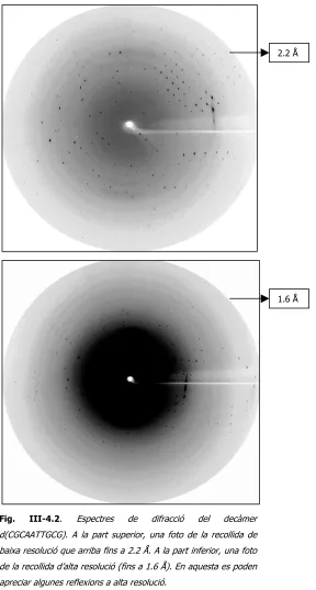
2.1 ESTRUCTURA d(CGCAATTGCG)
**************************************************************************** **************************************************************************** 3DNA (v1.5, Nov. 2002) by Xiang-Jun Lu at Wilma K. Olson's Lab. **************************************************************************** 1. The list of the parameters given below correspond to the 5' to 3' direction of strand I and 3' to 5' direction of strand II.
2. All angular parameters, except for the phase angle of sugar pseudo- rotation, are measured in degrees in the range of [-180, +180], and all displacements are measured in Angstrom units.
**************************************************************************** File name: duplex_nuria_r1.pdb
Date and time: Tue Jul 22 13:04:07 2003 Number of base-pairs: 8
Number of atoms: 328
**************************************************************************** **************************************************************************** RMSD of the bases (--- for WC bp, + for isolated bp, x for helix change) Strand I Strand II Helix
1 (0.020) A:...2_:[..G]G---C[..C]:...9_:A (0.023) | 2 (0.022) A:...3_:[..C]C---G[..G]:...8_:A (0.016) | 3 (0.022) A:...4_:[..A]A---T[..T]:...7_:A (0.022) | 4 (0.010) A:...5_:[..A]A---T[..T]:...6_:A (0.018) | 5 (0.018) A:...6_:[..T]T---A[..A]:...5_:A (0.010) | 6 (0.022) A:...7_:[..T]T---A[..A]:...4_:A (0.022) | 7 (0.016) A:...8_:[..G]G---C[..C]:...3_:A (0.022) | 8 (0.023) A:...9_:[..C]C---G[..G]:...2_:A (0.020) |
**************************************************************************** Detailed H-bond information: atom-name pair and length [ON]
1 G---C [3] N2 - O2 2.71 N1 - N3 2.93 O6 - N4 2.95 2 C---G [3] O2 - N2 2.70 N3 - N1 2.88 N4 - O6 2.90 3 A---T [2] N1 - N3 2.83 N6 - O4 3.08
4 A---T [2] N1 - N3 2.90 N6 - O4 3.19 5 T---A [2] N3 - N1 2.90 O4 - N6 3.19 6 T---A [2] N3 - N1 2.83 O4 - N6 3.08
7 G---C [3] N2 - O2 2.70 N1 - N3 2.88 O6 - N4 2.90 8 C---G [3] O2 - N2 2.71 N3 - N1 2.93 N4 - O6 2.95
**************************************************************************** Overlap area in Angstrom^2 between polygons defined by atoms on successive bases. Polygons projected in the mean plane of the designed base-pair step. Values in parentheses measure the overlap of base ring atoms only. Those outside parentheses include exocyclic atoms on the ring. Intra- and inter-strand overlap is designated according to the following diagram: i2 3' 5' j2
step i1-i2 i1-j2 j1-i2 j1-j2 sum 1 GC/GC 4.58( 2.46) 0.00( 0.00) 0.00( 0.00) 4.11( 1.99) 8.69( 4.44) 2 CA/TG 0.66( 0.00) 0.00( 0.00) 0.13( 0.00) 1.82( 0.13) 2.61( 0.13) 3 AA/TT 3.81( 2.42) 0.00( 0.00) 0.00( 0.00) 5.32( 0.13) 9.13( 2.55) 4 AT/AT 5.41( 1.68) 0.00( 0.00) 0.00( 0.00) 5.41( 1.68) 10.82( 3.36) 5 TT/AA 5.32( 0.13) 0.00( 0.00) 0.00( 0.00) 3.81( 2.42) 9.13( 2.55) 6 TG/CA 1.82( 0.13) 0.00( 0.00) 0.13( 0.00) 0.66( 0.00) 2.60( 0.13) 7 GC/GC 4.11( 1.99) 0.00( 0.00) 0.00( 0.00) 4.59( 2.46) 8.69( 4.45) **************************************************************************** Origin (Ox, Oy, Oz) and mean normal vector (Nx, Ny, Nz) of each base-pair in the coordinate system of the given structure
bp Ox Oy Oz Nx Ny Nz 1 G-C 0.00 0.00 0.00 0.00 0.00 1.00 2 C-G 0.17 -0.12 3.15 0.00 0.00 1.00 3 A-T 0.05 -0.33 6.47 0.09 0.06 0.99 4 A-T 0.56 -0.01 9.71 0.06 0.05 1.00 5 T-A 1.18 0.28 12.95 0.09 0.00 1.00 6 T-A 1.77 0.48 16.19 0.11 0.03 0.99 7 G-C 1.56 0.42 19.51 0.01 -0.02 1.00 8 C-G 1.59 0.56 22.67 0.01 -0.02 1.00 **************************************************************************** Local base-pair parameters
bp Shear Stretch Stagger Buckle Propeller Opening 1 G-C -0.35 -0.16 0.04 -2.71 -1.19 -0.03 2 C-G 0.26 -0.17 0.04 5.53 -5.60 0.89 3 A-T -0.15 -0.13 -0.11 3.80 -15.21 1.59 4 A-T -0.02 -0.06 0.08 1.31 -16.60 4.77 5 T-A 0.02 -0.06 0.08 -1.28 -16.60 4.78 6 T-A 0.15 -0.13 -0.11 -3.82 -15.20 1.59 7 G-C -0.26 -0.17 0.04 -5.51 -5.59 0.89 8 C-G 0.35 -0.16 0.04 2.73 -1.22 -0.04 ~~~~~~~~~~~~~~~~~~~~~~~~~~~~~~~~~~~~~~~~~~~~~~~~~~~~~~~~~~~~ ave. 0.00 -0.13 0.01 0.01 -9.65 1.81 s.d. 0.24 0.05 0.08 3.93 6.91 1.94 **************************************************************************** Local base-pair step parameters
step Shift Slide Rise Tilt Roll Twist 1 GC/GC 0.13 -0.15 3.15 0.14 -0.04 31.85 2 CA/TG -0.41 0.00 3.30 1.70 6.08 33.24 3 AA/TT 0.17 -0.25 3.28 -2.05 -0.48 36.16 4 AT/AT 0.00 -0.44 3.29 0.01 -3.65 34.90 5 TT/AA -0.17 -0.25 3.28 2.03 -0.46 36.16 6 TG/CA 0.41 0.00 3.30 -1.70 6.08 33.25 7 GC/GC -0.13 -0.15 3.15 -0.14 -0.05 31.84 ~~~~~~~~~~~~~~~~~~~~~~~~~~~~~~~~~~~~~~~~~~~~~~~~~~~~~~~~~~~~ ave. 0.00 -0.18 3.25 0.00 1.07 33.91 s.d. 0.27 0.16 0.07 1.54 3.65 1.85 **************************************************************************** Local base-pair helical parameters
step X-disp Y-disp h-Rise Incl. Tip h-Twist 1 GC/GC -0.27 -0.22 3.15 -0.08 -0.25 31.85 2 CA/TG -0.99 0.98 3.23 10.51 -2.94 33.82 3 AA/TT -0.34 -0.56 3.27 -0.77 3.30 36.22 4 AT/AT -0.17 0.00 3.32 -6.07 -0.02 35.09 5 TT/AA -0.34 0.56 3.27 -0.74 -3.27 36.21 6 TG/CA -0.99 -0.98 3.23 10.51 2.94 33.83 7 GC/GC -0.27 0.22 3.15 -0.09 0.26 31.84 ~~~~~~~~~~~~~~~~~~~~~~~~~~~~~~~~~~~~~~~~~~~~~~~~~~~~~~~~~~~~ ave. -0.48 0.00 3.23 1.90 0.00 34.12 s.d. 0.35 0.66 0.06 6.24 2.55 1.84 **************************************************************************** Structure classification:
This is a right-handed nucleic acid structure
**************************************************************************** lambda: virtual angle between C1'-YN1 or C1'-RN9 glycosidic bonds and the base-pair C1'-C1' line
4 A-T 54.2 54.3 10.5 8.8 9.9 5 T-A 54.4 54.1 10.5 8.8 9.9 6 T-A 55.7 52.0 10.6 8.9 9.8 7 G-C 54.2 55.1 10.6 8.9 9.8 8 C-G 53.4 53.3 10.7 8.9 9.8
**************************************************************************** Classification of each dinucleotide step in a right-handed nucleic acid structure: A-like; B-like; TA-like; intermediate of A and B, or other cases step Xp Yp Zp XpH YpH ZpH Form
1 GC/GC -3.42 9.26 -0.63 -3.69 9.26 -0.64 B 2 CA/TG -3.06 8.98 -0.30 -4.04 8.89 1.31 B 3 AA/TT -3.19 9.08 -0.39 -3.49 9.07 -0.51 B 4 AT/AT -3.41 9.14 -0.36 -3.57 9.05 -1.28 B 5 TT/AA -3.19 9.08 -0.39 -3.49 9.07 -0.50 B 6 TG/CA -3.06 8.98 -0.30 -4.04 8.89 1.31 B 7 GC/GC -3.42 9.26 -0.63 -3.69 9.26 -0.65 B
**************************************************************************** Minor and major groove widths: direct P-P distances and refined P-P distances which take into account the directions of the sugar-phosphate backbones (Subtract 5.8 Angstrom from the values to take account of the vdw radii of the phosphate groups, and for comparison with FreeHelix and Curves.) Ref: M. A. El Hassan and C. R. Calladine (1998). ``Two Distinct Modes of Protein-induced Bending in DNA.'' J. Mol. Biol., v282, pp331-343. Minor Groove Major Groove
P-P Refined P-P Refined 1 GC/GC --- --- --- --- 2 CA/TG --- --- --- --- 3 AA/TT 9.8 --- 18.7 --- 4 AT/AT 9.0 9.0 16.9 16.9 5 TT/AA 9.8 --- 18.7 --- 6 TG/CA --- --- --- --- 7 GC/GC --- --- --- ---
**************************************************************************** Global linear helical axis defined by equivalent C1' and RN9/YN1 atom pairs Deviation from regular linear helix: 3.31(0.25)
Helix: 0.048 0.013 0.999
HETATM 9998 XS X X 999 0.208 0.179 -0.254 HETATM 9999 XE X X 999 1.329 0.472 22.923 Average and standard deviation of helix radius: P: 9.71(0.39), O4': 6.53(0.23), C1': 6.06(0.20) Global parameters based on C1'-C1' vectors:
disp.: displacement of the middle C1'-C1' point from the helix
angle: inclination between C1'-C1' vector and helix (subtracted from 90) twist: helical twist angle between consecutive C1'-C1' vectors
rise: helical rise by projection of the vector connecting consecutive C1'-C1' middle points onto the helical axis
bp disp. angle twist rise 1 G-C 2.69 0.07 28.38 3.54 2 C-G 2.89 -2.33 35.50 3.53 3 A-T 3.25 -3.42 35.47 3.07 4 A-T 2.93 -3.26 34.59 2.92 5 T-A 2.93 -3.27 35.47 3.08 6 T-A 3.25 -3.43 35.49 3.53 7 G-C 2.89 -2.33 28.38 3.54 8 C-G 2.69 0.07 --- ---
**************************************************************************** Main chain and chi torsion angles:
Note: alpha: O3'(i-1)-P-O5'-C5' beta: P-O5'-C5'-C4' gamma: O5'-C5'-C4'-C3' delta: C5'-C4'-C3'-O3' epsilon: C4'-C3'-O3'-P(i+1) zeta: C3'-O3'-P(i+1)-O5'(i+1) chi for pyrimidines(Y): O4'-C1'-N1-C2 chi for purines(R): O4'-C1'-N9-C4 Strand I
2 C -60.8 168.7 49.7 101.9 168.2 -92.5 -116.4 3 A -47.8 -177.8 44.9 136.9 176.9 -96.8 -110.4 4 A -47.2 177.9 42.0 127.8 179.1 -93.8 -111.5 5 T -58.2 174.1 55.1 118.9 -178.3 -98.8 -121.1 6 T -50.8 172.8 51.6 122.0 -173.5 -87.8 -110.1 7 G -65.4 179.0 52.7 123.8 -161.4 -99.4 -108.4 8 C -50.1 169.6 35.6 132.7 --- --- -104.6 Strand II
base alpha beta gamma delta epsilon zeta chi 1 C -50.1 169.6 35.6 132.7 --- --- -104.6 2 G -65.3 179.0 52.7 123.8 -161.5 -99.3 -108.4 3 T -50.8 172.8 51.7 121.9 -173.5 -87.8 -110.2 4 T -58.1 174.1 55.1 118.8 -178.3 -98.8 -121.1 5 A -47.2 177.9 42.1 127.7 179.1 -93.8 -111.5 6 A -47.8 -177.8 44.9 137.0 176.9 -96.9 -110.4 7 C -60.9 168.8 49.7 102.1 168.2 -92.5 -116.3 8 G --- -142.0 51.0 145.1 179.8 -95.6 -96.5
**************************************************************************** Sugar conformational parameters:
Note: v0: C4'-O4'-C1'-C2' v1: O4'-C1'-C2'-C3' v2: C1'-C2'-C3'-C4' v3: C2'-C3'-C4'-O4' v4: C3'-C4'-O4'-C1'
tm: amplitude of pseudorotation of the sugar ring P: phase angle of pseudorotation of the sugar ring Strand I
base v0 v1 v2 v3 v4 tm P Puckering 1 G -10.3 26.6 -32.3 26.7 -10.6 32.3 180.3 C3'-exo 2 C -37.4 27.4 -7.8 -13.7 31.8 36.6 102.2 O4'-endo 3 A -25.4 35.3 -31.2 16.9 5.0 35.0 153.1 C2'-endo 4 A -27.0 35.1 -28.4 13.2 8.4 33.9 146.7 C2'-endo 5 T -37.8 38.9 -25.2 3.9 21.1 39.5 129.6 C1'-exo 6 T -32.0 36.6 -27.6 9.9 13.7 36.3 139.6 C1'-exo 7 G -37.0 40.2 -28.4 7.5 18.6 40.4 134.7 C1'-exo 8 C -35.4 45.9 -38.1 18.4 10.9 45.1 147.8 C2'-endo Strand II
base v0 v1 v2 v3 v4 tm P Puckering 1 C -35.4 45.9 -38.2 18.4 10.9 45.1 147.9 C2'-endo 2 G -37.1 40.3 -28.5 7.6 18.6 40.4 134.8 C1'-exo 3 T -32.0 36.5 -27.6 9.9 13.6 36.2 139.7 C1'-exo 4 T -37.8 38.8 -25.2 3.9 21.1 39.5 129.6 C1'-exo 5 A -26.9 35.1 -28.4 13.2 8.3 33.9 146.8 C2'-endo 6 A -25.4 35.3 -31.2 16.9 5.0 35.0 153.1 C2'-endo 7 C -37.4 27.4 -7.8 -13.7 31.8 36.7 102.2 O4'-endo 8 G -10.3 26.5 -32.3 26.7 -10.5 32.3 180.2 C3'-exo **************************************************************************** Same strand P--P and C1'--C1' virtual bond distances
Strand I Strand II
base P--P C1'--C1' base P--P C1'--C1' 1 G/C 6.3 4.5 1 C/G 6.5 4.7 2 C/A 6.6 5.2 2 G/T 6.7 4.8 3 A/A 6.9 4.8 3 T/T 6.7 4.9 4 A/T 6.8 4.8 4 T/A 6.8 4.8 5 T/T 6.7 4.9 5 A/A 6.9 4.8 6 T/G 6.7 4.8 6 A/C 6.6 5.2 7 G/C 6.5 4.7 7 C/G 6.4 4.5
**************************************************************************** Helix radius (radial displacement of P, O4', and C1' atoms in local helix frame of each dimer)
for each dinucleotide step
bp Px Py Pz Hx Hy Hz 1 GC/GC 0.28 0.21 1.58 0.00 0.00 1.00 2 CA/TG 1.43 -0.13 4.86 -0.05 0.18 0.98 3 AA/TT -0.18 0.22 8.13 0.08 0.00 1.00 4 AT/AT 0.79 0.28 11.33 0.16 0.07 0.98 5 TT/AA 1.41 1.00 14.55 0.05 0.05 1.00 6 TG/CA 0.87 -0.61 17.78 0.13 -0.16 0.98 7 GC/GC 1.24 0.47 21.09 0.01 -0.02 1.00
2.2 ESTRUCTURA d(GAATTCG)
**************************************************************************** **************************************************************************** 3DNA (v1.5, Nov. 2002) by Xiang-Jun Lu at Wilma K. Olson's Lab. **************************************************************************** 1. The list of the parameters given below correspond to the 5' to 3' direction of strand I and 3' to 5' direction of strand II.
2. All angular parameters, except for the phase angle of sugar pseudo- rotation, are measured in degrees in the range of [-180, +180], and all displacements are measured in Angstrom units.
**************************************************************************** File name: duplex_new.pdb
Date and time: Fri Dec 12 18:03:36 2003 Number of base-pairs: 6
Number of atoms: 240
**************************************************************************** **************************************************************************** RMSD of the bases (--- for WC bp, + for isolated bp, x for helix change) Strand I Strand II Helix
1 (0.024) A:...1_:[..G]G---C[..C]:..12_:B (0.016) | 2 (0.016) A:...2_:[..A]A---T[..T]:..11_:B (0.025) | 3 (0.028) A:...3_:[..A]A---T[..T]:..10_:B (0.046) | 4 (0.026) A:...4_:[..T]T---A[..A]:...9_:B (0.021) | 5 (0.032) A:...5_:[..T]T---A[..A]:...8_:B (0.038) | 6 (0.019) A:...6_:[..C]C---G[..G]:...7_:B (0.033) |
**************************************************************************** Detailed H-bond information: atom-name pair and length [ON]
1 G---C [3] N2 - O2 2.72 N1 - N3 2.72 O6 - N4 2.66 2 A---T [2] N1 - N3 2.72 N6 - O4 2.66
3 A---T [2] N1 - N3 2.60 N6 - O4 2.72 4 T---A [2] N3 - N1 2.66 O4 - N6 2.69 5 T---A [2] N3 - N1 2.66 O4 - N6 2.68
6 C---G [3] O2 - N2 2.86 N3 - N1 2.82 N4 - O6 2.82
**************************************************************************** Overlap area in Angstrom^2 between polygons defined by atoms on successive bases. Polygons projected in the mean plane of the designed base-pair step. Values in parentheses measure the overlap of base ring atoms only. Those outside parentheses include exocyclic atoms on the ring. Intra- and inter-strand overlap is designated according to the following diagram: i2 3' 5' j2
/|\ | | | Strand I | | II | | | | | \|/ i1 5' 3' j1
bp Ox Oy Oz Nx Ny Nz 1 G-C 31.22 8.12 17.19 -0.58 -0.68 0.45 2 A-T 28.83 6.65 19.37 -0.51 -0.67 0.53 3 A-T 27.17 4.14 20.77 -0.50 -0.68 0.54 4 T-A 25.86 1.38 22.31 -0.49 -0.65 0.58 5 T-A 24.67 -0.97 24.23 -0.50 -0.65 0.57 6 C-G 22.23 -2.52 26.42 -0.46 -0.62 0.63 **************************************************************************** Local base-pair parameters
bp Shear Stretch Stagger Buckle Propeller Opening 1 G-C 0.24 -0.29 0.02 -1.38 1.66 -3.10 2 A-T 0.77 -0.29 0.10 0.84 -6.17 -9.09 3 A-T 0.34 -0.33 0.04 2.51 -7.75 -5.37 4 T-A -0.46 -0.33 0.09 -0.97 -8.37 -6.62 5 T-A -0.74 -0.32 0.16 0.37 -7.88 -8.88 6 C-G -0.64 -0.21 0.09 1.31 2.33 -3.68 ~~~~~~~~~~~~~~~~~~~~~~~~~~~~~~~~~~~~~~~~~~~~~~~~~~~~~~~~~~~~ ave. -0.08 -0.29 0.08 0.44 -4.36 -6.12 s.d. 0.62 0.05 0.05 1.45 4.98 2.54 **************************************************************************** Local base-pair step parameters
step Shift Slide Rise Tilt Roll Twist 1 GA/TC -0.91 0.63 3.38 -5.44 3.32 34.86 2 AA/TT -0.11 -0.46 3.28 -0.05 0.58 35.73 3 AT/AT -0.13 -0.70 3.35 -1.08 2.20 25.98 4 TT/AA 0.01 -0.49 3.23 -0.99 -0.03 36.59 5 TC/GA 0.77 0.66 3.48 4.04 2.18 32.93 ~~~~~~~~~~~~~~~~~~~~~~~~~~~~~~~~~~~~~~~~~~~~~~~~~~~~~~~~~~~~ ave. -0.07 -0.07 3.34 -0.70 1.65 33.22 s.d. 0.60 0.66 0.10 3.38 1.36 4.27 **************************************************************************** Local base-pair helical parameters
step X-disp Y-disp h-Rise Incl. Tip h-Twist 1 GA/TC 0.52 0.64 3.52 5.48 8.99 35.42 2 AA/TT -0.84 0.17 3.27 0.94 0.08 35.74 3 AT/AT -2.17 -0.02 3.28 4.88 2.39 26.09 4 TT/AA -0.78 -0.15 3.23 -0.05 1.57 36.60 5 TC/GA 0.76 -0.62 3.58 3.81 -7.08 33.24 ~~~~~~~~~~~~~~~~~~~~~~~~~~~~~~~~~~~~~~~~~~~~~~~~~~~~~~~~~~~~ ave. -0.50 0.01 3.38 3.01 1.19 33.42 s.d. 1.18 0.46 0.16 2.45 5.74 4.28 **************************************************************************** Structure classification:
This is a right-handed nucleic acid structure
**************************************************************************** lambda: virtual angle between C1'-YN1 or C1'-RN9 glycosidic bonds and the base-pair C1'-C1' line
C1'-C1': distance between C1' atoms for each base-pair RN9-YN1: distance between RN9-YN1 atoms for each base-pair RC8-YC6: distance between RC8-YC6 atoms for each base-pair bp lambda(I) lambda(II) C1'-C1' RN9-YN1 RC8-YC6 1 G-C 55.4 55.0 10.5 8.8 9.7 2 A-T 52.1 46.0 10.8 8.9 9.7 3 A-T 50.5 54.3 10.6 8.8 9.6 4 T-A 49.8 51.0 10.7 8.8 9.6 5 T-A 46.7 53.6 10.7 8.9 9.6 6 C-G 54.1 56.0 10.6 9.0 9.8
**************************************************************************** Classification of each dinucleotide step in a right-handed nucleic acid structure: A-like; B-like; TA-like; intermediate of A and B, or other cases step Xp Yp Zp XpH YpH ZpH Form
1 GA/TC -2.47 8.89 0.05 -1.95 8.85 0.80 B 2 AA/TT -2.78 8.96 -0.20 -3.58 8.96 -0.06 B 3 AT/AT -2.92 9.03 -0.27 -5.03 9.02 0.47 B 4 TT/AA -2.76 8.90 -0.16 -3.50 8.90 -0.16 B 5 TC/GA -2.40 8.86 -0.01 -1.64 8.84 0.47 B
Ref: M. A. El Hassan and C. R. Calladine (1998). ``Two Distinct Modes of Protein-induced Bending in DNA.'' J. Mol. Biol., v282, pp331-343. Minor Groove Major Groove
P-P Refined P-P Refined 1 GA/TC --- --- --- --- 2 AA/TT --- --- --- --- 3 AT/AT 12.3 --- 16.6 --- 4 TT/AA --- --- --- --- 5 TC/GA --- --- --- ---
**************************************************************************** Global linear helical axis defined by equivalent C1' and RN9/YN1 atom pairs Deviation from regular linear helix: 3.29(0.30)
Helix: -0.581 -0.568 0.582
HETATM 9998 XS X X 999 31.802 7.469 17.442 HETATM 9999 XE X X 999 22.309 -1.806 26.947 Average and standard deviation of helix radius: P: 9.64(0.30), O4': 6.86(0.40), C1': 6.21(0.39) Global parameters based on C1'-C1' vectors:
disp.: displacement of the middle C1'-C1' point from the helix
angle: inclination between C1'-C1' vector and helix (subtracted from 90) twist: helical twist angle between consecutive C1'-C1' vectors
rise: helical rise by projection of the vector connecting consecutive C1'-C1' middle points onto the helical axis
bp disp. angle twist rise 1 G-C 3.21 8.19 33.13 3.52 2 A-T 3.43 4.22 37.81 3.20 3 A-T 3.20 5.92 31.18 2.98 4 T-A 3.12 5.63 38.04 3.11 5 T-A 3.38 3.78 33.61 3.52 6 C-G 3.09 7.45 --- ---
**************************************************************************** Main chain and chi torsion angles:
Note: alpha: O3'(i-1)-P-O5'-C5' beta: P-O5'-C5'-C4' gamma: O5'-C5'-C4'-C3' delta: C5'-C4'-C3'-O3' epsilon: C4'-C3'-O3'-P(i+1) zeta: C3'-O3'-P(i+1)-O5'(i+1) chi for pyrimidines(Y): O4'-C1'-N1-C2 chi for purines(R): O4'-C1'-N9-C4 Strand I
base alpha beta gamma delta epsilon zeta chi 1 G --- --- 76.6 163.2 -135.2 -148.5 -96.3 2 A -78.1 123.8 79.5 128.1 -153.3 -117.7 -137.7 3 A -55.4 157.7 52.5 113.6 168.8 -83.4 -121.4 4 T -96.7 -176.7 85.7 120.5 -170.0 -104.7 -124.3 5 T -70.5 -174.7 54.8 123.9 -175.6 -98.2 -116.8 6 C -58.2 178.6 53.1 156.5 --- --- -91.7 Strand II
base alpha beta gamma delta epsilon zeta chi 1 C -56.7 179.9 52.9 154.5 --- --- -89.6 2 T -60.9 178.7 47.6 124.1 -175.8 -102.3 -117.2 3 T -86.4 179.7 74.2 121.4 -166.2 -107.3 -126.3 4 A -46.9 150.8 50.4 115.3 175.4 -86.2 -123.2 5 A -74.3 128.7 76.0 131.0 -154.5 -121.7 -135.7 6 G --- --- 76.0 163.9 -137.5 -157.1 -95.4
**************************************************************************** Sugar conformational parameters:
Note: v0: C4'-O4'-C1'-C2' v1: O4'-C1'-C2'-C3' v2: C1'-C2'-C3'-C4' v3: C2'-C3'-C4'-O4' v4: C3'-C4'-O4'-C1'
tm: amplitude of pseudorotation of the sugar ring P: phase angle of pseudorotation of the sugar ring Strand I
1 G -10.0 31.3 -40.8 35.5 -16.0 41.0 184.7 C3'-exo 2 A -46.9 43.5 -26.2 -0.5 29.5 47.0 123.8 C1'-exo 3 A -45.5 37.0 -17.7 -8.5 35.5 44.8 113.4 C1'-exo 4 T -47.1 44.1 -25.0 -1.5 30.0 47.1 122.1 C1'-exo 5 T -43.3 40.1 -22.3 -2.4 28.8 43.4 120.9 C1'-exo 6 C -21.3 41.3 -44.4 33.2 -7.7 45.0 171.0 C2'-endo Strand II
base v0 v1 v2 v3 v4 tm P Puckering 1 C -15.1 37.8 -44.6 37.1 -14.1 44.6 179.3 C2'-endo 2 T -44.3 41.5 -23.2 -2.0 29.7 44.7 121.3 C1'-exo 3 T -49.1 42.8 -24.0 -3.2 32.8 48.0 120.0 C1'-exo 4 A -49.8 44.4 -25.2 -2.9 34.1 49.5 120.6 C1'-exo 5 A -49.4 45.5 -28.4 0.9 30.0 49.3 125.2 C1'-exo 6 G -12.9 35.5 -41.9 36.9 -15.1 41.9 181.6 C3'-exo **************************************************************************** Same strand P--P and C1'--C1' virtual bond distances
Strand I Strand II
base P--P C1'--C1' base P--P C1'--C1' 1 G/A --- 4.6 1 C/T 6.4 5.0 2 A/A 6.7 5.3 2 T/T 6.5 5.0 3 A/T 6.6 4.7 3 T/A 6.6 4.7 4 T/T 6.7 5.1 4 A/A 6.9 5.1 5 T/C 6.4 4.9 5 A/G --- 4.7
**************************************************************************** Helix radius (radial displacement of P, O4', and C1' atoms in local helix frame of each dimer)
Strand I Strand II step P O4' C1' P O4' C1' 1 GA/TC 9.6 7.0 6.1 8.6 5.6 5.1 2 AA/TT 9.7 6.9 6.2 9.6 6.7 6.1 3 AT/AT 10.3 7.4 6.9 10.3 7.3 6.8 4 TT/AA 9.6 6.6 6.1 9.6 6.7 6.1 5 TC/GA 8.6 5.6 5.0 9.5 6.8 6.0 **************************************************************************** Position (Px, Py, Pz) and local helical axis vector (Hx, Hy, Hz)
for each dinucleotide step
2.3 ESTRUCTURES d(CGTACG)
Complex CGTACG/antraquinona
**************************************************************************** **************************************************************************** 3DNA (v1.5, Nov. 2002) by Xiang-Jun Lu at Wilma K. Olson's Lab. **************************************************************************** 1. The list of the parameters given below correspond to the 5' to 3' direction of strand I and 3' to 5' direction of strand II.
2. All angular parameters, except for the phase angle of sugar pseudo- rotation, are measured in degrees in the range of [-180, +180], and all displacements are measured in Angstrom units.
**************************************************************************** File name: cc4_3DNA.pdb
Date and time: Tue Feb 10 11:54:25 2004 Number of base-pairs: 5
Number of atoms: 202
**************************************************************************** **************************************************************************** RMSD of the bases (--- for WC bp, + for isolated bp, x for helix change) Strand I Strand II Helix
1 (0.018) A:...1_:[..G]G---C[..C]:..10_:B (0.019) | 2 (0.015) A:...2_:[..T]T---A[..A]:...9_:B (0.025) | 3 (0.027) A:...3_:[..A]A---T[..T]:...8_:B (0.015) | 4 (0.011) A:...4_:[..C]C---G[..G]:...7_:B (0.013) | 5 (0.013) A:...5_:[..G]G-**+-C[..C]:...6_:B (0.014) | Note: This structure contains 1[1] non-Watson-Crick base-pair.
**************************************************************************** Detailed H-bond information: atom-name pair and length [ON]
1 G---C [3] N2 - O2 2.65 N1 - N3 2.89 O6 - N4 2.89 2 T---A [2] N3 - N1 2.93 O4 - N6 3.07
3 A---T [2] N1 - N3 2.82 N6 - O4 2.78
4 C---G [3] O2 - N2 2.61 N3 - N1 2.71 N4 - O6 2.76 5 G-**+-C [0]
**************************************************************************** Overlap area in Angstrom^2 between polygons defined by atoms on successive bases. Polygons projected in the mean plane of the designed base-pair step. Values in parentheses measure the overlap of base ring atoms only. Those outside parentheses include exocyclic atoms on the ring. Intra- and inter-strand overlap is designated according to the following diagram: i2 3' 5' j2
/|\ | | | Strand I | | II | | | | | \|/ i1 5' 3' j1
step i1-i2 i1-j2 j1-i2 j1-j2 sum 1 GT/AC 6.94( 2.79) 0.00( 0.00) 0.00( 0.00) 4.92( 3.27) 11.86( 6.06) 2 TA/TA 0.98( 0.01) 0.00( 0.00) 0.00( 0.00) 0.58( 0.00) 1.57( 0.01) 3 AC/GT 5.77( 4.02) 0.00( 0.00) 0.00( 0.00) 5.62( 1.41) 11.39( 5.43) 4 CG/CG 4.04( 1.65) 0.00( 0.00) 0.00( 0.00) 0.00( 0.00) 4.04( 1.65) **************************************************************************** Origin (Ox, Oy, Oz) and mean normal vector (Nx, Ny, Nz) of each base-pair in the coordinate system of the given structure
bp Ox Oy Oz Nx Ny Nz 1 G-C 19.84 12.42 10.01 -0.61 -0.79 0.03 2 T-A 18.17 9.69 9.91 -0.65 -0.75 0.10 3 A-T 16.58 6.89 10.34 -0.60 -0.79 0.12 4 C-G 14.65 4.85 11.78 -0.66 -0.74 0.14 5 G+C 14.68 1.23 20.21 -0.90 0.01 0.44 **************************************************************************** Local base-pair parameters
2 T-A -0.07 -0.04 0.34 -0.66 -1.11 1.58 3 A-T 0.17 -0.15 -0.16 -1.11 1.82 -4.48 4 C-G 0.27 -0.30 -0.08 15.71 -1.56 -0.13 5 G+C -0.29 22.36 3.95 4.33 -89.81 91.56 ~~~~~~~~~~~~~~~~~~~~~~~~~~~~~~~~~~~~~~~~~~~~~~~~~~~~~~~~~~~~ ave. -0.04 4.35 0.78 1.35 -17.05 18.15 s.d. 0.25 10.07 1.79 9.87 40.77 41.12 **************************************************************************** Local base-pair step parameters
step Shift Slide Rise Tilt Roll Twist 1 GT/AC -0.15 -0.50 3.16 -4.31 2.79 24.40 2 TA/TA -0.05 -0.52 3.21 4.05 0.39 38.90 3 AC/GT 0.87 -0.65 2.97 -2.85 4.20 27.31 4 CG/CG -0.95 -8.15 4.09 4.82 49.50 12.91 ~~~~~~~~~~~~~~~~~~~~~~~~~~~~~~~~~~~~~~~~~~~~~~~~~~~~~~~~~~~~ ave. -0.07 -2.46 3.36 0.43 14.22 25.88 s.d. 0.74 3.79 0.50 4.68 23.57 10.68 **************************************************************************** Local base-pair helical parameters
step X-disp Y-disp h-Rise Incl. Tip h-Twist 1 GT/AC -1.97 -0.88 3.06 6.51 10.06 24.92 2 TA/TA -0.83 0.55 3.18 0.59 -6.06 39.10 3 AC/GT -2.25 -2.41 2.74 8.81 5.97 27.77 4 CG/CG -6.97 0.86 -6.86 76.01 -7.40 51.28 ~~~~~~~~~~~~~~~~~~~~~~~~~~~~~~~~~~~~~~~~~~~~~~~~~~~~~~~~~~~~ ave. -3.00 -0.47 0.53 22.98 0.64 35.77 s.d. 2.72 1.50 4.93 35.52 8.69 12.02 **************************************************************************** Structure classification:
This nucleic acid structure is *unusual*
**************************************************************************** lambda: virtual angle between C1'-YN1 or C1'-RN9 glycosidic bonds and the base-pair C1'-C1' line
C1'-C1': distance between C1' atoms for each base-pair RN9-YN1: distance between RN9-YN1 atoms for each base-pair RC8-YC6: distance between RC8-YC6 atoms for each base-pair bp lambda(I) lambda(II) C1'-C1' RN9-YN1 RC8-YC6 1 G-C 51.6 54.7 10.7 8.9 9.8 2 T-A 56.3 57.1 10.5 8.9 9.9 3 A-T 52.0 49.3 10.9 9.0 9.8 4 C-G 56.3 51.2 10.5 8.7 9.6 5 G+C 93.1 171.0 19.8 21.4 23.4
**************************************************************************** Classification of each dinucleotide step in a right-handed nucleic acid structure: A-like; B-like; TA-like; intermediate of A and B, or other cases step Xp Yp Zp XpH YpH ZpH Form
1 GT/AC -3.55 9.26 -0.29 -5.47 9.24 0.71 2 TA/TA -2.83 8.90 0.64 -3.57 8.89 0.71 3 AC/GT -2.50 9.02 0.97 -4.70 8.77 2.37 4 CG/CG -2.22 8.58 0.38 -8.65 2.10 8.12
**************************************************************************** Minor and major groove widths: direct P-P distances and refined P-P distances which take into account the directions of the sugar-phosphate backbones (Subtract 5.8 Angstrom from the values to take account of the vdw radii of the phosphate groups, and for comparison with FreeHelix and Curves.) Ref: M. A. El Hassan and C. R. Calladine (1998). ``Two Distinct Modes of Protein-induced Bending in DNA.'' J. Mol. Biol., v282, pp331-343. Minor Groove Major Groove
P-P Refined P-P Refined 1 GT/AC --- --- --- --- 2 TA/TA --- --- --- --- 3 AC/GT --- --- --- --- 4 CG/CG --- --- --- ---
**************************************************************************** Global linear helical axis defined by equivalent C1' and RN9/YN1 atom pairs Deviation from regular linear helix: 2.81(1.14)
**************************************************************************** Main chain and chi torsion angles:
gamma: O5'-C5'-C4'-C3' delta: C5'-C4'-C3'-O3' epsilon: C4'-C3'-O3'-P(i+1) zeta: C3'-O3'-P(i+1)-O5'(i+1) chi for pyrimidines(Y): O4'-C1'-N1-C2 chi for purines(R): O4'-C1'-N9-C4 Strand I
base alpha beta gamma delta epsilon zeta chi 1 G --- -178.3 39.8 147.0 -168.6 -99.7 -112.2 2 T -53.5 162.1 45.1 104.0 164.3 -81.8 -122.9 3 A -73.7 -172.5 53.4 123.7 -173.6 -87.0 -113.8 4 C -60.8 172.2 43.9 114.5 -156.5 -130.8 -118.9 5 G 79.3 153.5 -58.2 111.7 --- --- -70.0 Strand II
base alpha beta gamma delta epsilon zeta chi 1 C -69.8 175.4 50.2 131.6 --- --- -100.3 2 A -64.4 -160.7 39.5 141.7 -166.3 -88.1 -113.5 3 T -62.4 178.8 58.3 146.6 -173.5 -101.7 -123.6 4 G 68.1 134.4 45.4 81.4 -166.1 -75.8 -165.2 5 C --- --- -165.8 146.3 -113.6 72.7 -119.2
**************************************************************************** Sugar conformational parameters:
Note: v0: C4'-O4'-C1'-C2' v1: O4'-C1'-C2'-C3' v2: C1'-C2'-C3'-C4' v3: C2'-C3'-C4'-O4' v4: C3'-C4'-O4'-C1'
tm: amplitude of pseudorotation of the sugar ring P: phase angle of pseudorotation of the sugar ring Strand I
base v0 v1 v2 v3 v4 tm P Puckering 1 G -17.0 29.2 -31.4 22.3 -3.4 32.1 168.0 C2'-endo 2 T -39.1 28.7 -9.0 -13.3 32.3 37.9 103.7 O4'-endo 3 A -29.6 29.6 -19.0 2.5 16.7 30.5 128.5 C1'-exo 4 C -43.4 34.2 -12.7 -11.8 34.8 42.3 107.4 O4'-endo 5 G 18.4 -24.1 20.4 -10.6 -4.8 23.6 329.8 C2'-exo Strand II
base v0 v1 v2 v3 v4 tm P Puckering 1 C -45.2 48.5 -35.6 10.8 21.5 49.2 136.3 C1'-exo 2 A -15.7 28.2 -29.3 21.6 -3.9 29.9 168.5 C2'-endo 3 T -24.7 37.4 -36.7 22.3 1.5 39.1 159.9 C2'-endo 4 G -3.4 -20.4 35.0 -37.8 26.1 38.2 23.5 C3'-endo 5 C -18.3 34.6 -37.1 27.7 -6.0 37.6 170.4 C2'-endo **************************************************************************** Same strand P--P and C1'--C1' virtual bond distances
Strand I Strand II
base P--P C1'--C1' base P--P C1'--C1' 1 G/T 6.7 4.6 1 C/A 6.4 4.8 2 T/A 6.8 5.0 2 A/T 7.0 5.3 3 A/C 6.4 4.5 3 T/G 6.1 5.6 4 C/G 6.3 7.4 4 G/C --- 8.9
**************************************************************************** Helix radius (radial displacement of P, O4', and C1' atoms in local helix frame of each dimer)
Strand I Strand II step P O4' C1' P O4' C1' 1 GT/AC 10.0 7.0 6.3 11.5 8.2 7.6 2 TA/TA 10.3 7.3 6.7 8.9 6.5 5.8 3 AC/GT 8.5 5.9 5.2 11.6 10.0 9.1 4 CG/CG 12.7 11.6 11.6 5.7 7.8 7.4 **************************************************************************** Position (Px, Py, Pz) and local helical axis vector (Hx, Hy, Hz)
for each dinucleotide step
Complex CGTACG/acridina
**************************************************************************** **************************************************************************** 3DNA (v1.5, Nov. 2002) by Xiang-Jun Lu at Wilma K. Olson's Lab. **************************************************************************** 1. The list of the parameters given below correspond to the 5' to 3' direction of strand I and 3' to 5' direction of strand II.
2. All angular parameters, except for the phase angle of sugar pseudo- rotation, are measured in degrees in the range of [-180, +180], and all displacements are measured in Angstrom units.
**************************************************************************** File name: b6_3DNA.pdb
Date and time: Fri Feb 13 13:16:17 2004 Number of base-pairs: 5
Number of atoms: 202
**************************************************************************** **************************************************************************** RMSD of the bases (--- for WC bp, + for isolated bp, x for helix change) Strand I Strand II Helix
1 (0.033) A:...1_:[..G]G---C[..C]:..10_:B (0.019) | 2 (0.016) A:...2_:[..T]T---A[..A]:...9_:B (0.032) | 3 (0.019) A:...3_:[..A]A---T[..T]:...8_:B (0.015) | 4 (0.013) A:...4_:[..C]C---G[..G]:...7_:B (0.014) | 5 (0.017) A:...5_:[..G]G-**--C[..C]:...6_:B (0.014) | Note: This structure contains 1[1] non-Watson-Crick base-pair.
**************************************************************************** Detailed H-bond information: atom-name pair and length [ON]
1 G---C [3] N2 - O2 2.81 N1 - N3 2.92 O6 - N4 2.84 2 T---A [2] N3 - N1 2.87 O4 - N6 2.99
3 A---T [2] N1 - N3 2.92 N6 - O4 2.91
4 C---G [3] O2 - N2 2.78 N3 - N1 2.91 N4 - O6 2.97 5 G-**--C [0]
**************************************************************************** Overlap area in Angstrom^2 between polygons defined by atoms on successive bases. Polygons projected in the mean plane of the designed base-pair step. Values in parentheses measure the overlap of base ring atoms only. Those outside parentheses include exocyclic atoms on the ring. Intra- and inter-strand overlap is designated according to the following diagram: i2 3' 5' j2
/|\ | | | Strand I | | II | | | | | \|/ i1 5' 3' j1
step i1-i2 i1-j2 j1-i2 j1-j2 sum 1 GT/AC 6.84( 2.68) 0.00( 0.00) 0.00( 0.00) 4.57( 2.84) 11.41( 5.52) 2 TA/TA 0.89( 0.00) 0.00( 0.00) 0.00( 0.00) 0.79( 0.00) 1.68( 0.00) 3 AC/GT 5.68( 3.95) 0.00( 0.00) 0.00( 0.00) 5.28( 1.25) 10.96( 5.20) 4 CG/CG 2.28( 0.64) 0.00( 0.00) 0.00( 0.00) 0.00( 0.00) 2.28( 0.64) **************************************************************************** Origin (Ox, Oy, Oz) and mean normal vector (Nx, Ny, Nz) of each base-pair in the coordinate system of the given structure
bp Ox Oy Oz Nx Ny Nz 1 G-C 19.47 12.58 10.02 -0.57 -0.82 0.03 2 T-A 17.86 9.87 10.06 -0.60 -0.80 0.09 3 A-T 16.38 6.85 10.33 -0.56 -0.82 0.12 4 C-G 14.48 4.81 11.84 -0.63 -0.76 0.15 5 G-C 14.33 1.16 20.48 -0.02 -1.00 0.07 **************************************************************************** Local base-pair parameters
~~~~~~~~~~~~~~~~~~~~~~~~~~~~~~~~~~~~~~~~~~~~~~~~~~~~~~~~~~~~ ave. -2.57 3.65 -0.26 -8.42 11.53 34.14 s.d. 5.87 8.44 0.40 24.85 24.92 78.24 **************************************************************************** Local base-pair step parameters
step Shift Slide Rise Tilt Roll Twist 1 GT/AC -0.06 -0.32 3.14 -3.23 2.76 26.94 2 TA/TA -0.21 -0.52 3.33 2.90 0.94 36.75 3 AC/GT 0.95 -0.65 2.95 -1.93 5.35 28.34 4 CG/CG 5.51 -6.12 4.48 26.35 -28.58 -40.05 ~~~~~~~~~~~~~~~~~~~~~~~~~~~~~~~~~~~~~~~~~~~~~~~~~~~~~~~~~~~~ ave. 1.55 -1.90 3.48 6.02 -4.88 13.00 s.d. 2.69 2.82 0.69 13.80 15.90 35.63 **************************************************************************** Local base-pair helical parameters
step X-disp Y-disp h-Rise Incl. Tip h-Twist 1 GT/AC -1.33 -0.65 3.08 5.87 6.87 27.27 2 TA/TA -0.95 0.73 3.29 1.48 -4.58 36.87 3 AC/GT -2.34 -2.28 2.71 10.80 3.88 28.89 4 CG/CG 7.29 6.58 -2.25 33.72 31.08 -55.25 ~~~~~~~~~~~~~~~~~~~~~~~~~~~~~~~~~~~~~~~~~~~~~~~~~~~~~~~~~~~~ ave. 0.67 1.10 1.71 12.97 9.31 9.45 s.d. 4.45 3.86 2.65 14.35 15.30 43.33 **************************************************************************** Structure classification:
This nucleic acid structure is *unusual*
**************************************************************************** lambda: virtual angle between C1'-YN1 or C1'-RN9 glycosidic bonds and the base-pair C1'-C1' line
C1'-C1': distance between C1' atoms for each base-pair RN9-YN1: distance between RN9-YN1 atoms for each base-pair RC8-YC6: distance between RC8-YC6 atoms for each base-pair bp lambda(I) lambda(II) C1'-C1' RN9-YN1 RC8-YC6 1 G-C 50.6 57.0 10.7 8.9 9.8 2 T-A 54.6 53.5 10.5 8.8 9.8 3 A-T 52.1 50.3 10.9 9.0 9.9 4 C-G 55.8 54.3 10.6 8.9 9.8 5 G-C 96.0 169.2 19.8 21.4 23.5
**************************************************************************** Classification of each dinucleotide step in a right-handed nucleic acid structure: A-like; B-like; TA-like; intermediate of A and B, or other cases step Xp Yp Zp XpH YpH ZpH Form
1 GT/AC -3.47 9.16 -0.23 -4.73 9.14 0.66 B 2 TA/TA -2.85 8.93 0.44 -3.73 8.92 0.65 B 3 AC/GT -2.50 9.04 0.85 -4.77 8.73 2.51 4 CG/CG -3.66 7.50 0.97 3.85 5.32 6.67
**************************************************************************** Minor and major groove widths: direct P-P distances and refined P-P distances which take into account the directions of the sugar-phosphate backbones (Subtract 5.8 Angstrom from the values to take account of the vdw radii of the phosphate groups, and for comparison with FreeHelix and Curves.) Ref: M. A. El Hassan and C. R. Calladine (1998). ``Two Distinct Modes of Protein-induced Bending in DNA.'' J. Mol. Biol., v282, pp331-343. Minor Groove Major Groove
P-P Refined P-P Refined 1 GT/AC --- --- --- --- 2 TA/TA --- --- --- --- 3 AC/GT --- --- --- --- 4 CG/CG --- --- --- ---
**************************************************************************** Global linear helical axis defined by equivalent C1' and RN9/YN1 atom pairs Deviation from regular linear helix: 3.02(1.08)
**************************************************************************** Main chain and chi torsion angles:
chi for pyrimidines(Y): O4'-C1'-N1-C2 chi for purines(R): O4'-C1'-N9-C4 Strand I
base alpha beta gamma delta epsilon zeta chi 1 G --- -174.6 48.4 143.1 -178.9 -95.4 -108.6 2 T -54.9 167.7 51.4 107.4 174.2 -89.8 -121.1 3 A -63.4 -171.3 43.0 134.6 -169.0 -98.5 -108.5 4 C -60.3 166.3 46.0 104.4 -165.2 -92.8 -117.8 5 G -65.2 -144.4 46.8 82.9 --- --- -78.9 Strand II
base alpha beta gamma delta epsilon zeta chi 1 C -71.2 178.4 48.5 128.4 --- --- -94.9 2 A -63.6 -165.5 45.6 135.8 -168.5 -87.0 -113.1 3 T -66.9 -176.7 63.4 140.4 178.6 -95.6 -121.5 4 G 71.3 141.2 53.3 83.0 -169.8 -76.9 -160.2 5 C --- --- -27.0 155.7 -116.6 54.9 -114.0
**************************************************************************** Sugar conformational parameters:
Note: v0: C4'-O4'-C1'-C2' v1: O4'-C1'-C2'-C3' v2: C1'-C2'-C3'-C4' v3: C2'-C3'-C4'-O4' v4: C3'-C4'-O4'-C1'
tm: amplitude of pseudorotation of the sugar ring P: phase angle of pseudorotation of the sugar ring Strand I
base v0 v1 v2 v3 v4 tm P Puckering 1 G -13.9 26.3 -28.6 21.1 -4.7 28.9 170.7 C2'-endo 2 T -40.0 35.7 -17.0 -5.8 28.7 39.7 115.4 C1'-exo 3 A -23.4 31.5 -27.3 14.3 5.7 31.2 151.1 C2'-endo 4 C -43.3 29.5 -6.8 -17.6 38.4 42.4 99.3 O4'-endo 5 G -0.3 -20.8 33.2 -33.8 21.5 35.1 18.8 C3'-endo Strand II
base v0 v1 v2 v3 v4 tm P Puckering 1 C -37.2 42.3 -32.0 11.9 15.7 41.9 139.8 C1'-exo 2 A -22.4 32.6 -31.2 18.2 2.5 33.7 157.8 C2'-endo 3 T -21.3 33.1 -32.2 20.4 0.5 34.1 160.8 C2'-endo 4 G -7.8 -16.7 32.5 -38.4 29.6 37.8 30.5 C3'-endo 5 C -18.3 40.8 -46.2 37.1 -12.0 46.3 176.0 C2'-endo **************************************************************************** Same strand P--P and C1'--C1' virtual bond distances
Strand I Strand II
base P--P C1'--C1' base P--P C1'--C1' 1 G/T 6.8 4.5 1 C/A 6.5 4.8 2 T/A 6.6 5.1 2 A/T 6.9 5.2 3 A/C 6.4 4.6 3 T/G 6.3 5.6 4 C/G 6.1 7.2 4 G/C --- 8.8
**************************************************************************** Helix radius (radial displacement of P, O4', and C1' atoms in local helix frame of each dimer)
Strand I Strand II step P O4' C1' P O4' C1' 1 GT/AC 9.7 6.7 6.0 10.9 7.6 7.0 2 TA/TA 10.5 7.5 6.9 8.8 6.4 5.7 3 AC/GT 8.6 6.1 5.4 11.5 9.9 9.0 4 CG/CG 12.4 11.3 11.1 7.0 5.6 6.0 **************************************************************************** Position (Px, Py, Pz) and local helical axis vector (Hx, Hy, Hz)
for each dinucleotide step
2.4 ESTRUCTURA d(CAATTAATTG)
**************************************************************************** **************************************************************************** 3DNA (v1.5, Nov. 2002) by Xiang-Jun Lu at Wilma K. Olson's Lab. **************************************************************************** 1. The list of the parameters given below correspond to the 5' to 3' direction of strand I and 3' to 5' direction of strand II.
2. All angular parameters, except for the phase angle of sugar pseudo- rotation, are measured in degrees in the range of [-180, +180], and all displacements are measured in Angstrom units.
**************************************************************************** File name: last_3_3DNA.pdb
Date and time: Fri Jun 11 10:49:43 2004 Number of base-pairs: 10
Number of atoms: 404
**************************************************************************** **************************************************************************** RMSD of the bases (--- for WC bp, + for isolated bp, x for helix change) Strand I Strand II Helix
1 (0.009) A:...1_:[CYT]C---G[GUA]:..20_:A (0.016) | 2 (0.024) A:...2_:[ADE]A---T[THY]:..19_:A (0.009) | 3 (0.016) A:...3_:[ADE]A---T[THY]:..18_:A (0.013) | 4 (0.010) A:...4_:[THY]T---A[ADE]:..17_:A (0.027) | 5 (0.008) A:...5_:[THY]T---A[ADE]:..16_:A (0.016) | 6 (0.014) A:...6_:[ADE]A---T[THY]:..15_:A (0.012) | 7 (0.016) A:...7_:[ADE]A---T[THY]:..14_:A (0.010) | 8 (0.009) A:...8_:[THY]T---A[ADE]:..13_:A (0.013) | 9 (0.010) A:...9_:[THY]T---A[ADE]:..12_:A (0.014) | 10 (0.018) A:..10_:[GUA]G---C[CYT]:..11_:A (0.012) |
**************************************************************************** Detailed H-bond information: atom-name pair and length [ON]
1 C---G [3] O2 - N2 2.84 N3 - N1 2.90 N4 - O6 2.86 2 A---T [2] N1 - N3 2.70 N6 - O4 2.65
3 A---T [2] N1 - N3 2.95 N6 - O4 3.17 4 T---A [2] N3 - N1 2.89 O4 - N6 3.67 5 T---A [2] N3 - N1 2.82 O4 - N6 2.98 6 A---T [2] N1 - N3 2.81 N6 - O4 2.67 7 A---T [2] N1 - N3 2.70 N6 - O4 2.71 8 T---A [2] N3 - N1 3.02 O4 - N6 3.25 9 T---A [2] N3 - N1 2.88 O4 - N6 3.18
10 G---C [3] N2 - O2 2.89 N1 - N3 2.90 O6 - N4 2.92
**************************************************************************** Overlap area in Angstrom^2 between polygons defined by atoms on successive bases. Polygons projected in the mean plane of the designed base-pair step. Values in parentheses measure the overlap of base ring atoms only. Those outside parentheses include exocyclic atoms on the ring. Intra- and inter-strand overlap is designated according to the following diagram: i2 3' 5' j2
/|\ | | | Strand I | | II | | | | | \|/ i1 5' 3' j1
step i1-i2 i1-j2 j1-i2 j1-j2 sum 1 CA/TG 0.68( 0.00) 0.00( 0.00) 0.33( 0.00) 1.97( 0.04) 2.98( 0.04) 2 AA/TT 3.56( 2.35) 0.00( 0.00) 0.00( 0.00) 4.48( 0.00) 8.03( 2.35) 3 AT/AT 5.68( 2.23) 0.00( 0.00) 0.00( 0.00) 6.78( 3.39) 12.46( 5.62) 4 TT/AA 4.98( 0.11) 0.00( 0.00) 0.00( 0.00) 3.31( 2.01) 8.29( 2.12) 5 TA/TA 0.65( 0.00) 0.00( 0.00) 0.15( 0.14) 0.80( 0.00) 1.60( 0.14) 6 AA/TT 2.35( 1.24) 0.00( 0.00) 0.00( 0.00) 5.42( 0.37) 7.77( 1.61) 7 AT/AT 5.22( 1.57) 0.00( 0.00) 0.00( 0.00) 6.72( 3.37) 11.94( 4.93) 8 TT/AA 4.02( 0.00) 0.00( 0.00) 0.00( 0.00) 4.67( 3.16) 8.69( 3.16) 9 TG/CA 6.25( 1.40) 0.00( 0.00) 0.00( 0.00) 1.64( 0.00) 7.89( 1.40) **************************************************************************** Origin (Ox, Oy, Oz) and mean normal vector (Nx, Ny, Nz) of each base-pair in the coordinate system of the given structure
1 C-G -1.16 36.68 9.04 -0.46 -0.85 -0.26 2 A-T -3.05 33.43 8.06 -0.43 -0.90 -0.08 3 A-T -4.24 30.41 8.21 -0.46 -0.88 -0.11 4 T-A -5.17 27.73 8.36 -0.53 -0.84 -0.08 5 T-A -6.67 24.79 8.40 -0.51 -0.85 -0.14 6 A-T -8.65 21.51 8.68 -0.53 -0.84 -0.11 7 A-T -10.60 18.97 8.60 -0.51 -0.85 -0.17 8 T-A -12.84 16.35 8.28 -0.48 -0.87 -0.10 9 T-A -14.78 13.87 8.14 -0.46 -0.87 -0.14 10 G-C -15.34 10.37 9.39 -0.50 -0.85 -0.18 **************************************************************************** Local base-pair parameters
bp Shear Stretch Stagger Buckle Propeller Opening 1 C-G -0.66 -0.10 -0.40 14.69 -17.70 0.12 2 A-T 0.33 -0.31 -0.55 -4.48 -18.79 -1.77 3 A-T -0.93 -0.25 -0.29 -5.24 -22.66 0.99 4 T-A 0.20 0.05 -0.01 9.73 -22.92 10.20 5 T-A 0.18 -0.12 -0.04 8.36 -9.20 2.94 6 A-T 0.38 -0.17 -0.38 -10.23 -18.87 -4.38 7 A-T 0.05 -0.28 -0.21 -11.38 -16.19 -3.12 8 T-A 0.40 0.01 0.25 -14.33 -12.56 0.77 9 T-A -0.24 -0.05 0.12 -5.87 -13.84 1.49 10 G-C 0.48 -0.09 0.22 8.16 -13.30 -0.93 ~~~~~~~~~~~~~~~~~~~~~~~~~~~~~~~~~~~~~~~~~~~~~~~~~~~~~~~~~~~~ ave. 0.02 -0.13 -0.13 -1.06 -16.60 0.63 s.d. 0.48 0.12 0.28 10.31 4.44 4.02 **************************************************************************** Local base-pair step parameters
step Shift Slide Rise Tilt Roll Twist 1 CA/TG -0.37 0.09 3.87 -1.16 10.81 43.33 2 AA/TT 0.38 -0.38 3.20 -2.28 -0.94 26.48 3 AT/AT -0.08 -0.66 2.76 -2.52 4.46 36.73 4 TT/AA -0.19 -0.44 3.27 -2.95 -1.88 36.16 5 TA/TA -0.39 -0.65 3.77 2.12 0.45 37.92 6 AA/TT -0.19 -0.46 3.16 -2.86 2.69 34.64 7 AT/AT -0.25 -0.61 3.40 -2.54 -3.71 34.00 8 TT/AA -0.54 -0.23 3.09 0.83 2.41 31.43 9 TG/CA 0.83 1.99 3.09 2.76 -2.05 41.62 ~~~~~~~~~~~~~~~~~~~~~~~~~~~~~~~~~~~~~~~~~~~~~~~~~~~~~~~~~~~~ ave. -0.09 -0.15 3.29 -0.96 1.36 35.81 s.d. 0.43 0.84 0.35 2.26 4.41 5.08 **************************************************************************** Local base-pair helical parameters
step X-disp Y-disp h-Rise Incl. Tip h-Twist 1 CA/TG -1.06 0.36 3.80 14.37 1.54 44.60 2 AA/TT -0.58 -1.41 3.17 -2.04 4.97 26.60 3 AT/AT -1.54 -0.16 2.66 7.03 3.98 37.08 4 TT/AA -0.45 -0.11 3.29 -3.02 4.75 36.32 5 TA/TA -1.07 0.91 3.73 0.68 -3.25 37.98 6 AA/TT -1.15 -0.10 3.12 4.50 4.79 34.86 7 AT/AT -0.42 0.00 3.46 -6.31 4.31 34.28 8 TT/AA -0.84 1.13 3.05 4.44 -1.53 31.53 9 TG/CA 3.00 -0.89 3.04 -2.88 -3.88 41.76 ~~~~~~~~~~~~~~~~~~~~~~~~~~~~~~~~~~~~~~~~~~~~~~~~~~~~~~~~~~~~ ave. -0.46 -0.03 3.26 1.87 1.74 36.11 s.d. 1.35 0.80 0.36 6.39 3.67 5.30 **************************************************************************** Structure classification:
This is a right-handed nucleic acid structure
**************************************************************************** lambda: virtual angle between C1'-YN1 or C1'-RN9 glycosidic bonds and the base-pair C1'-C1' line