1
History and perspective of stem cell research
Ariff Bongso*
DSc, DVM, MSc, PhD, FRCOG (ad eundem)Mark Richards
BScDepartment of Obstetrics and Gynaecology, National University Hospital, Kent Ridge, Singapore 119074
Several types of stem cell have been discovered from germ cells, the embryo, fetus and adult. Each of these has promised to revolutionize the future of regenerative medicine through the provision of cell-replacement therapies to treat a variety of debilitating diseases. Stem cell research is politically charged, receives considerable media coverage, raises many ethical and religious debates and generates a great deal of public interest. The tremendous versatility of embryonic stem cells versus the unprecedented reports describing adult stem cell plasticity have ignited debates as to the choice of one cell type over another for future application. However, the biology of these mysterious cells have yet to be understood and a lot more basic research is needed before new therapies using stem-cell-differentiated derivatives can be applied. Stem cell research opens-up the new field of ‘cell-based therapies’ and, as such, several safety measures have also to be evaluated.
Key words:adult stem cell; differentiation; embryonic stem cell; hESC; multipotent; plasticity; pluripotent; reparative medicine; self-renewal; stem cell; totipotent; transdifferentiation.
Man has long been fascinated by the regenerative abilities of certain animals. Regeneration is a remarkable physiological process in which remaining tissues organize to reform a missing body part. Several invertebrates, such as planarian flatworms and
Hydra, regenerate tissues with speed and precision, whereas the majority of higher vertebrates are incapable of any form of whole-organ regeneration, even though they had all the necessary instructions and machinery to generate the tissue during embryonic development.1,2Of the higher vertebrates, mammals appear to have lost the most regenerative ability, a trade-off perhaps for more proficient wound healing ability. The most striking example of whole-organ regeneration in mammals is that of antler regeneration in elks, and in humans, liver regeneration after partial hepatectomy.2
Most tissue repair events in mammals are dedifferentiation-independent events resulting from the activation of pre-existing stem cells or progenitor cells. By contrast,
1521-6934/$ - see front matterQ2004 Elsevier Ltd. All rights reserved. Vol. 18, No. 6, pp. 827–842, 2004
doi:10.1016/j.bpobgyn.2004.09.002 available online athttp://www.sciencedirect.com
some vertebrates, like the salamanders, regenerate lost body parts through the dedifferentiation of specialized cells into new precursor cells. These dedifferentiated cells then proliferate and later form new specialized cells of the regenerated organ. Stem cells or progenitor cells are the common denominator for nearly all types of regeneration. They are either already pre-existing, as is the case for mammals, or created by the process of dedifferentiation. Stem cells can also be found in plants in the root and shoot meristems. Etymological origins of the term ‘stem cell’ can be traced back to early botanical monographs documenting the regenerative competence of plant meristems.2,3
Every day we read and listen to news reports about how stem cells promise to revolutionize medicine and change our lives with panaceas for every imaginable disease, including rhetoric that stem cell therapy will some day delay the process of ageing. Embroiled in the hype and media frenzy are also political agendas and numerous religious and genuine ethical concerns. To further fuel the debate, embryonic stem cell research is often unjustly associated with reproductive cloning.
The hope that someday many debilitating human diseases will be treated with stem cell therapy is inspired by these remarkable examples of whole-organ and limb regeneration in animals, as well as the historical success of bone marrow transplants, which have improved the lives of many patients suffering from leukaemia and immunological and other blood disorders. Clearly, stem cell research leading to prospective therapies in reparative medicine has the potential to affect the lives of millions of people around the world for the better and there is good reason to be optimistic. However, the road towards the development of an effective cell-based therapy for widespread use is long and involves overcoming numerous technical, legislative, ethical and safety issues.
WHAT IS A STEM CELL?
Three basic categories of cells make-up the human body: germ cells, somatic cells and stem cells. Somatic cells include the bulk of the cells that make-up the human adult and each of these cells in its differentiated state has its own copy, or copies, of the genome; the only exception being cells without nuclei, i.e. red blood cells. Germ cells are cells that give rise to gametes, i.e. eggs and sperm. The canonical definition of a stem cell is a cell with the ability to divide indefinitely in culture and with the potential to give rise to mature specialized cell types.4When a stem cell divides, the daughter cells can either enter a path leading to the formation of a differentiated specialized cell or self-renew to remain a stem cell, thereby ensuring that a pool of stem cells is constantly replenished in the adult organ. This mode of cell division characteristic of stem cells is asymmetric and is a necessary physiological mechanism for the maintenance of the cellular composition of tissues and organs in the body.
Other attributes of stem cells include the ability to differentiate into cell types beyond the tissues in which they normally reside. This is often referred to as stem cell plasticity. Stem cells are also believed to be slow cycling but highly clonogenic and generally represent a small percentage of the total cellular make-up of a particular organ.5
the molecular level. These essential attributes of ‘stemness’ are proposed to include: (i) active Janus kinase signal transducers and activators of transcription, TGFb and Notch signalling; (ii) the capacity to sense growth factors and interaction with the extracellular matrix via integrins; (iii) engagement in the cell cycle, either arrested in G1 or cycling; (iv) a high resistance to stress with upregulated DNA repair, protein folding, ubiquitination and detoxifier systems; (v) a remodeled chromatin, acted upon by DNA helicases, DNA methylases and histone deacetylases; and (vi) translation regulated by RNA helicases of the Vasa type.6
SOURCES OF STEM CELLS
Mammalian stem cells are usually classified according to their tissue of origin. The ovary and testis contain oogonia and spermatogonia, which have been referred to as the stem cells of the gonads. In adult mammals, only the germ cells undergo meiosis to produce male and female gametes, which fuse to form the zygote that retains the ability to make a new organism thereby ensuring the continuation of the germ line. In fact, the zygote is at the top of the hierarchical stem cell tree being the most primitive and producing the first two cells by cleavage. This unique characteristic of germ cells is known as ‘developmental totipotency’. Intriguingly, Oct 4—an embryonic transcription factor critical for the maintenance of pluripotency—continues to be expressed in the germ cells but is absent in other peripheral tissues.7,8
In mammals, the fertilized egg, zygote and the first 2, 4, 8, and 16 blastomeres resulting from cleavage of the early embryo are examples of totipotent cells. Proof that these cells are indeed totipotent arises from the observation that identical twins are produced from splitting of the early embryo. However, the expression ‘totipotent stem cell’ is perhaps a misnomer because the fertilized egg and the ensuing blastomeres from early cleavage events cannot divide to make more of them. Although these cells have thepotentialto give rise to the entire organism, they do not have thecapabilityto self-renew and, by strict definition therefore, the totipotent cells of the early embryo should not be called stem cells.
Embryonic stem (ES) cells, however, are derived from the isolated inner cell masses (ICM) of mammalian blastocysts. The continuous in vitro subculture and expansion of an isolated ICM on an embryonic fibroblast feeder layer (human or murine) leads to the development of an embryonic stem cell line. In nature, however, embryonic stem cells are ephemeral and present only in the ICM of blastocysts. The cells of the ICM are destined to differentiate into tissues of the three primordial germ layers (ectoderm, mesoderm and endoderm) and finally form the complete soma of the adult organism. ES cells can be expanded in vitro very easily and, under optimal culture conditions, divide symmetrically to give two daughter cells. ES cell lines express the telomerase gene, the protein product of which ensures that the telomere ends of the chromosomes are retained at each cell division, preventing the cells from undergoing senescence. These cells also retain a normal karyotype after continuous passage in vitro, thus making them truly immortal. The earliest human embryonic stem cell (hESC) lines derived in our laboratory have been maintained continuously in culture for over 300 population doublings, a figure that surpasses the theoretical Hayflick limit of 50 population doublings.9–11
embryos appears to be an important determinant of success in deriving hESC lines. Nevertheless, protocols for hESC line derivation have been reproduced in many labs and are relatively easy to follow.11,13,14
To qualify as a bona fide ES cell line, the following criteria must be satisfied: (i) immortality and telomerase expression; (ii) pluripotentiality and teratoma formation; (iii) maintenance of stable karyotype after extended in vitro passage; (iv) clonality; (v) Oct 4 and other pluripotent marker expression; and (vi) ability to contribute to chimera formation through blastocyst injection. hESCs have fulfilled all criteria with the exception of chimera contribution. For obvious ethical reasons, experiments involving blastocyst injections and ectopic grafting in adult hosts cannot be performed in the human.
Primordial germ cells (PGCs) are diploid germ cell precursors that transiently exist in the embryo before they enter into close association with the somatic cells of the gonad and become irreversibly committed as germ cells. Human embryonic germ (hEG) cells, also a form of stem cell, are isolates of PGCs from the developing gonadal ridge of 5- to 9-week-old fetuses of elective abortions. Shamblott et al15,16reported the successful isolation and characterization of hEG cell lines. hEG cells are pluripotent and are capable of forming all three primordial germ layers.
Fetal stem cells are cell types in the fetus that eventually develop into the various organs of the body. Research with fetal stem cells has thus far been limited to only a few cell types because of the unavailability of abortuses. These include neural crest stem cells, fetal hematopoietic stem cells, fetal mesenchymal stem cells and pancreatic islet progenitors.17 Fetal neural stem cells are abundant in the fetal brain and have been shown to differentiate into both neurons and glial cells.18,19Fetal blood, placenta and umbilical cord are rich sources of fetal hematopoietic stem cells. Several commercial enterprises trying to capitalize on the theoretical potential of fetal hematopoietic stem cells as a source of stem cells for cell-replacement therapy have been established in the last few years. Although working with umbilical cord blood appears to circumvent the majority of the ethical issues associated with research on fetal material, fetal stem cell research is in many ways underdeveloped and is still in its infancy.
The molecular marking and lineage tracing of pancreatic cells has revealed that some organs, like the islet component of the pancreas, appear not to contain any stem cells.22
TOTIPOTENCY, PLURIPOTENCY AND MULTIPOTENCY
Stem cells can also be classified as totipotent, pluripotent and multipotent. Totipotency is the ability to form all cell types of the conceptus, including the entire fetus and placenta. Such cells have unlimited capability; they can basically form the whole organism. Early mammalian embryos are clusters of totipotent cells. Pluripotency is the ability to form several cell types of all three germ layers (ectoderm, mesoderm and endoderm) but not the whole organism. In theory, pluripotent stem cells have the ability to form all the 200 or so cell types in the body. There are four classes of pluripotent stem cells. These are embryonic stem cells, embryonic germ cells, embryonic carcinoma cells and recently the discovery of a fourth class of pluripotent stem cell, the multipotent adult progenitor cell from bone marrow.23
It is generally assumed that the range of potential fates for hEGCs will be relatively limited compared to hESCs because hEGCs are much further along in the schema of embryonic development. The number of groups working with hESCs continues to expand rapidly. This, coupled with the deluge of exciting experimental reports and publications on hESCs, appears to have overshadowed much of the interest in hEGCs. Human embryonal carcinoma (hEC) cell lines are derived from tumours of germ cell origin and have long served as the human counterpart of murine EC cells for studying human development and differentiation in vitro.24hEC cell lines are capable of multi-lineage differentiation in vitro but, being of tumour origin, are unfortunately mostly aneuploid, which makes them unsuitable for cell-replacement therapeutics. Both hESC and hEC cell lines express similar stage-specific embryonic antigens and tumour rejection antigens on the surfaces of their cells. hEC lines also express Oct 4, grow in colonies and are morphologically similar to hESC, with individual cells displaying a high nuclear to cytoplasmic ratio. Several hEC cell lines also require the support of a feeder layer to retain pluripotent characteristics. Not all hEC cell lines are pluripotent and some feeder-independent hEC lines have been reported to be nullipotent.
Do pluripotent adult stem cells exist? In a series of experiments, Jiang et al25isolated mouse multipotent adult progenitor cells (MAPCs) from murine bone marrow and demonstrated that these cells express telomerase and that a single MAPC could be expanded clonally into a large number of daughter cells. Additionally, under appropriate conditions, MAPCs differentiate into ectoderm, endoderm and mesoderm and are capable of generating chimaeric mice when injected into mouse blastocysts. Also, reporter gene marked MAPCs contribute to adult tissues when injected into the veins of adult mice.25 Although extremely promising, MAPCs are rare cells in the bone marrow and difficult to isolate. It is also still unclear if these cells are truly biologically equivalent to hESCs and if they can be expanded indefinitely while retaining their long-term differentiation potential. More data needs to be collected from human MAPCs as most of the current experimental data are derived from studies in the murine model. Multipotency is the ability of giving rise to a limited range of cells and tissues appropriate to their location, e.g. blood stem cells give rise to red blood cells, white blood cells and platelets, whereas skin stem cells give rise to the various types of skin cells.
and differentiate into cells of a different tissue.26–29 This phenomenon of unprecedented adult stem cell plasticity has been termed ‘transdifferentiation’ and appears to defy canonical embryological rules of strict lineage commitment during embryonic development.
DIFFERENTIATION, TRANSDIFFERENTIATION AND CELL FUSION
Differentiation is the process whereby an unspecialized early embryonic cell acquires the features of a specialized cell such as a heart, liver or muscle. Differentiation in vitro can be spontaneous or controlled. From a teleological perspective there appears to be no limit to the types of cell that can be formed from hESC differentiation. This is in contrast to the practical and theoretical constraints levied on somatic stem cells by virtue of their position in embryonic development.
In vitro, hESCs spontaneously differentiate in high-density cultures or, when culture conditions are suboptimal, to yield a mixed milieu of differentiated cell types. Representative tissues from all three germ layers, including neurons, cardiomyocytes and primitive endoderm, have been identified in differentiating hESC cultures. However, to fully appreciate the plasticity of hESCs one has to look at the teratomas formed in immune compromised mice when undifferentiated hESCs are injected into these hosts to allow spontaneous differentiation and tumour formation. Histological sections of non-malignant teratomas reveal complex, well-organized, organ-like structures representative of tissues from the ectoderm, endoderm and mesoderm. Gut-like structures, bone and cartilage, neural rosettes and glandular epithelium with secretions are commonly found in hESC formed teratomas.
Controlled in vitro differentiation studies have been reported by several groups working on hESCs. Typically, hESCs are induced to form embryoid bodies (EBs) by removal of the feeder layer and the disaggregation into single cells in suspension culture. Alternatively, the hanging drop method is used to induce EB formation. EBs and hESCs have been found to differentiate in response to treatment with an array of protein-based cytokines and growth factors.30However, homogenous differentiation into specific cell types was not achievable in any of these studies and, instead, the final population of cells consisted of mixed cell types representative of two or three germ layers.
To date, several studies have been published on the specific differentiation of hESCs. Kehat et al31described a reproducible method based on spontaneous differentiation to derive cardiomyocytes, whereas Mummery et al32used co-culture techniques with isolates of primitive endoderm to induce cardiomyocyte formation in hESCs. Reubinoff et al33and Zhang et al34described methods for the isolation of neural precursors from differentiating hESC cultures and showed incorporation of these precursor cells in animal hosts and Assady et al35reported the ability of hESCs to differentiate into insulin-secreting cells. More recently, hESCs have also been shown to be capable of differentiating into germ cell-like derivatives.36
BMP4 induction of hESCs into the formation of trophoblast cells where up to 40% conversion of hESCs into trophoblasts was reported.37
Nevertheless, these early reports of hESC differentiation lay an excellent framework for the establishment of true, efficient, directed, differentiation strategies for the large-scale derivation of differentiated specialized cell types from hESCs and the subsequent functional testing of these cells in primate models.
The possibility that cell fusion events might be an alternative explanation for some remarkable reports of somatic stem cell transdifferentiation has been highlighted by some studies. Ying et al38found that neural stem cells co-cultured with ES cells could contribute to non-neural tissues not by dedifferentiation but via fusion with the ES cells, and Terada et al39 carried out similar co-culture experiments with bone marrow cells and ES cells and found that the resulting ES-like cells, which could differentiate to many different cell types in vitro, were aneuploid. Several scientists believe that the in vivo environment might be permissive for cell fusion, and that cell fusion could be an alternative explanation for some of the reported somatic stem cell transdifferentia-tion events.
The transdifferentiation phenomenon is not as straightforward as it seems. We currently have no understanding of the developmental mechanisms regulating transdifferentiation and its physiological significance. Genuine rare transdifferentiation events could be a reflection of an error rate in cell specification that was not previously detected or perhaps could represent a facultative repair mechanism in response to severe tissue damage.
The phenomenon of transdifferentiation is also intimately linked with the debate on adult versus ES cells. Despite several studies showing that many multipotent adult stem cells are capable of forming a wider variety of cell types than previously thought, it is unlikely that they can make the full range of cell types made by embryo-derived pluripotent stem cells.
HISTORY OF ES CELL RESEARCH
Pluripotent EC cells were the first kind of stem cells that were recognized in terminally differentiated tissues of spontaneously occurring murine tumours (teratocarcino-mas).24They can be stimulated to differentiate in vivo as well as in vitro. Their similar characteristics and behaviour to ES cells served as a model to isolate comparable cells from mammalian embryos.
phosphatase positiveness and normal karyotype for two passages in 17 of the embryos.9,10
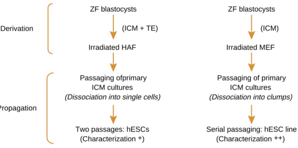
Later, ES cell lines were successfully produced for the rhesus monkey41 and the human.12 Irradiated murine embryonic fibroblast (MEF) feeders, immunosurgery to separate the ICM and passaging of clumps of hES cells instead of disaggregation into single cells were used. Immunosurgery, mitomycin-C-treated MEFs and a similar ‘cut and paste’ method was later used to derive and propagate hES cell lines that could be spontaneously differentiated into neuronal cells.13 Amit and Itskovitz-Eldor42 confirmed that the whole embryo culture worked as well as the immunosurgery protocol to produce hES cell lines. Given the social nature of hES cells as known today, the disaggregration of ICM and hESC colonies with trypsin into single cells during early passage, rather than a ‘cut and paste approach’, might have been responsible for the hES cells differentiating after two passages in the early reports of Bongso et al (Figure 1).9,10 Reliance on a xeno-support system such as MEF introduces considerable disadvantages with respect to exploiting the therapeutic potential of hES cells. A major drawback is the risk of transmitting pathogens from the animal feeder cells or conditioned medium to hES cells. After Bongso et al9,10showed the successful support of hES cells on human feeders, this group reported the production of the first xeno-free hES cell line where hES cells were derived and propagated on mitomycin-C-treated human fetal muscle feeders in the absence of hLIF and in the presence of human-based ingredients in the culture medium. There was no exposure to animal feeders, matrices or animal-based products in the in vitro system.11The same group refined the system further by ranking a variety of human feeders that successfully support the propagation of hES cells. In addition to human fetal muscle, human fetal and adult skin from in-house-derived and commercial sources also supported hES cells very well.43It is therefore possible to collect a non-invasive skin biopsy, expand the epidermis in vitro as epithelial cells in primary culture, then allow the epithelial cells to transform to fibroblasts in passage and use these transformed fibroblasts as hES cell supports. This approach has the advantage that the same in vitro fertilization (IVF) patient donating an embryo for hESC derivation could give her skin biopsy for hESC support and, as she would have been previously screened for HIV and hepatitis B, the risk of cross-contamination of the hESCs with pathogens from the human feeders is eliminated.
ZF blastocysts (ICM + TE)
ZF blastocysts (ICM) Derivation
Irradiated HAF Irradiated MEF
Passaging ofprimary ICM cultures (Dissociation into single cells)
Passaging of primary ICM cultures (Dissociation into clumps)
Two passages: hESCs (Characterization +)
Serial passaging: hESC line (Characterization ++) Propagation
POTENTIAL APPLICATIONS OF STEM CELLS
The most important potential use of hESCs is in transplantation medicine. It is this aspect of hESC research that attracts the most media attention, as well as the most private research funding. In theory, it should be possible to coax hESCs to form a pletheora of differentiated cell types. These derivative cell types can in turn be used to develop cell-replacement therapies for a variety of human diseases. Although in principle this implies that hESCs should have the potential to provide a source of tissues for replacement for all diseases in which native cell types are inactivated or destroyed, the diverse nature of human diseases and technical shortcomings will limit the promise of hESC-based cell-replacement therapy to a few common human diseases. Diseases that affect multiple organs are unlikely to be cured by any form of hESC replacement therapy. Type 1 diabetes and parkinsonism emerge as prime candidates for hESC-based cell-replacement therapy. The success of the Edmonton protocol and cadaver islet transplantation augur well for the success of an analogous strategy using hESC-derived islet progenitor cells. A similar approach utilizing hESC-derived dopaminergic neurons could hypothetically be used to treat Parkinson’s disease.
By contrast, the value of hESCs as a developmental model in helping us gain insight to virtually all human diseases with a genetic basis appears to be limitless. For example, hESC lines are excellent in vitro models for studies on the early events of human development, the causes of early pregnancy loss and certain aspects of embryonic ageing. hESCs also prove useful in the study of the toxicological effects of new drugs. ES cells are very sensitive to culture conditions and differentiate or senesce readily when the culture environment is suboptimal. The greater sensitivity of ES cells compared to adult cells may be an advantage for drug screening studies.
CHALLENGES AND HURDLES IN STEM CELL RESEARCH
Treatment via stem cell research will be a ‘cell-based therapy’. Currently, doctors administer fluids (injections), solids (pills) or carry out surgical intervention to correct disease. For the first time, treatment via stem cell research will be the administration of a cell directly into the body. Thus, several added precautions have to be taken before cell-based therapeutic products can be released into the market (Figure 2).
Stringent tests have to be conducted to ensure that the specialized cell types, which are returned to the patient, are 100% pure. No contaminating undifferentiated hESCs should be present because any undetected transferred renegade hESC has the potential to produce tumours. Specialized cell types derived from hESCs and awaiting transplantation to the patient must be rigorously tested in vitro and in animal or primate models in vivo to show that they can restore normal physiological function in disease models.
Several hurdles in the manipulation and differentiation of hESCs must also be overcome before the technology can be successfully transferred to the bedside. Cell-replacement therapies require the growth of massive numbers of hESCs, so large-scale hESC culture strategies utilizing bioreactors and perfusion systems must be developed to generate sufficient numbers of cells. High efficiency directed differentiation strategies, safer and purer populations of hESCs and their differentiated progeny and clinically compliant ‘xeno-free’ hESC cell lines must be produced.
Screen egg and sperm donors for HIV1, HIV2, Hep B, CJD and other infectious diseases
Derivation of clinical grade ‘xeno-free’ hESC lines in CGMP conditions on human feeder layers or in feeder-free culture
Genetic testing of hESC lines for chromosomal abnormalities and other single-gene defects
Expansion of early passage clinical grade ‘xeno-free’ hESC lines in strict CGMP conditions and detailed phenotypic, genomic and
proteomic characterization tests for pluripotency
Periodic testing of hESC lines for impurities, infectious agents and karyotypic stability
Detailed in vitro characterization of differentiated hESC therapeutic progenitors:
Morphological evaluation
Detection of appropriate cell surface antigens Detailed gene and protein expression analysis Test biological activity in vitro
Check MHC/HLA expression
Detailed in vivo characterization of differentiated hESC therapeutic progenitors:
Test physiological and biological functions in murine and primate models
Demonstrate efficacy and efficiency Demonstrate safety – no tumour formation Test methods to prevent rejection
Phase I clinical trials in human subjects •
• • • •
•
• • •
achieved recently by Hwang et al.44This is one approach in obtaining rejection-free, customised ‘tailor made’ hESC lines. At present, however, the widespread use of nuclear transfer technology to create such cell lines for individual patients seems untenable because of the extremely low success rates of a highly inefficient procedure, the paucity of donor human oocytes and the unknown repercussions of absence of the sperm-imprinting mechanisms.45Instead, given the large number of supernumerary IVF embryos available world wide, deriving and maintaining banks of HLA-typed hESC lines from different genetic and ethnic backgrounds might be a more feasible solution in overcoming the tissue rejection problem. Alternatively, it has been suggested that hESCs could be made less reactive by genetically engineering histocompatibility complexes through the introduction or removal of the appropriate cell surface antigens, thereby creating a universal donor hESC line. Still unknown is whether hESCs are immuno-privileged because of their embryonic origin.
ANIMAL VERSUS HUMAN STEM CELLS
Although many somatic stem cells have been very well characterized and isolated in rodents, the human equivalents of these adult stem cells have been difficult to identify and difficult to expand in vitro. This could reflect innate differences between human and rodent cell physiology. Human somatic stem cells appear to display telomere-dependent replicative senescence, whereas rodent stem cells do not.46
ES cell lines have been established in the mouse, chicken, hamster, rabbit, pig, cow, fish (medaka), primates and humans. However, only mouse and chicken ES cells appear to have germ-line competence and the ability to contribute to chimera formation, which makes them true embryonic stem cell lines. Strikingly, no group has yet been able to derive bona fide rat ES cell lines. In general, rat rather than mouse physiology is believed to be a closer parallel to human physiology. Therefore, the rat model would in theory be a closer representative for the study of human disease. For example, there is a rat model for hypertension but no similar mouse model. The rat asthma model also mimics many features of human asthma and, given the same level of cholesterol and triglycerides, the rat atherosclerosis model demonstrates coronary artery disease and decreased survival comparable to that of humans.47,48 Therefore, if rat ES cell lines can be established they will perhaps benefit medical science in more ways than mESCs have, through the use of knock-out technology.
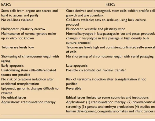
ADULT VERSUS EMBRYONIC STEM CELLS
The differences between adult and ES cells are summarised in Table 1. The contention that somatic stem cells alone will provide for the development of long-sought-after cell-based therapies, and that somatic stem cells are an equivalent and perfect substitute for embryonic stem cells, is a dated assertion. Substantial problems exist with the manipulation of adult stem cells. Some of these problems are possibly technical and might be overcome in the near future, but many perhaps reflect the inherent biology of somatic stem cells.
Most of the literature describing the plasticity of somatic stem cells derives from studies in rodent models. Not all of this work is directly applicable to human stem cell biology. Stem cells in adult human tissues are known to be notoriously difficult to isolate and characterize. In addition, few somatic stem cell types have been confirmed to exist in human tissues and those that can be isolated with relative ease are unfortunately difficult to scale-up in culture and their true latent plasticity has also not been established clearly. These difficulties, coupled with an innate reduced plasticity and cell fusion rather than transdifferentiation properties, have marred progress in the field. Although in the long term research on hESCs might be the best in realizing the therapeutic potential of stem cells, somatic stem cell research and ES cell research can complement each other in many ways and thus both directions should be actively pursued.
Table 1.Differences between human adult stem cells (hASCs) and human embryonic stem cells (hESCs).
hASCs hESCs
Stem cells from organs are scarce and hard to access and purify
Once derived and propagated, stem cells exhibit prolific cell growth and are abundant
No cell-lines available Cell-lines available; easy to scale-up using bulk culture protocol
Multipotent; plasticity narrow Pluripotent; versatile and plasticity wide Maintenance of normal genetic
make-up in vitro not known
Normal karyotype in late passages in ‘cut and paste’ protocol; changes in karyotype in late passage in high density bulk culture protocol
Telomerase levels low Telomerase levels high and consistent; unlimited self-renewal of cells
Shortening of chromosome length with ageing
No shortening of chromosome length with serial passaging
Early apoptosis Late apoptosis
Customizing stem cells/differentiated tissues not possible
Possible via somatic cell nuclear transfer
No risk of teratoma induction after accidental transplantation
Risk of teratoma induction after transplantation if not purified
Epigenetic genomic changes difficult to reverse
Reversible
No ethical issues Ethical issues limited to some countries and institutions Applications: transplantation therapy Applications: (1) transplantation therapy; (2) pharmaceutical
POLITICS AND THE FUTURE OF STEM CELL RESEARCH
As often happens in science, stem cell research has raised as many new questions as it has answered. The field is advancing but several difficult hurdles in the science still need to be overcome. Additionally, legislation and restrictions on hESC research in some countries are slowing progress. Socially, scientists have a responsibility to dispel misconceptions about ES cell research. Myths that hESCs are derived from aborted fetuses and have the potential to form the whole human being need to be dismissed. The public needs to be reassured of the soundness of the science, that there are regulatory frameworks that can govern hESC research and that punitive measures can be put in place to censure rogue scientists attempting to clone whole human beings.
In the US, federal funds can be used for work on only 21 hESC lines created before August 2001 under a partial ban decreed by the US President (see: http://www. stemcells.nih.gov/research/registry). All of them have been exposed to mouse feeder layers, making them undesirable for therapeutic endeavours. This has prompted scientists in the US to rely on private funding to circumvent these legislative issues and derive new hESC lines to proceed with their work.14
Political and religious disagreements about stem cells and their use are everywhere, but nowhere is there a more bewildering array of positions than in Europe, where four different models are emerging. The first model, developing in the UK, permits the generation and use of hESCs as well as therapeutic cloning, with certain restrictions. The second, visible in the Netherlands, permits the generation and use of hESCs but forbids therapeutic cloning. The third, seen in Germany, forbids the generation of hESCs and therapeutic cloning but allows, under exceptional conditions, the use of existing hESC lines for research only. The fourth, evident in Ireland and Austria, forbids all generation and use of hESCs and therapeutic cloning as well.52At the opposite end of the spectrum, hESC research in countries like the UK, Singapore, Israel, South Korea, China, and Japan enjoy generous government support.
A recurrent statement in stem cell biology today is the importance of standardizing culture conditions. Culture conditions have a profound effect on stem cell self-renewal, differentiation, and possibly on stem cell plasticity. A re-acquisition of plasticity in somatic stem cell transdifferentiation might arise because of in vitro culture conditions that actively promote reactivation and dedifferentiation.
For future work on hESCs, it is important to identify a common set of molecular markers and understand how different hESC lines from diverse genetic backgrounds derived in different labs differ from each other. Indeed, there appear to be several significant detectable differences at the molecular level between different hESC lines.51 Some hESC lines seem predisposed to differentiate along a particular cell lineage and form, for example, cardiomyocytes readily when undergoing spontaneous differen-tiation, whereas other hESC lines might form neural precursors more readily.53The karyotypic stability of hESC lines over hundreds of population doublings in vitro, and the frequency of occurrence of aneuploidy, need to be accurately determined.
Recent work suggests that there might be new ways of generating stem cells. mESCs can differentiate into oocyte-like cells that are potential recipients for nuclear transfer.54If hESCs could be coaxed to do the same, this would not only eliminate the need for oocyte donations in nuclear transfer experiments but would also allow the generation of patient-specific stem cells that would not be rejected by the patient’s immune system. The possible derivation of gametes from hESCs warrants further research.
Another area of future research entails the delivery of stem cells to the tissues in which they are needed. Current practice involves either the injection of stem cells directly into the targeted tissue, or injection of the stem cells into the bloodstream without any guarantee that they will actually home-in on the appropriate tissues. ‘Targeted delivery’ would ensure that the therapeutic stem cells are introduced only to organs and tissues that need them. Research should also be aimed at identifying and understanding the in vivo somatic stem cell niche. In particular, a more thorough understanding of how niche cells influence stem cell-fate decisions will lead to the development of better isolation and expansion techniques.
SUMMARY
Both embryonic and adult stem cells have enormous potential to further our understanding of basic developmental processes but the exceptional properties of hESCs make them uniquely powerful tools for the development of cell-based therapies in reparative medicine, as well as invaluable models for the study of early human embryogenesis.
Although there are fewer moral objections associated with adult stem cell work, the assertion that both adult and embryonic stem cells are equivalent is tenuous. Attempts have been made to hype adult stem cells at the expense of hESCs but hESCs clearly have greater differentiation potential over adult stem cells by virtue of their position in embryonic development. It is this fact, and the ability to culture hESCs easily in vitro in large numbers, that makes hESCs the current best hope for the development of cell-replacement therapies. Nevertheless, research on adult stem cells and embryonic stem cells should be energetically pursued in tandem because some diseases might benefit from one, and some from the other.
It is important to identify the exact nature of the pluripotent state in hESCs and hEGCs: How it is acquired, maintained and propagated and which genes confer pluripotency. Furthermore, we will need to unravel how epigenetic modifications permit a switch in patterns of gene expression that are central to plasticity and transdifferentiation in adult stem cells. Finally, understanding the fundamental mechanisms by which cell-fate is determined during embryonic development will prove informative for the in vitro manipulation of stem cells.
REFERENCES
1. Wolpert L, Hicklin J & Hornbruch A. Positional information and pattern regulation in regeneration of hydra.Symp Soc Exp Biol1971;25:391–415.
4. Alison MR, Poulsom R, Forbes S & Wright NA. An introduction to stem cells.J Pathol2002;197:419–423. 5. Gardner RL. Stem cells: potency, plasticity and public perception.J Anat2002;200:277–282. *6. Ramalho-Santos M, Yoon S, Matsuzaki Y et al. ‘Stemness’: transcriptional profiling of embryonic and adult
stem cells.Science2002;298:597–600.
7. Yoshimizu T, Sugiyama N, De Felice M et al. Germline-specific expression of the Oct-4/green fluorescent protein (GFP) transgene in mice.Dev Growth Differ1999;41:675–684.
8. Pesce M, Wang X, Wolgemuth DJ & Scholer H. Differential expression of the Oct-4 transcription factor during mouse germ cell differentiation.Mech Dev1998;71:89–98.
9. Bongso A, Fong CY, Ng SC et al. The growth of inner cell mass cells from human blastocysts (Abstract). Theriogenology1993;41:161.
*10. Bongso A, Fong CY, Ng SC et al. Isolation and culture of inner cell mass cells from human blastocysts. Hum Reprod1994;9:2110–2117.
*11. Richards M, Fong CY, Chan WK et al. Human feeders support prolonged undifferentiated growth of human inner cell masses and embryonic stem cells.Nat Biotechnol2002;20:933–936.
*12. Thomson JA, Itskovitz-Eldor J, Shapiro SS et al. Embryonic stem cell lines from human blastocysts.Science 1998;282:1145–1147.
*13. Reubinoff BE, Pera MF, Fong CY et al. Embryonic stem cell lines from human blastocysts: somatic differentiation in vitro.Nat Biotechnol2000;18:399–404.
14. Cowan CA, Klimanskaya I, McMahon JP et al. Derivation of embryonic stem-cell lines from human blastocysts.N Engl J Med2004;350:1353–1356.
*15. Shamblott MJ, Axelman J, Wang S et al. Derivation of pluripotent stem cells from cultured human primordial germ cells.Proc Natl Acad Sci USA1998;95:13726–13731.
16. Shamblott MJ, Axelman J, Wang S et al. Human embryonic germ cell derivatives express a broad range of developmentally distinct markers and proliferate extensively in vitro.Proc Natl Acad Sci USA2001;98:
113–118.
17. Beattie GM, Otonkoski T, Lopez AD & Hayek A. Functional beta-cell mass after transplantation of human fetal pancreatic cells: differentiation or proliferation?Diabetes1997;46:244–248.
18. Brustle O, Choudary K, Karram K et al. Chimeric brains generated by intraventricular transplantation of human brain cells into embryonic rats.Nat Biotechnol1998;16:1040–1044.
19. Villa A, Snyder EY, Vescovi A & Martinez-Serrano A. Establishment and properties of a growth factor dependent perpetual neural stem cell line from the human CNS.Exp Neurol2000;161:67–84. 20. Spradling A, Drummond-Barbosa D & Kai T. Stem cells find their niche.Nature2001;414:98–104. 21. Short B, Brouard N, Occhiodoro-Scott T et al. Mesenchymal stem cells.Arch Med Res2003;34:565–571. 22. Dor Y, Brown J, Martinez OI & Melton DA. Adult pancreatic beta-cells are formed by self-duplication
rather than stem-cell differentiation.Nature2004;429:41–46.
23. Smith AG. Embryo-derived stem cells- of mice and men.Annu Rev Cell Dev Biol2001;17:435–462. 24. Andrews PW. From teratocarcinomas to embryonic stem cells.Philos Trans R Soc Lond B Biol Sci2002;
357:405–417.
25. Jiang Y, Jahagirdar BN, Reinhardt RL et al. Pluripotency of mesenchymal stem cells derived from adult marrow.Nature2002;418:41–49.
26. Bjornson CR, Rietze RL, Reynolds BA et al. Turning brain into blood: a hematopoietic fate adopted by adult neural stem cells in vivo.Science1999;283:534–537.
27. Jackson KA, Mi T & Goodell MA. Hematopoietic potential of stem cells isolated from murine skeletal muscle.Proc Natl Acad Sci1999;96:14482–14486.
28. Clarke DL, Johansson CB, Wilbertz J et al. Generalized potential of adult neural stem cells.Science2000;
288:1660–1663.
29. Krause DS, Theise ND, Collector MI et al. Multi-organ, multi-lineage engraftment by a single bone-marrow derived stem cell.Cell2001;105:369–377.
30. Schuldiner M, Yanuka O, Itskovitz-Eldor J et al. Effects of eight growth factors on the differentiation of cells derived from human embryonic stem cells.Proc Natl Acad Sci2000;97:11307–11312.
31. Kehat I, Kenyagin-Karsenti D, Snir M et al. Human embryonic stem cells can differentiate into myocytes with structural and functional properties of cardiomyocytes.J Clin Invest2001;108:407–414. *32. Mummery C, Ward-van Oostwaard D, Doevendans P et al. Differentiation of human embryonic
stem cells to cardiomyocytes: role of coculture with visceral endoderm-like cells.Circulation2003;107:
*33. Reubinoff BE, Itsykson P, Turetsky T et al. Neural progenitors from human embryonic stem cells.Nat Biotechnol2001;19:1134–1140.
34. Zhang SC, Wernig M, Duncan ID et al. In vitro differentiation of transplantable neural precursors from human embryonic stem cells.Nat Biotechnol2001;19:1129–1133.
35. Assady S, Maor G, Amit M et al. Insulin production by human embryonic stem cells.Diabetes2001;50:
1691–1697.
36. Clark AT, Bodnar MS, Fox M et al. Spontaneous differentiation of germ cells from human embryonic stem cells in vitro.Hum Mol Genet2004;13:723–727.
37. Xu RH, Chen X, Li DS et al. BMP4 initiates human embryonic stem cell differentiation to trophoblast. Nat Biotechnol2002;20:1261–1264.
*38. Ying QL, Nichols J, Evans EP et al. Changing potency by spontaneous fusion.Nature2002;416:545–548. 39. Terada N, Hamazaki T, Oka M et al. Bone marrow cells adopt the phenotype of other cells by
spontaneous fusion.Nature2002;416:542–545.
40. Evans MJ & Kaufman M. Establishment in culture of pluripotential stem cells from mouse embryos.Nature 1981;292:151–156.
41. Thomson JA, Kalishman J, Golos TG et al. Isolation of a primate embryonic stem cell line.Proc Natl Acad Sci1995;92:7844–7848.
42. Amit M & Itskovitz-Eldor J. Derivation and spontaneous differentiation of human embryonic stem cells. J Anat2002;200(Pt 3):225–232.
43. Richards M, Tan S, Fong CY et al. Comparative evaluation of various human feeders for prolonged undifferentiated growth of human embryonic stem cells.Stem Cells2003;21:546–556.
*44. Hwang WS, Ryu YJ, Park JH et al. Evidence of a pluripotent human embryonic stem cell line derived from a cloned blastocyst.Science2004;303:1669–1674.
45. Mann JR. Imprinting in the germ line.Stem Cells2001;19:287–294.
46. Wright WE & Shay JW. Fundamental differences in human and mouse telomere biology.Nat Med2000;6:
849–851.
47. Stoll M & Jacob HJ. Genetic rat models of hypertension: relationship to human hypertension.Curr Hypertens Rep2001;3:157–164.
48. Bice DE, Seagrave J & Green FH. Animal models of asthma: potential usefulness for studying health effects of inhaled particles.Inhal Toxicol2000;12:829–862.
49. Smith AG, Heath JK, Donaldson DD et al. Inhibition of pluripotential embryonic stem cell differentiation by purified polypeptides.Nature1988;336:688–690.
50. Williams RL, Hilton DJ, Pease S et al. Myeloid leukaemia inhibitory factor maintains the developmental potential of embryonic stem cells.Nature1988;336:684–687.
51. Richards M, Tan SP, Tan JH et al. The transcriptome profile of human embryonic stem cells as defined by SAGE.Stem Cells2004;22:51–64.
52. Lanza R & Rosenthal N. The stem cell challenge.Sci Am2004;June.
53. Abeyta MJ, Clark AT, Rodriguez RT et al. Unique gene expression signatures of independently-derived human embryonic stem cell lines.Hum Mol Genet2004;13:601–608.