Effect of Vitamin D Serum Levels and
GC Gene Polymorphisms in
Liver Fibrosis Due to Chronic Hepatitis C
Laura A. Azevedo,* Ursula Matte,**,*** Themis R. Silveira*,*** Jacqueline W. Bonfanti,**** Juliana P. Bruch,* Mário R. Álvares-da-Silva*,****
* Graduate Program in Gastroenterology and Hepatology Sciences, Universidade Federal do Rio Grande do Sul, Porto Alegre, Brazil. ** Department of Genetics; Graduate Program in Genetics and Molecular Biology, Universidade Federal do Rio Grande do Sul, Porto Alegre, Brazil. *** Graduate Program in Child and Adolescence Health, Universidade Federal do Rio Grande do Sul; Experimental Research Center, Hospital de Clínicas de Porto Alegre, Porto Alegre, Brazil. **** Department of Internal Medicine, Universidade Federal do Rio Grande do Sul; Division of Gastroenterology, Hospital de Clínicas de Porto Alegre, Porto Alegre, Brazil.
September-October, Vol. 16 No. 5, 2017: 742-748
INTRODUCTION
Liver fibrosis is defined by extracellular matrix (ECM) accumulation with consequent loss of liver function, due to acute or chronic damage.1 Chronic hepatitis C (CHC) leads to liver fibrosis and 20-30% of patients progress to cirrhosis after 25-30 years of infection.2 Several factors in-fluence the evolution of CHC and formation of liver fi-brosis, such as age, obesity, virus C genotype, alcohol consumption, duration of infection, presence of viral co-infections and metabolic disorders.2
Vitamin D has been associated with chronic liver dis-eases and low vitamin levels may contribute to progres-sion of CHC.3 Vitamin D is mainly synthesized in the skin by 7-dehydrocholesterol redutase (DHCR7) in pre-vitamin D, which is converted into pre-vitamin D by isomeri-zation. Vitamin D and its metabolites circulate bounded to vitamin D-binding protein (GC-globulin). Vitamin D goes under hydroxylation in the liver by cytochrome P450 enzymes forming 25-hydroxyvitamin D (25(OH)D) and is used to evaluate vitamin D status. The metabolite 25(OH)D is hydroxylated by CYP27B1 in the kidney, The Official Journal of the Mexican Association of Hepatology,
the Latin-American Association for Study of the Liver and the Canadian Association for the Study of the Liver
Manuscript received: Manuscript received: Manuscript received: Manuscript received:
Manuscript received: October 22, 2016. Manuscript accepted:Manuscript accepted:Manuscript accepted:Manuscript accepted:Manuscript accepted: April 02, 2017.
DOI:.
A B S T R A C T A B S T R A C T A B S T R A C T A B S T R A C T A B S T R A C T
Introduction and aim. Introduction and aim.Introduction and aim. Introduction and aim.
Introduction and aim. Vitamin D has been associated with chronic liver diseases and low vitamin levels may contribute to pro-gression of chronic hepatitis C. The aim of this study was to evaluate the influence of vitamin D serum levels and GC gene polymor-phisms in the severity of liver fibrosis in patients with chronic hepatitis C genotype 1. Material and methods.Material and methods.Material and methods.Material and methods.Material and methods. Cross-sectional study that enrolled 132 adult patients with chronic hepatitis C genotype 1 attended at the outpatient Clinic of Gastroenterology Divi-sion at Hospital de Clínicas de Porto Alegre. At the time of enrollment patients had a blood withdraw for serum 25(OH)D determina-tion and genotypic analysis of rs7041 and rs4588 polymorphisms in GC gene. None/mild fibrosis was considered as METAVIR F0, F1 and F2 and severe fibrosis as METAVIR F3 and F4. Results.Results.Results.Results.Results. Median 25(OH)D levels in the sample were 19.9 ng/mL (P25-P75: 14.0-29.4). Fifty percent of patients presented vitamin D deficiency (< 20 ng/mL). In stepwise multiple linear regression the var-iables associated with 25(OH)D levels were blood withdrawn in Winter/spring season, the haplotypes AT/AT + AG/AT of rs7041 and rs4588 and female sex. For evaluation of severe fibrosis, variables associated in logistic regression were age, vitamin D severe defi-ciency (< 10 ng/mL), glucose levels, BMI and platelets count. Conclusions.Conclusions.Conclusions.Conclusions.Conclusions. Vitamin D levels are associated with severity of liver fibrosis in chronic hepatitis C genotype 1 patients. Although the rs7041 and rs4588 GC polymorphisms are strong predictors of vita-min D levels, they do not play a direct role in liver fibrosis.
Key words. Key words.Key words. Key words.
producing 1,25-dihydroxyvitamin D (1,25(OH)D), the ac-tive form that binds to vitamin D receptor (VDR).4 Vita-min D deficiency is a risk factor for all-cause mortality in the general population5 as well as for cirrhotic patients.6
GC-globulin is coded by GC gene. The three protein isoforms are coded by different alleles, which are formed by single nucleotide polymorphisms (SNPs). Polymor-phism rs7041 has two alternative alleles, containing either A or C, and polymorphism rs4588 has either G or T. Each person carries two copies of the GC gene; therefore pos-sible genotypes for rs7041 are AA, AC or CC, for example. Different SNPs at the same gene are inherited together forming a haplotype. For rs7041 and rs4588 possible haplo-types are CT, CG or AT. These two SNPs are responsible for the three major circulating isoforms of GC-globulin Gc1F (CT), Gc1S (CG) and Gc2rs (AT).7 The Gc-2 iso-form is described to have the lowest affinity for its ligand.8 The three common haplotype variants combined form CT-CT, CT-CG, CG-CG, CG-AT, CT-AT and AT-AT.
It is known that several polymorphisms involved in the vitamin D metabolism are associated with its serum con-centrations.9,10 The aim of this study was to evaluate the influence of vitamin D serum levels and GC polymor-phisms in the severity of liver fibrosis in patients with chronic hepatitis C genotype 1.
MATERIAL AND METHODS
This was a cross-sectional study that enrolled 132 adult patients with chronic hepatitis C genotype 1 attended at the outpatient Clinic of Gastroenterology Division at Hospital de Clínicas de Porto Alegre, Brazil, from June 2013 to February 2015. Hepatitis C virus infection was confirmed by anti-HCV ELISA 3 and by detection of viral RNA by polymerase chain reaction (HCV RNA PCR). Patients infected by the human immunodeficiency virus (HIV) or hepatitis B virus (HBV), alcohol abusers (> 40g/ day), chronic renal injured, liver transplanted, decompen-sated cirrhotic and those with hepatocellular carcinoma (HCC) or in immunosuppressive or antiviral treatment were excluded. Both naïve and previously PEG-INF treated patients were included.
At the time of the enrollment patients had a blood with-draw for vitamin D serum levels determination and geno-typic analysis. Alanine aminotransferase (ALT), aspartate aminotransferase (AST), gamma-glutamyl-transferase (GGT), platelets, glucose, cholesterol and body mass in-dex (BMI) were extracted from patient’s chart from the previous 6 months. Severity of fibrosis was evaluated by the last liver biopsy done by the outpatients Clinic rou-tine. Liver biopsies were evaluated by the same patholo-gist and scored with METAVIR system.11 Serum 25(OH)D levels were determined using a
chemolumines-cent immunoassay on a Liaison automatic analyzer (Dia-Sorin). Vitamin D levels below 10 ng/mL were considered severe deficiency; 10 to 20 ng/mL deficiency; 20 to 30 ng/ mL insufficiency; and above 30 ng/mL sufficiency.12 Date of sample collection was categorized into two seasons, Summer/autumn (December-May) and Winter/spring (June-November). The study was conducted according to the principles of the Declaration of Helsinki and was ap-proved by the hospital Ethical Committee. All patients gave written informed consent to participate in the study.
Genotypic analysis
The genomic DNA was extracted from whole blood EDTA using Wizard® Genomic DNA Purification Kit (Promega) according to manufacturer’s instructions. The polymorphisms rs7041 and rs4588 from the GC gene were identified using hydrolysis probes (TaqMan Assays; ABI), IDs: C_3133594_30 and C_8278879_10, respectively. A to-tal of 10 ng of DNA was used in a final reaction volume of 12.5 μL for real-time PCR on StepOne™ System (Thermo Fisher).
Statistical analysis
data.13,14 Statistical analysis was performed using SPSS 20.0 (SPSS Inc, Chicago, Il, USA). Significance level was set at
α< 0.05.
RESULTS
Patient features
Demographical, clinical, biochemical and genotypic features of enrolled patients are described in table 1. Pa-tients' age varied from 26 to 74 years. The majority of pa-tients were cirrhotic (33.3%). Median 25(OH)D levels were 19.9 ng/mL (14.0 - 29.4). Fifty percent of patients presented deficiency or severe deficiency of vitamin D.
Genotypes and alleles frequencies of rs7041 and rs4588 were in Hardy Weinberg Equilibrium. The most frequent genotypes were AC for rs7041 and GG for rs4588 (Table 2). Finally, for haplotypes, GC/TA, GC/GC and GC/GC were the most common ones.
Predictors of 25(OH)D serum levels
Vitamin D levels were higher in patients enrolled in Summer/Autumn than in Winter/Fall [31.2ng/mL (IQR 19.8-35.0) vs. 18.4ng/mL (IQR 12.8-24.1); P < 0.001] (Figure 1). Median 25(OH)D levels was 18.0 ng/mL (12.7 - 23.2) for females and 24.0 ng/mL (17.2 - 31.8) for males (P = 0.002). Vitamin D did not differ between näive and PEG-INF treated patients (P = 0.921). When
Table 1. Demographic, clinical and biochemical features of chronic hepatitis C genotype 1 patients.
Variable Chronic hepatitis C
genotype 1 (n = 132)
Sex (male)a 53.8% (71)
Age (years)b 53 (± 9)
Body mass index (kg/m2)b 28.5 (± 5.2)
ALT (IU/L)c 64 (42-100)
AST(IU/L)c 53 (36-84)
GGT(IU/L)c 90 (43-156)
Platelets (103/μL)b 186 (± 51)
Cholesterol (mg/dL)c 164 (147-185)
Glucose (mg/dL) c 100 (91-111)
DM presence (yes)a 21.2% (28)
METAVIRa
F0 12.9% (17)
F1 22.7% (30)
F2 18.9% (25)
F3 12.1% (16)
F4 33.3% (44)
Näivea 56.8% (75)
25OH vitamin D (ng/mL)c 19.9 (14.0-29.4)
Vitamin D statusa
Severe deficiency 13.6% (18)
Deficiency 36.4% (48)
Insufficiency 27.3% (36)
Sufficiency 22.7% (30)
Season of blood withdrawn
Summer/Autumn 25% (33)
DM: diabetes mellitus. a Percentage (n). b Mean (standard deviation). c Median (IQR P25-P75). Total sample for: BMI = 125; platelets = 131.
Table 2. Allelic, genotypic and haplotypic data of rs7041 and rs4588 polymorphisms in chronic hepatitis C genotype 1 pa-tients.
Chronic hepatitis C genotype 1 (n = 129)
rs7041a
C 46.1%
CC 19.4% (25)
AC 53.5% (69)
A A 27.1% (35)
rs4588a
T 21.3%
TT 6.2% (8)
GT 30.2% (39)
GG 63.6% (82)
Haplotypesa
AT/AT 6.2% (8)
AG/AT 9.3% (12)
AG/AG 11.6% (15)
CG/AT 20.9% (27)
CG/CG 19.4% (25)
AG/CG 32.6% (42)
a Percentage (n).
Figure 1. Figure 1.Figure 1.
Figure 1.Figure 1. Median 25(OH)D serum levels of the enrolled patients according to investigation month.
Median 25(OH)D (ng/mL)
40.0
30.0
20.0
10.0
0.0
25(OH)D serum levels were stratified among rs7041 and rs4588 genotypes and haplotypes, there was no statistical significance (P > 0.05), however a trend is graphically ob-served for AA genotype of rs7041 and TT genotype of rs4588, as well as for AT/AT and AG/AT haplotypes (Fig-ure 2). Transforming the mentioned genotypes and haplo-types into dichotomous variables, it is observed that 25(OH)D levels are lower for AA genotype of rs7041, TT genotype of rs4588 and AT/AT + AG/AT haplotypes when compared to their counterparts (Table 3). In multiple lin-ear regression the variables associated with diminished
Table 4. Stepwise forward multiple linear regression analysis for variables associated with 25(OH)D serum levels.
Variables β SE P value
Winter/Spring vs. Summer/Autumn -0.375 2.003 < 0.001
Female sex -0.261 1.740 0.001
Haplotypes AT/AT + AG/AT vs. others -0.259 1.254 0.001
Variables included in the model: sex, age, fibrosis (none/mild or severe), diabetes mellitus, BMI, ALT, AST, GGT, cholesterol, glucose, platelets, rs4588 and rs7041 haplotypes (AT/AT + AG/AT vs. others), season of blood withdrawn (Summer/Autumn vs. Winter/Spring). β: standardized coefficient beta. SE: standard error.
25(OH)D circulating levels were blood withdrawn in Winter/Spring, the risk haplotypes AT/AT + AG/AT and female sex (Table 4).
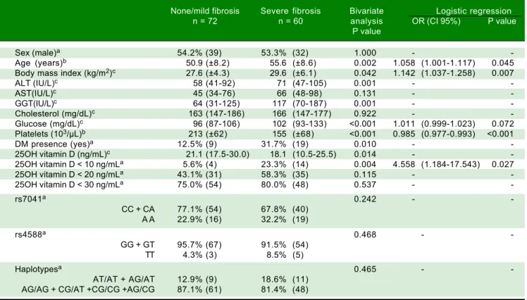
Variables related to liver fibrosis
In table 5 are presented the association of studied varia-bles with severity of liver fibrosis. Older age; higher BMI, ALT, GGT and glucose; lower platelets and 25(OH)D; di-abetes and severe vitamin D deficiency were related to se-vere liver fibrosis in bivariate analysis. None of the
Table 3. Median 25 (OH)D stratified according to risky genotypes and haplotypes of rs7041 and rs4588.
25(OH) D (ng/mL)* P value
rs7041 0.026
CC + CA 22.8 (16.2-30.1)
A A 17.0 (8.9-26.6)
rs4588 0.023
GG + GT 20.2 (15.8-29.8)
TT 9.6 (8.1-20.9)
Haplotypes 0.002
AT/AT + AG/AT 12.3 (8.1-19.2)
AG/AG + CG/AT +CG/CG +AG/CG 21.2 (16.2-30.1)
* Median (IQR P25-P75) Mann-Whitney. Figure 2.
Figure 2.Figure 2.
Figure 2.Figure 2. Box plots showing the distribution of 25(OH) D serum levels stratified according to (AAAA) rs7041 genotypes, (BA BBB) rs4588 genotypes and (C)B C)C)C)C) rs7041 and rs4588 haplotypes. Upper horizontal line of box, 75th percentile; lower horizontal line of box, 25th percentile; horizontal bar within box, median; upper horizon-tal bar outside box, 90th percentile; lower horizonhorizon-tal bar outside box, 10th percentile. * Circles represent outliers. P values for Kruskal-Wallis test.
25(OH)D levels (ng/mL)
60
40
20
0
CC AC AA
rs7041 genotypes
B BB B B
TT GT GG
rs4588 genotypes
C C C C C
AT/AT CG/AT CG/CG AG/AT AG/CG AG/AG
rs7041 and rs4588 haplotypes A
A A A A
P = 0.075 P = 0.057 P = 0.073
60
40
20
0
60
40
20
0
evaluated genotypes and haplotypes of rs7041 and rs4588 were associated with fibrosis severity. In the logistic re-gression analysis were included sex, age, 25(OH)D > 10 ng/mL, season of blood withdrawn (Summer/Autumn vs. Winter/Spring), diabetes mellitus, BMI, ALT, AST, GGT, cholesterol, glucose, platelets, rs4588 and rs7041 haplo-types (AT/AT + AG/AT vs. others). The variables that re-mained in the model were age, 25(OH)D < 10 ng/mL, glucose levels, BMI and platelets count. When 25(OH)D is included as a continuous variable it loses statistical sig-nificance in the model.
DISCUSSION
In recent years, the influence of vitamin D in liver dis-eases has been broadly discussed since it undergoes hepat-ic metabolism. Herein we evaluated the association of vitamin D serum levels and of rs7041 and rs4588 polymor-phisms from GC gene in severe liver fibrosis in a sample of 132 chronic hepatitis C genotype 1 patients.
In this sample, vitamin D median levels were 19.9 ng/ mL and deficiency prevalence was 50%. In a study with 496
Table 5. Bivariate and logistic regression analysis of risk factors associated with severe fibrosis in chronic hepatitis C genotypes 1 patients.
None/mild fibrosis Severe fibrosis Bivariate Logistic regression
n = 72 n = 60 analysis OR (CI 95%) P value
P value
Sex (male)a 54.2% (39) 53.3% (32) 1.000 -
-Age (years)b 50.9 (±8.2) 55.6 (±8.6) 0.002 1.058 (1.001-1.117) 0.045
Body mass index (kg/m2)c 27.6 (±4.3) 29.6 (±6.1) 0.042 1.142 (1.037-1.258) 0.007
ALT (IU/L)c 58 (41-92) 71 (47-105) 0.001 -
-AST(IU/L)c 45 (34-76) 66 (48-98) 0.131 -
-GGT(IU/L)c 64 (31-125) 117 (70-187) 0.001 -
-Cholesterol (mg/dL)c 163 (147-186) 166 (147-177) 0.922 -
-Glucose (mg/dL)c 96 (87-106) 102 (93-133) <0.001 1.011 (0.999-1.023) 0.072
Platelets (103/μL)b 213 (±62) 155 (±68) <0.001 0.985 (0.977-0.993) <0.001
DM presence (yes)a 12.5% (9) 31.7% (19) 0.010 -
-25OH vitamin D (ng/mL)c 21.1 (17.5-30.0) 18.1 (10.5-25.5) 0.014 -
-25OH vitamin D < 10 ng/mLa 5.6% (4) 23.3% (14) 0.004 4.558 (1.184-17.543) 0.027
25OH vitamin D < 20 ng/mLa 43.1% (31) 58.3% (35) 0.115 -
-25OH vitamin D < 30 ng/mLa 75.0% (54) 80.0% (48) 0.537 -
-rs7041a 0.242 -
-CC + CA 77.1% (54) 67.8% (40)
A A 22.9% (16) 32.2% (19)
rs4588a 0.468 -
-GG + GT 95.7% (67) 91.5% (54)
TT 4.3% (3) 8.5% (5)
Haplotypesa 0.465 -
-AT/AT + AG/AT 12.9% (9) 18.6% (11)
AG/AG + CG/AT +CG/CG +AG/CG 87.1% (61) 81.4% (48)
Bivariate analysis: a Percentage (n) Fisher’s Exact Test. b Mean (standard deviation) T test. c Median (IQR P25-P75) Mann-Whitney. Variables included in the
stepwise logistic regression model with forward approach: sex, age, 25(OH)D > 10 ng/mL, season of blood withdrawn (summer/autumn vs. winter/spring), diabetes mellitus, BMI, ALT, AST, GGT, cholesterol, glucose, platelets, rs4588 and rs7041 haplotypes (AT/AT + AG/AT vs. others). OR: odds ratio. CI: confidence interval.
CHC patients from Swiss hospitals, median 25(OH)D was 13.4 ng/mL, and 74% presented levels below 20.0 ng/ mL.15 It has been described that the prevalence of vitamin D insufficiency and deficiency in patients with chronic liver disease ranges between 64 and 92%.16
same results in healthy Brazilian girls.18 The Gc-2 isoform is described to have the lowest affinity for its ligand, which is in accordance confirming the lowest levels of vi-tamin D described in both studies.10
Vitamin D deficiency seems to be associated with a poorer prognosis of the liver disease. In this sample, a dif-ference in 25(OH)D serum levels was observed in none/ mild and severe fibrosis (21.1 vs. 18.1 ng/mL; P = 0.014). Baur, et al. reported that individuals with CHC METAVIR 2 presented 15.5 ng/mL 25(OH)D levels compared to 22.2 ng/mL of individuals METAVIR 0/1.19 Meanwhile, Petta described that CHC G1 with Scheuer scoring 0-2 present-ed 25.5ng/mL vs. 21.9 ng/mL from patients Scheuer scor-ing of 3-4.20 In our sample the overall prevalence of severe deficiency was 13.6% (18/132), in patients with none/mild fibrosis was 5.6% (4/72) and in patients with severe fibro-sis was 23.3% (14/60). In a cohort of German CHC pa-tients, severe deficiency prevalence was 25%.21 In logistic regression, severe vitamin D deficiency was associated with severe fibrosis along with age, BMI and platelets count. Although the studied polymorphisms have present-ed association with vitamin D circulating levels, they were not related to liver fibrosis. There are studies that have shown that polymorphisms of vitamin D metabolism relate to the stages of liver disease. Studies from Petta and Grünhage identified the polymorphism rs12785878 in the gene of cholesterol reductase (DHCR7) associated with a greater degree of liver fibrosis.22,23 This polymorphism was not evaluated in the present sample. Another gene that is important in liver fibrosis is VDR, since it can down-regulate the activation of hepatic stellate cells by compet-ing for several genomic sites related to fibrosis, which are activated by SMAD3, an effector of the transforming growth factor beta 1 (TGFβ1).24 The rs1544410, rs7975232 and rs731236 polymorphisms have been associated with the rate of progression of fibrosis and cirrhosis.19
As limitation of this study we could cite that some co-founding factors were not analyzed, such as smoking, in-travenous drug use, duration of HCV infection. Also, it was a cross-sectional study that analyzed only two poly-morphisms. Although the sample size is relatively small, we considered as a main strength the use of a well-defined cohort of HCV genotype 1 patients that represent the real-world experience. Our results corroborate the literature findings, whereas lower vitamin D levels are found among patients with higher liver fibrosis. Identifying those pa-tients is important since oral supplementation of vitamin D is a very simple therapeutic measure. Several studied have demonstrated that vitamin D deficiency I is likely to be related to lower odds of sustained virological response (SVR) to treatment with pegylated interferon plus ribavi-rin.25 and recently, in a Japanese trial, vitamin D3 supple-mentation increased SVR rates on refractory CHC patients
on treatment with simeprevir, pegylated interferon plus ribavirin.26
In summary, vitamin D levels were associated with severity of liver fibrosis in chronic hepatitis C genotype 1 patients. Although the rs7041 and rs4588 GC gene poly-morphisms are strong predictors of vitamin D levels, they do not play a direct role in liver fibrosis.
ABBREVIATIONS
• 1,25(OH)D: 1,25-dihydroxyvitamin D. • 25(OH)D: 25-hydroxyvitamin D. • ALT: alanine aminotransferase. • AST: aspartate aminotransferase. • BMI: body mass index.
• CHC: chronic hepatitis C.
• DHCR7: 7-dehydrocholesterol redutase. • ECM: extracellular matrix.
• GGT: gamma-glutamyl-transferase. • SVR: sustained virological response. • VDR: vitamin D receptor.
FINANCIAL SUPPORT
The authors would like to thank Fundo de Incentivo à Pesquisa do Hospital de Clínicas de Porto Alegre (FIPE-HCPA), Programa de Apoio à Pós-Graduação da Coorde-nação de Aperfeiçoamento de Pessoal de Nível Superior (PROAP-CAPES) and Conselho Nacional de Desen-volvimento Científico e Tecnológico (CNPq).
AUTHORS CONTRIBUTION
Study conception and design: LAA; UM; TRS; MRAS. Performed the experiments: LAA; JWB; JPB. Analyzed the data: LAA. Wrote the paper: LAA; UM; TRS; JPB; JWB; MRAS. Critical revision: UM; TRS; MRAS.
REFERENCES
1. Friedman SL. Mechanisms of hepatic fibrogenesis. Gastro-enterology 2008; 134: 1655-69.
2. Lingala S, Ghany MG. Natural History of Hepatitis C. Gastro-enterol Clin North Am 2015; 44: 717-34.
3. Iruzubieta P, Teran A, Crespo J, Fabrega E. Vitamin D defi-ciency in chronic liver disease. World J Hepatol 2014; 6: 901-15.
4. Stokes CS, Volmer DA, Grunhage F, Lammert F. Vitamin D in chronic liver disease. Liver International 2013; 33: 338-52. 5. Schottker B, Jorde R, Peasey A, Thorand B, Jansen EH,
Groot L, Streppel M, et al. Vitamin D and mortality: meta-anal-ysis of individual participant data from a large consortium of cohort studies from Europe and the United States. BMJ 2014; 348: g3656.
with advanced liver cirrhosis. Eur J Clin Invest 2014; 44: 176-83.
7. Christiansen M, Jorgensen CS, Laursen I, Hirschberg D, Ho-jrup P, Houen G. Protein chemical characterization of Gc globulin (vitamin D-binding protein) isoforms; Gc-1f, Gc-1s and Gc-2. Biochim Biophys Acta 2007; 1774: 481-92. 8. Arnaud J, Constans J. Affinity differences for vitamin D
me-tabolites associated with the genetic isoforms of the human serum carrier protein (DBP). Hum Genet 1993; 92: 183-8. 9. Wang TJ, Zhang F, Richards JB, Kestenbaum B, van Meurs
JB, Berry D, Kiel DP, et al. Common genetic determinants of vitamin D insufficiency: a genome-wide association study. Lancet 2010; 376: 180-8.
10. Ahn J, Yu K, Stolzenberg-Solomon R, Simon KC, McCullough ML, Gallicchio L, Jacobs EJ, et al. Genome-wide association study of circulating vitamin D levels. Hum Mol Genet 2010; 19: 2739-45.
11. Bedossa P, Poynard T. An algorithm for the grading of activi-ty in chronic hepatitis C. The METAVIR Cooperative Study Group. Hepatology 1996; 24: 289-93.
12. Pearce SH, Cheetham TD. Diagnosis and management of vi-tamin D deficiency. BMJ 2010; 340: b5664.
13. Stephens M, Smith NJ, Donnelly P. A new statistical method for haplotype reconstruction from population data. Am J Hu-man Genetics 2001; 68: 978-89.
14. Stephens M, Scheet P. Accounting for decay of linkage dis-equilibrium in haplotype inference and missing-data imputa-tion. Am J Human Genetics 2005; 76: 449-62.
15. Lange CM, Bibert S, Kutalik Z, Burgisser P, Cerny A, Dufour JF, Geier A, et al. A genetic validation study reveals a role of vitamin D metabolism in the response to interferon-alfa-based therapy of chronic hepatitis C. Plos One 2012; 7: e40159.
16. Konstantakis C, Tselekouni P, Kalafateli M, Triantos C. Vita-min D deficiency in patients with liver cirrhosis. Ann Gastro-enterol 2016; 29: 297-306. Doi: 10.20524/aog.2016.0037. 17. Batai K, Murphy AB, Shah E, Ruden M, Newsome J, Agate S,
Dixon MA, et al. Common vitamin D pathway gene variants reveal contrasting effects on serum vitamin D levels in Afri-can AmeriAfri-cans and European AmeriAfri-cans. Hum Genet 2014; 133: 1395-405.
18. Santos BR, Mascarenhas LPG, Boguszewski MCS, Spritzer PM. Variations in the Vitamin D-Binding Protein (DBP) Gene Are Related to Lower 25-Hydroxyvitamin D Levels in Healthy Girls: A Cross-Sectional Study. Hormone Research in Pae-diatrics 2013; 79: 162-8.
19. Baur K, Mertens JC, Schmitt J, Iwata R, Stieger B, Eloranta JJ, Frei P, et al. Combined effect of 25-OH vitamin D plasma levels and genetic Vitamin D Receptor (NR 1I1) variants on fibrosis progression rate in HCV patients. Liver International 2012; 32: 635-43.
20. Petta S, Grimaudo S, Di Marco V, Scazzone C, Macaluso FS, Camma C, Cabibi D, et al. Association of vitamin D serum lev-els and its common genetic determinants, with severity of liver fibrosis in genotype 1 chronic hepatitis C patients. J Vi-ral Hepat 2013; 20: 486-93.
21. Lange CM, Bojunga J, Ramos-Lopez E, von Wagner M, Has-sler A, Vermehren J, Herrmann E, et al. Vitamin D deficiency and a CYP27B1-1260 promoter polymorphism are associat-ed with chronic hepatitis C and poor response to interferon-alfa based therapy. J Hepatol 2011; 54: 887-93.
22. Petta S, Grimaudo S, Di Marco V, Scazzone C, Macaluso FS, Camma C, Cabibi D, et al. Association of Vitamin D Serum Levels and Its Common Genetic Determinants, with Severity of Liver Fibrosis in Genotype 1 Chronic Hepatitis C Patients. J Hepatol 2013; 58: S194.
23. Grunhage F, Hochrath K, Krawczyk M, Hoblinger A, Ober-mayer-Pietsch B, Geisel J, Trauner M, et al. Common genetic variation in vitamin D metabolism is associated with liver stiffness. Hepatology 2012; 56: 1883-91.
24. Ding N, Yu RT, Subramaniam N, Sherman MH, Wilson C, Rao R, Leblanc M, et al. A Vitamin D Receptor/SMAD Genomic Circuit Gates Hepatic Fibrotic Response. Cell 2013; 153: 601-13.
25. Garcia-Alvarez M, Pineda-Tenor D, Jimenez-Sousa MA, Fernandez-Rodriguez A, Guzman-Fulgencio M, Resino S. Relationship of vitamin D status with advanced liver fibrosis and response to hepatitis C virus therapy: a meta-analysis. Hepatology 2014; 60: 1541-50.
26. Atsukawa M, Tsubota A, Shimada N, Yoshizawa K, Abe H, Asano T, Ohkubo Y, et al. Effect of native vitamin D3 supple-mentation on refractory chronic hepatitis C patients in simeprevir with pegylated interferon/ribavirin. Hepatol Res 2016; 46: 450-58.
Correspondence and reprint request:
Laura Alencastro de Azevedo, Master, Ph.D. Student Avenida Ipiranga 2752, sala 303. Porto Alegre, RS, Brazil. CEP:
90610-000.